العربية
Part of the Master Guide

AAOS Orthopedic MCQs (Set 1): Spine Disorders, Sports Medicine & Arthroplasty | 2026 Board Review

AAOS Orthopedic MCQs (Set 3): Pediatric Femur Fractures, DDH & Scoliosis | Board Review

27 Apr 2026 57 min read 93 Views
Pediatrics 2007 MCQs - Part 3

Key Takeaway

This high-yield question set for AAOS/ABOS/OITE exams focuses on crucial pediatric orthopedic topics. Questions cover diagnosis, classification, and management of pediatric femur fractures, developmental dysplasia of the hip (DDH), and various aspects of pediatric scoliosis, essential for board preparation.

AAOS Orthopedic MCQs (Set 3): Pediatric Femur Fractures, DDH & Scoliosis | Board Review

Comprehensive 100-Question Exam


00:00

Start Quiz

Question 1

A 14-year-old girl has a painful hallux valgus deformity that has not responded to shoe modifications. Figure 21 shows a standing AP radiograph. What is the most appropriate surgical procedure?

Pediatrics 2007 Practice Questions: Set 3 (Solved) - Figure 1


Explanation

The radiograph reveals an increased first-second intermetatarsal angle and a congruent metatarsophalangeal joint with an abnormal distal metatarsal articular angle. Correction of both of these abnormalities requires a proximal and distal first metatarsal osteotomy. Coughlin M: Juvenile bunions, in Coughlin MJ, Mann RA (eds): Surgery of the Foot and Ankle, ed 6. St Louis, MO, Mosby, 1993, pp 297-339.

Question 2

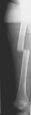
Figure 22 shows the radiograph of a 7-year-old boy who underwent retrograde elastic nailing of a femoral shaft fracture. What is the most common problem following this procedure?

Pediatrics 2007 Practice Questions: Set 3 (Solved) - Figure 2


Explanation

Several large clinical studies have shown that the most common problem after elastic nailing of a femoral shaft fracture is persistent pain and irritation at the nail insertion site. Unacceptable shortening and malunion are very rare in a 7-year-old patient. Rotational malalignment also is unusual. Osteonecrosis has been reported in solid antegrade nailing but not with elastic nailing of femoral shaft fractures in skeletally immature patients. Flynn JM, Luedtke LM, Ganley TJ, et al: Comparison of titanium elastic nails with traction and a spica cast to treat femoral fractures in children. J Bone Joint Surg Am 2004;86:770-777. Flynn JM, Hresko T, Reynolds RA, et al: Titanium elastic nails for pediatric femur fractures: A multicenter study of early results with analysis of complications. J Pediatr Orthop 2001;21:4-8.

Question 3

A newborn girl with an isolated unilateral dislocatable hip is placed in a Pavlik harness with the hips flexed 100 degrees and at resting abduction. Figure 23 shows an ultrasound obtained 2 weeks later. What is the next step in management?

Pediatrics 2007 Practice Questions: Set 3 (Solved) - Figure 3


Explanation

The infant has a well-positioned hip in the Pavlik harness and treatment should be continued in the current position. The success rate is over 90% with the use of this device for a dislocatable hip. Ultrasound is a useful tool to confirm appropriate positioning of the cartilaginous femoral head during treatment. If the femoral head is not reduced after 2 to 3 weeks in the harness, this mode of treatment should be abandoned. Forceful extreme abduction can cause osteonecrosis of the femoral epiphysis and should be avoided. Closed reduction, arthrography, and spica casting are indicated if the hip cannot be maintained in a reduced position with the harness. Lehmann HP, Hinton R, Morello P, et al: Developmental dysplasia of the hip practice guideline: Technical report. Committee on Quality Improvement, and Subcommittee on Developmental Dysplasia of the Hip. Pediatrics 2000;105:E57.

Question 4

During the early swing phase of the normal gait cycle, what lower extremity muscle is primarily contracting?


Explanation

Electromyography during walking reveals the tibialis anterior muscle is active during early swing, allowing the foot to clear the ground. All of the other muscles are quiet, as the limb moves forward through space with minimal muscular effort. The other muscles are primarily active during weight acceptance or push-off. Gage JR: An overview of normal walking. Instr Course Lect 1990;39:291-303.

Question 5

A 6-month-old child is seen in the emergency department with a spiral fracture of the tibia. The parents are vague about the etiology of the injury. There is no family history of a bone disease. In addition to casting of the fracture, initial management should include


Explanation

Unwitnessed spiral fractures should raise the possibility of child abuse, especially prior to walking age. With nonaccidental trauma being considered in the differential diagnosis, a skeletal survey is indicated to determine if there are other fractures in various stages of healing. Kempe CH, Silverman FN, Steele BF, et al: The battered-child syndrome. JAMA 1962;181:17-24.

Question 6

What is the primary indication for performing an arthroscopic synovectomy on a patient with hemophilia that is the result of factor VIII deficiency?


Explanation

Improved medical management has changed musculoskeletal outcomes for individuals with hemophilia. Patients with severe hemophilia receiving prophylactic administration of factor VIII may never develop a target joint that requires further orthopaedic intervention. Patients with moderate hemophilia and those patients with severe hemophilia not receiving prophylactic treatment will still develop joints that have recurrent hemarthroses. When recurrent hemarthrosis continues despite optimal medical management, synovectomy is indicated. While synovectomy is predictable in its ability to decrease joint bleeding, it does not necessarily improve joint range of motion or prevent the development of hemophilic arthropathy over time. It will not reverse articular damage to the joint once it has developed. Dunn AL, Busch MT, Wyly JB; et al: Arthroscopic synovectomy for hemophilic joint disease in a pediatric population. J Pediatr Orthop 2004;24:414-426.

Question 7

The rate of complications after in situ pinning of a chronic slipped capital femoral epiphysis is highest with placement of the screw in what quadrant of the femoral head?


Explanation

The rate of complications increases as the pin moves farther from the ideal position, which is the center of the head. This is the strongest argument for the use of a single pin. The highest rate of complications, primarily osteonecrosis and pin penetration, is associated with pin placement in the anterior superior quadrant. Raney EM, Ogden JA: Slipped capital femoral epiphysis. Current Ortho 1995;9:111-116.

Question 8

What is the incidence and significance of anterior cruciate ligament laxity following tibial eminence fractures in skeletally immature individuals?


Explanation

Measurable anterior cruciate ligament laxity, while frequently seen after tibial eminence fractures, usually does not cause symptoms. It is found even in patients whose fractures have been anatomically reduced and fixed, leading to speculation that it is due to stretching of the ligament at the time of injury. Willis R, Blokker C, Stall TM, et al: Long-term follow-up of anterior eminence fractures. J Pediatr Orthop 1993;13:361-364.

Question 9

A full-term newborn has webbing at the knees, rigid clubfeet, a Buddha-like posture of the lower extremities, and no voluntary or involuntary muscle action at and below the knees. Radiographs of the spine and pelvis reveal an absence of the lumbar spine and sacrum. What maternal condition is associated with this diagnosis?


Explanation

The history, physical examination, and radiographic findings are consistent with type IV sacral agenesis or caudal regression syndrome. These children are born with no lumbar spine or sacrum. The T12 vertebra is often prominent posteriorly. Popliteal webbing and knee flexion contractures are common with this diagnosis. There is a higher incidence of this diagnosis when the mother has diabetes mellitus. Maternal drug abuse and alcoholism can produce phenotypically unique children but without the findings described here. Maternal idiopathic scoliosis is not associated with caudal regression syndrome. Chan BW, Chan KS, Koide T, et al: Maternal diabetes increases the risk of caudal regression caused by retinoic acid. Diabetes 2002;51:2811-2816.

Question 10

Figure 24 shows the sitting AP and lateral spinal radiographs of a nonambulatory 12½-year-old boy with Duchenne muscular dystrophy who is being evaluated for scoliosis. The lumbar curve from T12 to L5 measures 36 degrees, and the thoracic curve from T3 to T12 measures 24 degrees on the AP radiograph. He has 5 degrees of pelvic obliquity. His forced vital capacity is 45% of predicted for height and weight. What is the most appropriate treatment for the spinal deformity?

Pediatrics 2007 Practice Questions: Set 3 (Solved) - Figure 4


Explanation

Posterior spinal fusion is the treatment of choice for scoliosis in patients with Duchenne muscular dystrophy once they are no longer able to walk. This treatment improves quality of life and upright wheelchair positioning. Its effect on pulmonary function is less clear, as pulmonary function will continue to decline because of the underlying muscle disease. While bracing and wheelchair modifications may slow the progression of the curve, progression will continue. Surgical intervention at this stage does not have to include the pelvis, which, in general, is indicated in curves of greater than 40 degrees, and when pelvic obliquity is greater than 10 degrees. Fixation to the pelvis should also be considered in lumbar curves where the apex is lower than L1. Surgical treatment usually can be safely performed if the vital capacity is greater than 35%. Hahn GV, Mubarak SJ: Muscular dystrophy, in Weinstein SL (ed): The Pediatric Spine, ed 2. Philadelphia, PA, Lippincott Williams & Wilkins, 2001, pp 819-832.

Question 11

A 3-year-old child has refused to walk for the past 2 days. Examination in the emergency department reveals a temperature of 102.2 degrees F (39 degrees C) and limited range of motion of the left hip. An AP pelvic radiograph is normal. Laboratory studies show a WBC count of 9,000/mm3, an erythrocyte sedimentation rate (ESR) of 65 mm/h, and a C-reactive protein level of 10.5 mg/L (normal < 0.4). What is the next most appropriate step in management?


Explanation

Examination reveals an irritable hip, creating a differential diagnosis of transient synovitis versus pyogenic hip arthritis. Kocher and associates described four criteria to help predict the presence of infection: inability to bear weight, fever, ESR of more than 40 mm/h, and a peripheral WBC count of more than 12,000/mm3. This patient meets three of the four criteria, with a positive predictive value of 73% to 93% for joint infection. Therefore, aspiration of the hip is warranted, with a high likelihood that emergent hip arthrotomy will be indicated. Ideally, intravenous antibiotics should be administered after culture material has been obtained from needle aspiration of the hip. An urgent bone scan is better indicated as a screening test for sacroiliitis or diskitis. If the arthrocentesis proves negative, CT or MRI of the pelvis may be indicated to rule out a pelvic or psoas abscess. Del Beccaro MA, Champoux AN, Bockers T, et al: Septic arthritis versus transient synovitis of the hip: The value of screening laboratory tests. Ann Emerg Med 1992;21:1418-1422. Kocher MS, Mandiga R, Zurakowski D, et al: Validation of a clinical prediction rule for the differentiation between septic arthritis and transient synovitis of the hip in children. J Bone Joint Surg Am 2004;86:1629-1635.

Question 12

A 2-year-old girl has had a swollen right knee for the past 7 weeks. There is no history of significant trauma, and she has not had a fever or been ill. Her parents report that she is stiff in the morning but otherwise does not report pain. A CBC count and erythrocyte sedimentation rate are normal. Treatment with naproxen at appropriate doses for the past 2 weeks has resulted in some improvement. Radiographs show only soft-tissue swelling. Examination reveals a healthy-appearing child with a warm and swollen right knee that is only slightly tender but lacks full extension by 20 degrees. What is the next most appropriate step in management?


Explanation

Up to 30% of children with juvenile rheumatoid arthritis (increasingly known now as juvenile idiopathic arthritis or JIA) already have potentially damaging uveitis at the time of diagnosis. This patient has typical oligoarticular JRA (JIA) and therefore is at significant risk for uveitis. MRI, radioisotope scanning, or an ACE level most likely would not provide additional useful diagnostic information because intra-articular derangement, osteomyelitis, or sarcoidosis are all unlikely. Arthrocentesis and triamcinolone hexacetonide joint injection might be indicated if continued use of nonsteroidal medication does not result in improvement, but should be held off for at least an additional 4 to 6 weeks to see if continued use of naproxen results in control of the arthritis. Wolf MD, Lichter PR, Ragsdale CG: Prognostic factors in the uveitis of juvenile rheumatoid arthritis. Ophthalmology 1987;94:1242. Cassidy JT, Petty RE: Textbook of Pediatric Rheumatology. Philadelphia, PA, WB Saunders, 2001, p 220.

Question 13

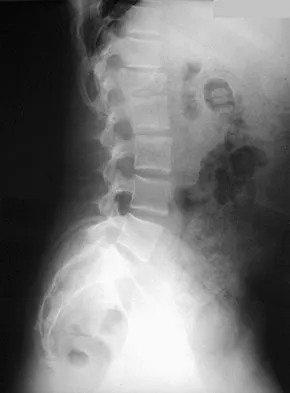
A 12-year-old girl has back pain after falling 20 feet and landing in the sitting position. She has no fractures or other injuries, and her neurologic examination is normal. A lateral radiograph, transverse CT scan, and reformatted sagittal CT scan are shown in Figures 25a through 25c. Which of the following methods is associated with the best long-term outcome?
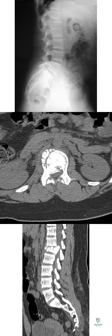


Explanation

The patient has a displaced burst fracture. Fusion with instrumentation has shown better results than casting alone. Posterior fusion with instrumentation, with sagittal plane correction, yields the best results. Decompression occurs indirectly with correction of the kyphosis. Anterior decompression is unnecessary. Lalonde F, Letts M, Yang JP, et al: An analysis of burst fractures of the spine in adolescents. Am J Orthop 2001;30:115-120. Clark P, Letts M: Trauma to the thoracic and lumbar spine in the adolescent. Can J Surg 2001;44:337-345.

Question 14

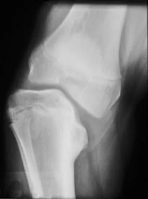
Figure 26 shows the radiograph of an otherwise healthy Caucasian 5-year-old boy who has a painless limp. What is the best treatment option?

Pediatrics 2007 Practice Questions: Set 3 (Solved) - Figure 8


Explanation

The prognosis of Legg-Perthes disease in children younger than age 6 years is good. There is no indication that surgical treatment will improve the outcome. Range-of-motion exercises to prevent contracture may be helpful. Herring JA, Kim HT, Browne R: Legg-Calve-Perthes disease: Prospective multicenter study of the effect of treatment on outcome. J Bone Joint Surg Am 2004;86:2121-2134.

Question 15

A 12-year-old girl who has a history of frequent tripping and falling also has bilateral symmetric hand weakness, high arched feet, absent patellar and Achilles tendon reflexes, and excessive wear on the lateral border of her shoes. She reports that she has multiple paternal family members with similar deformities. She most likely has a defect of what protein?


Explanation

The girl shows clinical features of hereditary motor sensory neuropathy type 1, Charcot-Marie-Tooth disease. The most common type of this autosomal-dominant disease is due to an underlying defect in the gene coding for peripheral myelin protein-22 on chromosome 17. Many other less common mutations have been identified in this family of neuropathies. Dystrophin is a protein that is abnormal in Duchenne's muscular dystrophy, which affects males and is diagnosed earlier. Type I collagen is defective in osteogenesis imperfecta. Alpha-L-iduronidase is defective in mucopolysaccharidosis type I, Hurler's syndrome. Defective cartilage oligomeric matrix protein is associated with some forms of multiple epiphyseal dysplasia. Patel PI, Roa BB, Welcher AA, et al: The gene for the peripheral myelin protein PMP-22 is a candidate for Charcot-Marie-Tooth disease type 1A. Nat Genet 1992;1:159-165.

Question 16

A 12-year-old boy with an ankle fracture undergoes closed reduction under sedation in the emergency department. Figure 27 shows a lateral radiograph of the ankle after two attempts at closed reduction. Based on these findings, treatment should now consist of

Pediatrics 2007 Practice Questions: Set 3 (Solved) - Figure 9


Explanation

The widening of the physis associated with incomplete reduction of this fracture suggests that periosteum is interposed at the fracture site. Clinical and animal study findings suggest that the interposed periosteum may lead to premature physeal closure. Repeated forceful attempts at reduction may subject the physis to further injury and should be avoided. Growth problems are common in children with Salter-Harris type I fractures of the lower extremities. Nonunions are rare in children with Salter-Harris type I fractures. Barmada A, Gaynor T, Mubarak SJ: Premature physeal closure following distal tibial physeal fractures: A new radiographic predictor. J Pediatr Orthop 2003;23:733-739.

Question 17

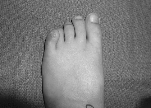
A 9-year-old girl has pain over the fifth toe that is aggravated by shoe wear. Clinical photographs are shown in Figures 28a and 28b. Treatment of this deformity should consist of
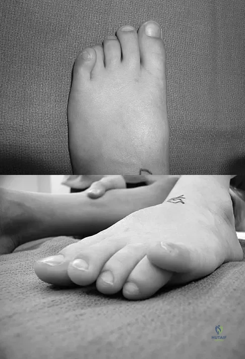


Explanation

The major obstacle to overcome in the surgical treatment of this cock-up deformity is recurrence. Dorsal releases can be performed; however, chronic dislocation of the fifth MTP joint usually needs to be addressed with plantar release as well. Chronic dorsal soft-tissue contractures may be overcome with translation of the toe into a plantar-based incision, as described originally by Cockin and accredited to Butler. This is the treatment of choice. Resection of the proximal phalanx improves symptoms but induces a secondary deformity; this procedure is usually reserved for skeletally mature individuals. Black GB, Grogan DP, Bobechko WP: Butler arthroplasty for correction of adducted fifth toe: A retrospective study of 36 operations between 1968 and 1982. J Pediatr Orthop 1985;5:439-441. Paton RW: V-Y plasty for correction of varus fifth toe. J Pediatr Orthop 1990;10:248-249.

Question 18

What acetabular procedure for developmental dysplasia of the hip does not require a concentric reduction of the femoral head in the acetabulum?


Explanation

All of the reorientation innominate osteotomies require a concentric reduction of the hip. The Staheli shelf procedure may be performed even with the hip subluxated, but it is a salvage procedure that covers a portion of the femoral head with capsular fibrocartilage rather than hyaline cartilage. Staheli LT, Chew DE: Slotted acetabular augmentation in childhood adolescence. J Pediatr Orthop 1992;12:569-580.

Question 19

Figure 29 shows the AP radiograph of a 14-year-old boy. The radiographic findings are most consistent with what pathologic process?

Pediatrics 2007 Practice Questions: Set 3 (Solved) - Figure 12


Explanation

The severe depression of the proximal medial tibial epiphysis is most consistent with the diagnosis of neglected infantile Blount's disease. Blount's disease in adolescents produces a deformity in the metaphyseal region. Septic arthritis and JRA affect both sides of the joint. Hemophilia produces a characteristic widening of the intercondylar notch. Thompson GH, Carter JR: Late-onset tibia vara (Blount's Disease). Clin Orthop 1990;255:24-35.

Question 20

A 5-year-old boy has had pain in the right foot for the past month. Examination reveals tenderness and mild swelling in the region of the tarsal navicular. Radiographs are shown in Figure 30. Management should consist of

Pediatrics 2007 Practice Questions: Set 3 (Solved) - Figure 13


Explanation

The child has the classic findings of Kohler's disease or osteochondrosis of the tarsal navicular. The cause of this condition is not known, but osteonecrosis and mechanical compression have been proposed. Children generally report midfoot pain over the tarsal navicular and limping. Physical findings include tenderness, swelling, and occasionally redness in the region of the tarsal navicular. Radiographs show sclerosis and narrowing of the tarsal navicular. The natural history of the condition is spontaneous resolution and reconstitution of the navicular. Symptomatic treatment with restriction of weight bearing or casting is recommended. Karp M: Kohler's disease of the tarsal scaphoid. J Bone Joint Surg 1937;19:84-96.

Question 21

A 9-year-old child sustained a fracture-dislocation of C-5 and C-6 with a complete spinal cord injury. What is the likelihood that scoliosis will develop during the remaining years of his growth?


Explanation

The incidence of late spinal deformity after complete spinal cord injury in children depends on the level of the spinal cord injury and the age of the patient at the time of injury. If a cervical level injury occurs before age 10 years, paralytic scoliosis will develop in virtually 100% of patients. Brown JC, Swank SM, Matta J, et al: Late spinal deformity in quadriplegic children and adolescents. J Pediatr Orthop 1984;4:456-461. Lancourt JE, Dickson JH, Carter RE: Paralytic spinal deformity following traumatic spinal-cord injury in children and adolescents. J Bone Joint Surg Am 1981;63:47-53.

Question 22

Figures 31a and 31b show the radiograph and MRI scan of an otherwise normal 3-month-old infant who has a spinal deformity. MRI reveals no intraspinal anomalies. What is the next step in management?
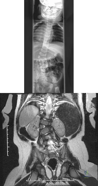


Explanation

Congenital scoliosis in an infant warrants evaluation of the renal, cardiac, and neurologic systems because frequently there is concurrent pathology. Progression in this instance is possible but not certain; therefore, progression must be documented prior to any surgical intervention. Close observation with serial radiographs every 4 to 6 months is appropriate. All of the surgical options listed may be reasonable choices in the future, but cardiac evaluation is the most important issue at this time. Basu PS, Elsebaie H, Noordeen MH: Congenital spinal deformity: A comprehensive assessment at presentation. Spine 2002;27:2255-2259.

Question 23

A 22-month-old girl has cerebral palsy. Which of the following findings is a good prognostic indicator of the child's ability to walk in the future?


Explanation

For the parachute test, the examiner holds the child prone and then lowers the child rapidly toward the floor. The parachute reaction is normal or positive if the child reaches toward the floor. The Moro or startle reflex should not be present beyond age 6 months. Asymmetric tonic neck reflex, extensor thrust, and absent foot placement are abnormal findings at any age. Bleck EE: Orthopaedic Management in Cerebral Palsy. Lavenham, Suffolk, The Lavenham Press, 1987, pp 121-139.

Question 24

The husband of a 22-year-old woman has hypophosphatemic rickets. The woman has no orthopaedic abnormalities, but she is concerned about her chances of having a child with the same disease. What should they be told regarding this disorder?


Explanation

Hypophosphatemia is a rare genetic disease usually inherited as an X-linked dominant trait. The fact that the woman has no skeletal manifestations would indicate that the husband has the X-linked mutation. The disease is more severe in boys than it is in girls. The husband will not transmit the disease to his sons. However, all of their daughters will be affected either with the disease or as carriers. If the woman has the disease or the trait, there is a 50% chance that her sons will inherit the disease and a 50% chance that her daughters will be carriers or have a milder form of the disease. Parents should be advised to have genetic counseling so they can be informed when deciding whether to have children. Herring JA: Metabolic and endocrine bone diseases, in Tachdjian's Pediatric Orthopaedics, ed 3. New York, NY, WB Saunders, 2002, pp 1685-1743. Sillence DO: Disorders of bone density, volume, and mineralization, in Rimoin DL, Conner JM, Pyerite RE, et al (eds): Principles and Practice of Medical Genetics, ed 4. New York, NY, Churchill Livingstone, 2002.

Question 25

A 9-year-old boy sustained a traumatic brain injury and right lower extremity trauma in an accident involving a motor vehicle and a pedestrian. Initial evaluation in the emergency department reveals an obtunded patient who is breathing spontaneously and withdraws appropriately to painful stimuli. After initial resuscitation and stabilization, a CT scan reveals a right parietal intracranial hemorrhage. Radiographs of the swollen right thigh are shown in Figures 32a and 32b. Management of the fractured femur should ultimately consist of
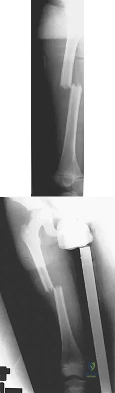


Explanation

A child with a traumatic brain injury generally achieves significant neurologic recovery and has a more favorable prognosis than an adult. Early stabilization of fractures facilitates transportation of the child for diagnostic tests and decreases the incidence of shortening and malunion. Surgical treatment of the fracture is indicated when cerebral perfusion pressure has stabilized. Casting or traction is not the most appropriate treatment of a femoral fracture in a child of this age with a brain injury. Fracture reduction is difficult to maintain if the brain injury leads to spasticity, and transportation within the hospital for tests is more difficult. Insertion of a reamed antegrade intramedullary nail inserted at the piriformis fossa is associated with a small risk of osteonecrosis of the femoral head. The transverse femoral fracture in this patient is ideally suited for stabilization with flexible intramedullary nails. Ligier and associates treated 123 femoral shaft fractures in children with flexible intramedullary nails, including 35 patients with head injury. In one patient with hemiplegia and a urinary tract infection, a deep wound infection developed, necessitating nail removal. The remaining patients all healed without major complications. Heinrich and associates treated 78 diaphyseal femoral fractures with flexible intramedullary nails, including 14 with head injury. No major complications were reported and all fractures healed. Tolo VT: Management of the multiply injured child, in Rockwood CA, Wilkins KE, Beaty JH (eds): Fractures in Children, ed 4. Philadelphia, PA, Lippincott-Raven, 1996, pp 83-95. Ligier JN, Metaizeau JP, Prevot J, et al: Elastic stable intramedullary nailing of femoral shaft fractures in children. J Bone Joint Surg Br 1988;70:74-77. Heinrich MS, Drvaric DM, Darr K, et al: The operative stabilization of pediatric diaphyseal femur fractures with flexible intramedullary nails: A prospective analysis. J Pediatric Orthop 1994;14:501-507.

Question 26

An 11-year-old boy sustains a transverse diaphyseal femur fracture. The surgeon elects to proceed with rigid intramedullary nailing. To minimize the risk of iatrogenic avascular necrosis (AVN) of the femoral head, which entry point is most appropriate?


Explanation

In pediatric patients with open physes, a trochanteric entry point (specifically the lateral edge or tip of the greater trochanter) is preferred for rigid nailing. Utilizing the piriformis fossa disrupts the medial circumflex femoral artery anastomosis, significantly increasing the risk of AVN.

Question 27

A 4-week-old infant with developmental dysplasia of the hip (DDH) has been treated in a Pavlik harness for 1 week. The mother brings the child in, noting that the infant is no longer actively kicking or extending the knee on the affected side. What is the most appropriate next step in management?


Explanation

The clinical presentation describes a femoral nerve palsy, a known complication of excessive hip flexion in a Pavlik harness. The most appropriate management is to remove or loosen the harness and observe until nerve function recovers.

Question 28

A 12-year-old pre-menarcheal girl presents with a right thoracic adolescent idiopathic scoliosis (AIS).

Her Risser stage is 0, and her curve measures 32 degrees on a standing PA radiograph. What is the most evidence-based management strategy?


Explanation

The patient is a growing child (Risser 0, pre-menarcheal) with a curve between 25 and 45 degrees. According to the BRAIST trial, full-time bracing (TLSO for 16-23 hours/day) is highly effective in preventing curve progression to surgical thresholds in this population.

Question 29

A 3-year-old child sustains an isolated spiral fracture of the femoral shaft and is managed with a hip spica cast. During reduction, how much initial fracture shortening is most appropriate to accept to compensate for anticipated overgrowth?


Explanation

In children aged 2 to 10 years, femoral fractures typically stimulate a hyperemic response leading to 1 to 2 cm of overgrowth. Therefore, accepting 10 to 15 mm of bayonet apposition (shortening) during casting is optimal to prevent long-term leg length discrepancy.

Question 30

A 7-month-old girl presents for her first pediatric orthopedic evaluation and is diagnosed with bilateral DDH. Ultrasound confirms bilateral dislocated hips that are irreducible on dynamic examination. What is the most appropriate initial treatment?


Explanation

The Pavlik harness has a high failure rate in children older than 6 months and in those with irreducible dislocations. Closed reduction with spica casting under anesthesia is the most appropriate initial intervention for late-presenting DDH at this age.

Question 31

A 9-month-old boy is referred for a 25-degree left thoracic scoliosis.

Radiographs demonstrate a rib-vertebral angle difference (RVAD) of 25 degrees. What is the most likely natural history and appropriate treatment?


Explanation

This is infantile idiopathic scoliosis. According to Mehta's criteria, an RVAD greater than 20 degrees indicates a high likelihood of curve progression. The gold standard for progressive infantile curves is early serial derotational casting.

Question 32

An 8-year-old boy is treated with titanium elastic nails for a midshaft transverse femur fracture.

What is the most commonly reported complication associated with this specific surgical intervention?


Explanation

Insertion site pain or skin irritation from prominent nail ends at the distal femur is the most common complication of flexible intramedullary nailing in pediatric femur fractures. It often necessitates premature or planned hardware removal.

Question 33

A 4-year-old girl with residual DDH requires an open reduction and pelvic osteotomy. The surgeon plans a Salter innominate osteotomy. This osteotomy relies on a hinge at which of the following structures to provide anterolateral coverage?


Explanation

The Salter osteotomy is a complete, redirectional transiliac osteotomy that redirects the entire acetabulum to improve anterolateral coverage. It hinges symmetrically on the pubic symphysis to achieve its corrective rotation.

Question 34

A 12-year-old boy with Duchenne muscular dystrophy presents with a 45-degree sweeping thoracolumbar neuromuscular scoliosis. His forced vital capacity (FVC) is 40% of predicted. What is the most appropriate management?


Explanation

Bracing is ineffective in halting curve progression in Duchenne muscular dystrophy. Posterior spinal fusion to the pelvis is indicated for curves over 20-30 degrees to halt progressive respiratory decline, maintain sitting balance, and prevent severe pelvic obliquity.

Question 35

A 6-week-old female infant is evaluated for DDH. Coronal ultrasound of the right hip reveals an alpha angle of 55 degrees and a beta angle of 65 degrees. According to the Graf classification, what is the appropriate management?


Explanation

An alpha angle between 50 and 59 degrees in a child younger than 3 months represents a Graf Type IIa hip (physiologic immaturity). The standard of care is observation with a follow-up ultrasound, as the majority will resolve spontaneously.

Question 36

A 6-year-old girl sustains a closed, completely displaced subtrochanteric femur fracture.

What is the most appropriate fixation strategy to minimize complications while facilitating early mobilization?


Explanation

Submuscular or open plating is preferred for pediatric subtrochanteric fractures. Elastic nails have a high rate of failure, malunion, and varus collapse in the subtrochanteric region due to poor biomechanical control, and rigid nails carry AVN risk in young children.

Question 37

A 10-year-old boy with a history of DDH treated by closed reduction at age 1 presents for follow-up. Radiographs demonstrate a progressive coxa valga deformity and a short femoral neck, but the medial physis remains open. According to the Kalamchi-MacEwen classification, what type of vascular insult occurred?


Explanation

The Kalamchi-MacEwen classification describes AVN after DDH treatment. Type II involves damage to the lateral portion of the physis, resulting in a progressive valgus deformity of the femoral head and neck.

Question 38

A 10-year-old boy presents with a 30-degree left-sided thoracic scoliosis.

His neurological examination is normal. What is the most appropriate next step in his diagnostic workup?


Explanation

Left-sided thoracic curves, particularly in juvenile patients or males, have a higher association with intraspinal anomalies such as syringomyelia or Chiari malformations. An MRI of the neuroaxis is indicated to rule out underlying neural axis abnormalities prior to treatment.

Question 39

A 10-month-old infant who is not yet cruising or walking presents to the emergency department with a swollen, painful thigh. Radiographs reveal a closed spiral fracture of the mid-diaphyseal femur. What is the most critical next step in management?


Explanation

Femur fractures in non-ambulatory infants are highly suspicious for non-accidental trauma (child abuse). The standard of care mandates immediate involvement of child protective services and a full skeletal survey to identify other hidden injuries.

Question 40

A 5-year-old girl with residual DDH undergoes preoperative planning for a pelvic osteotomy. The chosen technique is an incomplete pericapsular osteotomy that hinges on the triradiate cartilage, dynamically reducing the volume of the acetabulum. Which osteotomy is described?


Explanation

The Pemberton osteotomy is an incomplete pericapsular procedure that hinges at the triradiate cartilage. By changing the shape of the acetabular roof, it inherently reduces the acetabular volume, making it highly effective for true dysplastic, capacious acetabula.

Question 41

When classifying adolescent idiopathic scoliosis using the Lenke system, the lumbar spine modifier is determined by the relationship of the center sacral vertical line (CSVL) to the lumbar vertebrae. What defines a Lumbar Modifier B?


Explanation

In the Lenke classification, Modifier A indicates the CSVL passes between the lumbar pedicles. Modifier B indicates the CSVL touches the apical vertebra between the medial pedicle and the lateral vertebral margin. Modifier C means the CSVL falls completely lateral to the vertebra.

Question 42

A 4-week-old infant sustains a closed midshaft femur fracture after a roll-over injury in a stroller. What is the most appropriate initial management?


Explanation

The Pavlik harness is the standard of care for infants younger than 6 months with femur fractures. It provides excellent outcomes, is easier to apply than a spica cast, and has lower rates of skin complications.

Question 43

A 10-year-old boy weighing 55 kg (121 lbs) sustains a length-unstable, comminuted midshaft femur fracture. Which of the following surgical options is most appropriate?


Explanation

Submuscular plating is ideal for length-unstable femur fractures in children weighing over 50 kg. ESIN in children over 50 kg is associated with unacceptable rates of loss of reduction and malunion.

Question 44

A 7-year-old girl sustains a Delbet type II (transcervical) femoral neck fracture. Despite urgent closed reduction and internal fixation, she is at the highest risk for which of the following complications?


Explanation

AVN is the most common and devastating complication of pediatric femoral neck fractures. The risk is highest with Delbet type I and type II fractures due to disruption of the intracapsular blood supply.

Question 45

A 3-year-old child sustains a closed midshaft femur fracture and is treated with a one-and-a-half spica cast. What is the maximum acceptable amount of fracture overriding to anticipate the overgrowth phenomenon?


Explanation

In children aged 2 to 10 years, femoral shaft fractures typically stimulate a hyperemic response causing 1 to 2 cm of overgrowth. Accepting 1.5 cm of overriding in a cast prevents a clinically significant leg length discrepancy.

Question 46

A 4-month-old girl is being treated in a Pavlik harness for developmental dysplasia of the hip (DDH). Her parents report that she is no longer actively extending her right knee. What is the most likely cause of this complication?


Explanation

Femoral nerve palsy in a Pavlik harness is caused by excessive hip flexion, leading to nerve impingement against the inguinal ligament. It usually resolves completely after temporarily releasing the anterior flexion straps.

Question 47

A 6-week-old infant undergoes hip ultrasonography for suspected developmental dysplasia. The alpha angle is measured at 62 degrees and the beta angle is 50 degrees. According to the Graf classification, what is the appropriate management?


Explanation

An alpha angle greater than 60 degrees and a beta angle less than 55 degrees indicates a Graf Type I (normal) hip. No treatment is necessary, and routine pediatric care should continue.

Question 48

A 12-year-old premenarchal girl (Risser 0) has a right thoracic adolescent idiopathic scoliosis (AIS) curve of 35 degrees. Which of the following is the most appropriate management?


Explanation

Bracing is indicated for skeletally immature patients (Risser 0-2) with idiopathic curves between 25 and 45 degrees. A TLSO brace significantly decreases the risk of curve progression to the surgical threshold.

Question 49

A 2-year-old boy presents with congenital scoliosis secondary to a fully segmented hemivertebra at T8. In addition to full spine radiographs, what routine screening must be obtained?


Explanation

Congenital scoliosis is frequently associated with VACTERL anomalies. Screening for genitourinary anomalies (renal US) and cardiac defects (echocardiogram), along with a total spine MRI for intraspinal anomalies, is standard.

Question 50

An 11-year-old boy requires rigid intramedullary nailing for a transverse femur fracture. To minimize the risk of iatrogenic avascular necrosis (AVN), what is the optimal entry point?


Explanation

In pediatric and adolescent patients, a piriformis entry portal risks damaging the medial circumflex femoral artery, leading to AVN. A trochanteric entry (tip or lateral to it) minimizes this risk.

Question 51

A 9-month-old infant undergoes closed reduction and spica casting for late-diagnosed DDH. Postoperatively, the hip is noted to be immobilized in 65 degrees of abduction. What complication is the child at highest risk for?


Explanation

Immobilizing the hip in excessive abduction (greater than 60 degrees) dramatically increases the risk of avascular necrosis of the femoral head. This is due to compression of the extracapsular epiphyseal vessels.

Question 52

A 10-year-old boy presents with a left-sided thoracic scoliosis of 25 degrees. His neurologic examination is unremarkable. What is the most appropriate next step in evaluation?


Explanation

A left-sided thoracic curve is atypical for adolescent idiopathic scoliosis and raises high suspicion for an intraspinal anomaly, such as a syrinx, Chiari malformation, or tethered cord. A total spine MRI is strongly indicated.

Question 53

An 18-month-old girl presents with untreated developmental dysplasia of the hip (DDH). She has a positive Galeazzi sign and limited hip abduction. What is the most appropriate initial intervention?


Explanation

For DDH presenting at 18 months or older, open reduction is generally the initial treatment of choice. Closed reduction at this age has a high failure rate and an increased risk of AVN.

Question 54

In an infant with idiopathic scoliosis, which radiographic parameter most strongly predicts the likelihood of curve progression?


Explanation

Mehta's rib-vertebral angle difference (RVAD) is the primary prognostic indicator in infantile idiopathic scoliosis. An RVAD greater than 20 degrees strongly predicts progressive deformity rather than spontaneous resolution.

Question 55

A 2-year-old child is placed in a 90/90 spica cast for a midshaft femur fracture. This specific positioning (90 degrees hip flexion, 90 degrees knee flexion) is primarily chosen to control which of the following?


Explanation

The 90/90 spica cast position effectively controls rotational alignment of the femur. It also helps lock the pelvis to prevent the child from sliding distally within the cast, maintaining overall fracture reduction.

Question 56

A 6-year-old boy sustains a midshaft femur fracture and is treated with flexible intramedullary nails. Which of the following is the most common complication associated with this treatment modality?


Explanation

The most common complication of flexible intramedullary nailing for pediatric femur fractures is soft-tissue irritation at the insertion site, occurring in up to 15% of cases. Avascular necrosis is rare unless a rigid antegrade nail with a piriformis entry is improperly used in a child.

Question 57

A 6-week-old female is treated with a Pavlik harness for developmental dysplasia of the hip (DDH). After 1 week, the mother notes the child is no longer kicking her right leg, and the knee rests in extension. What is the most likely cause?


Explanation

Hyperflexion of the hips in a Pavlik harness can compress the femoral nerve, leading to a transient femoral nerve palsy, indicated by a lack of active knee extension. The harness should be temporarily removed or adjusted (flexion reduced) to resolve the palsy.

Question 58

Which of the following factors indicates the highest risk for curve progression in a patient with Adolescent Idiopathic Scoliosis (AIS)?


Explanation

The highest risk for curve progression in AIS occurs during the period of peak height velocity. Skeletal immaturity (e.g., Risser 0, open triradiate cartilage), female gender, and larger initial curve magnitudes are also significant risk factors.

Question 59

A 9-month-old boy presents with a spiral fracture of the femoral shaft. He is not yet walking independently. What is the most appropriate next step in management?


Explanation

A femur fracture in a non-ambulatory infant is highly suspicious for non-accidental trauma (child abuse). A full skeletal survey and mandatory reporting to child protective services must be initiated before or concurrently with definitive orthopedic management.

Question 60

When evaluating a 4-week-old infant's hip using Graf's ultrasound method, an alpha angle of 45 degrees is noted. What does this angle primarily measure?


Explanation

The alpha angle reflects the bony concavity (roof) of the acetabulum, with a normal angle being greater than 60 degrees. An alpha angle of 45 degrees indicates significant bony dysplasia. The beta angle measures the cartilaginous roof coverage.

Question 61

A 9-month-old boy is diagnosed with infantile idiopathic scoliosis. His rib-vertebral angle difference (RVAD) of Mehta is measured at 25 degrees. What is the most likely clinical course?


Explanation

An RVAD greater than 20 degrees indicates a high likelihood of curve progression in infantile idiopathic scoliosis (progressive phase). Treatment typically involves serial elongation-derotation-flexion (Mehta) casting to control the curve and allow spinal growth.

Question 62

A 13-year-old boy weighing 65 kg sustains a length-unstable transverse femoral shaft fracture. If a rigid locked intramedullary nail is selected, which entry point minimizes the risk of iatrogenic avascular necrosis?


Explanation

To avoid iatrogenic avascular necrosis from damage to the medial circumflex femoral artery branches, rigid nailing in adolescents should use a lateral trochanteric entry point. A piriformis fossa entry is strictly contraindicated in skeletally immature patients.

Question 63

A 4-year-old girl with untreated DDH is scheduled for open reduction and an innominate osteotomy. The planned procedure involves a complete transiliac osteotomy that redirects the entire acetabulum by hinging on the pubic symphysis. Which osteotomy is being described?


Explanation

The Salter osteotomy is a complete transiliac osteotomy that redirects the acetabulum to improve anterolateral coverage, hinging at the pubic symphysis. Pemberton and Dega are incomplete osteotomies that hinge on the triradiate cartilage.

Question 64

A 3-year-old girl is found to have congenital scoliosis due to a fully segmented hemivertebra. Which of the following screening tests must be obtained to evaluate for commonly associated anomalies?


Explanation

Congenital scoliosis has a high association with VACTERL anomalies, intraspinal anomalies (e.g., tethered cord, diastematomyelia), and genitourinary defects. A spinal MRI and renal ultrasound are mandatory screening tools for these patients.

Question 65

A 3-year-old boy is treated in a 90-90 hip spica cast for a femoral shaft fracture. He presents 2 weeks later with excessive crying, bilious vomiting, and food refusal. What is the most likely diagnosis?


Explanation

SMA syndrome (cast syndrome) occurs when the third portion of the duodenum is compressed by the SMA, often precipitated by body casts or rapid weight loss. Symptoms include severe abdominal pain, vomiting, and food refusal, requiring cast univalving or bi-valving.

Question 66

During closed reduction of a dislocated hip in a 9-month-old child, the Ramsey safe zone is assessed. What defines the boundaries of this zone?


Explanation

The Ramsey safe zone is the arc of abduction between the angle of re-dislocation (lower limit) and the angle of maximal abduction (upper limit). Excessive abduction past this safe zone dramatically increases the risk of avascular necrosis.

Question 67

In the Lenke classification for Adolescent Idiopathic Scoliosis, how is a structural minor curve defined on supine side-bending radiographs?


Explanation

A minor curve is considered structural in the Lenke classification if it does not bend out to less than 25 degrees on supine side-bending radiographs. Additionally, regional kyphosis greater than +20 degrees also classifies a curve as structural.

Question 68

A 7-year-old child weighing 45 kg sustains a highly comminuted, length-unstable femur fracture. What is the most appropriate fixation method to maintain length and alignment?


Explanation

For length-unstable, comminuted femur fractures in heavier school-aged children, submuscular bridge plating provides excellent stability and maintains length while preserving the fracture hematoma. Flexible nails are relatively contraindicated here due to a high risk of unacceptable shortening.

Question 69

A 12-year-old girl with a history of DDH treated with closed reduction at infancy presents with a limp. Radiographs show a shortened, widened femoral neck and an enlarged, flat femoral head. This deformity is most likely a sequela of which complication?


Explanation

The classic radiographic findings of coxa magna, coxa brevis, and a flat femoral head in a patient with a history of DDH treatment are indicative of iatrogenic avascular necrosis. This is typically sustained during early immobilization, often due to excessive hip abduction.

Question 70

A 14-year-old non-ambulatory male with Duchenne muscular dystrophy has a 60-degree thoracolumbar scoliosis and severe pelvic obliquity. What is the most appropriate surgical strategy?


Explanation

In non-ambulatory patients with Duchenne muscular dystrophy and severe scoliosis with pelvic obliquity, posterior spinal fusion must typically extend to the pelvis. This is necessary to correct the obliquity, prevent pressure sores, and provide a stable sitting balance.

Question 71

A 6-month-old female presents with an asymmetric thigh crease. An AP pelvis radiograph is obtained. Which of the following acetabular index measurements is considered normal for this age?


Explanation

The normal acetabular index in a 6-month-old is generally less than 25 degrees. Values greater than 28-30 degrees are highly suspicious for developmental dysplasia of the hip requiring intervention.

Question 72

A 4-year-old boy undergoes application of a hip spica cast for a transverse midshaft femur fracture. The surgeon intentionally positions the fracture with 1.5 cm of overlap. What is the primary rationale for this?


Explanation

In children aged 2 to 10 years, a femur fracture stimulates longitudinal overgrowth (averaging 1-2 cm) due to fracture hyperemia. Bayonet apposition with 1-1.5 cm of overlap is intentionally accepted to compensate for this expected overgrowth.

Question 73

In a young child with early-onset scoliosis and multiple fused ribs, vertical expandable prosthetic titanium rib (VEPTR) surgery is indicated primarily to treat or prevent which condition?


Explanation

VEPTR devices are primarily used in early-onset scoliosis with associated rib fusions to expand the constrained hemithorax. This expansion treats or prevents thoracic insufficiency syndrome, allowing for more normal lung development and volume.

Question 74

When performing an anterior (Smith-Petersen) approach for open reduction of a DDH, which structure is typically released at the pelvic brim rather than its insertion to protect the primary blood supply to the femoral head?


Explanation

During an anterior open reduction, the iliopsoas tendon is typically released at the pelvic brim rather than the lesser trochanter. This technique avoids injuring the medial circumflex femoral artery, which is the primary blood supply to the femoral head.

Question 75

A 12-year-old premenarchal girl (Risser 0) is diagnosed with Adolescent Idiopathic Scoliosis. Her right thoracic curve measures 32 degrees. What is the most appropriate recommended management?


Explanation

Bracing is indicated in skeletally immature patients (Risser 0-2, premenarchal) with curve magnitudes between 25 and 45 degrees. A TLSO worn at least 16-18 hours a day has been shown to significantly decrease the risk of curve progression to surgical magnitude.

Question 76

A 12-year-old boy weighing 55 kg (121 lbs) sustains a midshaft femur fracture. The plan is to proceed with antegrade rigid intramedullary nailing. Which entry point is recommended to minimize the risk of iatrogenic avascular necrosis (AVN) of the femoral head?


Explanation

In pediatric patients, using a piriformis fossa entry point risks damaging the medial circumflex femoral artery, leading to AVN. A lateral entry point to the tip of the greater trochanter is recommended to avoid the retinacular vessels and minimize this devastating complication.

Question 77

A 4-month-old female with developmental dysplasia of the hip (DDH) is being treated with a Pavlik harness. During a follow-up exam, she exhibits absent active knee extension on the treated side. What is the most likely cause of this finding?


Explanation

Hyperflexion of the hips in a Pavlik harness can lead to femoral nerve palsy, presenting as decreased active knee extension. The harness must be adjusted or temporarily discontinued until full nerve function recovers.

Question 78

A 13-year-old premenarchal girl presents with a right thoracic adolescent idiopathic scoliosis. Standing AP radiographs demonstrate a Cobb angle of 32 degrees. Her Risser stage is 1. What is the most appropriate management?


Explanation

Full-time TLSO bracing is indicated for growing children (premenarchal, Risser 0-2) with a Cobb angle between 25 and 45 degrees. Clinical trials have proven that full-time bracing significantly decreases the risk of curve progression to the surgical threshold.

Question 79

A 4-year-old boy is treated with a hip spica cast for a transverse midshaft femur fracture. Which of the following describes the expected maximum amount of femoral overgrowth and the typical timeframe it peaks following the injury?


Explanation

Femoral overgrowth following a shaft fracture in young children averages 1 to 1.5 cm due to hyperemia stimulating the physes. This accelerated growth phase typically peaks around 18 to 24 months post-injury.

Question 80

An 18-month-old girl presents with a painless limp and delayed walking. Examination reveals asymmetric thigh folds and a positive Galeazzi sign. Radiographs confirm a unilateral dislocated hip. What is the most appropriate initial treatment?


Explanation

For DDH presenting between 6 and 18 months of age, closed reduction and spica casting under general anesthesia is the standard first-line treatment. A Pavlik harness is ineffective at this age due to infant size and mobility.

Question 81

A 2-year-old boy is evaluated for congenital scoliosis secondary to a fully segmented hemivertebra at T8. What screening studies are mandatory prior to definitive orthopedic management?


Explanation

Congenital scoliosis is highly associated with VACTERL anomalies. A renal ultrasound and echocardiogram are mandatory to rule out genitourinary and cardiovascular abnormalities, which occur in up to 30% and 10% of these patients, respectively.

Question 82

A 9-year-old girl weighing 48 kg (105 lbs) sustains a length-unstable, highly comminuted spiral fracture of the femoral shaft. Which of the following is the most appropriate surgical fixation method?


Explanation

Submuscular plating is ideal for length-unstable or comminuted femur fractures in older, heavier children (near 50 kg) where flexible nails have high failure rates. It preserves the fracture hematoma while providing rigid length and rotational control.

Question 83

On an AP pelvis radiograph of a 6-month-old evaluated for DDH, the proximal femoral metaphysis is located in the upper outer quadrant formed by Hilgenreiner's and Perkin's lines. What does this radiographic finding indicate?


Explanation

Perkin's line (vertical) and Hilgenreiner's line (horizontal) divide the hip into quadrants. In a normal hip, the medial beak of the metaphysis is in the lower inner quadrant; presence in the upper outer quadrant signifies a dislocated hip.

Question 84

A 14-year-old boy with Duchenne muscular dystrophy presents with a progressive 55-degree thoracolumbar scoliosis. His forced vital capacity (FVC) is 40% of predicted. What is the recommended management?


Explanation

In Duchenne muscular dystrophy, scoliosis is relentlessly progressive and bracing is poorly tolerated and ineffective. Posterior spinal fusion to the pelvis is indicated for progressive curves >20-30 degrees while pulmonary function (FVC >30-40%) is still adequate for surgery.

Question 85

A 7-month-old infant is brought to the emergency department with a spiral midshaft femur fracture. The parents state the child rolled off a low couch. What is the most appropriate next step in management?


Explanation

A femur fracture in a non-ambulatory infant is highly suspicious for non-accidental trauma (child abuse). A complete skeletal survey and consultation with child protective services are mandatory to ensure the child's safety before definitive fracture management.

Question 86

An ultrasound of a 6-week-old infant's hip reveals a rounded bony promontory, an alpha angle of 48 degrees, and a beta angle of 60 degrees. According to the Graf classification, what is the best description of this hip?


Explanation

A Graf Type II hip is characterized by an alpha angle between 43 and 59 degrees, indicating mild dysplasia or physiologic delay. An alpha angle greater than 60 degrees is considered normal (Type I).

Question 87

In the Lenke classification for Adolescent Idiopathic Scoliosis, what radiographic criterion defines a "structural" minor curve that must be included in the operative fusion construct?


Explanation

The Lenke classification defines a minor curve as structural if it does not correct to less than 25 degrees on side-bending radiographs (or if there is a regional kyphosis of at least 20 degrees). Structural curves generally must be included in the fusion.

Question 88

You are performing retrograde titanium elastic nailing for a pediatric femur fracture. To achieve optimal three-point fixation and biomechanical stability, how should the nails be contoured and positioned within the canal?


Explanation

Elastic nails should be pre-bent into a C-shape and inserted such that the apices face in opposite directions, creating an O-shape inside the medullary canal. This provides maximum three-point fixation and rotational stability at the fracture site.

Question 89

What is the most critical technical risk factor for the development of iatrogenic avascular necrosis (AVN) of the femoral head during closed reduction and spica casting for DDH?


Explanation

Immobilizing the hip in excessive abduction (greater than 60 degrees, the "frog-leg" position) places severe tension on the medial circumflex femoral artery. This is the most significant preventable risk factor for iatrogenic AVN in DDH treatment.

Question 90

A 6-month-old boy has a left-sided infantile idiopathic scoliosis with a Cobb angle of 35 degrees. The Rib-Vertebral Angle Difference (RVAD) of Mehta is measured at 25 degrees. What is the most likely natural history of this curve without treatment?


Explanation

Mehta's Rib-Vertebral Angle Difference (RVAD) predicts progression in infantile idiopathic scoliosis. An RVAD greater than 20 degrees is highly predictive of rapid curve progression, necessitating aggressive early treatment such as serial casting.

None

Clinic OS
Medically Verified Content by
Prof. Clinic OS
Consultant Orthopedic & Spine Surgeon
Chapter Index