العربية
Part of the Master Guide

AAOS Orthopedic MCQs (Set 1): Spine Disorders, Sports Medicine & Arthroplasty | 2026 Board Review

AAOS Spine Surgery MCQs (Set 2): Degenerative Cervical, Lumbar Disc, & Trauma | ABOS Review

23 Apr 2026 64 min read 104 Views
Spine 2009 MCQs - Part 2

Key Takeaway

This high-yield Set 2 of spine surgery MCQs prepares you for AAOS/ABOS exams. Questions cover crucial topics like degenerative cervical spine pathology, lumbar disc herniation, and acute spinal trauma management. Enhance your diagnostic and treatment planning skills for challenging spine cases.

AAOS Spine Surgery MCQs (Set 2): Degenerative Cervical, Lumbar Disc, & Trauma | ABOS Review

Comprehensive 100-Question Exam


00:00

Start Quiz

Question 1

A previously healthy 35-year-old man was involved in a rollover motor vehicle accident 2 days ago. He was placed in a semi-rigid cervical orthosis. He now reports mostly axial neck pain with attempted range of motion. Examination reveals the mechanical neck pain but no obvious neurologic deficits. AP, flexion, and extension radiographs are shown in Figures 10a through 10c, and sagittal and coronal CT scans are shown in Figures 10d and 10e. What is the most appropriate management at this time?


Explanation

Odontoid fractures can be classified based on the anatomic position of the fracture within the dens itself. Type I is an oblique fracture through the upper part of the odontoid process. Type II is a fracture that occurs at the base of the odontoid as it attaches to the body of C2; type III occurs when the fracture line extends through the body of the axis. Type 1 fractures typically can be treated nonsurgically with 6 to 8 weeks of immobilization with a semi-rigid cervical orthosis. Nondisplaced, deep type III fractures generally are treated with skeletal halo fixation. Deep, displaced, and angled type III fractures can be treated with closed reduction and skeletal halo fixation. Shallow type III fractures are sometimes amenable to anterior odontoid screw fixation. Type II fractures can be managed nonsurgically or surgically. Treatment options include halo immobilization, internal fixation (odontoid screw fixation), and posterior atlantoaxial arthrodesis. Management with the halo vest usually is considered if the initial dens displacement is less than 6 mm, the reduction is performed within 1 week of the injury and is able to be maintained, and the patient is younger than age 60 years. Halo vest immobilization can lead to a healing rate of more than 90%. Posterior surgical fusion techniques provide high fusion success rates but do so at the expense of cervical rotation. Up to 50% of rotation is lost with these techniques. Anterior odontoid single screw fixation is often tolerated better than skeletal halo fixation and also is noted to preserve the normal rotation at C1/C2. Studies have shown less of a malunion and nonunion rate in the treatment of type II odontoid fractures with anterior odontoid screw fixation. Osteoporosis, short neck and barrel-chested anatomy, and fractures that are more than 4 weeks old preclude anterior odontoid fixation. Shilpakar S, McLaughlin MR, Haid RW Jr, et al: Management of acute odontoid fractures: Operative techniques and complication avoidance. Neurosurg Focus 2000;8:e3. Subach BR, Morone MA, Haid RW Jr, et al: Management of acute odontoid fractures with single-screw anterior fixation. Neurosurgery 1999;45:812-819.

Question 2

Which of the following palpable bony landmarks is correctly matched with its corresponding vertebral level?


Explanation

The carotid tubercle is usually located at the level of C6. The angle of the mandible is at C1-C2; the hyoid is at C4; the superior portion of the thyroid cartilage is C4-C5; and the cricoid cartilage is at C6. Smith GW, Robinson RA: The treatment of certain cervical-spine disorders by anterior removal of the intervertebral disc and interbody fusion. J Bone Joint Surg Am 1958;40:607.


Question 3

What root is most commonly involved with a segmental root level palsy after laminoplasty?


Explanation

The postoperative incidence of C5 root palsy after laminoplasty ranges from 5% to 12%. Other roots also may be affected. The palsies tend to be motor dominant, although sensory dysfunction and radicular pain are also possible. The palsy may arise during the immediate postoperative period or up to 20 days later. C5 may be preferentially involved because it is at the apex of the cervical lordosis. Recovery usually occurs over weeks to months. Spivak JM, Connolly PJ (eds): Orthopaedic Knowledge Update: Spine 3. Rosemont, IL, American Academy of Orthopaedic Surgeons, 2006, pp 235-249.


Question 4

Up to what time frame are the risks minimized in anterior revision disk replacement surgery?


Explanation

Revision anterior exposure within 2 weeks of total disk replacement incurs relatively little additional morbidity because adhesion formation is minimal. Surgeons should have a low threshold for revising implants that are clearly dangerously malpositioned or show early migration within this 2-week window. Beyond this time period, a revision strategy must be individualized to the particular clinical situation. A posterior fusion with instrumentation with or without a laminectomy is currently the most effective salvage procedure.


Question 5

Which of the following best describes the use of epidural morphine and steroid paste after laminectomy?


Explanation

Kramer and associates conducted a retrospective review during an "epidemic" period to identify the risk factors associated with a sudden increase in the rate of surgical site infections. They found in a multivariate analysis that the use of morphine nerve paste resulted in a 7.6-fold increase in postoperative surgical wound debridement, and an 11% rate of surgical site complications. Kramer MH, Mangram AJ, Pearson ML, et al: Surgical-site complications associated with a morphine nerve paste used for postoperative pain control after laminectomy. Infect Control Hosp Epidemiol 1999;20:183-186.


Question 6

Figures 11a and 11b show the T2-weighted MRI scans of the lumbar spine of a 53-year-old woman who has low back and right lower extremity pain. What structure is the arrow pointing to in Figure 11a?


Explanation

The arrow is pointing to a cystic-appearing structure with high signal intensity on T2-weighted image sequencing. It appears to be contiguous with the hypertrophied right facet joint, which appears to also have high signal intensity. The mass significantly narrows the right lateral recess. The high signal intensity suggests that this is a fluid-filled mass. In addition, the facet joints are degenerative and there is a very mild degree of anterolisthesis on the sagittal image. These findings make a lumbar synovial cyst the most likely diagnosis. Most lumbar juxtafacet cysts are observed at the L4-5 level, extradurally and adjacent to the degenerative facet joint. They may contain synovial fluid and/or extruded synovium. Presentation is indistinguishable from that of a herniated disk. The etiology of spinal cysts remains unclear, but there appears to be a strong association between their formation and worsening spinal instability. They occasionally regress spontaneously and may respond to aspiration and injection of corticosteroids, though there is a high recurrence rate with nonsurgical management. Synovial cysts resistant to nonsurgical management should be treated surgically. If the patient's symptoms can be attributable to radicular findings, a microsurgical decompression that limits further destabilization should suffice. However, if there is significant low back pain attributable to spinal instability, decompression and fusion remains an appropriate option. Banning CS, Thorell WE, Leibrock LG: Patient outcome after resection of lumbar juxtafacet cysts. Spine 2001;26:969-972. Deinsberger R, Kinn E, Ungersbock K: Microsurgical treatment of juxta facet cysts of the lumbar spine. J Spinal Disord Tech 2006;19:155-160.


Question 7

A 38-year-old man reports a 2-week history of acute lower back pain with radiation into the left lower extremity. There is no history of trauma and no systemic signs are noted. Examination reveals a positive straight leg test at 35 degrees on the left side and a contralateral straight leg raise on the right side. Motor testing demonstrates mild weakness of the gluteus medius and weakness of the extensor hallucis longus of 3+/5. Sensory examination demonstrates decreased sensation along the lateral aspect of the calf and top of the foot. Knee and ankle reflexes are intact and symmetrical. Radiographs demonstrate no obvious abnormality. MRI scans show a posterolateral disk hernation. The diagnosis at this time is consistent with a herniated nucleus pulposus at


Explanation

The patient's history and physical examination findings are consistent with a lumbar disk herniation at the L4-5 level. Weakness of the extensor hallucis longus and gluteus medius are consistent with an L5 lumbar radiculopathy. Nerve root tension signs are also consistent with sciatica from a lumbar disk herniation. The MRI scans confirm a posterolateral disk herniation at L4-5, which typically affects the exiting L5 nerve root. Hoppenfeld S: Orthopedic Neurology. Philadelphia, PA, JB Lippincott, 1977, pp 45-74.


Question 8

A 42-year-old woman is brought to the emergency department following a motor vehicle accident. She has sustained multiple injuries, and she is intubated and pharmacologically paralyzed. Sagittal cervical CT scans through the right cervical facets, the left cervical facets, and the midline are shown in Figures 12a through 12c, respectively. Definitive management of her cervical injury should consist of


Explanation

The CT scans reveal an occipital-cervical dissociation with subluxation of the occipitocervical joints bilaterally. Definitive management should consist of an occipital-cervical fusion with instrumentation. Immobilization in a Philadelphia collar is inadequate for this highly unstable injury, and halo immobilization, while affording adequate temporary immobilization, is not appropriate definitive management for this ligamentous injury. The patient does not have an injury at C4-C5 or C6-C7. Jackson RS, Banit DM, Rhyne AL III, et al: Upper cervical spine injuries. J Am Acad Orthop Surg 2002;10:271-280.


Question 9

A 32-year-old motorcycle rider is involved in a motor vehicle accident and radiographs show a burst fracture at L2 with 20 degrees of kyphosis. The neurologic examination is consistent with unilateral motor and sensory involvement of the L5, S1, S2, S3, and S4 nerve roots. He has no other injuries. CT demonstrates 20% anterior canal compromise with displaced laminar fractures at the level of injury. What is the best option for management of this patient?


Explanation

The patient has a burst fracture with probable unilateral entrapment of the cauda equina within the elements of the fractured lamina. A dural tear is likely in this scenario as well. It is recommended that this type of burst fracture be treated surgically with laminectomy, freeing of the entrapped nerve roots, and dural repair followed by stabilization of the fracture by either a posterior or combined approach. The degree of kyphosis and the extent of anterior canal compromise does not warrant corpectomy in this patient. Therefore, after completing the laminectomy and dural repair, posterior fusion and instrumentation should be sufficient to stabilize the fracture. Cammisa FP Jr, Eismont FJ, Green BA: Dural laceration occurring with burst fractures and associated laminar fractures. J Bone Joint Surg Am 1989;71:1044-1052.

Question 10

A patient who underwent a L4-L5 hemilaminotomy and partial diskectomy for radiculopathy 8 weeks ago now reports increasing low back pain without neurologic symptoms. A sagittal T2-weighted MRI scan is shown in Figure 13a, and a contrast enhanced T1-weighted MRI scan is shown in Figure 13b. What is the most appropriate management for the patient's symptoms?


Explanation

The MRI scans show Modic changes in the L4-L5 vertebral bodies due to spondylosis. There is no increased fluid signal or enhancement in the L4-L5 disk to suggest infection or any other pathologic process. Therefore, the patient's pain should be treated with a course of physical therapy and rehabilitation. There is no infection; therefore, IV antibiotics and debridement are not indicated. Similarly, a pseudomeningocele is not present. A revision diskectomy is useful for recurrent radiculopathy but would not be helpful for degenerative low back pain. Spivak JM, Connolly PJ (eds): Orthopaedic Knowledge Update: Spine 3. Rosemont, IL, American Academy of Orthopaedic Surgeons, 2006, pp 319-329.


Question 11

What is the heaviest weight that can be safely applied to the adult cervical spine via Gardner-Wells tong traction?


Explanation

Cotler and associates reported on the use of awake skeletal traction to reduce facet fracture-dislocations in 24 patients. Seventeen patients required more than 50 pounds of traction (the "traditional" limit) to achieve reduction. More than 100 pounds of traction was safely used in one-third of the patients in this study. A cadaver study has supported the safe use of traction with weights in excess of 100 pounds. Cotler JM, Herbison GJ, Nasuti JF, et al: Closed reduction of traumatic cervical spine dislocation using traction weights up to 140 pounds. Spine 1993;18:386-390.

Question 12

A 68-year-old man reports a 4-week history of progressive left-sided lower back and hip pain. The pain is in the posterior buttock region with radiation to the groin and to the left anterior knee region. The pain is aggravated with walking and improves with rest. There is no history of previous trauma. Radiographs are seen in Figures 14a and 14b, and MRI scans are seen in Figures 14c through 14e. What is the most appropriate treatment option at this time?


Explanation

Although the imaging reveals generalized lumbar spondylosis and stenosis, in particular at L4-5, the MRI scan of the left hip clearly reveals a stress fracture of the femoral neck. Therefore, the treatment of choice is non-weight-bearing of the left lower extremity. During the evaluation of acute back pain, clinicians must include other possibilities within the differential diagnosis that may mimic mechanical axial back pain; thus, potential complications from a missed diagnosis can be avoided. Wong DA, Transfeldt E: Macnab's Backache, ed 4. Philadelphia, PA, Lippincott Williams and Wilkins, 2007, pp 339-361.


Question 13

A 79-year-old woman reports a history of left leg pain with walking. Her pain is exacerbated with walking and stair climbing, and her symptoms are improved by standing after she stops walking. Lumbar flexion does not provide any significant improvement of the symptoms and sitting does not significantly change symptoms. Her leg pain is worse at night and she obtains relief by hanging her leg over the side of the bed. The neurologic examination is essentially normal. Examination of the lower extremities demonstrates mild early trophic changes, and her pulses distally are palpable but are diminished bilaterally. Radiographs are shown in Figures 15a and 15b. What is the next most appropriate step in management?


Explanation

The patient has symptoms that are more consistent with vascular claudication than with the pseudoclaudication anticipated from lumbar spinal stenosis. Therefore, the patient is a candidate for further vascular work-up. The radiographs reveal early spinal stenosis and spondylolisthesis at L4-5 but also show significant calcification of the iliac arteries, suggestive of peripheral vascular disease. Vascular claudication is a manifestation of peripheral vascular disease and presents with crampy leg pain that is exacerbated by physical exertion. The pain is easily relieved by standing still or sitting. Unlike pseudoclaudication, a forward-flexed posture and/or sitting does not improve the symptoms. Night pain is common in vascular claudication due to the elevation of the extremities and patients often report pain improvement by hanging their extremities in a dependent position. In evaluation of a patient with suspected vascular claudication, the five "P's" of vascular insufficiency should be monitored, including pulselessness, paralysis, paresthesia, pallor, and pain. While pain and paresthesias can be common in both vascular claudication and pseudoclaudication, the presence of any of the remaining symptoms is suggestive of vascular disease. Aufderheide TP: Peripheral arteriovascular disease, in Rosen P, Barkin R (eds): Emergency Medicine: Concepts and Clinical Practice, ed 4. St Louis, MO, Mosby, 1998, pp 1826-1844.


Question 14

Figure 16 shows the MRI scan of a 43-year-old man who has had worsening low back pain for the past 4 months. What is the most likely diagnosis?


Explanation

Tuberculosis of the spine is seen in 50% to 60% of skeletal disease and is most commonly found in the lower thoracic or upper lumbar spine. Typically two or more adjacent bodies are involved as seen in this MRI scan. The disk space is narrowed but still relatively preserved as opposed to pyogenic infections (black arrow). Epidural extensions often spread from vertebrae to vertebrae (white arrow); however, the posterior elements are not frequently involved (arrowhead). Tumors rarely spread to adjacent vertebrae. The anterior and posterior spread of the infectious process rules out trauma. Boachie-Adjei O, Squillante RG: Tuberculosis of the spine. Orthop Clin North Am 1996;27:95-103.


Question 15

In patients without spondylolisthesis or scoliosis undergoing laminectomy for lumbar spinal stenosis, spinal fusion is generally recommended if


Explanation

With the notable exception of fusion for degenerative spondylolisthesis and scoliosis, there is a paucity of evidence on the indications for spinal fusion in patients undergoing laminectomy for spinal stenosis. However, it is generally recommended that if the spine is destabilized (for example by removal of one complete facet joint or by an iatrogenic pars fracture), spinal fusion should be considered. Although fusion can be considered for a very long laminectomy, a two-level laminectomy does not represent, by itself, a clear indication for the addition of a spinal fusion. The repair of a dural tear and the use of nicotine by the patient play no role in the determination of whether or not to add fusion to a laminectomy procedure. Spivak JM, Connolly PJ (eds): Orthopaedic Knowledge Update: Spine 3. Rosemont, IL, American Academy of Orthopaedic Surgeons, 2006, pp 299-409.

Question 16

An 18-year-old collegiate basketball player has had a 3-month history of activity-related back pain. She describes isolated low back pain without radiation that increases with training and playing basketball. Her pain resolves with rest. Physical therapy for 6 weeks has failed to provide relief. An axial CT scan is shown in Figure 17a, and Figures 17b and 17c show sagittal CT reconstructions through the right and left lumbar facets, respectively. Further management should consist of which of the following?


Explanation

The sagittal and axial CT scans show a bilateral spondylolysis at L5. The defect is in the pars interarticularis on the right side but at the base of the pedicle on the left. Having failed a trial of physical therapy with only a 3-month history of pain, the next most appropriate step in management should consist of activity modification and bracing in an antilordotic lumbosacral orthosis. Surgical intervention is reserved for patients who have failed to respond to a trial of bracing and activity restriction. Debnath UK, Freeman BJ, Grevitt MP, et al: Clinical outcome of symptomatic unilateral stress injuries of the lumbar pars interarticularis. Spine 2007;32:995-1000.


Question 17

Radiating pain associated with a posterolateral thoracic disk herniation typically follows what pattern?


Explanation

Although symptomatic thoracic disk herniations can affect more caudal structures, even to the point of paralysis, the pattern of radiating pain has been described as either following the dermatomal band around the chest or feeling to the patient as if the pain passes straight anteriorly to the chest wall.

Question 18

A 53-year-old man reports a 5-week history of worsening low back pain accompanied by bilateral knee and ankle pain and swelling. He also reports a lesser degree of neck and left elbow pain. He denies any history of trauma or provocative episodes. His medical history is significant for Reiter's syndrome more than 25 years ago, with no subsequent exacerbations. Furthermore, he has recently returned from a vacation in Costa Rica and noted the development of infectious gastroenteritis with diarrhea within 1 week of his return. This was treated with a 10-day course of oral antibiotics and has since resolved. He denies any significant bowel or urinary symptoms at this time. His neurologic examination is essentially within normal limits, but is somewhat limited by his low back and leg pain. What further investigation is most appropriate at this time?


Explanation

The patient has pain involving the cervical and lumbar spine as well as pain and swelling in both the knees and ankles. As such, this can be classified as polyarticular arthritis. The presence of multiple joint symptoms in the lower extremities, the absence of a history of trauma, and the multiple joints involved direct attention away from the spine as the etiology of this patient's pain. Radiographs of the involved joints are not likely to yield much useful information to assist with a diagnosis. Likewise, an MRI scan of the lumbar spine is not likely to provide much information regarding the etiology of the patient's condition. When a rheumatologic illness is suspected, the selective use of confirmatory laboratory testing can aid in arriving at a correct diagnosis. A presumed case of gout or chondrocalcinosis can be confirmed by the presence of the appropriate crystals in a joint-fluid aspiration. Because of the patient's recent trip to Costa Rica and the subsequent gastroenteritis, a CBC count, ESR, and CRP should be ordered to rule out infectious and inflammatory versus noninflammatory conditions. Rheumatoid factor (RF) in general should only be ordered for patients with polyarticular joint inflammation for more than 6 weeks. The presence of rheumatoid factor does not indicate rheumatoid arthritis. Antinuclear antibodies (ANA) should be ordered when a connective tissue disease such as systemic lupus erythematosus (SLE) is suspected on the basis of specific history and physical examination findings, such as inflammatory arthritis. Human leukocyte antigen-B27 (HLA-B27) should be ordered only when the patient's history is compatible with ankylosing spondylitis or Reiter's syndrome and this patient had a history of Reiter's syndrome. Gardner GC, Kadel NJ: Ordering and interpreting rheumatologic laboratory tests. J Am Acad Orthop Surg 2003;11:60-67.

Question 19

The 5-year outcome for patients with sciatica secondary to lumbar disk herniation shows which of the following results?


Explanation

Atlas and associates, in the Maine Lumbar Spine Study, reported that overall, patients treated initially with surgery reported better outcomes. By 5 years, 19% of surgical patients had undergone at least one additional lumbar spine operation, and 16% of nonsurgical patients had opted for at least one lumbar spine operation. At the 5-year follow-up, 70% of patients initially treated surgically reported improvement in their predominant symptom (back or leg pain) versus 56% of those initially treated nonsurgically. They also noted that there was no difference in the proportion of patients receiving disability compensation at the 5-year follow-up.

Question 20

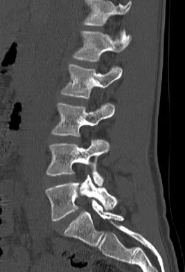
What is one of the principle concerns when a fracture such as the one seen in Figure 18 is encountered?


Explanation

The injury shown is a fracture-dislocation and it is highly unstable. In addition to this concern, spinal epidural hematomas have a much higher incidence in people with ankylosing spondylitis following knee fracture. It is felt to be due to disrupted epidural veins, with hypervascular epidural soft tissue in the setting of a rigid spinal canal. Patients with ankylosing spondylitis may have other significant comorbidities, especially cardiac and pulmonary, and these should be carefully assessed. Ludwig S, Zarro CM: Complications encountered in the management of patients with ankylosing spondylitis, in Vaccaro AR, Regan JJ, Crawford AH, et al (eds): Complications of Pediatric and Adult Spine Surgery. New York, NY, Marcel Dekker, 2004, pp 279-290.


Question 21

Retrograde ejaculation is most commonly associated with what surgical approach?


Explanation

Retrograde ejaculation is the sequela of an injury to the superior hypogastric plexus. This structure needs protection, especially during anterior exposure of the lumbosacral junction. Although the superior hypogastric plexus can be injured with anterior or anterolateral spine surgery at any lumbar level, it is most at risk with anterior transperitoneal approaches to the lumbosacral junction. To avoid this complication, the use of monopolar electrocautery should be avoided during deep dissection in this region. The ideal anterior exposure starts with blunt dissection just to the medial aspect of the left common iliac vein sweeping the prevertebral tissues toward the patient's right side. Flynn JC, Price CT: Sexual complications of anterior fusion of the lumbar spine. Spine 1984;9:489-492. Watkins RG (ed): Surgical Approaches to the Spine. New York, NY, Springer-Verlag, 1983, p 107.

Question 22

What nerve is most likely to be injured during the anterior exposure of C2-3?


Explanation

The hypoglossal nerve exits from the ansa cervicalis at approximately the C2-3 level and can be injured during retraction up to the C2 level. The superior laryngeal nerve lies at about C4-5. The facial nerve is much higher. The vagus nerve runs with the internal jugular and carotid much more laterally. The phrenic nerve exits posteriorly. Chang U, Lee MC, Kim DH: Anterior approach to the midcervical spine, in Kim DH, Henn JS, Vaccaro AR, et al (eds): Surgical Anatomy and Techniques to the Spine. Philadelphia, PA, Saunders Elsevier, 2006, pp 45-54.

Question 23

A 24-year-old man sustains the injury shown in Figures 19a through 19e in a paragliding accident. He is neurologically intact. He also sustained fractures of his left femur and right distal radius. Which of the following represents the best option for management of the spinal injury?


Explanation

The injury pattern is that of a burst fracture at L1 contiguous with a compression fracture at T12. There is associated kyphosis and slight spondylolisthesis of T12 on L1. Treatment of this type of burst fracture in neurologically intact patients is somewhat controversial, with at least one study demonstrating equal long-term results comparing nonsurgical treatment to surgical treatment. In this study, however, body casts were used initially in the nonsurgical group. Moreover, because this patient has multiple fractures, spinal fracture stabilization should be considered to facilitate early mobilization. Surgical stabilization and fusion via a posterior approach is the best treatment option in this patient. Anterior decompression is not necessary since the patient is neurologically intact. McLain RF, Benson DR: Urgent surgical stabilization of spinal fractures in polytrauma patients. Spine 1999;24:1646-1654. Wood K, Butterman G, Mehbod A, et al: Operative compared with nonoperative treatment of a thoracolumbar burst fracture without neurological deficit: A prospective, randomized study. J Bone Joint Surg Am 2003;85:773-781.


Question 24

An 82-year-old man is seen in consultation after being admitted for a fall from ground level. There was no loss of consciousness and the patient recalls striking his head and sustaining a hyperextension-type injury to the cervical spine. Examination reveals an 8-cm head laceration with only mild axial neck tenderness. He has generalized weakness throughout the upper extremities and maintained motor function of the lower extremities. There are no obvious sensory deficits, and the bulbocavernous reflex and deep tendon reflexes are maintained. What is the most appropriate diagnosis at this time?


Explanation

Incomplete cord syndromes have variable neurologic findings with partial loss of sensory and/or motor function below the level of injury. Incomplete cord syndromes include the anterior cord syndrome, the Brown-Séquard syndrome, central cord syndrome, and posterior cord syndrome. Central cord syndrome is characterized with greater motor weakness in the upper extremities than in the lower extremities. The pattern of motor weakness shows greater distal involvement in the affected extremity than proximal muscle weakness. Anterior cord syndrome involves a variable loss of motor function and pain and/or temperature sensation, with preservation of proprioception. The Brown-Séquard syndrome involves a relatively greater ipsilateral loss of proprioception and motor function, with contralateral loss of pain and temperature sensation. Posterior cord syndrome is a rare injury and is characterized by preservation of motor function, sense of pain, and light touch, with loss of proprioception and temperature sensation below the level of the lesion. Spinal shock is the period of time, usually 24 hours, after a spinal injury that is characterized by absent reflexes, flaccidity, and loss of sensation below the level of the injury. Penrod LE, Hegde SK, Ditunno JF: Age effect on prognosis for functional recovery in acute, traumatic central cord syndrome. Arch Phys Med Rehab 1990;71:963-968.

Question 25

Kyphosis from a vertebral osteoporotic compression fracture often results in progressive kyphosis due to


Explanation

Kayanja and associates, in a number of biomechanical studies, showed that in a kyphotic spine the strain is located at the apex of the deformity, the force is transmitted to the superior adjacent vertebrae, and that realignment and cement augmentation effectively normalize the load transfer. Kayanja MM, Ferrara LA, Lieberman IH: Distribution of anterior cortical shear strain after a thoracic wedge compression fracture. Spine J 2004;4:76-87. Kayanja MM, Togawa D, Lieberman IH: Biomechanical changes after the augmentation of experimental osteoporotic vertebral compression fractures in the cadaveric thoracic spine. Spine J 2005;5:55-63. Kayanja MM, Schlenk R, Togawa D, et al: The biomechanics of 1, 2, and 3 levels of vertebral augmentation with polymethylmethacrylate in multilevel spinal segments. Spine 2006;31:769-774.

Question 26

A 45-year-old male presents with right arm pain radiating to his thumb. He has weakness in wrist extension and an absent brachioradialis reflex. Which cervical nerve root is most likely affected?


Explanation

C6 radiculopathy typically presents with pain and numbness in the thumb, weakness in wrist extension and elbow flexion, and an absent or diminished brachioradialis reflex.

Question 27

A 55-year-old male presents with progressive clumsiness of his hands and difficulty walking. Examination shows hyperreflexia in the lower extremities, a positive Hoffmann sign bilaterally, and an inability to tandem walk. What is the most appropriate next step in management?


Explanation

The patient's clinical presentation is classic for cervical spondylotic myelopathy. An MRI of the cervical spine is the gold standard diagnostic step to evaluate for spinal cord compression and cord signal changes.

Question 28

A 25-year-old female is involved in a high-speed MVC. She is neurologically intact but complains of severe neck pain. CT scan shows a fracture through the pars interarticularis of C2 bilaterally with 2 mm of displacement and no angulation. What is the most appropriate initial management?


Explanation

This describes a Type I traumatic spondylolisthesis of the axis (Hangman's fracture), which features a pars fracture with less than 3 mm of translation and no angulation. It is a stable fracture pattern best managed non-operatively with a rigid cervical collar.

Question 29

An 80-year-old male with pre-existing cervical spondylosis falls forward and strikes his forehead. He presents with profound weakness in his bilateral upper extremities (1/5) and mild weakness in his lower extremities (4/5). Perianal sensation and rectal tone are intact. What is the most likely diagnosis?


Explanation

Central cord syndrome is the most common incomplete spinal cord injury, typically occurring after a hyperextension injury in older adults with cervical spondylosis. It classically causes greater motor impairment in the upper extremities compared to the lower extremities.

Question 30

A 40-year-old male complains of severe left anterior thigh pain. Examination reveals weakness in left knee extension and a decreased patellar reflex. Which of the following lumbar disc herniations is most likely responsible?


Explanation

A far lateral (extraforaminal) disc herniation at L4-L5 compresses the exiting L4 nerve root. L4 radiculopathy manifests with anterior thigh pain, quadriceps weakness, and an asymmetrical or diminished patellar reflex.

Question 31

What is the most common neurologic complication following cervical laminectomy and laminoplasty?


Explanation

C5 nerve root palsy is the most common neurologic complication following cervical decompression without fusion (such as laminoplasty), occurring in up to 10% of cases. It is believed to be caused by tethering of the C5 nerve root as the spinal cord drifts posteriorly.

Question 32

A 30-year-old unrestrained driver is involved in an MVC. CT of the lumbar spine shows a flexion-distraction injury (Chance fracture) at L1. Which of the following associated injuries must be carefully ruled out?


Explanation

Chance fractures are flexion-distraction injuries commonly associated with seatbelt use. Up to 50% of patients with these fractures have concomitant intra-abdominal injuries, most commonly involving hollow viscous organs like the small bowel.

Question 33

A 65-year-old man undergoes a C3-C7 posterior cervical laminectomy and fusion for cervical spondylotic myelopathy. On postoperative day 2, he develops profound weakness of the deltoid and biceps unilaterally, with no other neurologic deficits. Which of the following is the most likely etiology of this complication?


Explanation

C5 palsy is a known complication of posterior cervical decompression procedures, occurring in roughly 5-10% of cases. It is widely thought to be caused by tethering of the C5 nerve root due to the posterior drift of the spinal cord after decompression.

Question 34

A 45-year-old man presents with severe sharp right-leg pain radiating to the anterior thigh and medial leg. Examination reveals 4/5 strength in right knee extension and a diminished patellar reflex. MRI of the lumbar spine reveals a far-lateral disc herniation at L4-L5. Which nerve root is most likely compressed?


Explanation

In the lumbar spine, a far-lateral (extraforaminal) disc herniation compresses the exiting nerve root at that level. Therefore, an L4-L5 far-lateral disc herniation compresses the L4 nerve root, leading to anterior thigh pain and quadriceps weakness.

Question 35

A 72-year-old woman with a history of cervical stenosis sustains a hyperextension injury to her neck in a motor vehicle collision. She complains of severe weakness in her bilateral upper extremities and mild weakness in her lower extremities. Proprioception and sensation are partially preserved. What is the most likely diagnosis?


Explanation

Central cord syndrome typically occurs after a hyperextension injury in a patient with pre-existing cervical spondylosis. It classically presents with motor deficits that are more severe in the upper extremities than in the lower extremities.

Question 36

A 24-year-old man wearing a lap belt is involved in a high-speed motor vehicle collision. Radiographs demonstrate a flexion-distraction injury (Chance fracture) at L1. Which of the following associated injuries is most critical to rule out in this patient?


Explanation

Chance fractures (flexion-distraction injuries) are highly associated with intra-abdominal injuries, particularly to hollow viscous organs like the bowel, due to the acute compression from the lap belt. A thorough abdominal evaluation is mandatory.

Question 37

Traumatic spondylolisthesis of the axis (Hangman's fracture) is typically caused by which of the following mechanisms of injury?


Explanation

A Hangman's fracture involves bilateral pars interarticularis fractures of C2. In modern scenarios like motor vehicle collisions, it is most commonly caused by hyperextension combined with axial loading.

Question 38

A 78-year-old woman falls and sustains a Type II odontoid fracture. Which of the following factors is most strongly associated with an increased risk of nonunion for this injury?


Explanation

Risk factors for nonunion of a Type II odontoid fracture include age greater than 50 years, initial displacement greater than 5 mm, posterior displacement, and significant angulation.

Question 39

A 32-year-old man dives into a shallow pool and sustains a Jefferson burst fracture of C1. On the open-mouth odontoid view, the sum of the lateral mass displacement of C1 on C2 is measured. A transverse ligament rupture is highly suspected if this combined displacement exceeds which of the following thresholds?


Explanation

The Rule of Spence states that a combined lateral mass overhang of C1 on C2 of greater than 6.9 mm on an AP open-mouth radiograph indicates a high likelihood of rupture of the transverse alar ligament.

Question 40

A 28-year-old man sustains a stab wound to the midthoracic spine. He exhibits loss of motor function and proprioception in his right lower extremity, and loss of pain and temperature sensation in his left lower extremity. Which of the following incomplete spinal cord syndromes does he have?


Explanation

Brown-Séquard syndrome results from spinal cord hemisection, characterized by ipsilateral loss of motor function and proprioception, and contralateral loss of pain and temperature sensation. It carries the best prognosis for ambulation among incomplete spinal cord injuries.

Question 41

A 35-year-old unrestrained driver is involved in a head-on collision. Lateral cervical spine radiographs show a C5 on C6 translation of approximately 60%. What is the most likely mechanism of this injury?


Explanation

Bilateral facet dislocations are typically caused by severe hyperflexion injuries. They present with greater than 50% anterior subluxation of the vertebral body on the lateral radiograph.

Question 42

A 68-year-old man presents with bilateral leg and buttock pain that worsens with walking. Which of the following clinical features most strongly differentiates neurogenic claudication from vascular claudication?


Explanation

Neurogenic claudication is typically relieved by lumbar flexion, such as leaning on a shopping cart or sitting, because flexion increases the cross-sectional area of the spinal canal. Vascular claudication is typically relieved simply by resting or standing still.

Question 43

A 42-year-old man presents with sudden onset of severe lower back pain, bilateral sciatica, saddle anesthesia, and urinary retention. Which of the following is the most appropriate next step in management?


Explanation

This patient presents with classic signs of cauda equina syndrome. This is a surgical emergency requiring an urgent MRI followed by emergent surgical decompression to prevent permanent neurologic deficits, particularly bladder and bowel dysfunction.

Question 44

A 55-year-old man with a long history of Ankylosing Spondylitis sustains a minor fall. He complains of severe neck pain without neurologic deficits. Radiographs appear unchanged from his baseline showing a "bamboo spine." What is the most appropriate next step?


Explanation

Patients with ankylosing spondylitis have a rigid, osteoporotic spine and are at high risk for highly unstable fractures even after minor trauma. If a fracture is suspected and plain films are negative or obscured, a CT scan of the spine is mandatory to rule out an occult fracture.

Question 45

According to the Thoracolumbar Injury Classification and Severity (TLICS) score, which of the following factors is given the highest point value when determining the need for surgical stabilization?


Explanation

In the TLICS system, an unequivocally disrupted posterior ligamentous complex (PLC) is assigned 3 points. Incomplete spinal cord injury is also assigned 3 points. Surgical management is generally indicated for a total score of 5 or more.

Question 46

A 25-year-old man is brought to the trauma bay after a high-speed motorcycle accident. Lateral cervical spine radiographs show a basion-dens interval of 14 mm. Which of the following is the most appropriate definitive management for this injury?


Explanation

A basion-dens interval >12 mm indicates atlanto-occipital dissociation (AOD). AOD is a highly unstable ligamentous injury with a high mortality rate. Definitive treatment is occipitocervical fusion, as halo immobilization is insufficient for this level of instability.

Question 47

A 50-year-old woman presents with neck pain radiating down her right arm. Physical examination reveals weakness in wrist extension and decreased sensation over the dorsal aspect of the thumb and index finger. The brachioradialis reflex is diminished. Which cervical disc herniation is most likely responsible?


Explanation

A C5-C6 disc herniation compresses the C6 nerve root. C6 radiculopathy typically presents with weakness in wrist extension, diminished brachioradialis reflex, and sensory deficits in the thumb and index finger.

Question 48

A patient presents with triceps weakness, absent triceps reflex, and numbness in the middle finger. Which nerve root is affected?


Explanation

C7 radiculopathy, typically caused by a C6-C7 disc herniation, presents with triceps weakness, wrist flexion weakness, diminished triceps reflex, and sensory changes in the middle finger.

Question 49

A 65-year-old man presents with stiffness in his mid-back and mild dysphagia. Radiographs of the thoracic spine demonstrate flowing ossification along the anterolateral aspect of five contiguous vertebral bodies. The disc heights are preserved, and the sacroiliac joints are normal. What is the most likely diagnosis?


Explanation

DISH is characterized by flowing anterolateral ossification of at least four contiguous vertebral bodies with preserved disc spaces and an absence of sacroiliitis. Dysphagia can occur due to prominent anterior cervical osteophytes.

Question 50

A 65-year-old man presents with bilateral upper extremity weakness and numbness following a hyperextension injury to his neck. His lower extremity strength is nearly normal. MRI of the cervical spine demonstrates central cord edema with multilevel congenital stenosis but no fracture or ligamentous instability. What is the most appropriate initial management?


Explanation

This patient has Central Cord Syndrome. In the absence of acute fracture or instability, current guidelines recommend conservative management with hemodynamic optimization (MAP > 85 mmHg for 5-7 days) to ensure adequate spinal cord perfusion.

Question 51

A 45-year-old man complains of severe right anterior thigh pain and weakness in knee extension. He has no significant back pain. Examination reveals a diminished right patellar reflex and decreased sensation over the anterior thigh. MRI shows a far lateral disc herniation. At which lumbar level is this herniation most likely located?


Explanation

A far lateral disc herniation compresses the exiting nerve root at the same level. An L3-L4 far lateral disc herniation compresses the L3 nerve root, resulting in anterior thigh pain, quadriceps weakness, and a diminished patellar reflex.

Question 52

An 82-year-old man with a history of falls presents with neck pain. CT of the cervical spine reveals a Type II odontoid fracture with 2 mm of posterior displacement. He is neurologically intact. What is the most appropriate management?


Explanation

In elderly patients with Type II odontoid fractures and minimal displacement (< 5 mm), a rigid cervical collar is the preferred treatment. Surgery has higher perioperative risks, and halo vests carry high morbidity and mortality in the elderly.

Question 53

A 55-year-old man with long-standing ankylosing spondylitis presents to the emergency department with acute back pain after tripping and falling on level ground. Plain radiographs of the thoracic and lumbar spine are inconclusive. What is the most appropriate next step in management?


Explanation

Patients with ankylosing spondylitis have a rigid, osteopenic spine making them highly susceptible to unstable fractures even from minor trauma. Advanced imaging (CT or MRI) of the entire spine is mandatory as occult or non-contiguous fractures are common.

Question 54

A 30-year-old man is brought to the trauma bay following a motor vehicle collision. He is awake, alert, and cooperative but has no motor or sensory function below the C5 level (ASIA A). Imaging shows a bilateral facet dislocation at C5-C6. What is the next best step in management?


Explanation

In an awake and testable patient with a cervical facet dislocation and a neurologic deficit, urgent closed cranial traction for reduction is indicated to rapidly decompress the spinal cord. MRI should not delay closed reduction in an awake patient.

Question 55

A 25-year-old woman falls from a height and sustains an L1 burst fracture. CT shows 40% retropulsion into the spinal canal. She is neurologically intact, and MRI confirms the posterior ligamentous complex is intact. What is the most appropriate management?


Explanation

Neurologically intact patients with a thoracolumbar burst fracture and an intact posterior ligamentous complex (PLC) have a stable injury pattern. They are best managed non-operatively with an orthosis and early mobilization.

Question 56

A 65-year-old man complains of bilateral calf, thigh, and buttock pain that worsens with walking and improves when he leans forward over a shopping cart. Lower extremity pulses are normal. Which of the following physical examination findings is most characteristic of his condition?


Explanation

This patient has neurogenic claudication secondary to lumbar spinal stenosis. Symptoms are characteristically exacerbated by lumbar extension, which further decreases the cross-sectional area of the spinal canal.

Question 57

A 50-year-old man of East Asian descent presents with progressive clumsiness in his hands and a broad-based gait. CT of the cervical spine shows continuous ossification of the posterior longitudinal ligament (OPLL) from C3 to C6. On sagittal imaging, the OPLL mass crosses the "K-line" (a negative K-line). What is the recommended surgical approach?


Explanation

A negative K-line indicates that the OPLL mass is large and kyphotic, preventing the spinal cord from shifting posteriorly after a posterior decompression. Therefore, an anterior approach (or combined anterior-posterior) is required for adequate decompression.

Question 58

A 40-year-old man underwent an L4-L5 microdiscectomy 2 years ago with excellent relief of his right leg pain. He now presents with a 6-week history of recurrent right L5 radicular pain that has failed conservative therapy. MRI shows a recurrent L4-L5 right paracentral disc herniation. He has minimal back pain and no instability on dynamic radiographs. What is the recommended surgical intervention?


Explanation

For a first-time recurrent lumbar disc herniation with isolated radiculopathy and no mechanical instability or significant back pain, a revision microdiscectomy is indicated. It provides similar outcomes to fusion with lower morbidity.

Question 59

A 22-year-old man wearing a lap-only seatbelt is involved in a high-speed motor vehicle collision. Imaging reveals an L2 fracture extending transversely through the vertebral body, pedicles, and spinous process. He is neurologically intact. What is the most appropriate management?


Explanation

This is a purely bony Chance fracture (flexion-distraction injury). Because bony injuries have excellent healing potential, this can be successfully managed non-operatively in a hyperextension orthosis (TLSO) to reduce the fracture.

Question 60

A 68-year-old woman with neurogenic claudication and an L4-L5 Grade 1 degenerative spondylolisthesis has failed non-operative treatment. What is the primary advantage of performing a lumbar decompression with fusion rather than a decompression alone?


Explanation

Adding fusion to a decompression in the setting of degenerative spondylolisthesis prevents postoperative slip progression. It significantly decreases the long-term need for revision surgery due to secondary instability.

Question 61

A 55-year-old male intravenous drug user presents with severe back pain, subjective fevers, and progressive paraparesis. MRI reveals a large posterior spinal epidural abscess at T8-T10 causing severe cord compression. What is the most appropriate immediate management?


Explanation

A spinal epidural abscess causing neurologic deficits is a surgical emergency. Because the abscess is located posteriorly in the thoracic spine, an emergent posterior laminectomy with debridement is the standard of care.

Question 62

A 30-year-old man sustains a C1 burst fracture. An open-mouth odontoid radiograph demonstrates that the lateral masses of C1 overhang the lateral masses of C2 by a combined total of 8 mm. What does this finding most likely indicate?


Explanation

According to the Rule of Spence, a combined lateral mass overhang of 6.9 mm or greater (often cited as 7 mm on standard X-rays due to magnification) indicates incompetence or rupture of the transverse ligament.

Question 63

A 45-year-old woman presents with pain radiating down her posterior arm into her middle finger. Physical examination reveals weakness in triceps extension and wrist flexion, with a diminished triceps reflex. Which nerve root is most likely compressed?


Explanation

C7 radiculopathy is the most common cervical radiculopathy. It presents with pain radiating to the middle finger, a diminished triceps reflex, and weakness in the triceps and wrist flexors.

Question 64

A patient is evaluated for a traumatic spondylolisthesis of the axis (Hangman's fracture). Imaging shows significant angulation with minimal translation, and widening of the posterior C2-C3 disc space. According to the Levine-Edwards classification, what treatment modality is absolutely contraindicated in the initial management?


Explanation

This describes a Levine-Edwards Type IIA Hangman's fracture, which is primarily a flexion-distraction injury. Application of longitudinal traction will exacerbate the deformity and is strictly contraindicated.

Question 65

A 42-year-old man presents with acute onset urinary retention, saddle anesthesia, and bilateral sciatica. MRI confirms a massive L4-L5 disc herniation compressing the cauda equina. To maximize the chance of complete recovery of bowel and bladder function, surgical decompression should ideally be performed within what maximum timeframe from symptom onset?


Explanation

Cauda equina syndrome is a surgical emergency. Evidence indicates that surgical decompression performed within 48 hours of symptom onset yields the best outcomes for recovery of motor and sphincter function.

Question 66

A 28-year-old woman was rear-ended in a motor vehicle collision. She has posterior neck pain without neurologic deficits. CT of the cervical spine is normal, and dynamic flexion-extension views show no instability. Which of the following is the most appropriate management?


Explanation

For a cervical sprain (whiplash) without structural instability or neurologic deficits, the best evidence supports early mobilization, active range of motion, and physical therapy to optimize recovery and prevent chronic pain.

Question 67

A 35-year-old man presents with severe right-sided back and leg pain extending to the lateral aspect of his right foot. Examination reveals profound weakness in right plantar flexion and an absent right Achilles reflex. Which disc herniation pattern is most likely responsible for his symptoms?


Explanation

An L5-S1 paracentral disc herniation compresses the traversing S1 nerve root. S1 radiculopathy classically presents with pain radiating to the lateral foot, weakness in ankle plantar flexion, and an absent Achilles reflex.

Question 68

Which of the following physical examination findings is highly specific for spinal cord compression localizing to the C5-C6 level rather than a higher cervical level?


Explanation

The inverted brachioradialis reflex indicates a lower motor neuron lesion at C5-C6 (absent brachioradialis response) combined with an upper motor neuron lesion below this level (hyperactive finger flexion). It is highly specific for C5-C6 myelopathy.

Question 69

A 25-year-old woman is brought in after falling from a third-story balcony. Imaging reveals a U-shaped sacral fracture with severe focal kyphosis and bilateral S1 nerve root deficits. Which surgical technique is most appropriate to stabilize this spinopelvic dissociation?


Explanation

U-shaped sacral fractures represent a severe form of spinopelvic dissociation. They are highly unstable and typically require robust lumbopelvic fixation (connecting the lower lumbar spine to the ilium) to restore alignment and permit mobilization.

Question 70

A 45-year-old man presents with severe right leg pain. Examination reveals weakness in knee extension and an absent patellar reflex. Sensation is decreased over the medial aspect of the lower leg. An MRI demonstrates a far-lateral disc herniation at L4-L5. Which nerve root is most likely compressed?


Explanation

In the lumbar spine, a far-lateral (extraforaminal) disc herniation compresses the exiting nerve root at that level. At L4-L5, the L4 nerve root exits the foramen and is affected, causing quadriceps weakness, an absent patellar reflex, and medial leg numbness.

Question 71

A 25-year-old man arrives at the trauma bay after a diving accident. He is awake, alert, and cooperative, but exhibits 0/5 motor strength in his lower extremities, bilateral hand weakness, and absent sensation below the clavicles. Lateral cervical spine radiographs demonstrate an anterolisthesis of C5 on C6 of approximately 50%. What is the most appropriate next step in management?


Explanation

In an awake, alert, and cooperative patient with a cervical dislocation and a neurologic deficit, urgent closed reduction via skeletal traction is indicated without obtaining an MRI first. An MRI is required before reduction only if the patient is obtunded or cannot participate in a reliable clinical exam.

Question 72

A 22-year-old woman is involved in a high-speed motor vehicle collision while wearing a lap belt. She complains of severe back pain. Radiographs reveal a flexion-distraction injury (Chance fracture) at L2. Which of the following associated injuries must be carefully evaluated for in this patient?


Explanation

Chance fractures are flexion-distraction injuries commonly caused by lap seatbelts acting as a fulcrum. They have a high association (up to 50%) with intra-abdominal injuries, particularly to hollow viscus organs such as the small bowel.

Question 73

A 65-year-old man undergoes a C3-C6 cervical laminectomy and instrumented fusion for severe cervical spondylotic myelopathy. Postoperatively, he develops profound weakness of the bilateral deltoid and biceps muscles (1/5 strength) but maintains full strength in his hands and lower extremities. What is the most likely etiology of this complication?


Explanation

C5 nerve root palsy is a known complication of posterior cervical decompression, due to the posterior drift of the spinal cord putting traction on the short, tethered C5 roots. Most cases resolve spontaneously within 6 to 12 months.

Question 74

An 80-year-old man falls and strikes his chin, hyperextending his neck. He presents with 2/5 strength in his upper extremities and 4/5 strength in his lower extremities. Sensation is decreased in the hands. Reflexes are brisk in the lower extremities. Which of the following is the most accurate prognostic statement regarding his neurologic recovery?


Explanation

The patient has central cord syndrome, characterized by greater weakness in the upper extremities than the lower extremities. The typical pattern of recovery is lower extremities first, followed by proximal upper extremities, with distal hand intrinsic function often remaining permanently impaired.

Question 75

A 75-year-old woman falls from a standing height and sustains a Type II odontoid fracture with 3 mm of posterior displacement. She is neurologically intact. Given her age, which of the following treatment options is associated with the highest risk of mortality and severe morbidity?


Explanation

In elderly patients (especially those >65 years), halo vest immobilization for odontoid fractures is poorly tolerated and associated with high rates of severe complications, including pneumonia, respiratory failure, and increased mortality.

Question 76

A 42-year-old man complains of right neck and arm pain. Physical examination reveals weakness in wrist extension, a diminished brachioradialis reflex, and numbness over the dorsal thumb and index finger. Which cervical nerve root is most likely compressed?


Explanation

Compression of the C6 nerve root presents with weakness in wrist extension and elbow flexion, a diminished brachioradialis reflex, and sensory changes in the thumb and index finger.

Question 77

During an anterior cervical discectomy and fusion (ACDF) at C6-C7 using a right-sided approach, the patient subsequently develops postoperative hoarseness. Which anatomical characteristic of the recurrent laryngeal nerve (RLN) makes it more susceptible to injury on the right side compared to the left?


Explanation

The right recurrent laryngeal nerve loops under the right subclavian artery and ascends obliquely into the neck, making its course variable and more susceptible to injury during a right-sided approach. The left RLN loops under the aortic arch and runs safely in the tracheoesophageal groove.

Question 78

A 55-year-old diabetic man presents with severe mid-back pain, low-grade fevers, and progressive bilateral leg weakness over the past 48 hours. He has a history of recent intravenous drug use. MRI reveals a large dorsal epidural mass at T8 with cord compression, hyperintense on T2 and showing peripheral enhancement. What is the most appropriate management?


Explanation

The patient presents with a spinal epidural abscess causing a progressive neurologic deficit. Emergent surgical decompression and debridement, along with culture-directed antibiotics, are indicated to prevent irreversible spinal cord injury.

Question 79

A 30-year-old man is involved in a motor vehicle collision and sustains a traumatic spondylolisthesis of the axis (Hangman's fracture). Radiographs reveal severe angulation of C2 on C3 with minimal anterior translation. The disc space is widened posteriorly. Which of the following treatments is strictly contraindicated?


Explanation

This describes a Levine and Edwards Type IIa Hangman's fracture, resulting from a flexion-distraction mechanism. Cervical traction is strictly contraindicated as it will further distract the fracture and exacerbate the instability.

Question 80

A 68-year-old man presents with bilateral leg cramping and pain that worsens with walking. Which of the following historical features most strongly suggests neurogenic claudication rather than vascular claudication?


Explanation

Neurogenic claudication (due to lumbar spinal stenosis) is exacerbated by spinal extension and relieved by flexion. Walking uphill flexes the spine and increases the canal area, relieving symptoms, whereas walking downhill extends the spine and worsens symptoms.

Question 81

A 10-year-old boy is struck by a car and presents intubated in the emergency department. Lateral cervical spine radiographs show a Basion-Dental Interval (BDI) of 14 mm. What is the definitive treatment for this injury?


Explanation

A Basion-Dental Interval (BDI) greater than 12 mm indicates atlanto-occipital dissociation (AOD), a highly unstable ligamentous injury. The definitive treatment for AOD is occipitocervical fusion, as ligamentous healing is predictably poor.

Question 82

A 40-year-old patient falls from a height and sustains an L1 burst fracture. Which of the following radiographic findings most strongly indicates a complete disruption of the posterior ligamentous complex (PLC), warranting operative stabilization?


Explanation

Widening of the interspinous distance (splaying of the spinous processes) on AP radiographs or sagittal MRI indicates disruption of the posterior ligamentous complex (PLC). PLC disruption in a burst fracture renders the spine mechanically unstable and is a strong indication for surgery.

Question 83

A 38-year-old woman presents with severe low back pain radiating down the posterior aspect of her left calf to the plantar surface of her foot. She has weakness in ankle plantarflexion and an absent Achilles reflex. An MRI demonstrates a typical paracentral disc herniation at the L5-S1 level. Which nerve root is being compressed?


Explanation

In the lumbar spine, a typical paracentral disc herniation at L5-S1 compresses the traversing S1 nerve root. This results in S1 radiculopathy, characterized by posterior leg pain, weakness in ankle plantarflexion, and a diminished or absent Achilles reflex.

Question 84

A 62-year-old man undergoes a C3-C6 laminectomy and instrumented fusion for cervical spondylotic myelopathy. On postoperative day 2, he develops isolated profound weakness in shoulder abduction and elbow flexion bilaterally. What is the most widely accepted pathophysiologic mechanism for this complication?


Explanation

C5 palsy after posterior cervical decompression is most commonly attributed to the posterior drift of the spinal cord, which tethers the relatively short C5 nerve roots. It presents as deltoid and biceps weakness.

Question 85

A 45-year-old man presents with severe right leg pain, numbness over the medial aspect of his calf, and weakness in knee extension. An MRI of the lumbar spine reveals a far lateral (extraforaminal) disc herniation at the L4-L5 level. Which nerve root is most likely compressed?


Explanation

A far lateral (extraforaminal) disc herniation compresses the exiting nerve root at the same level. Therefore, an L4-L5 far lateral disc compresses the L4 nerve root.

Question 86

A 28-year-old woman is involved in a motor vehicle collision. Radiographs and CT scans reveal a unilateral perched facet at C5-C6. What is the primary mechanism of injury for this specific pathology?


Explanation

Unilateral facet dislocations occur via a flexion-distraction mechanism combined with rotation. This leads to disruption of the posterior ligamentous complex and joint capsule on one side.

Question 87

A 58-year-old woman presents with progressive gait instability and hand clumsiness. Examination reveals positive Hoffman's signs bilaterally. MRI demonstrates multi-level cervical spondylotic myelopathy from C3-C6 with a fixed, rigid 15-degree cervical kyphosis. What is the most appropriate surgical management?


Explanation

In cervical spondylotic myelopathy with a rigid, kyphotic deformity, an anterior approach is required to decompress the cord and correct sagittal alignment. Posterior decompression alone is contraindicated in rigid kyphosis as the cord remains draped over the anterior pathology.

Question 88

A 42-year-old man presents to the emergency department with severe acute low back pain and bilateral sciatica following heavy lifting. Which of the following is typically the earliest clinical sign or symptom of cauda equina syndrome in this setting?


Explanation

Urinary retention is generally the most consistent and earliest sign of cauda equina syndrome. A post-void residual should be checked immediately if this is suspected.

Question 89

A 22-year-old man wearing a lap seatbelt is involved in a frontal motor vehicle collision. He has severe focal back pain but is neurologically intact. CT imaging shows a fracture extending horizontally through the spinous process, pedicles, and vertebral body of L1. What is the most appropriate definitive management?


Explanation

This is a bony Chance fracture (flexion-distraction injury). Because it is purely bony and the patient is neurologically intact, it has a high rate of healing when treated conservatively with an extension orthosis.

Question 90

A 60-year-old man presents with myelopathy. CT scan shows continuous ossification of the posterior longitudinal ligament (OPLL) from C3 to C5. If an anterior cervical corpectomy is planned, the patient is at highest risk for which of the following intraoperative complications?


Explanation

OPLL frequently involves ossification that merges with or penetrates the underlying dura. Surgical resection via an anterior approach carries a high risk of dural tearing and CSF leak.

Question 91

A 68-year-old woman returns to the clinic 4 years after undergoing a successful L4-L5 posterior lumbar interbody fusion. She now complains of new-onset neurogenic claudication. MRI demonstrates severe central canal stenosis at a new level. Which level is most commonly affected by symptomatic adjacent segment disease in this scenario?


Explanation

Symptomatic adjacent segment disease following a lumbar fusion most commonly occurs at the level immediately cephalad to the fusion. For an L4-L5 fusion, this is the L3-L4 level.

Question 92

An 82-year-old man presents with neck pain after a ground-level fall. CT scan reveals a displaced Type II odontoid fracture. He is neurologically intact. Given his age, what is the most appropriate management to minimize mortality and morbidity?


Explanation

Type II odontoid fractures in the elderly have a high rate of nonunion, and halo vest immobilization is associated with high mortality. Posterior C1-C2 instrumented fusion provides definitive stabilization with better outcomes.

Question 93

A 45-year-old woman undergoes an anterior cervical discectomy and fusion at C6-C7. Postoperatively, she is noted to have unilateral ptosis, miosis, and anhidrosis on the surgical side. Which structure was most likely injured during the surgical exposure?


Explanation

Horner's syndrome (ptosis, miosis, anhidrosis) results from injury to the cervical sympathetic chain. This structure is at risk during ACDF when dissection extends too far laterally over the longus colli muscles.

Question 94

A 72-year-old man with a history of cervical stenosis falls forward and strikes his chin, forcefully hyperextending his neck. He presents with severe motor weakness in his hands and arms, but is able to walk with only mild difficulty. What is the most likely diagnosis?


Explanation

Central cord syndrome classically occurs in older patients with pre-existing cervical stenosis who sustain a hyperextension injury. It presents with disproportionately greater motor impairment in the upper extremities.

Question 95

A 30-year-old man sustains an axial loading injury, resulting in a Jefferson burst fracture of C1. Which radiographic measurement on the open-mouth odontoid view best indicates a rupture of the transverse atlantal ligament?


Explanation

In a Jefferson fracture, an aggregate displacement of the C1 lateral masses on C2 greater than 6.9 mm indicates a rupture of the transverse atlantal ligament, rendering the injury highly unstable.

Question 96

A 50-year-old man presents with right-sided neck pain radiating down his arm. Physical examination reveals weakness in wrist extension, a diminished brachioradialis reflex, and decreased sensation over his thumb and index finger. A herniated disc at which cervical level is most likely responsible?


Explanation

The clinical presentation describes a C6 radiculopathy. This is most commonly caused by a disc herniation at the C5-C6 level compressing the exiting C6 nerve root.

Question 97

A 65-year-old woman presents with neurogenic claudication. Imaging reveals a Grade I degenerative spondylolisthesis. What is the most common anatomic level for this specific pathology to occur?


Explanation

Degenerative spondylolisthesis occurs most frequently at the L4-L5 level. This is in contrast to isthmic spondylolisthesis, which most commonly affects the L5-S1 level.

None

Clinic OS
Medically Verified Content by
Prof. Clinic OS
Consultant Orthopedic & Spine Surgeon
Chapter Index