العربية
Part of the Master Guide

AAOS Orthopedic MCQs (Set 1): Spine Disorders, Sports Medicine & Arthroplasty | 2026 Board Review

Pediatric Orthopedic MCQs (Set 4): DDH, SCFE, & Scoliosis for ABOS/OITE Exams

23 Apr 2026 64 min read 103 Views
Pediatrics 2007 MCQs - Part 4

Key Takeaway

This high-yield question set (Set 4) for AAOS, ABOS, and OITE exams focuses on critical pediatric orthopedic conditions. Questions cover the diagnosis, classification, and management of Developmental Dysplasia of the Hip, Slipped Capital Femoral Epiphysis, and common pediatric spine deformities such as scoliosis, preparing you for board success.

Pediatric Orthopedic MCQs (Set 4): DDH, SCFE, & Scoliosis for ABOS/OITE Exams

Comprehensive 100-Question Exam


00:00

Start Quiz

Question 1

Figure 33 shows the oblique radiograph of an 11-year-old boy who has a mild left flatfoot deformity. Examination reveals that subtalar motion is limited and painful. Despite casting for 6 weeks, the patient reports foot pain that limits participation in sport activities. A CT scan shows no subtalar joint abnormalities. Management should now include


Explanation

The radiograph shows an incompletely ossified calcaneonavicular coalition. When symptomatic, a trial of cast immobilization is reasonable. If this fails to provide relief, the preferred treatment is resection of the coalition. Before attempting surgery, a CT scan should be obtained to rule out ipsilateral subtalar coalition. Recurrence of the coalition is usually prevented with interposition of autogenous fat graft or with local interposition of the extensor digitorum brevis muscle. Approximately 80% of patients treated in this manner have decreased pain and improved subtalar motion. When the flatfoot deformity is mild, calcaneal lengthening or medial translation osteotomy is unnecessary. Primary triple arthrodesis may be indicated if degenerative changes are present in the subtalar or midfoot joints. Peroneal lengthening has been described for treatment of the peroneal spastic flatfoot without demonstrable tarsal coalition. Gonzalez P, Kumar SJ: Calcaneonavicular coalition treated by resection and interposition of the extensor digitorum brevis muscle. J Bone Joint Surg Am 1990;72:71-77. Vincent KA: Tarsal coalition and painful flatfoot. J Am Acad Orthop Surg 1998;6:274-281.

Question 2

A nonambulatory verbal 6-year-old child with spastic quadriplegic cerebral palsy has progressive bilateral hip subluxation of more than 50%. There is no pain with range of motion, but abduction is limited to 20 degrees maximum. An AP radiograph is seen in Figure 34. Management should consist of


Explanation

The natural history of the patient's hips, if left untreated, is gradual progression to dislocation. To prevent future pain, prevention of dislocation is often helpful. The patient is too old for soft-tissue releases alone. Therefore, the treatment of choice is medial release of both hips to obtain 45 degrees or better of hip abduction in conjunction with psoas tenotomy and bilateral femoral varus osteotomies. Presedo A, Oh CW, Dabney KY, et al: Soft-tissue releases to treat spastic hip subluxation in children with cerebral palsy. J Bone Joint Surg Am 2005;87:832-841.


Question 3

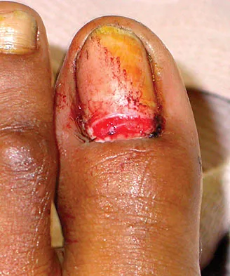
Figures 35a through 35c show the clinical photograph and radiographs of a 15-year-old boy who stubbed his toe 1 day ago while walking barefoot in the yard. Management should consist of


Explanation

The patient has an open fracture of the physis of the distal phalanx with a portion of the nail bed interposed in the physis. Seymour initially described this injury in the distal phalanges of fingers. Optimal treatment consists of removing the interposed tissue, irrigating the fracture, and a short course of antibiotics. The nail should be preserved to provide stability. Kensinger DR, Guille JT, Horn BD, et al: The stubbed great toe: Importance of early recognition and treatment of open fractures of the distal phalanx. J Pediatr Orthop 2001;21:31-34. Pinckney LE, Currarino G, Kennedy LA: The stubbed great toe: A cause of occult compound fracture and infection. Radiology 1981;138:375-377.


Question 4

Figure 36 shows the radiograph of a 14-year-old boy who has been treated in the past for Perthes' disease with an abduction brace. He now has hip pain that limits his activity, and nonsteroidal anti-inflammatory drugs have failed to provide relief. What is the most appropriate treatment?


Explanation

Several authors have reported good success in relieving pain with shelf acetabuloplasty. This patient's Perthes' disease is in the healed phase; therefore, proximal femoral varus and Salter innominate osteotomies aimed at improving containment are not indicated. The medial one half of the patient's femoral head is markedly deformed, and rotating it into a weight-bearing position with proximal femoral valgus osteotomy is unlikely to relieve pain. Hip arthrodesis can always be performed as a salvage procedure if the shelf acetabuloplasty fails. Daly K, Bruce C, Catterall A: Lateral shelf acetabuloplasty in Perthes' disease: A review of the end of growth. J Bone Joint Surg Br 1999;81:380-384.


Question 5

A newborn girl is referred for evaluation of suspected hip instability. What information from her history would place her in the highest risk category?


Explanation

Breech positioning has been noted as the risk factor that most increases the relative risk of developmental dysplasia of the hip in multiple series and meta-analysis. All the other factors also increase the risk but to a lesser magnitude. Lehmann HP, Hinton R, Morello P, et al: Developmental dysplasia of the hip practice guideline: Technical report. Committee on Quality Improvement, and Subcommittee on Developmental Dysplasia of the Hip. Pediatrics 2000;105:E57.

Question 6

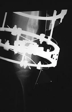
A teenager is undergoing a correction of deformity and lengthening of the femur. Distractions are proceeding as expected; however, during his 6-week follow-up examination, the patient reports that the distraction motors have become harder to turn over for the past 2 to 3 days. Figures 37a and 37b show current radiographs. What is the most likely complication being encountered?


Explanation

Premature consolidation is a complication that is unique to gradual bone lengthening after corticotomy. Causes include excessive latency period, inadequate distraction rate, exuberant bone formation, patient compliance problems, and mechanical failure of the distraction apparatus. The femur and fibula are most commonly involved. This patient did not have an incomplete corticotomy, as initial distraction occurred before the distraction device was noted to seize up. The radiographs show bowing of the Ilizarov wires and mature regenerate bone, both suggestive of premature consolidation. No wire breakage or joint subluxation is seen on the radiographs. Treatment for premature consolidation includes continuing distraction until the consolidation bridge ruptures, or additional surgery may include closed rotational osteoclasis or repeat corticotomy. Paley D: Problems, obstacles and complications of limb lengthening, in Maiocchi AB, Aronson J (eds): Operative Principles of Ilizarov. Baltimore, MD, Williams & Wilkins, 1991, p 360.


Question 7

A 4-year-old child was born with bilateral congenital radial clubhands. Which of the following associated conditions is a contraindication to centralization of the hands on the ulna?


Explanation

Patients born with bilateral radial clubhands may have difficulty getting their hands to their mouth. The centralization procedure would take away that ability if there is a lack of elbow flexion. Green DP, Hotchkiss RN, Pederson WC: Green's Operative Hand Surgery, ed 4. New York, NY, Churchill Livingstone, 1999, pp 344-349.

Question 8

A 6-year-old Little League pitcher has had pain in the right elbow for the past 2 weeks. Examination reveals mild lateral elbow joint tenderness with full range of motion and no effusion or collateral laxity. A radiograph is shown in Figure 38. Initial management should consist of


Explanation

The radiograph shows osteochondritis dissecans (OCD) of the capitellum, one manifestation of "pitcher's elbow." The lesion is nondisplaced, and healing is possible if the inciting throwing activities are curtailed. Long arm cast treatment may be reasonable for the noncompliant patient but should not exceed 6 weeks duration. Surgical treatment is indicated for loose bodies or cartilage flaps. Elbow OCD lesions are now being seen in younger children as more participate in organized sports, especially baseball and gymnastics. Bauer M, Jonsson K, Josefsson PO, et al: Osteochondritis dissecans of the elbow: A long-term follow-up study. Clin Orthop 1992;284:156-160. Takahara M, Ogino T, Sasaki I, et al: Long term outcome of osteochondritis dissecans of the humeral capitellum. Clin Orthop 1999;363:108-115.


Question 9

The parents of a 10-year-old boy with Down syndrome are seeking sports clearance for participation in the high jump at the Special Olympics. He is asymptomatic, and the neurologic examination is normal. The hips and patellae are clinically stable. Radiographs of the cervical spine in flexion and extension show a maximum atlanto-dens interval (ADI) of 6 mm. Based on these findings, what recommendation should be made?


Explanation

In approximately 15% of children with Down syndrome, atlantoaxial instability develops because of ligament laxity, making them susceptible to spinal cord injury with relatively minor trauma. The American Academy of Pediatrics recommends lateral flexion-extension views of the cervical spine in any patient with Down syndrome who wishes to participate in sports. A normal ADI is up to 4 mm. Patients with Down syndrome with an ADI of more than 5 mm should not participate in contact sports or sports with a high risk for neck injury, such as diving, gymnastics, high jump, or butterfly stroke. Cervical fusion has a very high rate of complications in patients with Down syndrome and is recommended only for patients who have myelopathic signs or symptoms. Atlantoaxial instability in Down syndrome: Subject review. American Academy of Pediatrics Committee on Sports Medicine and Fitness. Pediatrics 1995;96:151-154. Tredwell SJ, Newman DE, Lockitch G: Instability of the upper cervical spine in Down syndrome. J Pediatr Orthop 1990;10:602-606.

Question 10

An 18-month-old infant with myelomeningocele and rigid clubfeet has grade 5 quadriceps and hamstring strength, but no muscles are functioning below the knee. What is the best treatment option for the rigid clubfeet?


Explanation

This child has the potential to walk and therefore should have all the contracted structures in the feet released as necessary to place the feet in a plantigrade position for fitting of ankle-foot orthoses. Physical therapy, manipulation, and casting may provide some benefit in a newborn with flexible feet but are not effective in an older infant with rigid clubfeet. Botulinum injections and tendon transfers are of no use because there are no muscles functioning below the knee. Tendon releases are more effective than tendon transfers in children with myelomeningocele. Mazur JM: Management of foot and ankle deformities in the ambulatory child with myelomeningocele, in Sarwark JR, Lubicky JP (eds): Caring for the Child with Spina Bifida. Rosemont, IL, American Academy of Orthopaedic Surgeons, 2001, pp 155-160.

Question 11

Figures 39a and 39b show the current radiographs of an 8-year-old girl who has had pain in the left thigh for the past 3 months. She was recently diagnosed with hypothyroidism and started treatment 1 week ago. Examination reveals a mild abductor deficiency limp on the left side. She lacks 30 degrees internal rotation on the left hip compared with the right hip. Management should consist of


Explanation

The radiographs confirm a slipped capital femoral epiphysis of the left hip, as well as a widened growth plate on the contralateral hip. This is considered a stable slip because the patient is able to walk. Treatment options for stable slips include in situ pinning, bone graft epiphysiodesis, and in some centers severe slips are treated with primary osteotomy and epiphyseal fixation. Percutaneous in situ fixation is the most popular and widely used method of treatment. This juvenile patient has an endocrine condition and a widened growth plate on the right side; therefore, strong consideration should be given to pinning the contralateral hip "pre-slip." Muscle strengthening, hip spica casting, and closed reduction have no place in the primary treatment of a stable slipped capital femoral epiphysis. Loder RT, Richards BS, Shapiro PS, et al: Acute slipped capital femoral epiphysis: The importance of physeal stability. J Bone Joint Surg Am 1993;75:1134-1140. Loder R, Wittenberg B, DeSilva G: Slipped capital femoral epiphysis associated with endocrine disorders. J Pediatr Orthop 1995;15:349-356.


Question 12

A 7-year-old boy with spastic diplegia is a limited community ambulator. He has a moderately severe crouched gait. The parents request a treatment that will result in a permanent decrease in lower extremity muscle tone. This is best accomplished with


Explanation

Posterior rhizotomy provides a permanent reduction in tone of spastic muscles. Potential drawbacks of the procedure include excessive muscle weakness, hip dislocation, and spinal deformity. Intramuscular botulinum-A toxin results in permanent blockade of presynaptic release of acetylcholine across the neuromuscular junction. The clinical effect usually resolves after 3 to 6 months due to neural regeneration. Tone-reduction AFOs have not been shown to reduce tone. A baclofen pump could offer prolonged reduction in tone, but not a single intrathecal injection. Arens LJ, Peacock WJ, Peter J: Selective posterior rhizotomy: A long-term follow-up study. Childs Nerv Syst 1989;5:148-152. Koman LA, Paterson Smith B, Balkrishnan R: Spasticity associated with cerebral palsy in children: Guidelines for the use of botulinum-A toxin. Paediatr Drugs 2003;5:11-23.

Question 13

Figure 40 shows the radiographs of a 2-year-old boy who has a deformed leg. The patient is ambulatory and has no pain. What is the most appropriate management?


Explanation

The patient has a prefractured stage of congenital pseudarthrosis of the tibia and is at risk for fracture. The PTB orthosis may prevent or delay the fracture. Osteotomy is frequently complicated by nonunion. When established nonunion does not respond to intramedullary nailing and bone grafting, vascularized grafting may succeed. Amputation is a salvage procedure. Murray HH, Lovell WW: Congenital pseudarthrosis of the tibia: A long-term follow-up study. Clin Orthop 1982;166:14-20.


Question 14

Where is the most common site for tuberculosis (TB) spondylitis in children?


Explanation

In children, the main route of infection in skeletal TB is through hematogenous spread from a primary source. The mycobacterium is deposited in the end arterials in the vertebral body adjacent to the anterior aspect of the vertebral end plate. Thus, the anterior portion of the vertebral body is most commonly involved. The lower thoracic region is the most common segment; next in decreasing order of frequency are the lumbar, upper thoracic, cervical, and sacral regions. Teo HE, Peh WC: Skeletal tuberculosis in children. Pediatric Radiol 2004;34:853-860.

Question 15

Examination of a 13-year-old boy with asymptomatic poor posture reveals increased thoracic kyphosis that is fairly rigid and accentuates during forward bending. The neurologic examination is normal. Spinal radiographs show 10 degrees of scoliosis at Risser stage 2, and there is no evidence of spondylolisthesis. A standing lateral view of the thoracic spine is shown in Figure 41. The kyphosis corrects to 50 degrees. Management should consist of


Explanation

The radiograph shows excessive thoracic kyphosis (normal 20 degrees to 50 degrees) with multiple contiguous vertebral wedging and end plate irregularity, all consistent with the diagnosis of Scheuermann's kyphosis. The patient is skeletally immature; therefore, there is the potential for progression of the kyphotic deformity. Extension bracing has shown efficacy in the treatment of Scheuermann's kyphosis that measures 50 degrees to 74 degrees, and has actually reduced the curvature permanently in some patients. A thoracolumbosacral orthosis may be used if the apex of kyphosis is at T7 or lower. Indications for surgical treatment are controversial, but spinal fusion most likely should not be considered for a painless kyphosis measuring less than 75 degrees. Murray PM, Weinstein SL, Spratt KF: The natural history and long-term follow-up of Scheuermann kyphosis. J Bone Joint Surg Am 1993;75:236-248. Wenger DR, Frick SL: Scheuermann kyphosis. Spine 1999;24:2630-2639.


Question 16

What is the most important sign of impending modulation with rapid progression of a spinal deformity in neurofibromatosis?


Explanation

Rib penciling is the only singular factor; 87% of the curves progressed significantly in patients with three or more penciled ribs. Modulation in neurofibromatosis scoliosis implies the change from an idiopathic type to a dysplastic type of curve with rapid progression and the need for aggressive stabilization by fusion. Crawford AH, Schorry EK: Neurofibromatosis in children: The role of the orthopaedist. J Am Acad Orthop Surg 1999;7:217-230.

Question 17

A 6-year-old child has a fixed flexion deformity of the interphalangeal (IP) joint of the right thumb. The thumb is morphologically normal, with a nontender palpable nodule at the base of the metacarpophalangeal joint. Clinical photographs are shown in Figures 42a and 42b. Based on these findings, what is the treatment of choice?


Explanation

The child has a trigger thumb deformity. A trigger thumb is a developmental mechanical problem rather than a congenital deformity. The anomaly generally is not noted at birth. A fixed flexion deformity of the IP joint of the thumb most commonly occurs in children in the first 2 years of life. A stretching and splinting program may correct the deformity in the first year of life, but nonsurgical management after age 3 years results in a success rate of only 50%. Release of the proximal annular pulley of the flexor sheath is recommended at this age. Tan AH, Lam KS, Lee EH: The treatment outcome of trigger thumb in children. J Pediatric Orthop B 2002;11:256-259. Slakey JB, Hennrikus WL: Acquired thumb flexion contracture in children: Congenital trigger thumb. J Bone Joint Surg Br 1996;78:481-483.


Question 18

A 3-year-old boy had been treated with serial casting for a right congenital idiopathic clubfoot deformity. The parents are concerned because the child now walks on the lateral border of the right foot. Examination shows that the foot passively achieves a plantigrade position with neutral heel valgus and ankle dorsiflexion to 15 degrees. The forefoot inverts during active ankle dorsiflexion. Mild residual metatarsus adductus is present. Management should now consist of


Explanation

Dynamic midfoot supination that is the result of peroneal weakness is a common residual problem after cast correction or surgical reconstruction of a congenital idiopathic clubfoot. Dynamic supination is unlikely to resolve spontaneously. Most parents do not want to use brace support forever. Transfer of the posterior tibialis to the dorsum of the foot has shown poor results in clubfeet. Preferred treatments include: 1) transfer of the entire anterior tibialis tendon to the lateral cuneiform, or 2) split transfer of the anterior tibialis tendon to the cuboid or to the peroneus brevis tendon. Kuo KN, Hennigan SP, Hastings ME: Anterior tibial tendon transfer in residual dynamic clubfoot deformity. J Pediatr Orthop 2001;21:35-41. Garceau GJ: Anterior tibial tendon transfer for recurrent clubfoot. Clin Orthop 1972;84:61-65.

Question 19

Figures 43a and 43b show the clinical photographs of a 4-month-old child with bilateral popliteal pterygium. The fixed knee contractures measure 100 degrees bilaterally. What future treatment is most likely to successfully correct this deformity?


Explanation

Congenital popliteal webbing with contractures of 60 degrees is a difficult deformity to correct. The anatomy of the web is of considerable importance. MRI can delineate the extent of the posterior fibrous band that often stretches from the ischium to the calcaneus. The sciatic nerve, usually shortened, most often runs just anterior to this fibrous band. For mild contractures of less than 20 degrees, nonsurgical management is usually adequate. Hamstring lengthening and postoperative splinting are usually sufficient for contractures of 20 degrees to 40 degrees. Moderate contractures of up to 60 degrees usually require Z-plasties in the popliteal fossa and postoperative serial casting to avoid undue tension on neurovascular structures. Contractures of more than 60 degrees require a femoral shortening osteotomy or gradual correction with an external fixator. However, rapid recurrence following fixator removal is common if formal soft-tissue procedures and postoperative splinting are not performed. Parikh SN, Crawford AH, Do TT, et al: Popliteal pterygium syndrome: Implications for orthopaedic management. J Pediatr Orthop B 2004;13:197-201.


Question 20

A 15-year-old boy reports a 2-day history of progressive left buttock pain and severe limping. He denies any history of trauma or radiation of the pain. He has an oral temperature of 100.4 degrees F (38 degrees C). Examination reveals that the lumbar spine and left hip have unguarded motion. The abdomen is nontender. There is moderate tenderness of the left sacroiliac region with no palpable swelling. Pain is elicited when the left lower extremity is placed in the figure-4 position (FABER test). Laboratory studies show a peripheral WBC count of 11,500/mm3 (normal to 10,500/mm3) and an erythrocyte sedimentation rate of 38 mm/h (normal up to 20 mm/h). Radiographs of the pelvis, hips, and lumbar spine are normal. A nucleotide bone scan (posterior view) is shown in Figure 44. Initial management should consist of


Explanation

The symptoms, physical findings, and laboratory studies are most consistent with a diagnosis of infectious sacroiliitis, usually caused by Staphylococcus aureus. Initial radiographs will be normal, and the diagnosis of sacroiliitis is often delayed. A technetium Tc 99m bone scan will localize the problem in 90% of patients but may occasionally give a false-negative result in early cases. If suspicion is high, a gallium scan or MRI scan may help confirm the diagnosis of sacroiliitis. Needle aspiration of the sacroiliac joint is difficult; therefore, antibiotic selection is usually empiric or based on blood cultures. Sacroiliitis that is the result of connective tissue inflammatory disease is usually bilateral and without fever or leukocytosis. The lack of hip irritability, spinal rigidity, and abdominal tenderness helps to rule out other causes of limping with fever, such as psoas abscess, diskitis, and septic hip. Aprin H, Turen C: Pyogenic sacroiliitis in children. Clin Orthop 1993;287:98-106.


Question 21

A 12-month-old boy has right congenital fibular intercalary hemimelia with a normal contralateral limb. A radiograph of the lower extremities shows a limb-length discrepancy of 2 cm. All of the shortening is in the right tibia. Assuming that no treatment is rendered prior to skeletal maturity, the limb-length discrepancy will most likely


Explanation

Many congenital limb deficiencies and bowing deformities result in growth retardation. If unilateral, a gradually progressive limb-length discrepancy will result; however, the proportional lengths of the lower extremities will remain at a relatively constant ratio. For example, if the right foot is at the level of the left knee at birth, this will still be true at maturity. This concept can be useful for early prediction of limb-length discrepancy by using a "multiplier method," as described by Paley and associates. This method can facilitate early treatment decisions, such as the need for amputation, without having to wait for serial scanography measurements. Paley D, Bhave A, Herzenberg JE, et al: Multiplier method for predicting limb-length discrepancy. J Bone Joint Surg Am 2000;82:1432-1446.

Question 22

What is the preferred treatment of a symptomatic curly toe deformity in a 6-year-old child?


Explanation

While some curly toe deformities spontaneously improve in younger children, the deformity is likely to persist in a 6-year-old child. Taping techniques result in no change or only a temporary decrease in deformity. Studies have shown that simple flexor tenotomy is as effective as flexor tendon transfer. Arthrodesis is rarely indicated. Hamer A, Stanley D, Smith TW: Surgery for curly toe deformity: A double-blind, randomized, prospective trial. J Bone Joint Surg Br 1993;75:662-663.

Question 23

A 12-year-old girl who is Risser stage 3 has had intermittent mild midback pain for the past 4 weeks. The pain is worse after prolonged sitting and after carrying a heavy backpack at school. She occasionally takes acetaminophen, but the pain does not limit sport activities. Examination reveals a mild right rib prominence during forward bending. Neurologic examination is normal. Radiographs show a 20-degree right thoracic scoliosis with no congenital anomalies or lytic lesions. Management should consist of


Explanation

Mild scoliosis is not a painful condition, but it usually presents during adolescence. Intermittent back pain is reported by 25% to 30% of adolescents whether or not scoliosis is present. Such pain is often attributed to muscle strain from tight muscles, poor posture, or heavy school backpacks. The clinician must distinguish typical pain (mild, intermittent, nonlimiting) from atypical pain. The latter requires more careful examination and imaging studies (bone scan or MRI) to determine the source of pain. The patient's age and right thoracic curve pattern are typical for idiopathic scoliosis; therefore, imaging of the neuroaxis is not necessary to look for cord syrinx, tethering, or tumor. Brace treatment is not required for this small curve unless future progression is demonstrated. Ramirez N, Johnston CE, Browne RH: The prevalence of back pain in children who have idiopathic scoliosis. J Bone Joint Surg Am 1997;79:364-368. Hollingworth P: Back pain in children. Br J Rheum 1996;35:1022-1028.

Question 24

What zone of the physis is widened in rickets?


Explanation

Rickets causes widening of the hypertrophic layer of the physis because of the failure of mineralization and vascular invasion. The other zones of the physis may be altered in other disease conditions but remain relatively unchanged in rickets. Hunziker EB, Schenk RK, Cruz-Orive LM: Quantitation of chondrocyte performance in growth-plate cartilage during longitudinal bone growth. J Bone Joint Surg Am 1987;69:162-173.

Question 25

A 7-year-old boy has had low back pain for the past 3 weeks. Radiographs reveal apparent disk space narrowing at L4-5. The patient is afebrile. Laboratory studies show a WBC count of 9,000/mm3 and a C-reactive protein level of 10 mg/L. A lumbar MRI scan confirms the loss of disk height at L4-5 and reveals a small perivertebral abscess at that level. To achieve the most rapid improvement and to lessen the chances of recurrence, management should consist of


Explanation

The patient has diskitis. Administration of IV antibiotics speeds resolution and minimizes recurrence. Bed rest and cast immobilization have been successfully used to treat this disorder but can be associated with prolonged recovery and frequent recurrence, even when oral antibiotics are administered. A perivertebral abscess seen in association with this condition usually resolves without surgery. Ring D, Johnston CE II, Wenger DR: Pyogenic infectious spondylitis in children: The convergence of discitis and vertebral osteomyelitis. J Pediatr Orthop 1995;15:652-660.

Question 26

A 3-month-old girl is being treated for developmental dysplasia of the hip (DDH) with a Pavlik harness. During a follow-up visit, the parents report that the child has stopped actively extending her knee on the treated side. On examination, the patellar reflex is diminished. What is the most appropriate next step in management?


Explanation

The clinical presentation is consistent with a femoral nerve palsy, a known complication of excessive hip flexion in a Pavlik harness. Management requires immediate removal of the harness to allow for spontaneous neurological recovery.

Question 27

A 6-week-old infant presents for a routine screening hip ultrasound due to a breech presentation. The ultrasound report indicates an alpha angle of 48 degrees and a beta angle of 75 degrees. Which of the following is the most appropriate interpretation and management?


Explanation

An alpha angle less than 60 degrees and a beta angle greater than 55 degrees on a coronal ultrasound indicate developmental dysplasia of the hip (Graf Type IIc or worse). Initiation of a Pavlik harness is the standard of care for an infant at this age with these parameters.

Question 28

A 9-month-old child with DDH undergoes an attempted closed reduction in the operating room. An intraoperative arthrogram reveals an hourglass constriction that prevents the femoral head from seating fully into the true acetabulum. Which of the following structures is primarily responsible for this specific radiographic finding?


Explanation

An hourglass constriction seen on an arthrogram during DDH reduction is classic for an obstructing iliopsoas tendon. While the pulvinar, ligamentum teres, and transverse acetabular ligament can block reduction, they typically cause a medial dye pool rather than an hourglass shape.

Question 29

A 5-year-old girl with residual hip dysplasia following previous closed reduction requires a redirectional pelvic osteotomy. The surgeon opts for an osteotomy that hinges at the symphysis pubis to improve anterolateral coverage. Which of the following osteotomies is planned?


Explanation

The Salter innominate osteotomy is a complete transiliac cut that hinges at the pubic symphysis to provide anterolateral coverage. In contrast, the Pemberton osteotomy hinges at the triradiate cartilage.

Question 30

A 13-year-old obese boy presents with 2 days of severe left hip pain and inability to bear weight after a minor fall. Radiographs confirm a severe slipped capital femoral epiphysis (SCFE). According to the Loder classification, what is the most significant prognostic factor associated with his presentation?


Explanation

Under the Loder classification, a SCFE is unstable if the patient cannot bear weight, even with crutches. Unstable SCFE has a much higher rate of avascular necrosis (10% to 50%) compared to stable SCFE (less than 10%).

Question 31

An 8-year-old boy whose weight is in the 40th percentile presents with groin pain and an altered gait. Radiographs reveal a mild stable slipped capital femoral epiphysis (SCFE). Given the patient's age and body habitus, which of the following is the most appropriate next step in evaluation?


Explanation

SCFE typically occurs in obese adolescents during the pubertal growth spurt. Presentation in patients younger than 10 years or those who are not overweight strongly warrants an endocrine workup to rule out hypothyroidism or renal osteodystrophy.

Question 32

Which of the following radiographic signs is most sensitive for detecting an early, subtle Slipped Capital Femoral Epiphysis (SCFE) on an anteroposterior (AP) pelvis radiograph?


Explanation

Klein's line is drawn along the superior edge of the femoral neck on an AP radiograph. In a normal hip, it should intersect the lateral portion of the femoral epiphysis; failure to do so (Trethowan's sign) indicates a SCFE.

Question 33

A 4-month-old female with developmental dysplasia of the hip (DDH) is being treated with a Pavlik harness. At a follow-up visit, the mother notes the child is no longer kicking her left leg. On exam, there is an absent patellar reflex and decreased active knee extension. Which of the following is the most appropriate next step in management?


Explanation

The patient has developed a femoral nerve palsy, a known complication of hyperflexion in a Pavlik harness. The most appropriate immediate management is to discontinue the harness and observe for neurologic recovery.

Question 34

A 13-year-old obese male presents to the emergency department with acute left groin pain and inability to bear weight. He reports a 3-month history of mild intermittent knee pain. Radiographs reveal a left slipped capital femoral epiphysis (SCFE). Which of the following factors is the strongest predictor of developing avascular necrosis (AVN) in this patient?


Explanation

The Loder classification defines an unstable SCFE by the patient's inability to bear weight, even with crutches. Unstable slips have a significantly higher risk of developing avascular necrosis compared to stable slips.

Question 35

A 12-year-old girl is evaluated for adolescent idiopathic scoliosis. Which of the following radiographic parameters indicates the highest risk for curve progression?


Explanation

Curve progression risk is highest during the peak height velocity, which correlates closely with an open triradiate cartilage and lower Sanders maturity stages (1-3). Risser 4, closed triradiate cartilage, and post-menarcheal status indicate decreasing growth velocity.

Question 36

An 18-month-old female with neglected developmental dysplasia of the hip is scheduled for an open reduction via an anterior Smith-Petersen approach. During the procedure, several anatomical structures must be addressed to allow concentric reduction. Which of the following represents an extra-articular block to reduction?


Explanation

Blocks to reduction in DDH are categorized as extra-articular or intra-articular. The iliopsoas tendon and capsular constriction (hourglass capsule) are extra-articular blocks, whereas the ligamentum teres, transverse acetabular ligament, pulvinar, and inverted limbus are intra-articular blocks.

Question 37

An 8-year-old boy presents with bilateral slipped capital femoral epiphyses. His height is in the 5th percentile and his weight is in the 90th percentile. Which of the following laboratory studies is most critical in evaluating the underlying etiology of his condition?


Explanation

SCFE presenting in children under 10 years of age, or those with atypical body habitus (short stature), strongly suggests an underlying endocrinopathy. Hypothyroidism is the most common endocrine disorder associated with atypical SCFE.

Question 38

A 14-month-old boy is diagnosed with infantile idiopathic scoliosis. Radiographs demonstrate a left-sided thoracic curve of 35 degrees. The rib-vertebra angle difference (RVAD) is calculated at 28 degrees. What is the most appropriate management?


Explanation

In infantile scoliosis, an RVAD greater than 20 degrees strongly predicts curve progression (Mehta's criteria). Serial casting is the gold standard treatment for progressive infantile idiopathic scoliosis to delay or prevent the need for surgery.

Question 39

In the treatment of developmental dysplasia of the hip with a Pavlik harness or spica cast, maintaining the hip in excessive abduction significantly increases the risk of which of the following complications?


Explanation

Excessive abduction during DDH treatment places tension on the medial circumflex femoral artery against the margin of the acetabulum or iliopsoas. This vascular compromise leads to avascular necrosis of the femoral head.

Question 40

Prophylactic pinning of the contralateral hip is most strongly indicated in which of the following patients presenting with a unilateral slipped capital femoral epiphysis?


Explanation

Prophylactic contralateral pinning is indicated in patients with underlying metabolic or endocrine disorders, such as renal osteodystrophy or hypothyroidism. These patients have a high risk of developing a contralateral slip (up to 100% in some metabolic conditions).

Question 41

A 2-year-old girl is found to have a fully segmented hemivertebra at T8 on spine radiographs. Which of the following imaging studies is mandatory in the initial diagnostic workup of this patient?


Explanation

Congenital scoliosis is frequently associated with VACTERL anomalies, with genitourinary anomalies occurring in up to 30% of patients. A renal ultrasound and an MRI of the entire spine to evaluate for intraspinal anomalies are mandatory in the initial workup.

Question 42

A 5-year-old girl with persistent acetabular dysplasia following successful closed reduction of DDH at age 1 is planned for pelvic osteotomy. The surgeon opts for a Pemberton osteotomy over a Salter osteotomy. Which of the following is the primary biomechanical difference between a Pemberton and a Salter osteotomy?


Explanation

The Pemberton osteotomy is an incomplete pericapsular cut that hinges on the flexible triradiate cartilage, altering the shape and decreasing the volume of the acetabulum. The Salter osteotomy is a complete cut through the ilium that redirects the entire acetabulum, hinging at the pubic symphysis without changing acetabular volume.

Question 43

A 4-week-old female infant is undergoing treatment for developmental dysplasia of the hip (DDH) with a Pavlik harness. During a follow-up visit, the mother reports that the infant is no longer actively extending her knee on the affected side. Examination confirms absent active knee extension, though patellar reflexes are intact. What is the most appropriate next step in management?


Explanation

Femoral nerve palsy is a known complication of Pavlik harness treatment, typically caused by excessive hip flexion. The initial management is to adjust the anterior straps to decrease the degree of hip flexion or temporarily discontinue the harness until nerve function recovers.

Question 44

When evaluating coronal ultrasound images for developmental dysplasia of the hip (DDH) in a 6-week-old infant, the alpha angle is routinely measured. Which anatomic structure serves as the primary landmark for determining this angle?


Explanation

The alpha angle on a developmental hip ultrasound evaluates the bony acetabular roof. It is formed by the intersection of the baseline (iliac wing) and the bony roof line; an angle greater than 60 degrees is considered normal.

Question 45

An 8-month-old child with a late-presenting dislocated hip undergoes a closed reduction and application of a spica cast in the operating room. An arthrogram is utilized to determine the 'safe zone' of Ramsey. What defines this safe zone?


Explanation

The safe zone of Ramsey is the arc of motion between the maximum comfortable abduction and the point of redislocation as the hip is brought into adduction. Immobilizing the hip within this zone helps maintain reduction while minimizing the risk of avascular necrosis.

Question 46

A 12-year-old boy presents with an acute on chronic slipped capital femoral epiphysis (SCFE) of the left hip. Under which of the following circumstances is prophylactic in-situ pinning of the contralateral, asymptomatic hip most strongly indicated?


Explanation

Prophylactic pinning of the contralateral hip is highly recommended in patients with an elevated risk of bilateral disease. Risk factors for bilateral SCFE include underlying endocrinopathies (like hypothyroidism), metabolic disorders (renal osteodystrophy), or presentation at a young age (<10 years).

Question 47

A 13-year-old boy with a BMI of 35 presents with a 2-day history of severe right hip pain and absolute inability to bear weight, even with crutches. According to the Loder classification, what is his approximate risk of developing avascular necrosis (AVN) following treatment?


Explanation

Inability to bear weight defines an unstable SCFE according to the Loder classification. Unstable slips carry a significantly higher risk of avascular necrosis (AVN), historically reported to be up to 47-50%.

Question 48

A 12-year-old premenarchal female is diagnosed with Adolescent Idiopathic Scoliosis (AIS). Radiographs reveal a right thoracic curve of 25 degrees and a Risser stage of 0. Based on the Lonstein and Carlson nomogram, what is her approximate risk of curve progression?


Explanation

According to Lonstein and Carlson, a premenarchal female with a Risser 0 and a curve between 20 and 29 degrees has roughly a 68% risk of curve progression. This high risk warrants initiation of brace treatment.

Question 49

A 1-year-old boy presents with an infantile early-onset idiopathic scoliosis measuring 25 degrees in the thoracic spine. Which of the following radiographic parameters indicates a high likelihood of curve progression?


Explanation

In infantile idiopathic scoliosis, a Rib-Vertebral Angle Difference (RVAD) greater than 20 degrees, as described by Mehta, strongly predicts progressive disease. Phase II rib heads (overlapping the vertebral body) also indicate progression risk.

Question 50

A 14-year-old boy is evaluated 6 months after an uncomplicated in-situ percutaneous pinning for a stable SCFE. He now reports worsening hip pain and demonstrates a global loss of range of motion in the affected hip. Joint space narrowing is evident on radiographs. What is the most likely etiology?


Explanation

Global loss of motion and joint space narrowing after SCFE pinning strongly suggests chondrolysis. The most common iatrogenic cause of chondrolysis in this setting is unrecognized prominent hardware penetrating into the articular surface.

Question 51

A 4-year-old girl requires a pelvic osteotomy for residual acetabular dysplasia following prior closed reduction. The planned procedure aims to change the acetabular volume by hinging through the flexible triradiate cartilage. Which osteotomy fits this description?


Explanation

The Pemberton osteotomy is an incomplete, pericapsular osteotomy that hinges on the triradiate cartilage, reducing acetabular volume and improving anterolateral coverage. In contrast, the Salter osteotomy is a complete innominate osteotomy that hinges at the pubic symphysis.

Question 52

The BrAIST (Bracing in Adolescent Idiopathic Scoliosis Trial) study significantly impacted the management of AIS. Which variable was shown to be most highly correlated with treatment success (prevention of curve progression to surgery)?


Explanation

The BrAIST trial demonstrated a strong dose-response relationship between brace wear and success. Patients who wore the brace for more than 13-18 hours per day had significantly higher success rates in avoiding surgery.

Question 53

A 6-month-old girl has been treated with a Pavlik harness for 4 weeks due to a completely dislocated left hip (Developmental Dysplasia of the Hip). Serial ultrasounds demonstrate that the hip remains persistently dislocated despite confirmed appropriate strap tension and compliance. What is the most appropriate next step in management?


Explanation

Failure to achieve reduction in a Pavlik harness after 3-4 weeks is an indication to abandon the harness to prevent 'Pavlik harness disease' (posterior acetabular wear). The most appropriate next step is a closed reduction with an arthrogram and spica casting.

Question 54

A 12-year-old boy presents with a unilateral stable Slipped Capital Femoral Epiphysis (SCFE) of the left hip. Which of the following patient factors is the strongest absolute indication for prophylactic in situ pinning of the asymptomatic right hip?


Explanation

Endocrine disorders (such as hypothyroidism, growth hormone deficiency, or renal osteodystrophy) strongly predispose patients to bilateral SCFE, often sequentially. Prophylactic pinning of the contralateral hip is highly recommended in these patients.

Question 55

A 12-year-old premenarchal girl with Adolescent Idiopathic Scoliosis (AIS) is found to have a right thoracic curve of 32 degrees. Her Risser stage is 0. What is the most appropriate management?


Explanation

In a skeletally immature patient (Risser 0-2, premenarchal) with a curve between 25 and 45 degrees, full-time TLSO bracing (prescribed for 16-23 hours daily) is indicated to halt curve progression.

Question 56

A 2-year-old girl is brought to the clinic for a painless limp. She has a positive Trendelenburg gait. Radiographs show a dislocated right hip with a false acetabulum and hypoplastic femoral nucleus. Which of the following is the most appropriate treatment for this late-presenting developmental dysplasia of the hip (DDH)?


Explanation

In a child older than 18 months presenting with a dislocated hip, closed reduction is rarely successful. Open reduction combined with a pelvic osteotomy (and sometimes a femoral shortening osteotomy) is required to achieve and maintain a stable, concentric reduction.

Question 57

A 14-year-old boy underwent in situ pinning for a stable SCFE 6 months ago. He now presents with worsening hip pain, a severe limp, and profound global restriction of hip motion. Radiographs show concentric narrowing of the joint space to less than 3 mm. What is the most likely diagnosis?


Explanation

Chondrolysis is characterized by severe joint stiffness and diffuse joint space narrowing on radiographs. It is a known complication of SCFE, highly associated with unrecognized pin penetration into the joint space.

Question 58

A 6-month-old infant is incidentally noted to have a 20-degree left-sided thoracic scoliosis. The rib-vertebra angle difference (RVAD) of Mehta is measured at 12 degrees, and there is no vertebral rotation. What is the most likely natural history of this curve?


Explanation

Infantile idiopathic scoliosis with a curve under 25 degrees and a Mehta's Rib-Vertebral Angle Difference (RVAD) less than 20 degrees has a high likelihood of spontaneous resolution.

Question 59

A 13-year-old girl with a high BMI presents to the emergency department with acute right hip pain after a minor slip. She cannot bear weight on the right leg, even with the assistance of crutches. Radiographs confirm a slipped capital femoral epiphysis. Compared to a stable slip, this patient is at significantly higher risk for which of the following complications?


Explanation

The inability to bear weight even with crutches defines an unstable SCFE. Unstable slips carry a significantly higher risk of avascular necrosis (AVN), historically reported between 20% to 50%.

Question 60

During an open reduction for developmental dysplasia of the hip (DDH) via an anterior approach, several structures are identified that may block concentric reduction. Which of the following is considered an EXTRA-articular obstacle to reduction?


Explanation

The iliopsoas tendon is an extra-articular block to reduction that compresses the capsule into an hourglass shape. The pulvinar, inverted limbus, transverse acetabular ligament, and ligamentum teres are intra-articular obstacles.

Question 61

A newborn is diagnosed with congenital scoliosis secondary to a fully unsegmented unilateral bar with a contralateral hemivertebra. Renal ultrasound is normal. Before planning any surgical intervention, which imaging modality is strictly indicated?


Explanation

Up to 30% of patients with congenital scoliosis have associated neural axis abnormalities (e.g., tethered cord, diastematomyelia, syringomyelia). A total spine MRI is mandatory before surgical intervention.

Question 62

A 4-week-old infant is prescribed a Pavlik harness for developmental dysplasia of the hip. At a follow-up visit, the anterior straps are noted to be adjusted so tightly that the hips are held in 135 degrees of flexion. This excessive flexion puts the infant at greatest risk for which of the following?


Explanation

Hyperflexion of the hips in a Pavlik harness (>120 degrees) risks femoral nerve palsy. Conversely, excessive forced abduction risks avascular necrosis (AVN) of the femoral head.

Question 63

An 11-year-old overweight boy complains of left knee pain. Knee radiographs are unremarkable. An AP pelvis radiograph is obtained.

A line drawn along the superior margin of the left femoral neck fails to intersect any portion of the femoral epiphysis. What is the name of this radiographic line?


Explanation

Klein's line is drawn along the superior edge of the femoral neck. In a normal hip, it should intersect the lateral portion of the femoral epiphysis. Failure to do so is highly suggestive of a Slipped Capital Femoral Epiphysis (SCFE).

Question 64

A 13-year-old boy with Duchenne muscular dystrophy recently became wheelchair-dependent. He has developed a rapidly progressive thoracolumbar scoliosis that currently measures 45 degrees. His forced vital capacity (FVC) is 45% of predicted. What is the most appropriate management strategy?


Explanation

In Duchenne muscular dystrophy, scoliosis progresses rapidly once the child is wheelchair-bound. Bracing is ineffective and poorly tolerated. Posterior spinal fusion to the pelvis is indicated to maintain sitting balance and should be performed before the FVC drops below 30%.

Question 65

Which of the following surgical techniques is the most widely accepted standard to minimize the risk of complications when treating a typical stable Slipped Capital Femoral Epiphysis (SCFE)?


Explanation

The gold standard for a typical stable SCFE is in situ fixation using a single, central, partially or fully threaded screw. Forceful reduction increases the risk of AVN, and multiple pins increase the risk of chondrolysis and joint penetration without significant added biomechanical benefit.

Question 66

In a 12-year-old girl with Adolescent Idiopathic Scoliosis (AIS), which of the following radiographic markers indicates that she is currently at or very near the phase of peak height velocity, representing the highest risk for curve progression?


Explanation

Closure of the triradiate cartilage typically occurs just before or during the period of peak height velocity. An open triradiate cartilage indicates significant remaining growth and a high risk of curve progression.

Question 67

According to the AAOS Clinical Practice Guidelines, which of the following infants should routinely undergo a screening ultrasound for developmental dysplasia of the hip (DDH) at 6 weeks of age, assuming normal serial physical examinations?


Explanation

Breech presentation is the most significant single risk factor for DDH. The AAOS strongly recommends routine ultrasound screening at 4 to 6 weeks for infants born in the breech position, particularly females, even if physical exams are normal.

Question 68

A 4-week-old female is placed in a Pavlik harness for developmental dysplasia of the hip (DDH). Two weeks later, the parents report that the child has stopped kicking her right leg. On exam, there is an absent quadriceps reflex and no active knee extension. What is the most appropriate next step in management?


Explanation

This patient has a femoral nerve palsy, the most common neurologic complication of the Pavlik harness, typically caused by hyperflexion. The harness should be discontinued and the patient observed; function almost always returns within a few weeks.

Question 69

A 13-year-old obese male presents with 2 days of severe left hip pain and an inability to bear weight after a minor fall. Radiographs show a severe slipped capital femoral epiphysis (SCFE). He is treated with an urgent gentle closed reduction and pinning. Which of the following is the most significant risk associated with this specific presentation and intervention?


Explanation

This is an unstable SCFE, defined by the inability to bear weight even with crutches. Unstable slips, especially when treated with forceful or inadvertent closed reduction, carry a high risk of avascular necrosis (AVN), historically up to 47-50%.

Question 70

A 12-year-old premenarchal female with a Risser stage 0 presents with a right thoracic curve of 32 degrees on standing posteroanterior radiograph. She is prescribed a thoracolumbosacral orthosis (TLSO). What is the primary established goal of this treatment?


Explanation

The primary goal of bracing in Adolescent Idiopathic Scoliosis (AIS) is to halt curve progression and prevent it from reaching 50 degrees. Curves greater than 50 degrees often continue to progress in adulthood and typically require surgical intervention.

Question 71

An 18-month-old female presents with a waddling gait. Radiographs reveal a dislocated left hip with an acetabular index of 40 degrees. During the planned open reduction, which structure is considered the most inferior block to concentric reduction of the femoral head into the true acetabulum?


Explanation

Blocks to reduction in DDH include the pulvinar, ligamentum teres, inverted limbus, iliopsoas, and the transverse acetabular ligament. The transverse acetabular ligament spans the inferior acetabular notch and must often be incised or tensioned to allow concentric reduction.

Question 72

A 9-year-old boy presents with a unilateral stable slipped capital femoral epiphysis (SCFE). His height is in the 10th percentile and weight in the 90th percentile. Based on his age and body habitus, which of the following screening tests is most appropriate?


Explanation

Patients presenting with SCFE under the age of 10 or over the age of 16, or those with atypical body habitus (e.g., short stature), should be evaluated for endocrine disorders. Hypothyroidism is the most common endocrine disorder associated with atypical SCFE.

Question 73

In the Lenke classification system for Adolescent Idiopathic Scoliosis, a minor thoracic curve is defined as structural if it exhibits a Cobb angle of at least what magnitude on supine side-bending radiographs?


Explanation

In the Lenke classification, a minor curve is considered structural if it fails to correct to less than 25 degrees on supine side-bending radiographs. Alternatively, it is structural if there is kyphosis of at least +20 degrees in that region.

Question 74

A 6-month-old male with DDH undergoes a closed reduction and spica casting. A post-reduction MRI is obtained to confirm reduction. To minimize the risk of avascular necrosis (AVN), the hip must NOT be immobilized in which of the following excessive positions?


Explanation

To minimize the risk of AVN, the hip should be immobilized in the 'human position' of roughly 90-100 degrees of flexion and moderate abduction. Excessive abduction (e.g., >60 degrees) significantly increases the tension on the medial circumflex femoral artery, leading to AVN.

Question 75

A 14-year-old male presents with global hip stiffness and pain 8 months after undergoing in-situ single-screw fixation for a stable right SCFE. Radiographs show a joint space of 2 mm and profound osteopenia. The screw tip is positioned 3 mm from the subchondral bone. What is the most likely cause of his current symptoms?


Explanation

The patient has chondrolysis, characterized by diffuse joint space narrowing (<3 mm) and global loss of motion. The most common cause in the setting of SCFE treatment is unrecognized hardware penetration into the joint space.

Question 76

A 2-year-old boy presents with a 55-degree left thoracic curve. A complete neuroaxial MRI is unremarkable. What is the most appropriate initial treatment for this early-onset idiopathic scoliosis?


Explanation

For progressive Early-Onset Scoliosis (EOS) with a curve of this magnitude, serial casting (Mehta/EDF casting) is the initial treatment of choice. It can delay surgical intervention and, in some idiopathic infantile cases, completely resolve the curve.

Question 77

A 5-year-old girl with an untreated developmental dysplasia of the left hip requires an open reduction and pelvic osteotomy. The surgeon plans a redirectional osteotomy that hinges at the pubic symphysis to improve anterolateral coverage. Which of the following osteotomies is being described?


Explanation

The Salter innominate osteotomy is a redirectional osteotomy that hinges at the pubic symphysis, primarily providing anterior and lateral coverage. The Pemberton and Dega are incomplete, reshaping osteotomies that hinge at the flexible triradiate cartilage.

Question 78

During a surgical dislocation and subcapital realignment (modified Dunn procedure) for a severe SCFE, which of the following blood supply sources is most critical to protect while developing the retinacular flap?


Explanation

The deep branch of the medial femoral circumflex artery (MFCA) provides the primary blood supply to the femoral head. It must be meticulously protected within the retinacular flap during surgical hip dislocation to prevent AVN.

Question 79

A 3-year-old female is diagnosed with congenital scoliosis secondary to a fully segmented unilateral hemivertebra at T8. Which of the following screening tests is mandatory as part of her initial comprehensive workup due to common associated anomalies?


Explanation

Congenital scoliosis is highly associated with VACTERL anomalies. Renal anomalies (found in 20-30% of patients) and cardiac anomalies require mandatory screening with a renal ultrasound and an echocardiogram, respectively.

Question 80

A 6-week-old female infant born via breech presentation has a completely normal clinical hip examination with negative Barlow and Ortolani maneuvers. Which of the following is the most appropriate management regarding her hip development?


Explanation

Infants with major risk factors for DDH, such as breech presentation at term, should undergo a screening ultrasound at 6 weeks of age (or an AP radiograph at 4-6 months) even if the physical examination is completely normal.

Question 81

A 5-week-old female infant is currently being treated with a Pavlik harness for developmental dysplasia of the hip (DDH). During a follow-up visit, the parents report that the child has stopped actively kicking her left leg. On examination, the infant lacks active knee extension on the left side, though distal perfusion and sensation appear intact. What is the most appropriate next step in management?


Explanation

Decreased active knee extension in a Pavlik harness indicates a femoral nerve palsy, typically caused by hyperflexion of the hip. The appropriate management is to loosen the anterior straps or provide a 'harness holiday' until nerve function recovers.

Question 82

A 12-year-old boy presents with a 3-week history of right hip pain and a limp. He is diagnosed with a stable slipped capital femoral epiphysis (SCFE). Medical history is significant for primary hypothyroidism. Regarding surgical intervention, which of the following is the most appropriate management strategy?


Explanation

Patients with endocrine disorders (e.g., hypothyroidism, renal osteodystrophy) have a significantly higher risk of bilateral SCFE. Prophylactic pinning of the contralateral asymptomatic hip is strongly indicated in these populations.

Question 83

A 6-month-old infant is evaluated for a left-sided thoracic spinal curve. Radiographs demonstrate a Cobb angle of 28 degrees. The rib-vertebra angle difference (RVAD) of Mehta at the apical vertebra is 25 degrees, and the rib head overlaps the vertebral body (Phase 2). What is the most appropriate management?


Explanation

In infantile idiopathic scoliosis, an RVAD of Mehta greater than 20 degrees with a Phase 2 rib indicates a high likelihood of rapid progression. Serial elongating derotational casting is the gold standard of treatment for progressive infantile curves.

Question 84

A 24-month-old girl presents with a painless waddling gait. Radiographs reveal a unilaterally dislocated right hip with a false acetabulum and significant dysplasia of the true acetabulum. She has no prior treatment history for this condition. What is the most appropriate definitive management?


Explanation

In children older than 18 to 24 months presenting with neglected DDH, closed reduction is usually impossible or highly unstable. Open reduction combined with pelvic and/or femoral osteotomies is required to correct the bony dysplasia and maintain reduction.

Question 85

A 13-year-old boy presents to the emergency department unable to bear weight on his left leg after a minor fall. Radiographs demonstrate a severe slipped capital femoral epiphysis (SCFE). He is scheduled for urgent in situ percutaneous pinning. Based on the stability of his slip, what is the most significant anticipated complication?


Explanation

An unstable SCFE is defined clinically by the inability to bear weight, even with crutches. Unstable slips carry a high risk of avascular necrosis (AVN), which can occur in up to 50% of cases regardless of the treatment method.

Question 86

A 12-year-old premenarcheal girl is evaluated for adolescent idiopathic scoliosis (AIS). Upright standing radiographs reveal a right thoracic curve of 32 degrees. Her Risser stage is 0. What is the most appropriate management recommendation?


Explanation

Bracing is indicated in growing children (Risser 0-2, premenarcheal) with idiopathic scoliosis curves between 25 and 45 degrees. A TLSO brace aims to halt curve progression during the period of rapid adolescent growth.

Question 87

A 6-week-old infant undergoes a screening ultrasound for developmental dysplasia of the hip (DDH) due to a breech presentation. The report mentions the alpha and beta angles according to the Graf classification. What anatomical structure does the alpha angle primarily evaluate?


Explanation

In the Graf ultrasound classification for DDH, the alpha angle measures the concavity and depth of the bony acetabular roof. The beta angle measures the cartilaginous roof.

Question 88

A 13-year-old boy whose body mass index (BMI) is in the 95th percentile presents with a 4-month history of vague left knee pain. Examination of the knee shows no effusion, and there is full, painless range of motion. Examination of the left hip reveals obligatory external rotation when the hip is flexed to 90 degrees. What is the most appropriate next step in diagnosis?


Explanation

This classic presentation (knee pain, obesity, obligatory external rotation with hip flexion) is highly suspicious for a Slipped Capital Femoral Epiphysis (SCFE). Pain is often referred to the knee via the obturator nerve, making AP and frog-leg lateral pelvis radiographs mandatory.

Question 89

A 14-year-old boy with Duchenne muscular dystrophy is non-ambulatory and uses a motorized wheelchair. He has developed a progressive neuromuscular scoliosis, currently measuring 55 degrees with significant pelvic obliquity. His forced vital capacity (FVC) is 40% of predicted. What is the recommended surgical management?


Explanation

In non-ambulatory patients with Duchenne muscular dystrophy and progressive scoliosis, posterior spinal fusion from the upper thoracic spine to the pelvis is indicated to correct pelvic obliquity, improve sitting balance, and halt respiratory decline.

Question 90

A newborn with arthrogryposis multiplex congenita is noted to have bilateral rigid, dislocated hips on examination. Ultrasound confirms bilateral high dislocations. What is the most appropriate initial management for these hips?


Explanation

Teratologic hip dislocations (seen in arthrogryposis or severe syndromic conditions) are stiff and do not respond to Pavlik harness treatment. The standard of care is observation during early infancy, followed by open reduction (often with osteotomies) when the child is older and approaching walking age.

Question 91

A 14-year-old girl who underwent in situ pinning for a stable left SCFE 6 months ago presents with increasing left hip stiffness and pain. Radiographs demonstrate a diffuse 50% loss of the joint space in the left hip compared to the right, with no signs of hardware failure. What is the most likely diagnosis?


Explanation

Chondrolysis is characterized by diffuse joint space narrowing, severe stiffness, and pain after SCFE treatment. It is highly associated with unrecognized pin penetration into the hip joint during surgical fixation.

Question 92

A 3-year-old child is evaluated for a spinal deformity noted by the pediatrician. Radiographs reveal congenital scoliosis. Which of the following anatomic anomalies carries the highest risk for rapid curve progression?


Explanation

A unilateral unsegmented bar with a contralateral hemivertebra represents the highest risk of rapid curve progression in congenital scoliosis. This combination causes an extreme tethering effect on one side and unchecked growth on the opposite side.

Question 93

While performing a closed reduction and spica casting for a 9-month-old with developmental dysplasia of the hip (DDH), the surgeon evaluates the 'safe zone' of Ramsey. The hip dislocates in adduction. To minimize the risk of iatrogenic avascular necrosis (AVN) of the femoral head, what position must the surgeon strictly avoid when applying the cast?


Explanation

The safe zone of Ramsey lies between the angle of adduction where the hip dislocates and the angle of maximum abduction. Casting in extreme or forced abduction (the 'frog-leg' position) compromises the retinacular vessels, dramatically increasing the risk of avascular necrosis (AVN).

None

Clinic OS
Medically Verified Content by
Prof. Clinic OS
Consultant Orthopedic & Spine Surgeon
Chapter Index