العربية
Part of the Master Guide

AAOS & ABOS Basic Science MCQs (Set 3): Bone Biology, Biomechanics & Anatomy Review

Orthopedic Anatomy 2026 MCQs: Board Review Questions & Answers (Part 2)

23 Apr 2026 74 min read 83 Views
Figure for Anatomy 2005 MCQs - Part 2 - Question 26

Key Takeaway

This article provides essential research regarding Orthopedic Anatomy 2026 MCQs: Board Review Questions & Answers (Part 2). Top-rated Orthopedic Anatomy 2026 MCQs bank. Practice with clinical case questions, orthopedic surgery board review, and evidence-based answers updated for 2026.

Orthopedic Anatomy 2026 MCQs: Board Review Questions & Answers (Part 2)

Comprehensive 100-Question Exam


00:00

Start Quiz

Question 1

To adequately expose the volar plate of the proximal interphalangeal joint of the finger, which of following pulleys is typically incised?


Explanation

Full exposure of the volar plate of the proximal interphalangeal joint of the finger is best accomplished by incision of the distal C1, A3, and proximal C2 pulleys; followed by gentle retraction of the flexor digitorum superficialis and profundus tendons. Sacrifice of the A3 pulley, although associated with some biomechanic disadvantage, can be tolerated without causing functionally limiting bowstringing of the flexor tendon. Sacrifice of even a portion of the A2 or A4 pulleys can decrease the biomechanic leverage provided by the flexor tendon sheath, leading to bowstringing of the flexor tendons. Hoppenfeld S, deBoer P: Surgical Exposures in Orthopaedics, ed 2. Philadelphia, PA, Lippincott-Raven, 1994, pp 176-186. Strickland J: Flexor tendon-acute injuries, in Green DP, Hotchkiss RN, Pederson WC (eds): Green's Operative Hand Surgery, ed 4. New York, NY, Churchill Livingstone, 1999, vol 2, pp 1853-1855.

Question 2

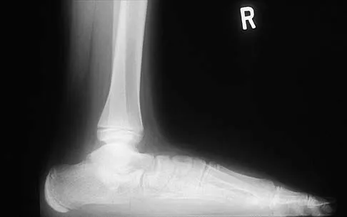
A 42-year-old patient has had a fever and low back pain for several days. Laboratory studies show an elevated erythrocyte sedimentation rate and a WBC count of 9,500 mm3 with 75% neutrophils. A CT scan is shown in Figure 15. Examination will most likely reveal what other findings?


Explanation

The CT scan reveals a left-sided psoas abscess. Irritation of the saphenous division of the femoral nerve can cause paresthesias along the medial aspect of the knee. Pain is usually improved with hip flexion. Cellier C, Gendre JP, Cosnes J, et al: Psoas abscess complication Crohn's disease. Gastroenterol Clin Biol 1992;16:235-238.

Question 3

Based on the diagram shown in Figure 16, what muscle derives its innervation from the nerve identified by the letter "A"?


Explanation

The nerve labeled A is the axillary nerve, a branch from the posterior cord. The posterior cord innervates the subscapularis, latissimus dorsi, teres major and minor, deltoid, triceps, anconeus, brachioradialis, and extensors of the forearm. The axillary nerve innervates the teres minor and deltoid. The pectoralis minor is innervated by the medial cord. The supraspinatus and the subclavius are innervated by the superior trunk. The brachialis is innervated by the lateral cord. Moore K: Anatomy, ed 3. Philadelphia, PA, Williams and Wilkins, 1992.

Question 4

In performing an opening wedge high tibial osteotomy at the tibial tubercle, the osteotome extends 5 mm posteriorly and centrally out of the bone as shown in Figures 17a and 17b. What is the first structure it enters?


Explanation

17b The major risk of performing a high tibial osteotomy is neurovascular injury. The new version of the high tibial osteotomy makes a transverse osteotomy at the level of the tibial tubercle. The osteotome is protected by the oblique belly of the popliteus muscle. The popliteal artery and vein and tibial nerve all lie posterior to the muscle. The soleus muscle originates below this level. Clement CD: Anatomy: A Regional Atlas of Human Anatomy, ed 3. Baltimore, MD, Munich, Germany, Urban and Schwarzberg, 1987, Figure 422.

Question 5

The arrow in the axial T1-weighted MRI scan shown in Figure 18 is pointing to which of the following structures?


Explanation

The arrow is pointing to the ulnar nerve within Guyon's canal. Guyon's canal is approximately 4 cm long, beginning at the proximal extent of the transverse carpal ligament and ending at the aponeurotic arch of the hypothenar muscles. Many structures comprise the boundaries of Guyon's canal. The floor, for example, consists of the transverse carpal ligament, the pisohamate and pisometacarpal ligaments, and the opponens digiti minimi. Within Guyon's canal, the ulnar nerve bifurcates into the superficial and deep branches, with the deep branch of the ulnar nerve persisting distal to the canal. The ulnar artery is immediately adjacent and radial to the ulnar nerve. The median nerve is visualized within the carpal tunnel. The radial artery is on the radial side of the wrist. The hook of the hamate is clearly seen in the figure, orienting the observer to the ulnar side of the wrist. Goss MS, Gelberman RH: The anatomy of the distal ulnar tunnel. Clin Orthop 1985;196:238-247.

Question 6

Osteonecrosis of the femoral head after intramedullary nailing in children is thought to be the result of injury to the


Explanation

All of these are possible explanations for the development of osteonecrosis following intramedullary nailing in children. However, the lateral ascending cervical artery, which supplies the epiphysis, is much more vulnerable to injury in children because it lies in the trochanteric fossa. Buckley SL: Current trends in the treatment of femoral shaft fractures in children and adolescents. Clin Orthop 1997;338:60-73.

Question 7

The illustration shown in Figure 19 shows a Chamberlain line. What is the most likely diagnosis?


Explanation

Basilar invagination is best defined as vertical or compressive instability at the occiput-C1 joint. Such invaginations most commonly occur in patients with rheumatoid arthritis but also can occur secondary to trauma or tumor. A Chamberlain line is used as a method to determine basilar invagination. The odontoid tip should not be more than 5 mm above a Chamberlain line. Wiesel SW, Rothman RH: Occipito-atlantal hypermobility. Spine 1979;4:187-191.

Question 8

Figures 20a and 20b show the sagittal and coronal T1-weighted MRI scans of a patient's left knee. Abnormal findings include


Explanation

20b The MRI scans show meniscal tissue extending across the entire lateral compartment, revealing a discoid lateral meniscus. The increased signal within the lateral meniscal tissue indicates a tear. Discoid lateral menisci are congenital variants that often present with mechanical symptoms in adolescents. The other structures in the knee are normal. Ahn JH, Shim JS, Hwang CH, et al: Discoid lateral meniscus in children: Clinical manifestations and morphology. J Pediatr Orthop 2001;21:812-816.

Question 9

An ulnar nerve palsy at the level of the wrist is typically associated with deficits in the palmaris brevis, the hypothenar muscles, and what other groups of muscles?


Explanation

The intrinsic muscles innervated by the ulnar nerve include the palmaris brevis, hypothenar muscles, all of the interossei, adductor pollicis, and the deep head of the flexor pollicis brevis. The superficial head of the flexor pollicis brevis is innervated by the median nerve. Goldfarb CA, Stern PJ: Low ulnar nerve palsy. JASSH 2003;3:14-26.

Question 10

Figures 21a and 21b show the radiographs of a 22-year-old man who has had progressive pain and swelling about the knee for the past 6 weeks. Examination reveals limited range of motion and fullness about the knee. What is the most likely diagnosis?


Explanation

21b The radiographs reveal a destructive lesion in the metaphysis of the distal femur with periosteal changes and an associated soft-tissue mass with subtle mineralization. This suggests an aggressive malignant process. In this age group, the most likely diagnosis is osteosarcoma. Giant cell tumor, which usually is in a more subchondral location, is not typically so aggressive. Aneurysmal bone cyst is usually more geographic, with a well-marginated reactive rim. Menendez LR (ed): Orthopaedic Knowledge Update: Musculoskeletal Tumors. Rosemont, IL, American Academy of Orthopaedic Surgeons, 2002, pp 175-186.

Question 11

The anterolateral (Watson-Jones) approach to the hip exploits the intermuscular interval between the


Explanation

The Watson-Jones approach to the hip uses the intermuscular interval between the gluteus medius and the tensor fascia lata. This is not a true internervous plane, as both muscles are supplied by the superior gluteal nerve. Hoppenfeld S, deBoer P: Surgical Exposures in Orthopaedics: The Anatomic Approach. Philadelphia, PA, JB Lippincott, 1984, pp 316-332.

Question 12

An 8-month-old infant has an infection of the fingertip as shown in Figure 22. If neglected, the anticipated path of ascending infection is the fingertip, the flexor sheath, and the


Explanation

The flexor sheaths are in continuity with the deep spaces of the hand. The flexor sheaths of the thumb and little finger communicate with the radial and ulnar bursae, respectively, and these two bursae commonly communicate. The central digits do not communicate as readily with deep spaces of the hand but if flexor tendon sheath infection of the index, long, and right fingers is neglected, the potential exists for rupture into the deep midpalmar spaces. Peimer CA (ed): Surgery of the Hand and Upper Extremity: Acute and Chronic Sepsis. New York, NY, Mcgraw Hill, 1996, pp 1735-1741.

Question 13

A 24-year-old man has had pain in the left knee for the past several months. He reports that initially the pain was associated with weight-bearing activities, but it has now become more constant. He denies any swelling but reports a lateral fullness at the tibial plateau. Figures 23a through 23e show radiographs, a bone scan, and T1- and T2-weighted MRI scans. What is the most likely diagnosis?


Explanation

23b 23c 23d 23e The radiographs reveal a lytic subchondral lesion that has a poorly defined margin and lacks mineralization. The bone scan confirms an active lesion that has central photopenia, producing the characteristic doughnut configuration. The MRI scans confirm the presence of a subchondral lesion that is modestly expansile at the lateral plateau and has low signal intensity on the T1-weighted image and a mixed high signal on the T2-weighted image. These features strongly suggest giant cell tumor of bone, more than 50% of which appear around the knee. Simple cyst is excluded by the MRI characteristics. Fibrous dysplasia is unlikely to be in a subchondral location and typically does not show this intensity of uptake on bone scan. Parsons TW: Benign bone tumors, in Fitzgerald RH, Kaufer H, Malkani AL (eds): Orthopaedics. St Louis, MO, Mosby, 2002, pp 1027-1035.

Question 14

Figure 24 shows an axial MRI scan of the ankle. The arrowhead is pointing to what structure?


Explanation

The peroneus brevis is easily identified by its location behind the fibula and its distal muscle belly. Axial MRI images provide a reliable guide even when one of the peroneals is completely ruptured, subluxated out of the peroneal groove, or absent. Sarrafian SK: Anatomy of the Foot and Ankle: Descriptive, Topographic, Functional, ed 2. New York, NY, Lippincott, 1993, pp 234-235.

Question 15

During total hip arthroplasty, profuse bleeding is noted following predrilling for placement of an acetabular component screw. The drill most likely penetrated too deep in the


Explanation

The acetabular quadrants are defined by two lines: one drawn from the anterosuperior iliac spine to the posterior fovea, forming acetabular halves, and a second drawn perpendicular to the first at the midpoint of the acetabulum, forming four quadrants. The anterior quadrants should be avoided because improper screw placement may injure the external iliac artery and vein, as well as the obturator nerve, artery, and vein. These structures lie close to the pelvic bone, with little protective interposition of soft tissue. Wasielewski RC, Cooperstein LA, Kruger MP, et al: Acetabular anatomy and the transacetabular fixation of screws in total hip arthroplasty. J Bone Joint Surg Am 1990;72:501-508.

Question 16

A posterolateral approach to the tibial plafond proceeds between what two muscles?


Explanation

A posterolateral approach to the posterior malleolus proceeds between the lateral and deep posterior compartments. Distally, the peroneus brevis muscle lies most medially within the lateral compartment, and the flexor hallucis longus lies most laterally in the deep posterior compartment. Henry AK: Extensile Exposure, ed 2. Edinburgh, UK, Churchill Livingstone, 1973, pp 269-270.

Question 17

The brachialis muscle is innervated by what two nerves?


Explanation

The brachialis is innervated by two nerves: medially, the musculocutaneous nerve; laterally, the radial nerve. The muscle is split longitudinally to approach the humerus anteriorly. Henry AK: The distal part of the humerus and front of the forearm, in Henry AK (ed): Extensile Exposure, ed 2. Edinburgh, UK, Churchill Livingstone, 1973, pp 90-115.

Question 18

Figure 25 shows the CT scan of an adult patient who has neck pain following a motor vehicle accident. What is the most likely diagnosis?


Explanation

If the atlanto-dens interval is greater than 3 mm in an adult, a transverse ligament rupture usually is suspected. The atlanto-dens interval can be seen with CT or in lateral radiographs of the upper cervical spine. Transverse ligament rupture can occur as an isolated entity or in association with an odontoid or a Jefferson's fracture. Patients with this type of injury usually require fusion. Dickman CA, Greene KA, Sonntag VK: Injuries involving the transverse atlantal ligament: Classification and treatment guidelines based upon experience with 39 injuries. Neurosurgery 1996;38:44-50.

Question 19

Which of the following best describes the course of the ulnar nerve in the midforearm?


Explanation

In the midforearm, the ulnar nerve travels deep to the flexor carpi ulnaris muscle and ulnar to the ulnar artery as it lies on the flexor digitorum profundus muscle. In this region, the ulnar nerve and artery lie side-by-side, whereas more proximal in the forearm, the ulnar artery originates from the brachial artery in the antecubital fossa, and the ulnar nerve lies within the cubital tunnel. Hoppenfeld S, deBoer P: Surgical Exposures in Orthopaedics, ed 2. Philadelphia, PA, Lippincott-Raven, 1994, pp 118-131.

Question 20

A 70-year-old former baseball catcher reports long-standing pain in the ring and little fingers. A gradient-echo MRI scan is shown in Figure 26. What is the most likely diagnosis?


Explanation

The gradient-echo MRI scan highlights the ulnar and radial arteries, as indicated by the arrow. This technique suppresses the signal of the surrounding fat and causes the stationary surrounding tissues to become intermediate in signal intensity. The flowing blood is then easily identified with a bright signal because it does not absorb the radiofrequency pulse. Based on the findings, the diagnosis is an ulnar artery aneurysm, most likely caused by years of repetitive trauma as the result of catching baseballs. Neurolemmoma and giant cell tumor of the tendon sheath would be intermediately enhanced on this image sequence, and the continuity with the ulnar artery, demonstrated here, would not be expected. Lipomas are not enhanced using the gradient-echo technique. The chronic nature of the patient's symptoms is not indicative of a hematoma, and the hematoma would be dark on this imaging sequence since it is stationary tissue. Koman LA, Ruch DS, Patterson Smith B, et al: Vascular disorders, in Green DP, Hotchkiss RN, Pederson WC (eds): Green's Operative Hand Surgery, ed 4. New York, NY, Churchill Livingstone, 1999, vol 2, pp 2254-2302.

Question 21

In a postganglionic brachial plexus lesion at Erb's point (point of formation of the upper trunk by the C5 and C6 nerve roots), which of the following nerves will still function normally?


Explanation

In a postganglionic injury to the brachial plexus, the rhomboid muscle, innervated by the dorsal scapular nerve, would still be expected to function. This is a useful clinical sign that the brachial plexus lesion is postganglionic as opposed to preganlionic. The musculocutaneous, axillary, and suprascapular nerves are all located distal to Erb's point (the most common location of an upper nerve root brachial plexus injury), and all contain fibers from the C5 and C6 nerve roots. Therefore, these nerves are not expected to function normally following a postganglionic C5 and C6 nerve root injury. Netter F: The Ciba Collection of Medical Illustrations: The Musculoskeletal System: Part 1, Anatomy, Physiology and Metabolic Disorders. West Caldwell, NJ, Ciba-Geigy, 1991, vol 8, pp 28-29.

Question 22

The posterior circumflex humeral artery and the axillary nerve usually lie in a space bordered superiorly by the


Explanation

The quadrangular space is bordered superiorly by the teres minor, medially by the long head of the triceps, laterally by the humerus, and inferiorly by the teres major. The posterior circumflex humeral artery and the axillary nerve lie in this space. Rockwood CA Jr, Matsen FA III: The Shoulder, ed 2. Philadelphia, PA, WB Saunders, 1998, vol 1, pp 70-71.

Question 23

A patient notes pain under the first metatarsophalangeal joint following a soccer injury. The MRI scans shown in Figures 27a and 27b reveal what pathologic finding?


Explanation

27b The MRI scans show a complete disruption of the sesamoid complex with proximal retraction of the medial sesamoid and high signal originating from the site normally occupied by the plantar plate (metatarsophalangeal ligament). This injury is the result of a hyperextension injury and is a severe variant of a turf toe. Watson TS, Anderson RB, Davis WH: Periarticular injuries to the hallux metatarsophalangeal joint in athletes. Foot Ankle Clin 2000;5:687-713.

Question 24

When performing the exposure for an anterior approach to the cervical spine, excessive retraction of the trachea and esophagus should be avoided to prevent injury of the


Explanation

The recurrent laryngeal nerve lies between the trachea and the esophagus and is subject to stretch injury if excessive retraction is applied. The vagus nerve lies in the carotid sheath. The sympathetic trunk lies anterior to the longus colli muscles. The hypoglossal nerve and superior laryngeal nerve are both at risk during the exposure but are not located between the trachea and esophagus. An HS: Principles and Techniques of Spine Surgery. Baltimore, MD, Williams and Wilkins, 1998, chapter 2.

Question 25

What is the first ossification center to appear radiographically in the pediatric elbow?


Explanation

The first ossification center to appear in the pediatric elbow is the capitellum. This ossification center generally appears between the first month and the 11th month in girls and between the first month and the 26th month in boys. The other ossification centers in the elbow appear in the following progression: radial head (3.8 to 4.5 years), medial epicondyle (5 to 6 years), olecranon (6 to 7 years), trochlea (9 to 10 years), and the lateral epicondyle (10 years). Wilkins KE, Beaty JH, Chambers HG, et al: Fractures and dislocation of the elbow region, in Rockwood CA, Green DP, Bucholz RW, et al (eds): Rockwood and Green's Fractures in Adults, ed 4. Philadelphia, PA, Lippincott-Raven, 1996, pp 657-662.

Question 26

A 28-year-old overhead athlete presents with insidious posterior shoulder pain and isolated weakness in external rotation. An MRI reveals a paralabral cyst located within the quadrangular space compressing the traversing nerve. Which of the following anatomic structures forms the inferior boundary of this space?


Explanation

The quadrangular space is bound superiorly by the teres minor, inferiorly by the teres major, medially by the long head of the triceps, and laterally by the surgical neck of the humerus. It contains the axillary nerve and the posterior circumflex humeral artery. A cyst in this space typically causes quadrilateral space syndrome, leading to axillary nerve compression (deltoid and teres minor weakness).

Question 27

During surgical reconstruction of a multi-ligamentous knee injury, the surgeon must anatomically restore the posterolateral corner (PLC). When identifying the native femoral footprint of the fibular collateral ligament (FCL) to place a graft, where is this attachment site located relative to the lateral femoral epicondyle?


Explanation

The anatomic femoral attachment of the fibular collateral ligament (FCL) is located approximately 1.4 mm proximal and 3.1 mm posterior to the lateral femoral epicondyle. The popliteus tendon insertion is situated approximately 18.5 mm anterior and distal to the FCL attachment. Precise anatomic knowledge is critical to prevent non-isometric graft placement during PLC reconstruction.

Question 28

A posterior approach (Kocher-Langenbeck) is utilized for an open reduction and internal fixation of a transverse acetabular fracture. The surgeon takes care to protect the primary blood supply to the femoral head. The primary vessel supplying the adult femoral head courses between which two muscles prior to piercing the posterior hip capsule?


Explanation

The medial circumflex femoral artery (MCFA) is the predominant blood supply to the adult femoral head. It branches from the profunda femoris, passes anteriorly between the pectineus and iliopsoas, and then courses posteriorly between the obturator externus and quadratus femoris. Protecting the obturator externus during posterior hip approaches helps prevent iatrogenic injury to the MCFA.

Question 29

A spine surgeon is performing a lateral transpsoas approach (LLIF/XLIF) to the lumbar spine at the L4-L5 level. To minimize the risk of iatrogenic injury to the lumbar plexus, the surgeon relies on anatomic safe zones. At the L4-L5 disc space, where is the lumbar plexus most commonly located within the substance of the psoas major muscle?


Explanation

The lumbar plexus forms within the substance of the psoas major muscle. As it descends from L1 to L5, it migrates from a more medial and dorsal position to a lateral and slightly more anterior position, but at the L4-L5 level, it is consistently found within the posterior one-third of the psoas muscle. Consequently, the surgical safe zone for a lateral transpsoas approach is in the anterior to middle third of the disc space to avoid lumbar plexus injury.

Question 30

A 45-year-old carpenter presents with a 4-month history of deep, aching pain in his proximal dorsal forearm and progressive weakness in extending his index and middle fingers. On examination, wrist extension is maintained but demonstrates radial deviation. Which of the following anatomic structures is the most common site of compression for the affected nerve?


Explanation

The patient's presentation is classic for Posterior Interosseous Nerve (PIN) syndrome. Wrist extension is preserved but deviates radially because the Extensor Carpi Radialis Longus (ECRL) is innervated by the radial nerve proper, proximal to its bifurcation into the PIN and superficial branch. The most common site of PIN compression is the Arcade of Frohse, which is the proximal fibrous edge of the superficial head of the supinator muscle.

Question 31

During a plantar approach for the excision of a severe, recurrent deep plantar fibromatosis, the surgeon carefully dissects near the midfoot and identifies the 'Master Knot of Henry.' At this anatomic crossover point, what is the spatial relationship of the crossing tendons?


Explanation

The 'Master Knot of Henry' is located in the midfoot at the level of the navicular and medial cuneiform. At this site, the Flexor Digitorum Longus (FDL) tendon crosses superficial (plantar) to the Flexor Hallucis Longus (FHL) tendon. Therefore, relative to the sole of the foot, the FHL is positioned dorsal (deep) to the FDL.

Question 32

A 32-year-old female presents with severe pain over the radial styloid, exacerbated by grasping and ulnar deviation of the wrist. Non-operative management fails, and surgical release is planned. To prevent a highly morbid postoperative complication characterized by painful neuromas, the surgeon must protect a specific nerve. This at-risk nerve characteristically courses superficial to the roof of which extensor compartment?


Explanation

The patient has De Quervain's tenosynovitis (involving the Abductor Pollicis Longus and Extensor Pollicis Brevis in the first dorsal compartment). The superficial branch of the radial nerve (SBRN) exits from beneath the brachioradialis in the distal forearm and courses directly over the roof of the first extensor compartment. Iatrogenic injury to the SBRN during first compartment release is a well-known complication causing severe neuroma pain.

Question 33

A 55-year-old male sustains an unstable pelvic ring injury. An anterior ilioinguinal approach is utilized for open reduction and internal fixation. During dissection along the superior pubic ramus, brisk arterial hemorrhage is encountered. This bleeding most likely originates from the 'Corona Mortis,' which represents an anastomosis between the obturator artery and which of the following vessels?


Explanation

The Corona Mortis (Crown of Death) is a critical vascular anastomosis located on the posterior aspect of the superior pubic ramus. It connects the obturator vessels (branch of the internal iliac system) with the inferior epigastric or external iliac vessels (external iliac system). It must be carefully identified and ligated during ilioinguinal or Stoppa approaches to the pelvis to prevent catastrophic hemorrhage.

Question 34

A regional anesthesia team performs an adductor canal (Hunter's canal) block for postoperative analgesia following a total knee arthroplasty, aiming to spare quadriceps motor function. The target nerve provides sensory innervation to the medial lower leg. This nerve typically exits the adductor canal by piercing which of the following anatomic structures?


Explanation

The adductor canal contains the superficial femoral artery, femoral vein, saphenous nerve, and the nerve to the vastus medialis. The saphenous nerve is a purely sensory branch of the femoral nerve. It exits the adductor canal by piercing its roof, which is formed by the vastoadductor membrane (a fascial expansion between the adductor longus/magnus and the vastus medialis), before traveling distally to supply the medial lower leg.

Question 35

A 30-year-old elite volleyball player develops progressive, isolated weakness of shoulder external rotation with no sensory deficits. Abduction strength is graded 5/5 and is symmetric to the contralateral shoulder. Given this specific clinical presentation, an entrapment neuropathy is suspected. Where is the most likely location of the nerve compression?


Explanation

The suprascapular nerve innervates the supraspinatus (shoulder abduction) and the infraspinatus (shoulder external rotation). Entrapment at the suprascapular notch affects BOTH muscles. Entrapment at the spinoglenoid notch (commonly due to a paralabral cyst associated with a posterior SLAP tear in overhead athletes) occurs distal to the motor branch to the supraspinatus, resulting in isolated infraspinatus atrophy and isolated external rotation weakness.

Question 36

A surgeon is performing a posterolateral approach to the distal tibia for fixation of a complex posterior malleolus fracture. To utilize a true internervous plane, the deep surgical dissection should occur between which of the following two muscle bellies?


Explanation

The posterolateral approach to the distal tibia and posterior malleolus utilizes an internervous plane between the flexor hallucis longus (innervated by the tibial nerve) medially, and the peroneus brevis (innervated by the superficial peroneal nerve) laterally. This approach safely exposes the posterior aspect of the tibia without devascularizing the bone or placing major neurovascular bundles at excessive risk.

Question 37

A 45-year-old man presents with severe right leg pain radiating to the anterior thigh. Magnetic resonance imaging reveals a far-lateral (extraforaminal) disc herniation at the L3-L4 level. Which nerve root is most likely compressed, and what clinical examination finding is expected?


Explanation

In the lumbar spine, a far-lateral (extraforaminal) disc herniation compresses the exiting nerve root at that corresponding level. Therefore, an L3-L4 far-lateral herniation will compress the L3 nerve root. A central or paracentral herniation at L3-L4 would compress the traversing L4 root. Compression of the L3 root commonly manifests with pain radiating to the anterior thigh and weakness in knee extension (quadriceps) and hip flexion (iliopsoas), accompanied by a diminished patellar tendon reflex.

Question 38

A 32-year-old construction worker presents with a deep space infection of the hand following a penetrating injury. The surgical team plans an incision to drain the midpalmar space. The midpalmar space is anatomically separated from the thenar space by which of the following structures?


Explanation

The deep spaces of the palm are divided into the midpalmar space and the thenar space. These two distinct potential spaces are separated by the midpalmar (oblique) septum, a fascial layer extending from the palmar aponeurosis to the anterior border of the third metacarpal shaft. Understanding this anatomy is critical for appropriately draining hand infections without spreading the purulence into uninvolved compartments.

Question 39

Following closed reduction and percutaneous pinning of a severely displaced supracondylar humerus fracture, a 6-year-old boy is noted to be unable to make an 'OK' sign, instead demonstrating a flat pinch mechanism. Which muscle's weakness is primarily responsible for this physical finding, and what is its innervation?


Explanation

The inability to form an 'OK' sign (pincer grasp) indicates weakness of the flexor pollicis longus (FPL) and the flexor digitorum profundus (FDP) to the index finger. This results in a flattened pinch because the interphalangeal joint of the thumb and the distal interphalangeal joint of the index finger cannot actively flex. These muscles are innervated by the anterior interosseous nerve (AIN), a motor branch of the median nerve that is frequently injured in pediatric supracondylar humerus fractures.

Question 40

During a surgical approach to the hip for internal fixation of a basicervical femoral neck fracture, the surgeon must exercise extreme caution to preserve the primary blood supply to the adult femoral head. Which of the following is the predominant source of this vascularity?


Explanation

The predominant blood supply to the adult femoral head is derived from the medial femoral circumflex artery (MFCA), specifically its deep branch, which gives rise to the posterior superior and posterior inferior retinacular vessels. While the lateral femoral circumflex artery and the foveal artery (via the ligamentum teres) contribute minimally in adults, the MFCA is the critical vessel at risk during femoral neck fractures and posterior surgical approaches.

Question 41

A 28-year-old professional volleyball player presents with insidious onset of vague posterior shoulder pain and paresthesias over the lateral deltoid. MRI of the shoulder demonstrates isolated atrophy of the teres minor muscle. Pathology within the quadrilateral space is suspected. Which structures traverse this anatomical space?


Explanation

Quadrilateral space syndrome occurs secondary to compression of the axillary nerve and posterior circumflex humeral artery. The boundaries of the quadrilateral space are the teres minor (superior), teres major (inferior), long head of the triceps (medial), and the surgical neck of the humerus (lateral). Compression here leads to axillary nerve palsy, often manifesting as teres minor atrophy on MRI, as well as deltoid weakness and lateral shoulder paresthesias.

Question 42

A surgeon is performing the volar (Henry) approach to the radius to fix a midshaft radius fracture. During the proximal superficial dissection, an internervous plane is developed. Which nerves supply the two muscles forming the boundaries of this proximal internervous plane?


Explanation

The volar (Henry) approach to the radius utilizes an internervous plane. Proximally, this plane is found between the brachioradialis (innervated by the radial nerve) and the pronator teres (innervated by the median nerve). Distally, the plane continues between the brachioradialis (radial nerve) and the flexor carpi radialis (median nerve). Utilizing this internervous interval allows for safe anterior exposure of the radius.

Question 43

While operatively stabilizing a syndesmotic injury of the ankle, the surgeon places a syndesmotic screw from the fibula to the tibia. Anatomically, the distal tibiofibular syndesmosis relies on multiple ligamentous structures for stability. Which of the following is considered the strongest and thickest primary stabilizer of this complex?


Explanation

The distal tibiofibular syndesmosis is stabilized by the AITFL, PITFL, and the interosseous ligament/membrane. Biomechanical studies have demonstrated that the posterior inferior tibiofibular ligament (PITFL) is the thickest and strongest component, providing approximately 42% of the strength of the syndesmosis and offering the greatest resistance to lateral displacement of the distal fibula.

Question 44

A 30-year-old gymnast complains of recurrent elbow instability, particularly when pushing out of a chair. Physical examination reveals a positive lateral pivot-shift test, indicating posterolateral rotatory instability (PLRI). The primary structure deficient in this condition originates from the lateral epicondyle and inserts onto which of the following osseous landmarks?


Explanation

Posterolateral rotatory instability (PLRI) of the elbow is primarily caused by insufficiency of the lateral ulnar collateral ligament (LUCL). The LUCL is the primary restraint to posterolateral rotatory subluxation. It originates on the lateral epicondyle of the humerus, courses posterior to the radial head, and inserts on the supinator crest of the proximal ulna.

Question 45

During an anterior intrapelvic (Stoppa) approach for an anterior column acetabular fracture, significant arterial hemorrhage is encountered as the dissection proceeds along the posterior aspect of the superior pubic ramus. This bleeding is most likely due to an injury to the 'corona mortis,' which is an anastomosis between which two vascular systems?


Explanation

The 'corona mortis' (crown of death) represents a vascular anastomosis between the external iliac system (usually the inferior epigastric artery or vein) and the internal iliac system (obturator artery or vein). It is typically located crossing the superior pubic ramus at an average distance of 5 to 6 cm from the pubic symphysis. Iatrogenic injury during pelvic or acetabular surgery can result in life-threatening hemorrhage that is difficult to control.

Question 46

During surgical reconstruction of the posterolateral corner of the knee, the surgeon must identify the popliteofibular ligament. Which of the following accurately describes the anatomy of the popliteofibular ligament?


Explanation

The popliteofibular ligament is a critical static stabilizer of the posterolateral corner of the knee. It originates from the musculotendinous junction of the popliteus and inserts on the posteromedial aspect of the fibular head. Its primary function is to resist excessive external rotation of the tibia and provide varus stability.

Question 47

A 28-year-old male sustains a displaced talar neck fracture (Hawkins Type III). He is at high risk for avascular necrosis. Which of the following arteries provides the majority of the blood supply to the talar body and is most likely injured in this fracture pattern?


Explanation

The main blood supply to the talar body is derived from the artery of the tarsal canal, which is a branch of the posterior tibial artery. The artery of the tarsal sinus (formed by branches of the perforating peroneal and dorsalis pedis arteries) and the deltoid branch provide supplementary blood supply. Displaced talar neck fractures often disrupt the artery of the tarsal canal, leading to a high rate of avascular necrosis of the talar body.

Question 48

A patient presents with a deep space infection of the hand localized strictly to the thenar space. Which of the following structures forms the ulnar border of the thenar space, separating it from the midpalmar space?


Explanation

The thenar space is a potential deep space of the hand bounded radially by the lateral palmar septum, dorsally by the adductor pollicis muscle, volarly by the index flexor tendons and the first lumbrical, and ulnarly by the midpalmar septum. The midpalmar septum typically attaches to the third metacarpal and separates the thenar space from the midpalmar space.

Question 49

In the distal extension of the anterolateral approach to the humerus, the brachialis muscle is split to expose the humeral shaft. What is the neurological basis that permits a safe, longitudinal split in the middle or lateral third of the brachialis muscle?


Explanation

The brachialis muscle has a unique dual innervation. The medial portion is innervated by the musculocutaneous nerve, while the lateral portion is innervated by the radial nerve. A longitudinal split in the brachialis utilizes this internervous plane, minimizing denervation to either half.

Question 50

A 45-year-old male presents with severe right anterior thigh pain and new-onset weakness in knee extension. MRI of the lumbar spine reveals a far lateral (extraforaminal) disc herniation at the L3-L4 level. Which nerve root is most likely compressed in this specific anatomical zone?


Explanation

In the lumbar spine, a typical posterolateral (paracentral) disc herniation at L3-L4 compresses the traversing L4 nerve root. However, a far lateral (extraforaminal) disc herniation at the L3-L4 level will impinge upon the exiting L3 nerve root as it passes through and exits the intervertebral foramen, leading to symptoms in the L3 distribution.

Question 51

During arthroscopic shoulder surgery, the rotator interval is evaluated. Which of the following correctly identifies the anatomical borders and contents of the rotator interval?


Explanation

The rotator interval is a triangular anatomic space in the anterosuperior shoulder. It is bounded superiorly by the anterior margin of the supraspinatus tendon, inferiorly by the superior margin of the subscapularis tendon, and medially by the base of the coracoid process. Its vital contents include the long head of the biceps tendon, the coracohumeral ligament, and the superior glenohumeral ligament.

Question 52

A 32-year-old female presents with pain and swelling over the dorsal aspect of her wrist, specifically at the level of Lister's tubercle. During surgical release for tenosynovitis, the tendon passing directly ulnar to Lister's tubercle is identified. This tendon belongs to which extensor compartment of the wrist?


Explanation

Lister's tubercle is a bony prominence on the distal radius that acts as a pulley for the extensor pollicis longus (EPL) tendon. The EPL tendon, which constitutes the third extensor compartment, travels just ulnar to Lister's tubercle before taking a sharp turn radially toward the thumb.

Question 53

The 'Master Knot of Henry' is a recognized surgical landmark in the plantar aspect of the midfoot. Which of the following correctly describes the anatomical intersection that defines this structure?


Explanation

The Master Knot of Henry is located in the medial plantar aspect of the foot at the level of the navicular bone. It is defined by the flexor digitorum longus (FDL) tendon crossing superficial (plantar) to the flexor hallucis longus (FHL) tendon. It is a critical landmark during tendon transfers and when dissecting near the medial and lateral plantar nerves.

Question 54

A 45-year-old cyclist complains of numbness in his ring and small fingers along with weakness in finger abduction. Compression of the ulnar nerve in Guyon's canal is diagnosed. Which of the following structures forms the floor of Guyon's canal?


Explanation

Guyon's canal (the ulnar tunnel) is bounded superficially (roof) by the volar carpal ligament, deeply (floor) by the transverse carpal ligament (flexor retinaculum) and the pisohamate ligament. The medial wall is formed by the pisiform, and the lateral wall is the hook of the hamate.

Question 55

During an ilioinguinal approach for the internal fixation of an anterior column acetabular fracture, the surgeon dissects near the superior pubic ramus and must be careful to ligate the 'corona mortis.' This structure represents an anastomosis between which two vascular systems?


Explanation

The corona mortis (crown of death) is a vascular anastomosis between the obturator vessels (from the internal iliac system) and the external iliac or inferior epigastric vessels. It crosses the superior pubic ramus at an average distance of 5 cm from the symphysis pubis and can cause massive hemorrhage if inadvertently torn during anterior pelvic surgery.

Question 56

A 35-year-old male presents with right shoulder pain and weakness after a direct blow to the posterior shoulder. On examination, he has weakness in external rotation and abduction. An MRI shows isolated atrophy of the teres minor. Which of the following anatomic borders defines the space through which the affected nerve passes?


Explanation

The axillary nerve passes through the quadrangular space, innervating the deltoid and teres minor. The borders of the quadrangular space are the teres minor superiorly, teres major inferiorly, long head of triceps medially, and the surgical neck of the humerus laterally. The axillary nerve is accompanied by the posterior circumflex humeral artery in this space.

Question 57

A 24-year-old football player sustains a contact injury to his knee, resulting in a varus and hyperextension moment. He complains of lateral knee pain and instability. Physical examination reveals a positive dial test at 30 degrees of flexion, which normalizes at 90 degrees. Which of the following structures is the primary static restraint to external rotation at 30 degrees of knee flexion?


Explanation

A positive dial test at 30 degrees of knee flexion that normalizes at 90 degrees indicates an isolated injury to the posterolateral corner (PLC). The primary restraints to external tibial rotation at 30 degrees of knee flexion are the popliteus complex (including the popliteofibular ligament) and the lateral collateral ligament (LCL). However, biomechanical studies demonstrate that the popliteofibular ligament is specifically the most critical static restraint to external rotation in this position.

Question 58

During a deep dissection of the palm for a complex tumor resection, the surgeon encounters the deep palmar arch. This vascular structure is primarily formed by the continuation of which artery, and it typically passes deep to which structure?


Explanation

The deep palmar arch is primarily formed by the terminal continuation of the radial artery and is completed medially by the deep palmar branch of the ulnar artery. It lies deep to the flexor tendons and their synovial sheaths, and superficial to the bases of the metacarpals and the interosseous muscles. In contrast, the superficial palmar arch is primarily formed by the ulnar artery and lies superficial to the flexor tendons.

Question 59

A 45-year-old patient presents with neck pain and occipital headaches after a motor vehicle collision. Flexion-extension radiographs of the cervical spine demonstrate an atlantodental interval (ADI) of 4 mm. An MRI is obtained to evaluate the ligamentous structures. The alar ligaments primarily prevent which of the following movements?


Explanation

The alar ligaments connect the posterolateral aspect of the dens to the medial surfaces of the occipital condyles. Their primary function is to limit contralateral axial rotation and lateral flexion of the occipito-atlanto-axial complex. Anterior translation of C1 on C2 is primarily prevented by the transverse ligament.

Question 60

A 32-year-old runner develops medial midfoot pain and numbness radiating to the plantar aspect of the hallux. Conservative management fails, and surgical exploration is planned. The surgeon targets the area near the 'Master Knot of Henry.' Which of the following describes the anatomical relationship at this location?


Explanation

The 'Master Knot of Henry' is located in the medial aspect of the midfoot, plantarly, at the level of the navicular base. At this location, the tendon of the flexor digitorum longus (FDL) crosses superficial (i.e., plantar) to the tendon of the flexor hallucis longus (FHL). This decussation is a potential site for tendinopathy or nerve entrapment.

Question 61

A 14-year-old boy undergoes a surgical dislocation of the hip for treatment of severe slipped capital femoral epiphysis (SCFE). During the exposure, the surgeon must protect the primary blood supply to the femoral head. Which of the following vessels provides the predominant blood supply to the femoral head in this age group, and what is its anatomical course?


Explanation

The predominant blood supply to the adult and adolescent femoral head is derived from the medial femoral circumflex artery (MFCA). Specifically, the posterosuperior and posteroinferior retinacular branches of the MFCA pierce the capsule and run along the femoral neck to enter the head. During surgical dislocation, it is vital to protect the external rotators (particularly the obturator externus) to avoid stretching or severing the MFCA, which courses between the quadratus femoris and the obturator externus.

Question 62

A 28-year-old man sustains a Monteggia equivalent fracture-dislocation and requires open reduction and internal fixation of the proximal radius. A volar (Henry) approach to the proximal radius is utilized. During deep dissection, the supinator muscle is identified. To safely expose the proximal radius and prevent injury to the posterior interosseous nerve (PIN), the forearm should be placed in which position, and what is the relationship of the PIN to the supinator?


Explanation

When utilizing the volar (Henry) approach to the proximal radius, the forearm should be supinated to displace the posterior interosseous nerve (PIN) radially and away from the surgical field. The PIN passes between the superficial and deep heads of the supinator muscle. Pronation brings the nerve ulnarly into the operative field, increasing the risk of iatrogenic transection.

Question 63

An orthopedic surgeon is performing an anterior (ilioinguinal) approach for the fixation of an anterior column acetabular fracture. While dissecting along the posterior aspect of the superior pubic ramus, brisk, difficult-to-control arterial bleeding is encountered. This bleeding is most likely originating from an anastomotic vessel connecting which two vascular systems?


Explanation

The 'corona mortis' (crown of death) is a highly variable anatomical anastomosis between the external iliac system (usually the inferior epigastric artery or vein) and the internal iliac system (obturator artery or vein). It crosses over the superior pubic ramus at an average of 5-7 cm from the symphysis pubis and is highly susceptible to injury during anterior pelvic exposures (like the ilioinguinal or Stoppa approach).

Question 64

A patient sustains a penetrating trauma to the anterior arm, resulting in inability to flex the elbow and loss of sensation over the lateral aspect of the forearm. The injured nerve is a terminal branch of which cord of the brachial plexus, and it typically pierces which muscle?


Explanation

The musculocutaneous nerve is the terminal branch of the lateral cord of the brachial plexus (C5, C6, C7). It typically pierces the coracobrachialis muscle in the upper arm, then descends between the biceps brachii and brachialis muscles, supplying all three. It then emerges lateral to the biceps tendon as the lateral antebrachial cutaneous nerve, providing sensation to the lateral forearm.

Question 65

A 40-year-old male develops acute compartment syndrome of the lower leg following a tibial plateau fracture. The surgeon proceeds with a two-incision four-compartment fasciotomy. When releasing the deep posterior compartment, which nerve is most closely associated with the contents of this compartment and at risk if dissection is too deep?


Explanation

The deep posterior compartment of the lower leg contains the tibialis posterior, flexor digitorum longus, and flexor hallucis longus muscles. The tibial nerve and posterior tibial artery run within this compartment, intimately associated with these muscles. When releasing the deep posterior compartment, especially from a medial incision, care must be taken to effectively decompress the fascia without injuring the neurovascular bundle.

Question 66

A 45-year-old male is undergoing open reduction and internal fixation of a proximal humerus fracture via an anterolateral (deltoid-splitting) approach. The surgeon must be careful to avoid iatrogenic injury to the axillary nerve. Which of the following best describes the typical anatomic distance of the axillary nerve distal to the lateral edge of the acromion?


Explanation

The axillary nerve runs transversely across the deep surface of the deltoid muscle, typically traversing approximately 5 to 7 cm distal to the lateral edge of the acromion. When utilizing an anterolateral (deltoid-splitting) approach, the split should not extend beyond 5 cm from the acromion to prevent denervation of the anterior aspect of the deltoid muscle.

Question 67

During a posterolateral corner (PLC) reconstruction of the knee, the surgeon isolates the fibular collateral ligament (FCL) to prepare for anatomic graft placement. Which of the following best describes the precise anatomic footprint of the FCL on the lateral femoral condyle relative to the popliteus tendon insertion?


Explanation

On the lateral femoral condyle, the footprint of the fibular collateral ligament (FCL) is located proximal and posterior to the attachment of the popliteus tendon. Recognizing this specific anatomic relationship is essential for accurate tunnel placement during anatomic posterolateral corner reconstructions.

Question 68

A cyclist presents with weakness of the intrinsic hand muscles and numbness limited to the volar ulnar aspect of the hand. You suspect entrapment of the ulnar nerve within Guyon's canal. At which specific anatomic landmark does the deep motor branch of the ulnar nerve diverge from the superficial sensory branch?


Explanation

Guyon's canal is divided into three zones. Zone 1 is proximal to the bifurcation of the ulnar nerve. The bifurcation into the deep motor branch and superficial sensory branch occurs just distal to the pisiform and proximal to the hook of hamate. The deep motor branch then dives between the abductor digiti minimi and flexor digiti minimi origins (Zone 2), while the sensory branch continues superficially (Zone 3).

Question 69

A 35-year-old male is undergoing open reduction and internal fixation of an anterior column acetabulum fracture via an ilioinguinal approach. The surgeon is working primarily through the 'middle window' to visualize the pelvic brim. Which structures define the medial and lateral borders of this surgical window?


Explanation

The ilioinguinal approach utilizes three primary anatomic windows. The lateral window is lateral to the iliopectineal fascia (containing the iliopsoas and femoral nerve). The middle window lies between the iliopectineal fascia laterally and the external iliac vessels medially. The medial window lies between the external iliac vessels laterally and the rectus abdominis/spermatic cord medially.

Question 70

A 60-year-old patient is undergoing a minimally invasive L4-L5 transforaminal lumbar interbody fusion (TLIF). To avoid injury to the exiting nerve root during facetectomy and disc preparation, the surgeon must be aware of its exact anatomical relationship to the pedicles. Which nerve root exits through the L4-L5 intervertebral foramen, and what is its anatomic relationship to the L4 pedicle?


Explanation

In the lumbar spine, the exiting nerve root takes the name of the pedicle immediately superior to it. Thus, the L4 nerve root exits through the L4-L5 foramen, immediately inferior (caudal) to the L4 pedicle. The L5 nerve root traverses the L4-L5 disc space centrally before exiting at the L5-S1 foramen.

Question 71

An orthopedic surgeon is performing an extensile lateral approach to the calcaneus for a displaced intra-articular calcaneus fracture. The sural nerve is at significant risk of injury during the creation of the full-thickness soft tissue flap. Which of the following best describes the typical anatomic course of the sural nerve at the level of the lateral malleolus?


Explanation

The sural nerve typically crosses approximately 10 to 15 mm (1.0 to 1.5 cm) posterior and inferior to the tip of the lateral malleolus. It is highly susceptible to injury during the extensile lateral approach to the calcaneus if the horizontal arm of the incision is misplaced or if the full-thickness subperiosteal flap is not properly maintained.

Question 72

During a standard Kocher-Langenbeck (posterior) approach to the hip for an associated posterior wall acetabular fracture, the surgeon performs a tenotomy of the short external rotators. To protect the main blood supply to the femoral head, which of the following muscles should ideally remain intact because its tendon directly overlies and protects the deep branch of the medial femoral circumflex artery (MFCA)?


Explanation

The deep branch of the medial femoral circumflex artery (MFCA) provides the predominant blood supply to the femoral head. It courses consistently posterior to the tendon of the obturator externus. Maintaining the obturator externus during a posterior approach to the hip protects this crucial vessel from iatrogenic injury.

Question 73

A 32-year-old male sustains a proximal third radius shaft fracture and undergoes open reduction and internal fixation via an anterior (Henry) approach.

During the approach, the supinator muscle must be elevated off the radius. To safely reflect the supinator and protect the posterior interosseous nerve (PIN), how should the forearm be positioned during muscle elevation?


Explanation

During the anterior (Henry) approach to the proximal radius, the forearm should be placed in full supination when reflecting the supinator muscle laterally. Full supination shifts the posterior interosseous nerve (PIN) further laterally and posteriorly, pulling it away from the anterior surgical field and minimizing the risk of traction or transection.

Question 74

A 28-year-old elite volleyball player presents with vague posterolateral shoulder pain and isolated weakness in external rotation. Shoulder abduction strength is normal.

An MRI reveals a paralabral cyst. Based on the clinical findings of isolated infraspinatus weakness with normal supraspinatus function, at which of the following anatomic locations is the nerve compression most likely occurring?


Explanation

The suprascapular nerve passes through the suprascapular notch, where it gives off motor branches to the supraspinatus, and then continues through the spinoglenoid notch to innervate the infraspinatus. Compression at the spinoglenoid notch (often due to a posterior labral tear and subsequent paralabral cyst) results in isolated infraspinatus denervation and external rotation weakness, sparing the supraspinatus.

Question 75

A 22-year-old male develops acute compartment syndrome of the lower leg following a high-energy tibial plateau fracture. The surgeon opts for a standard two-incision, four-compartment fasciotomy. Through the lateral incision, both the anterior and lateral compartments are released. Which key anatomic structure serves as the boundary dividing these two compartments and must be identified to ensure precise and complete release of both?


Explanation

The anterior intermuscular septum anatomically separates the anterior compartment from the lateral compartment in the leg. During a lateral fasciotomy incision, the surgeon must identify this septum to confidently incise the fascia anterior to it (releasing the anterior compartment) and posterior to it (releasing the lateral compartment). The superficial peroneal nerve courses within the lateral compartment and must be protected, but the septum itself is the defining boundary.

Question 76

During an anterolateral approach to the distal humerus, the radial nerve is at risk. Which of the following describes the correct anatomical plane and location to identify the radial nerve in this region?


Explanation

The radial nerve pierces the lateral intermuscular septum from the posterior to the anterior compartment approximately 10 cm proximal to the lateral epicondyle. It then travels distally in the anterior compartment within the interval between the brachialis (medial) and the brachioradialis (lateral) before dividing into the posterior interosseous nerve (PIN) and the superficial radial nerve.

Question 77

A 35-year-old sustains an intra-articular calcaneus fracture. The surgeon plans a lateral extensile approach. To prevent flap necrosis, a full-thickness subperiosteal flap must be created. What is the primary arterial supply to the apex (corner) of this lateral flap?


Explanation

The lateral calcaneal artery, a terminal branch primarily derived from the peroneal artery, provides the primary blood supply to the lateral heel skin and the apex of the flap in a lateral extensile approach to the calcaneus. The incision should be full-thickness straight to the bone to protect this delicate vascular supply.

Question 78

During an ilioinguinal approach to the acetabulum, the surgeon develops three distinct surgical windows. When working in the middle window to access the pelvic brim and quadrilateral surface, which of the following neurovascular structures is primarily mobilized and at highest risk?


Explanation

The ilioinguinal approach utilizes three windows: lateral, middle, and medial. The middle window is developed between the iliopectineal fascia (which overlies the iliopsoas and femoral nerve laterally) and the external iliac vessels medially. Therefore, the external iliac artery and vein are primarily manipulated and at risk in this specific window. The femoral nerve is in the lateral window.

Question 79

A 24-year-old athlete undergoes an anatomic reconstruction of the posterolateral corner (PLC) of the knee. The footprints of the fibular collateral ligament (FCL) and the popliteus tendon on the lateral femoral condyle must be accurately identified. What is the normal anatomic relationship of the FCL footprint relative to the popliteus tendon footprint?


Explanation

On the lateral femoral condyle, the popliteus tendon attaches at the anterior aspect of the popliteal sulcus. The fibular collateral ligament (FCL) attachment is located proximal and posterior to the popliteus tendon attachment. The average distance between the two insertions is 18.5 mm.

Question 80

A 30-year-old carpenter sustains a volar laceration over the proximal phalanx of his index finger (Zone II). Which of the following accurately describes the relationship of the flexor digitorum superficialis (FDS) and flexor digitorum profundus (FDP) tendons at the level of the A2 pulley?


Explanation

At the level of the A2 pulley (proximal phalanx, zone II), the flexor digitorum superficialis (FDS) tendon splits to form Camper's chiasm. The two slips wrap around the flexor digitorum profundus (FDP) tendon to insert on the volar aspect of the middle phalanx. The FDP passes through this split to continue distally to the distal phalanx.

Question 81

A spine surgeon is placing pedicle screws in the lumbar spine from L1 to L5. Which of the following describes the normal morphometric progression of the lumbar pedicles as one moves caudally from L1 to L5?


Explanation

From L1 to L5, the lumbar pedicles generally increase in transverse diameter. Additionally, the pedicle trajectory in the axial/coronal plane becomes more convergent (medially directed). At L1, the angle is approximately 10-15 degrees medial, increasing to 25-30 degrees at L5.

Question 82

During a posterior (Kocher-Langenbeck) approach to the hip, the short external rotators are detached to expose the posterior capsule. The deep branch of the medial femoral circumflex artery (MFCA), the primary blood supply to the femoral head, is at risk. Where does the main terminal branch of the MFCA lie in relation to the short external rotators?


Explanation

The deep branch of the medial femoral circumflex artery (MFCA) travels posteriorly to provide the primary blood supply to the femoral head. It consistently emerges posteriorly in the interval between the inferior gemellus and the superior border of the quadratus femoris muscle before traveling proximally and anterior to the short external rotators to perforate the capsule.

Question 83

During an arthroscopic evaluation of a shoulder, the surgeon visualizes the rotator interval. Which of the following options correctly identifies the anatomic borders and contents of the rotator interval?


Explanation

The rotator interval is a triangular space bounded superiorly by the anterior margin of the supraspinatus tendon and inferiorly by the superior margin of the subscapularis tendon. Its contents include the long head of the biceps tendon, the coracohumeral ligament, and the superior glenohumeral ligament.

Question 84

A 52-year-old patient undergoes surgical release for recalcitrant tarsal tunnel syndrome. The flexor retinaculum is carefully incised. Which of the following describes the correct anatomical order of structures within the tarsal tunnel from anterior-medial to posterior-lateral?


Explanation

The structures passing through the tarsal tunnel from anterior to posterior behind the medial malleolus can be recalled with the mnemonic 'Tom, Dick, And Very Nervous Harry': Tibialis posterior tendon, flexor Digitorum longus tendon, posterior tibial Artery, posterior tibial Vein, tibial Nerve, and flexor Hallucis longus tendon.

Question 85

A 28-year-old man develops acute compartment syndrome of the forearm following a crush injury. A volar Henry approach is utilized for fasciotomy. In the proximal third of the forearm, the radial artery must be identified and protected. Between which two muscles does the radial artery run in this specific proximal segment?


Explanation

In the proximal third of the forearm, the radial artery courses between the brachioradialis (laterally) and the pronator teres (medially). In the middle third, it lies between the brachioradialis and the flexor carpi radialis (FCR). In the distal third, it becomes more superficial between the tendons of the brachioradialis and FCR.

Question 86

During the volar (Henry) approach to the proximal radius, the surgeon develops an internervous plane to safely expose the underlying structures. Which of the following describes the correct proximal internervous interval and the respective nerve supply to the bordering muscles?


Explanation

The proximal portion of the volar (Henry) approach to the radius utilizes the internervous plane between the brachioradialis (innervated by the radial nerve) radially and the pronator teres (innervated by the median nerve) ulnarly. Distally, the interval transitions to between the brachioradialis and the flexor carpi radialis (median nerve). It is critical to identify and ligate the recurrent radial artery branches in this plane to safely mobilize the brachioradialis radially.

Question 87

A 35-year-old male requires autologous bone grafting for a recalcitrant tibial nonunion. A posterolateral approach to the tibia is chosen to avoid the compromised anteromedial soft tissue envelope. During this approach, the correct internervous plane is developed between the lateral gastrocnemius, soleus, and flexor hallucis longus posteriorly, and which of the following muscles anteriorly?


Explanation

The posterolateral approach to the tibia accesses the bone through a plane between the lateral compartment (peroneus longus and brevis, innervated by the superficial peroneal nerve) anteriorly and the superficial/deep posterior compartments (lateral gastrocnemius, soleus, and flexor hallucis longus, innervated by the tibial nerve) posteriorly. The tibialis posterior lies deep and is elevated directly off the interosseous membrane.

Question 88

A 45-year-old male sustains an anterior column and quadrilateral plate fracture of the acetabulum. The surgeon selects the modified Stoppa approach for reduction and fixation. During the initial dissection, a significant anastomotic vessel (corona mortis) is identified crossing the superior pubic ramus. This vessel typically connects the external iliac or inferior epigastric system to which of the following?


Explanation

The corona mortis ('crown of death') is a vascular anastomosis between the external iliac/inferior epigastric system and the obturator system. It traverses the superior pubic ramus at an average distance of 5-6 cm from the pubic symphysis. It is at significant risk during the modified Stoppa and ilioinguinal approaches to the acetabulum and must be meticulously identified and ligated to prevent catastrophic hemorrhage.

Question 89

A 28-year-old elite volleyball player presents with insidious onset of posterior shoulder pain and isolated weakness in external rotation. An MRI reveals a paralabral cyst causing nerve compression in the quadrangular space. The space through which this compressed nerve passes is anatomically bordered by which of the following structures?


Explanation

The quadrangular space is bordered superiorly by the teres minor (or subscapularis when viewed anteriorly), inferiorly by the teres major, medially by the long head of the triceps, and laterally by the surgical neck of the humerus. It transmits the axillary nerve and the posterior humeral circumflex artery. Compression here leads to axillary nerve neuropathy, commonly presenting with teres minor and deltoid denervation changes.

Question 90

A surgeon is performing an anterior (Smith-Petersen) approach to the hip for an open reduction of a slipped capital femoral epiphysis. After utilizing the superficial interval, the deep internervous plane is developed. Which two muscles form this deep internervous plane?


Explanation

The anterior (Smith-Petersen) approach to the hip features a true internervous plane. The superficial dissection is between the sartorius (femoral nerve) and the tensor fasciae latae (superior gluteal nerve). The deep dissection occurs between the rectus femoris (femoral nerve) and the gluteus medius (superior gluteal nerve). The ascending branch of the lateral femoral circumflex artery crosses the gap between the sartorius and TFL and must be ligated.

Question 91

A 32-year-old male sustains a high-energy trauma resulting in a Hawkins Type III talar neck fracture. The talar body is completely extruded and at a high risk of avascular necrosis. The majority of the blood supply to the talar body normally enters via the anastomotic sling in the tarsal canal. Which major artery is the primary contributor to the artery of the tarsal canal?


Explanation

The talus lacks muscle attachments and relies heavily on an extraosseous vascular ring. The artery of the tarsal canal, which supplies the majority of the talar body, is formed primarily by a branch of the posterior tibial artery (arising proximal to the bifurcation of the medial and lateral plantar arteries). The artery of the sinus tarsi is formed by an anastomosis between the peroneal and anterior tibial/dorsalis pedis arteries.

Question 92

During a late anatomical repair of a complex flexor tendon injury, the surgeon must evaluate the lumbrical muscles to prevent a 'lumbrical plus' finger deformity. Which of the following statements correctly describes the normal anatomy and innervation of the lumbrical muscles in the hand?


Explanation

The lumbricals originate from the tendons of the flexor digitorum profundus (FDP). The first and second (radial) lumbricals are unipennate and innervated by the median nerve. The third and fourth (ulnar) lumbricals are bipennate and innervated by the deep branch of the ulnar nerve. They insert onto the radial side of the extensor expansions, enabling flexion at the metacarpophalangeal (MCP) joints and extension at the interphalangeal (IP) joints.

Question 93

Following a motorcycle accident, a 25-year-old male complains of severe shoulder weakness. Physical examination reveals an inability to actively elevate the arm above 90 degrees and prominent medial winging of the scapula when he pushes against a wall. The injured nerve responsible for this clinical presentation originates directly from which anatomical level of the brachial plexus?


Explanation

Medial winging of the scapula indicates paralysis of the serratus anterior muscle, which is innervated by the long thoracic nerve. The long thoracic nerve arises directly from the anterior rami of the C5, C6, and C7 nerve roots. The dorsal scapular nerve (rhomboids) also originates from the root level (C5).

Question 94

A 22-year-old collegiate football player undergoes reconstruction of a multi-ligament knee injury. MRI confirms complete rupture of the primary static stabilizers of the posterolateral corner (PLC). The surgeon identifies the popliteofibular ligament for anatomical reconstruction. What are the correct origin and insertion sites of the native popliteofibular ligament?


Explanation

The major static stabilizers of the posterolateral corner are the fibular collateral ligament (LCL), the popliteus tendon, and the popliteofibular ligament (PFL). The PFL originates from the musculotendinous junction of the popliteus and runs distally to insert on the posteromedial aspect (down-slope) of the fibular styloid. It acts as a crucial restraint to posterior translation, varus opening, and external rotation.

Question 95

A 45-year-old female with long-standing rheumatoid arthritis presents with suboccipital neck pain and new-onset clumsiness in her hands. Dynamic cervical radiographs and a subsequent MRI reveal marked atlantoaxial instability and pannus formation. In evaluating the stability of the atlantoaxial joint, the alar ligaments serve as the primary restraints to which specific motion?


Explanation

The alar ligaments are strong, paired bands extending from the superolateral aspects of the dens to the medial aspects of the occipital condyles. They function as the primary restraints to axial rotation and lateral flexion of the cranium and atlas (C1) relative to the axis (C2). The transverse ligament, in contrast, is the primary restraint to anterior translation of the atlas on the axis.

Question 96

A surgeon is utilizing the volar (Henry) approach to the proximal radius for open reduction and internal fixation of a highly comminuted radial shaft fracture. During the initial superficial dissection, the surgeon develops the internervous plane between the brachioradialis and the pronator teres. Which of the following vascular structures must be identified and typically ligated to safely mobilize the brachioradialis laterally and expose the underlying supinator?


Explanation

In the proximal portion of the volar (Henry) approach to the radius, the internervous plane is between the brachioradialis (radial nerve) and the pronator teres (median nerve). To access the deeper structures (supinator) and fully mobilize the brachioradialis laterally and the radial artery medially, the radial recurrent artery and its associated veins (often termed the 'leash of Henry') must be identified, isolated, and ligated. Failure to do so can result in significant bleeding and limited surgical exposure of the proximal radius.

Question 97

A 45-year-old male undergoes open reduction and internal fixation of an anterior column acetabular fracture via an ilioinguinal approach. During dissection along the superior pubic ramus, brisk arterial bleeding is encountered approximately 6 centimeters from the pubic symphysis. This bleeding is most likely originating from an anastomotic vessel that connects which two vascular systems?


Explanation

The vessel described is the 'corona mortis' (crown of death), which is a common anatomical variant consisting of an anastomosis between the obturator artery (a branch of the internal iliac system) and the external iliac artery (or its branch, the inferior epigastric artery). It traverses the superior pubic ramus and is highly susceptible to iatrogenic injury during the ilioinguinal approach, Stoppa approach, or anterior pelvic trauma. Brisk, difficult-to-control bleeding from this vessel can be life-threatening if not quickly recognized and ligated.

Question 98

A 30-year-old female presents with an isolated laceration to the ulnar nerve at the level of the wrist (Zone 1 of Guyon's canal). Clinical examination demonstrates complete anesthesia over the volar small finger and ulnar half of the ring finger. Surprisingly, she retains strong, normal motor function in all the dorsal and volar interossei. Which of the following neural anatomic variants best explains this clinical finding?


Explanation

The Martin-Gruber anastomosis is a communicating nerve branch from the median nerve (or anterior interosseous nerve) to the ulnar nerve in the proximal forearm. It typically carries motor fibers that ultimately innervate the intrinsic muscles of the hand (often those usually supplied by the ulnar nerve). Consequently, a complete ulnar nerve laceration at the wrist may not result in complete intrinsic paralysis if these bypassing motor fibers re-entered the ulnar nerve distal to the injury, or if they travel with the median nerve entirely to the palm. Note that the Riche-Cannieu anastomosis occurs in the palm between the deep branch of the ulnar nerve and the recurrent motor branch of the median nerve.

Question 99

A 52-year-old male is scheduled for an anterior lumbar interbody fusion (ALIF) at the L4-L5 and L5-S1 levels. During the preoperative consent process, the surgeon discusses the risks of the procedure, specifically highlighting the potential for injury to a neural plexus that lies directly anterior to the L5-S1 intervertebral disc space. Iatrogenic injury to this structure will most likely result in which of the following complications?


Explanation

The superior hypogastric plexus is a continuation of the sympathetic chain that is situated directly anterior to the lower lumbar spine, typically bifurcating into the left and right hypogastric nerves anterior to the L5-S1 disc space. Injury to this plexus during an anterior approach to the L5-S1 disc (e.g., in ALIF procedures) can lead to sympathetic nervous system dysfunction. In males, this classic complication manifests as retrograde ejaculation because sympathetic innervation is responsible for the contraction of the internal urethral sphincter during ejaculation. Erectile dysfunction (parasympathetic: nervi erigentes S2-S4) is less commonly affected during an L5-S1 ALIF.

Question 100

A surgeon is performing a posterior approach to the shoulder to address a locked posterior glenohumeral fracture-dislocation. To safely access the posterior joint capsule and avoid denervating the dynamic stabilizers of the shoulder, an internervous plane is developed. Which of the following describes the correct internervous plane and its respective muscle innervations?


Explanation

The classic posterior approach to the shoulder exploits the true internervous plane between the infraspinatus (innervated by the suprascapular nerve) and the teres minor (innervated by the axillary nerve). Retracting the infraspinatus superiorly and the teres minor inferiorly safely exposes the posterior joint capsule while protecting the critical neurovascular supply to the rotator cuff musculature.

None

Clinic OS
Medically Verified Content by
Prof. Clinic OS
Consultant Orthopedic & Spine Surgeon
Chapter Index