العربية
Part of the Master Guide

AAOS & ABOS Basic Science MCQs (Set 3): Bone Biology, Biomechanics & Anatomy Review

Orthopedic Anatomy 2026 MCQs: Board Review Questions & Answers (Part 2)

23 Apr 2026 55 min read 78 Views
Figure for Anatomy 2008 MCQs - Part 2 - Question 26

Key Takeaway

In this comprehensive guide, we discuss everything you need to know about Orthopedic Anatomy 2026 MCQs: Board Review Questions & Answers (Part 2). Top-rated Orthopedic Anatomy 2026 MCQs bank. Practice with clinical case questions, orthopedic surgery board review, and evidence-based answers updated for 2026.

Orthopedic Anatomy 2026 MCQs: Board Review Questions & Answers (Part 2)

Comprehensive 100-Question Exam


00:00

Start Quiz

Question 1

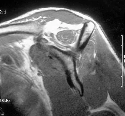
A 25-year-old tennis player has shoulder pain and weakness to external rotation. MRI scans are shown in Figures 16a and 16b. What is the most likely cause of his weakness?


Explanation

16b The MRI scans show a paralabral cyst, which is most commonly associated with labral tears. Compression of the suprascapular nerve results in weakness of the supraspinatus and/or infraspinatus depending on the level of compression. Piatt BE, Hawkins RJ, Fritz RC, et al: Clinical evaluation and treatment of spinoglenoid notch ganglion cysts. J Shoulder Elbow Surg 2002;11:600-604.

Question 2

The posterior approach to the proximal radius uses what intermuscular interval?


Explanation

Knowledge of intermuscular and internervous planes allows safe exposures throughout the body. The posterior (Thompson) approach to the proximal forearm uses the interval between the extensor carpi radialis brevis and extensor digitorum communis. The anterior (Henry) approach to the proximal forearm uses the interval between the brachioradialis and the flexor carpi radialis. Spinner M: Injuries to the Major Branches of Peripheral Nerves of the Forearm, ed 2. Philadelphia, PA, WB Saunders, 1978, pp 66-77.

Question 3

Which of the following statements best describes the anatomic considerations of the popliteal artery posterior to the knee joint?


Explanation

Popliteal artery injury during total knee arthroplasty is relatively rare. Knee flexion, the position that occurs during most of the arthroplasty procedure, allows the popliteal vessels to fall posteriorly, further away from harm. Anatomically, the popliteal artery lies anterior to the popliteal vein and 9 mm posterior to the posterior aspect of the tibial plateau in 90 degrees of flexion. Barrack RL, Booth RE Jr, Lonner JH, et al (eds): Orthopaedic Knowledge Update: Hip and Knee Reconstruction 3. Rosemont, IL, American Academy of Orthopaedic Surgeons, 2006, p 151.

Question 4

A 62-year-old woman reports diffuse aches and pains of the hip and pelvis. She denies any significant trauma but does have a history of chronic anemia. Figure 17a shows a radiograph of the pelvis, and Figures 17b and 17c show T2-weighted MRI scans. What is the most likely diagnosis?


Explanation

17b 17c The radiograph reveals diffuse osteopenia and areas in the proximal femora that are moth-eaten in appearance. The extent of the marrow-replacing process is evident on the MRI scans, which reveal signal abnormality throughout the entire pelvis and both proximal femora. This represents a marrow-packing process, of which multiple myeloma is the best choice. This diagnosis is also supported by the anemia noted on the patient's history. Metastatic carcinoma and lymphoma also may have a similar presentation.

Question 5

Involvement of what single muscle best distinguishes an L5 radiculopathy from a peroneal neuropathy?


Explanation

All of the muscles are innervated by the peroneal nerve with the exception of the tibialis posterior which is innervated by the tibial nerve. Tibialis posterior function is best tested with resistance to plantar flexion and inversion.

Question 6

What structure is located at the tip of the arrow in Figure 18?


Explanation

The structure shown is the exiting nerve root at the L3-4 disk, which is the right L3 root.

Question 7

A patient undergoes the procedure shown in Figure 19. An important part of this procedure is preservation of what wrist ligament?


Explanation

Proximal row carpectomy is a salvage wrist procedure that yields a surprisingly stable construct. This has been attributed to two factors: 1) the congruency of the head of the capitate in the lunate fossa (this articulation is less congruent than the native lunate/lunate fossa relationship, but surprisingly stable), and 2) preservation of the radioscaphocapitate ligament, the most radial of the palmar extrinsic ligaments, which prevents ulnar subluxation after proximal row carpectomy.

Question 8

A 23-year-old woman reports right knee pain and fullness. The pain is worse with activity but also present at rest. Radiographs are shown in Figures 20a and 20b. What is the most likely diagnosis?


Explanation

20b The radiographs reveal a predominantly lytic, destructive lesion of the distal femur, although there is a hint of some blastic change as well. The lesion has violated the cortex, and there is mineralization outside the cortex laterally. The lateral radiograph suggests a soft-tissue density. These aggressive changes on radiographs in this age group are strongly suggestive of osteosarcoma. Sanders TG, Parsons TW: Radiographic imaging of musculoskeletal neoplasia. Cancer Control 2001;8:221-231.

Question 9

What is the structure indicated by the letter "A" in Figure 21?


Explanation

The ligaments shown are the components of the lateral collateral ligament complex, and the structure indicated by the letter "A" is the radial collateral ligament. The lateral ulnar collateral ligament is the structure indicated by the letter "C" and the annular ligament is indicated by the letter "B." The transverse ligament is a component of the medial collateral ligament complex. Morrey BF: Anatomy of the elbow joint, in Morrey BF (ed): The Elbow and Its Disorders. Philadelphia, PA, WB Saunders, 1993, p 30.

Question 10

A 16-year-old boy sustains a twisting injury to the left knee while wrestling. MRI scans are shown in Figures 22a through 22c. What is the most likely diagnosis?


Explanation

22b 22c The MRI scans show a displaced bucket-handle medial meniscus tear that can be visualized on coronal, sagittal, and axial views. The sagittal view shows the typical "double posterior cruciate ligament sign," in which the low-signal bucket-handle fragment parallels the normal low-signal posterior cruciate ligament. The coronal and axial images both show the displaced medial meniscus in the notch. Sanders TG, Miller MD: A systematic approach to magnetic resonance imaging interpretation of sports medicine injuries of the knee. Am J Sports Med 2005;33:131-148.

Question 11

A 48-year-old woman reports bilateral thigh pain that is limiting her function as a librarian. A radiograph and a bone scan are shown in Figures 23a and 23b. What is the most likely diagnosis?


Explanation

23b The radiograph reveals bilateral severe acetabular protrusio. The bone scan and history confirm involvement of multiple joints, including the knees and the hindfoot. Although the first four choices can all cause the acetabular protrusio, the associated multiple joint involvement suggests the diagnosis of rheumatoid arthritis. Arthrokatadysis, or primary protrusio acetabuli, is often associated with osteomalacia but not other joint disease. Developmental dysplasia is a common cause of bilateral hip pathology but does not have acetabular protrusio. Resnick D: Diagnosis of Bone and Joint Disorders, ed 3. Philadelphia, PA, WB Saunders, 1995, pp 956-957. Wheeless' Textbook of Orthopaedics: Acetabular Protrusio. www.wheelessonline.com/ortho/acetabular_protrusio

Question 12

At the level of tibial bone resection in total knee arthroplasty, where does the common peroneal nerve lie?


Explanation

At the level of tibial bone resection in total knee arthroplasty, the common peroneal nerve lies superficial to the lateral head of the gastrocnemius and is therefore protected by this structure. In an MRI study of 60 knees, the mean distance from the bony posterolateral corner of the tibia to the nerve was 1.49 cm, with no distance less than 0.9 cm. The distance from the bone to nerve was greater in larger legs. Clarke HD, Schwartz JB, Math KR, et al: Anatomic risk of peroneal nerve injury with the "pie crust" technique for valgus release in total knee arthroplasty. J Arthroplasty 2004;19:40-44.

Question 13

Figures 24a through 24c show the coronal T1-weighted, T2-weighted fat-saturated, and T1-weighted fat-saturated gadolinium MRI scans of the proximal thigh of a 52-year-old woman who reports a mass in the medial thigh and groin area. She notes that the fullness has grown in size over the course of many months. Based on these findings, what is the most likely diagnosis?


Explanation

24b 24c The images show a complex, lobular lesion of the thigh that has signal characteristics that follow fat. The size of the lesion, the areas of stranding within the mass, along with mild uptake on the gadolinium sequences and the mild edema within the lesion on the T2-weighted image make liposarcoma the most likely diagnosis and simple intramuscular lipoma far less likely. All other diagnoses listed would not follow fat characteristics shown on the MRI sequences.

Question 14

The arrows in the axial T1-weighted MRI scan shown in Figure 25 show which of the following structures?


Explanation

The arrows in the figure show the deep branch of the ulnar nerve (more radial) and the superficial branch of the ulnar nerve within Guyon's canal. Guyon's canal is approximately 4 cm long beginning at the proximal extent of the transverse carpal ligament and ends at the aponeurotic arch of the hypothenar muscles. Many structures comprise the boundaries of Guyon's canal. The floor, for example, consists of the transverse carpal ligament, the pisohamate and pisometacarpal ligaments, and the opponens digiti minimi. Within Guyon's canal, the ulnar nerve bifurcates into the superficial and deep branches. The ulnar artery is immediately adjacent and radial to the ulnar nerve. The median nerve is visualized within the carpal tunnel, and the palmar cutaneous branch is more radial to Guyon's canal and volar to the carpal tunnel. The radial and ulnar digital nerves to the little finger are branches off of the superficial branch of the ulnar nerve distal to its emergence from Guyon's canal. The ulnar artery is the round structure located radial to the branches of the ulnar nerve within Guyon's canal. Adjacent to the ulnar artery are two small veins. The dorsal cutaneous branch of the ulnar nerve branches from the ulnar nerve in the distal forearm, well proximal to Guyon's canal. The common digital artery to the fourth web branches from the superficial palmar arch distal to Guyon's canal. The hook of the hamate is clearly seen in the figure, orienting the observer to the ulnar side of the wrist. Gross MS, Gelberman RH: The anatomy of the distal ulnar tunnel. Clin Orthop Relat Res 1985;196:238-247.

Question 15

An 82-year-old man has had episodic right thigh pain after undergoing a total hip arthroplasty 10 years ago. Initial postoperative radiographs are shown in Figures 26a and 26b, and current radiographs are shown in Figures 26c and 26d. What is the most likely cause of his pain?


Explanation

26b 26c 26d These radiographs are dominated by the subsidence of the femoral component. There is also evidence of polyethylene wear and femoral osteolysis in the region of the greater trochanter. There is no evidence of proximal (calcar) stress shielding, and there is a thick distal pedestal. Engh and associates defined two major signs of osseointegration - the absence of radiolucent lines around the porous-surfaced portion of the implant and new bone bridging the gap between the endosteal surface and the porous portion of the implant. Implant migration indicates failure of ingrowth. Osteolysis is a periprosthetic loss of bone secondary to particulate debris and it is often clinically silent unless it is accompanied by pathologic fracture. It is often globular. Acetabular loosening is based on radiolucent lines and implant migration. The current radiographs demonstrate subsidence of the stem with pedestal formation. Engh CA, Massin P, Suthers KE: Roentgenographic assessment of biologic fixation of porous-surface femoral components. Clin Orthop Relat Res 1990;257:107-128.

Question 16

A 37-year-old patient with type I diabetes mellitus has a flexor tenosynovitis of the thumb flexor tendon sheath following a kitchen knife puncture wound to the volar aspect of the thumb. Left unattended, this infection will likely first spread proximally creating an abscess in which of the following spaces of the palm?


Explanation

Flexor tenosynovitis of the thumb flexor tendon sheath can spread proximally and form an abscess within the thenar space of the palm. The flexor pollicis longus tendon does not pass through the central space of the palm or the hypothenar space of the palm. The flexor pollicis longus tendon does pass through the carpal tunnel, but this is not a palmar space. The three palmar spaces include the hypothenar space, the thenar space, and the central space. The posterior adductor space would likely only be involved secondarily after spread from a thenar space infection. Hollinshead W: Anatomy for Surgeons: The Back and Limbs, ed 3. Philadelphia, PA, Harper and Row, 1982, vol 3, pp 478-479.

Question 17

What tendon is closest to an appropriately placed anterolateral portal for ankle arthroscopy?


Explanation

The appropriate placement of the anterolateral portal provides access to the lateral gutter of the joint while avoiding the superficial peroneal nerve. The safest location for the portal is approximately 4 mm lateral to the peroneus tertius tendon, the closest of the tendons listed to the anterolateral portal. Because the superficial peroneal nerve location is variable, attempts to visualize, palpate, or transilluminate the nerve are mandatory.

Question 18

A 52-year-old woman reports nagging shoulder pain that has been present for months and is slowly progressive in nature. The patient also reports nocturnal pain and notes that the pain is not activity related. Figures 27a and 27b show the radiograph and bone scan, and Figures 27c through 27e show T1-weighted, T2-weighted, and gadolinium MRI scans, respectively. Based on these findings, what is the most likely diagnosis?


Explanation

27b 27c 27d 27e The radiograph reveals a metaphyseal lesion with some stippled mineralization suggesting a chondroid tumor. The bone scan shows increased uptake, beyond what is expected for a simple enchondroma, and beyond the limits of the lesion. The MRI sequences shows a lobular lesion on the T1- and T2-weighted (bright on the T2 sequence) images with inhomogeneous uptake of gadolinium; both findings are typical for a chondroid lesion. The history of pain, the positive bone scan, the age of the patient, the size of the lesion, and the central location (enostotic) of the lesion all suggest a malignant cartilage tumor. The images are not consistent with the other diagnoses. In particular, plasmacytoma is more uniformly bright on T2-weighted images and often has a negative bone scan. Menendez LR (ed): Orthopaedic Knowledge Update: Musculoskeletal Tumors. Rosemont, IL, American Academy of Orthopaedic Surgeons, 2002, pp 187-194.

Question 19

Figure 28 shows an arthroscopic view of a right shoulder in the lateral position through a posterior portal. What is the area between structure B (biceps) and SS (subscapularis tendon)?


Explanation

The rotator cuff interval is located between the supraspinatus and subscapularis and the biceps tendon is deep to the interval. It is a triangular area where the base is the coracoid process and the apex is the transverse humeral ligament at the biceps sulcus. Closure or tightening of this area is often helpful in patients with shoulder instability. Conversely, this area is often contracted in patients with adhesive capsulitis and may need to be released. Selecky MT, Tibone JE, Yang BY, et al: Glenohumeral joint translation after arthroscopic thermal capsuloplasty of the rotator interval. J Shoulder Elbow Surg 2003;12:139-143.

Question 20

New painful paresthesias near the site of the incision after an ulnar nerve transposition is the result of injury to what nerve?


Explanation

Branches of the medial antebrachial cutaneous nerve can often be identified during routine ulnar nerve surgery crossing the medial aspect of the elbow. It should be preserved to avoid development of painful paresthesias.

Question 21

A 23-year-old man reports pain on the superior aspect of his right shoulder with repetitive overhead activities and when lying on his right side. Figure 29 shows an axial MRI scan. What is the most likely diagnosis based on the MRI findings?


Explanation

Os acromiale represents a failure of fusion of the anterior acromial apophysis and has been reported in approximately 8% of the population. Patients with a symptomatic os acromiale often report impingement-type symptoms with pain over the superior acromion, especially with overhead activities or sleeping. When nonsurgical management is unsuccessful, surgical options include excision, open reduction and internal fixation, and arthroscopic decompression. Kurtz CA, Humble BJ, Rodosky MW, et al: Symptomatic os acromiale. J Am Acad Orthop Surg 2006;14:12-19.

Question 22

Following a chevron bunionectomy performed through a dorsal approach, a patient has persistent numbness on the dorsal and medial aspect of the hallux. What nerve has most likely been injured?


Explanation

The dorsomedial cutaneous nerve of the hallux, which is a distal branch of the superficial peroneal nerve, supplies sensation to the skin on the dorsal and medial half of the hallux and may be injured during a chevron bunionectomy. Injury to the nerve leads to particularly painful neuromas that directly impinge on the shoe. For this reason, direct medial approaches are typically preferred for access to the medial aspect of the metatarsophalangeal joint.

Question 23

A 74-year-old man reports progressive left hip pain with weight-bearing activities. A radiograph is shown in Figure 30. What is the most likely underlying diagnosis?


Explanation

The radiograph shows enlargement of the bone, coarse trabeculation, a blastic appearance, and thickening of the cortex, revealing the classic appearance of Paget's disease in the sclerotic phase, the most common presentation. While lymphoma may present as a blastic lesion, it will not have the same enlargement, coarse trabeculation of bone, and the significant sclerosis seen here. Menendez LR (ed): Orthopaedic Knowledge Update: Musculoskeletal Tumors. Rosemont, IL, American Academy of Orthopaedic Surgeons, 2002, pp 211-215.

Question 24

The anatomy of the sciatic nerve as it exits the pelvis is best described as exiting through the


Explanation

The sciatic nerve is formed by the roots of the lumbosacral plexus. It exits the pelvis through the greater sciatic notch and appears in the buttock anterior to the piriformus. From that point, the sciatic nerve passes posteriorly over the superior gemellus, obturator internus, inferior gemellus, and quadratus femoris before it passes deep to the biceps femoris. The tendon of the obturator internus passes through the lesser sciatic notch. Hoppenfeld S, deBoer P: Surgical Exposures in Orthopaedics: The Anatomic Approach. Philadelphia, PA, JB Lippincott, 1984, p 347. Anderson JE: Grant's Atlas of Anatomy, ed 7. Baltimore, MD, Lippincott Williams & Wilkins, 1978, pp 4-34, 4-36.

Question 25

What complication is more likely following excessive medial retraction of the anterior covering structures during the anterolateral (Watson-Jones) approach to the hip?


Explanation

The femoral nerve is the most lateral structure in the anterior neurovascular bundle. The femoral artery and vein lie medial to the nerve. Retractors placed in the anterior acetabular lip should be safe, although neurapraxia of the femoral nerve may occur if retraction is prolonged or forceful leading to quadriceps weakness. The femoral artery and nerve are well protected by the interposed psoas muscle. Damage to the lateral femoral cutaneous nerve, causing numbness over the anterolateral thigh, can occur while developing the interval between the tensor fascia latae and sartorious in the anterior (Smith-Petersen) approach but less likely in the Watson-Jones approach. Superior gluteal injury and accompanying abductor insufficiency may occur during excessive splitting of the glutei during the direct lateral (Hardinge) approach. Foot drop secondary to sciatic injury is more common with a posterior exposure or posterior retractor placement. Hoppenfeld S, deBoer P: Surgical Exposures in Orthopaedics: The Anatomic Approach. Philadelphia, PA, JB Lippincott, 1984, p 325.

Question 26

During a surgical dislocation of the hip, the surgeon must protect the deep branch of the medial femoral circumflex artery (MFCA). What is the anatomical path of this vessel as it courses toward the femoral head?


Explanation

The deep branch of the MFCA is the predominant blood supply to the femoral head. It consistently courses between the quadratus femoris and the inferior gemellus, passing anterior to the conjoined tendon.

Question 27

A patient sustains a midshaft radius fracture, and a volar (Henry) approach is chosen for fixation. What is the internervous plane for the proximal third of this approach?


Explanation

The proximal portion of the Henry approach to the radius utilizes the internervous plane between the brachioradialis (innervated by the radial nerve) and the pronator teres (innervated by the median nerve).

Question 28

A surgeon is performing an open Latarjet procedure. Which nerve is most at risk during the coracoid osteotomy and subsequent mobilization of the conjoined tendon?


Explanation

The musculocutaneous nerve typically penetrates the coracobrachialis muscle 2 to 8 cm distal to the tip of the coracoid process. Aggressive distal retraction can cause neurapraxia or permanent injury.

Question 29

During reconstruction of the posterolateral corner of the knee, the surgeon identifies the popliteofibular ligament. Which of the following accurately describes the origin and insertion of this structure?


Explanation

The popliteofibular ligament is a critical static stabilizer of the posterolateral corner. It originates from the popliteus musculotendinous junction and inserts distally onto the posteromedial fibular styloid.

Question 30

When establishing the anterolateral portal for ankle arthroscopy, the surgeon must be careful to avoid injuring a nerve that supplies sensation to the dorsum of the foot. Which nerve is this?


Explanation

The superficial peroneal nerve, specifically its intermediate dorsal cutaneous branch, is at highest risk during placement of the anterolateral ankle portal, which is typically made just lateral to the peroneus tertius.

Question 31

Which pulleys in the flexor tendon sheath of the digits are most critical to preserve in order to prevent mechanical bowstringing?


Explanation

The A2 (located over the proximal phalanx) and A4 (located over the middle phalanx) pulleys are the thickest and biomechanically most critical pulleys to prevent flexor tendon bowstringing.

Question 32

A patient presents with weakness in elbow flexion and forearm supination, but normal shoulder abduction and normal wrist extension. Which neural structure is most likely injured?


Explanation

The musculocutaneous nerve innervates the biceps brachii and brachialis, driving elbow flexion and aiding in supination. Normal shoulder abduction rules out an upper trunk injury.

Question 33

Which two tendons cross at the anatomical landmark known as the Master Knot of Henry in the plantar aspect of the foot?


Explanation

At the Master Knot of Henry, located in the plantar midfoot, the flexor hallucis longus tendon crosses dorsal (superior) to the flexor digitorum longus tendon.

Question 34

When placing lumbar pedicle screws using standard open anatomical landmarks, what is the accepted entry point?


Explanation

The standard entry point for a lumbar pedicle screw is the intersection of a horizontal line bisecting the transverse process and a vertical line tangent to the lateral border of the superior articular facet.

Question 35

The anterior bundle of the ulnar collateral ligament (UCL) of the elbow is the primary restraint to valgus stress. How do the distinct bands of the anterior bundle function during elbow range of motion?


Explanation

The anterior bundle consists of two bands: the anterior band is primarily tight in elbow extension, while the posterior band tightens progressively as the elbow is flexed.

Question 36

In total hip arthroplasty, placement of a screw in the anteroinferior quadrant of the acetabulum puts which of the following structures at greatest risk?


Explanation

The acetabulum is divided into four quadrants. The anteroinferior quadrant contains the obturator nerve and vessels, making it an unsafe zone for screw placement.

Question 37

During a posterior approach to the proximal humerus for a nonunion, you are identifying the axillary nerve in the quadrilateral space. Which of the following defines the superior border of this space when viewed posteriorly?


Explanation

Viewed posteriorly, the superior border of the quadrilateral space is the teres minor. The other borders are the teres major (inferior), long head of the triceps (medial), and the surgical neck of the humerus (lateral).

Question 38

A 45-year-old man undergoes an open reduction and internal fixation of a midshaft clavicle fracture. Postoperatively, he notes numbness over the anterolateral aspect of his shoulder. Which nerve was most likely injured during the procedure?


Explanation

The supraclavicular nerves (C3-C4) provide sensation over the anterior shoulder and clavicle. They are frequently injured during surgical approaches to the clavicle, leading to postoperative numbness.

Question 39

A surgeon is performing an extensile lateral approach to the calcaneus. The sural nerve is at risk during this exposure. The sural nerve is typically formed by the junction of the medial sural cutaneous nerve and the communicating branch of which nerve?


Explanation

The sural nerve is formed by the union of the medial sural cutaneous nerve (a branch of the tibial nerve) and the communicating branch of the lateral sural cutaneous nerve (a branch of the common peroneal nerve).

Question 40

When utilizing the standard anterolateral (Watson-Jones) approach to the hip, what is the intermuscular internervous interval?


Explanation

The Watson-Jones approach utilizes the interval between the tensor fasciae latae and the gluteus medius. Both muscles are innervated by the superior gluteal nerve, making this a non-true internervous plane.

Question 41

A patient sustains a posterior shoulder dislocation, and MRI reveals an isolated tear of the teres minor. During a posterior surgical approach, the surgeon must identify the quadrangular space. Which of the following best describes the inferior boundary of this space?


Explanation

The quadrangular space is bounded superiorly by the teres minor, inferiorly by the teres major, medially by the long head of the triceps, and laterally by the surgical neck of the humerus. It transmits the axillary nerve and posterior circumflex humeral artery.

Question 42

During a pectoralis major tendon repair following a weightlifting injury, the surgeon must recreate its anatomic insertion. Which of the following best describes the insertion of the sternal head of the pectoralis major?


Explanation

The pectoralis major tendon twists 90 degrees before its insertion. The sternocostal (inferior) head inserts deep and proximal to the clavicular (superior) head on the lateral lip of the intertubercular groove.

Question 43

Where is the main arterial supply to the femoral head most vulnerable during a posterior approach to the hip (Kocher-Langenbeck)?


Explanation

The deep branch of the medial circumflex femoral artery (MFCA) courses deep to the quadratus femoris. It can be injured if the superior portion of this muscle is aggressively divided or elevated during a posterior hip approach.

Question 44

During an anterolateral approach to the distal tibia for plating a pilon fracture, the surgeon must protect the superficial peroneal nerve. At what average distance proximal to the tip of the lateral malleolus does this nerve typically pierce the deep fascia to become superficial?


Explanation

The superficial peroneal nerve typically pierces the deep crural fascia in the anterolateral leg approximately 10 to 12 cm proximal to the tip of the lateral malleolus. It is highly vulnerable to injury during anterolateral approaches to the ankle.

Question 45

A patient undergoes percutaneous repair of an acute Achilles tendon rupture. To avoid trapping the sural nerve with a lateral suture pass, the surgeon must be aware of its anatomic course. Where does the sural nerve typically cross the lateral border of the Achilles tendon?


Explanation

The sural nerve typically courses from the midline of the posterior calf and crosses the lateral border of the Achilles tendon at an average of 10 cm proximal to the calcaneal tuberosity. Suture passes placed lateral to the tendon at this level pose a high risk of nerve entrapment.

Question 46

A 45-year-old man presents with numbness in his ring and small fingers and weakness in his intrinsic hand muscles. EMG testing localizes compression of the ulnar nerve in the distal arm. Which of the following structures is most likely responsible for this compression?


Explanation

The Arcade of Struthers is a fascial band extending from the medial head of the triceps to the medial intermuscular septum, located about 8 cm proximal to the medial epicondyle. It compresses the ulnar nerve, distinguishing it from the Ligament of Struthers which compresses the median nerve.

Question 47

A 30-year-old man sustains a closed tibial shaft fracture and develops compartment syndrome. During a four-compartment fasciotomy, the deep posterior compartment must be released. Which of the following muscles is NOT located in the deep posterior compartment of the leg?


Explanation

The deep posterior compartment of the leg contains the flexor hallucis longus, flexor digitorum longus, tibialis posterior, and popliteus muscles. The peroneus brevis is located in the lateral compartment.

Question 48

A 25-year-old woman presents with dorsal wrist pain with extension. Examination reveals swelling and tenderness over the 4th dorsal extensor compartment. Which of the following structures are contained within this compartment?


Explanation

The 4th dorsal extensor compartment of the wrist contains the extensor digitorum communis (EDC) and the extensor indicis proprius (EIP). The posterior interosseous nerve (PIN) is located deep to these tendons at the level of the radiocarpal joint.

Question 49

During an anterior (Smith-Petersen) approach to the hip, an internervous plane is utilized. Which of the following best describes the innervation of the muscles defining the superficial boundary of this plane?


Explanation

The superficial plane in the Smith-Petersen approach is between the tensor fasciae latae (superior gluteal nerve) and the sartorius (femoral nerve). The deep plane is between the rectus femoris (femoral nerve) and gluteus medius (superior gluteal nerve).

Question 50

A 28-year-old athlete sustains a multi-ligament knee injury. Examination reveals a positive dial test at 30 degrees of flexion but normal at 90 degrees. Injury to the posterolateral corner (PLC) is suspected. Which of the following structures attaches to the anteromedial aspect of the fibular styloid?


Explanation

The popliteofibular ligament attaches to the anteromedial aspect of the fibular styloid. The fibular collateral ligament (LCL) attaches laterally on the fibular head, and the biceps femoris surrounds the LCL.

Question 51

A 32-year-old overhead throwing athlete presents with poorly localized posterior shoulder pain and deltoid weakness. MRI reveals atrophy of the teres minor. Which of the following defines the borders of the space where the affected neurovascular bundle is likely compressed?


Explanation

Quadrilateral space syndrome involves compression of the axillary nerve and posterior circumflex humeral artery. The boundaries are the teres minor (superior), teres major (inferior), long head of the triceps (medial), and the surgical neck of the humerus (lateral).

Question 52

While performing an ilioinguinal approach for an anterior column acetabular fracture, significant hemorrhage is encountered posterior to the superior pubic ramus. This is most likely due to an injury to the "corona mortis", which is an anastomosis between which two vessel systems?


Explanation

The corona mortis is a vascular anastomosis between the external iliac system (specifically the inferior epigastric vessels) and the obturator system. It rests on the posterior aspect of the superior pubic ramus and is highly vulnerable during anterior pelvic exposures.

Question 53

A 12-year-old child presents with vague forearm pain and weakness in thumb flexion and forearm pronation. Radiographs reveal a bony spur on the anteromedial humerus, 5 cm proximal to the medial epicondyle. Which structure is most likely compressed by the ligament connecting this spur to the medial epicondyle?


Explanation

A supracondylar process can be connected to the medial epicondyle via the Ligament of Struthers. This fibrous band can compress the median nerve and brachial artery, leading to proximal median nerve entrapment.

Question 54

During a transforaminal endoscopic lumbar discectomy, the surgeon navigates instruments through Kambin's triangle to safely access the disc space. Which of the following structures forms the anterior border of Kambin's triangle?


Explanation

Kambin's triangle is a safe working zone for transforaminal access. Its borders are the exiting nerve root (anteriorly/superiorly), the superior articular process of the inferior vertebra (posteriorly), and the superior endplate of the inferior vertebra (inferiorly).

Question 55

A cyclist presents with weakness in finger abduction and adduction, but normal sensation in the little and ring fingers. A lesion in Guyon's canal is suspected. At which specific zone of Guyon's canal is the compression most likely located?


Explanation

Guyon's canal is divided into three zones. Zone 2 contains only the deep motor branch of the ulnar nerve; compression here results in isolated intrinsic muscle weakness without sensory deficits.

Question 56

A 28-year-old volleyball player has weakness in external rotation of the shoulder. Examination shows atrophy of the infraspinatus but normal bulk of the supraspinatus. Sensation is intact. Where is the most likely site of nerve compression?


Explanation

The suprascapular nerve innervates the supraspinatus before passing through the spinoglenoid notch to innervate the infraspinatus. Entrapment at the spinoglenoid notch (e.g., from a paralabral cyst) causes isolated infraspinatus weakness.

Question 57

During a surgical approach to the midfoot for an accessory navicular excision, the surgeon identifies a tendinous crossover beneath the navicular. At this location, known as the Master Knot of Henry, which anatomical relationship is correct?


Explanation

At the Master Knot of Henry, the flexor digitorum longus (FDL) tendon crosses superficial (plantar) to the flexor hallucis longus (FHL) tendon. This landmark is critical when harvesting the FDL for tendon transfers.

Question 58

The volar approach to the proximal radius (Henry approach) requires dissection between two muscles to expose the supinator. Which of the following nerves innervates the muscle forming the medial border of this proximal internervous plane?


Explanation

The proximal internervous plane in the volar (Henry) approach is between the brachioradialis (radial nerve) laterally and the pronator teres (median nerve) medially.

Question 59

A 45-year-old patient sustains a nondisplaced distal radius fracture and is treated in a cast. Six weeks later, she presents with sudden inability to extend her thumb interphalangeal joint. The ruptured tendon normally travels in which extensor compartment, and what is its radial boundary?


Explanation

The extensor pollicis longus (EPL) resides in the 3rd dorsal extensor compartment. It is bounded radially by Lister's tubercle, which acts as a fulcrum and makes the tendon vulnerable to rupture after distal radius fractures.

Question 60

The lateral femoral cutaneous nerve (LFCN) is at risk during anterior pelvic approaches. In the most common anatomical variant, where does the LFCN pass in relation to the anterior superior iliac spine (ASIS) and inguinal ligament?


Explanation

In its most common anatomic course, the LFCN passes under the inguinal ligament approximately 1-2 cm medial to the ASIS. Surgeons must remain vigilant for variants where it passes through the sartorius or over the iliac crest.

Question 61

In a patient with tarsal tunnel syndrome, a release of the flexor retinaculum is planned. From anterior to posterior, what is the correct order of structures passing behind the medial malleolus?


Explanation

The structures passing through the tarsal tunnel from anterior to posterior are the Tibialis posterior, Flexor Digitorum Longus, Posterior tibial Artery, Vein, Nerve, and Flexor Hallucis Longus (remembered by the mnemonic "Tom, Dick, AND Very Nervous Harry").

Question 62

During operative fixation of an ankle fracture, a surgeon evaluates the distal tibiofibular syndesmosis. The anterior inferior tibiofibular ligament (AITFL) originates from the Chaput tubercle. On which bone is the Chaput tubercle located, and where does the AITFL insert?


Explanation

The Chaput tubercle is located on the anterolateral aspect of the distal tibia. The AITFL originates here and inserts onto the Wagstaffe tubercle on the anterior aspect of the distal fibula.

Question 63

During the Smith-Petersen (anterior) approach to the hip, which internervous interval is utilized superficially?


Explanation

The superficial interval of the anterior approach to the hip is between the sartorius (innervated by the femoral nerve) and the tensor fasciae latae (innervated by the superior gluteal nerve). This provides a true internervous plane.

Question 64

Hypertrophy of the teres minor, teres major, long head of the triceps, and the surgical neck of the humerus creates a tight quadrangular space. Which of the following structures are at highest risk of compression?


Explanation

The quadrangular space is bounded by the teres minor (superior), teres major (inferior), long head of the triceps (medial), and surgical neck of the humerus (lateral). It contains the axillary nerve and the posterior circumflex humeral artery.

Question 65

Which of the following statements most accurately describes the primary blood supply to the scaphoid, predisposing it to proximal pole avascular necrosis?


Explanation

The primary blood supply to the scaphoid is retrograde, provided by the dorsal carpal branch of the radial artery, which enters the distal pole. Fractures at the waist or proximal pole disrupt this retrograde flow, leading to avascular necrosis.

Question 66

The volar (Henry) approach to the proximal radius utilizes an internervous interval between which two muscles?


Explanation

The proximal superficial interval in the Henry approach is between the brachioradialis (innervated by the radial nerve) and the pronator teres (innervated by the median nerve). Distally, the interval shifts between the brachioradialis and the flexor carpi radialis.

Question 67

Within Guyon's canal, the ulnar nerve bifurcates into superficial and deep branches. The deep branch dives into the hand by passing between which two muscular structures?


Explanation

The deep motor branch of the ulnar nerve dives deep into the palm by passing between the abductor digiti minimi and the flexor digiti minimi brevis. It then supplies the hypothenar muscles, interossei, the two ulnar lumbricals, and the adductor pollicis.

Question 68

What is the correct anatomic order of structures passing behind the medial malleolus within the tarsal tunnel, strictly from anterior to posterior?


Explanation

The structures from anterior to posterior are the Tibialis posterior, Flexor Digitorum Longus, posterior tibial Artery, posterior tibial Vein, tibial Nerve, and Flexor Hallucis Longus. This is famously remembered by the mnemonic "Tom, Dick, And Very Nervous Harry".

Question 69

The femoral attachment (footprint) of the anterior cruciate ligament (ACL) is anatomically located on the:


Explanation

The ACL originates from the posteromedial aspect of the lateral femoral condyle. It then courses distally, medially, and anteriorly to insert on the anterior intercondylar area of the tibia.

Question 70

During a posterolateral approach to the ankle for fixation of a posterior malleolus fracture, the sural nerve is at risk. It typically courses distally in close proximity to which vascular structure?


Explanation

The sural nerve travels down the posterolateral aspect of the leg and ankle, consistently running alongside the small saphenous vein. Surgeons must carefully identify and mobilize this neurovascular bundle during a posterolateral approach.

Question 71

The predominant blood supply to the adult femoral head is provided by the:


Explanation

The medial femoral circumflex artery (MFCA) is the primary blood supply to the adult femoral head, specifically via its lateral epiphyseal branches. Disruption of the MFCA significantly increases the risk of avascular necrosis.

Question 72

The roof of the cubital tunnel, which can become tightened and compress the ulnar nerve during elbow flexion, is formed by which structure?


Explanation

Osborne's ligament (or Osborne's fascia) forms the roof of the cubital tunnel. It spans between the olecranon and the medial epicondyle and is continuous with the aponeurosis of the two heads of the flexor carpi ulnaris.

Question 73

When placing a pedicle screw in the lumbar spine, the anatomic starting point is best described as the intersection of the:


Explanation

The starting point for a lumbar pedicle screw is located at the intersection of a vertical line dropped down the lateral border of the superior articular facet and a horizontal line bisecting the transverse process.

Question 74

The rotator interval is a clinically important anatomical space in the shoulder. What are its superior and inferior borders?


Explanation

The rotator interval is bordered superiorly by the anterior margin of the supraspinatus tendon and inferiorly by the superior margin of the subscapularis tendon. It contains the coracohumeral ligament, superior glenohumeral ligament, and the long head of the biceps tendon.

Question 75

Entrapment of the deep peroneal nerve beneath the inferior extensor retinaculum (anterior tarsal tunnel syndrome) typically causes isolated sensory loss in which anatomic area?


Explanation

The deep peroneal nerve provides motor innervation to the short toe extensors and sensory innervation strictly to the first dorsal web space. Compression at the ankle primarily manifests as pain and numbness in this specific web space.

Question 76

During a standard deltopectoral approach to the shoulder, the cephalic vein is typically identified and retracted in which direction to best preserve its primary venous drainage?


Explanation

The cephalic vein is typically retracted laterally with the deltoid to preserve its major venous tributaries from the deltoid muscle. If injured or ligated, patients may experience increased postoperative swelling of the shoulder.

Question 77

The flexor digitorum superficialis (FDS) tendon bifurcates at the level of the proximal phalanx to allow the flexor digitorum profundus (FDP) tendon to pass through. What is the anatomic term for this bifurcation structure?


Explanation

Camper's chiasm is the structural splitting of the FDS tendon that allows the FDP tendon to emerge superficially to insert on the distal phalanx. This transition occurs within the flexor sheath near the proximal interphalangeal joint.

Question 78

During a posterolateral corner reconstruction of the knee, identifying the femoral footprints of the stabilizing structures is crucial. What is the anatomic relationship of the popliteus tendon (PT) insertion relative to the fibular collateral ligament (FCL) origin on the lateral femoral condyle?


Explanation

On the lateral femoral condyle, the popliteus tendon inserts an average of 18.5 mm distal and anterior to the origin of the fibular collateral ligament. This anatomical relationship is critical to recreate during posterolateral corner reconstructions.

Question 79

The anterior approach to the hip (Smith-Petersen) utilizes a superficial internervous plane between two muscles. Which of the following nerves innervate these two muscles?


Explanation

The superficial interval for the anterior approach to the hip is between the tensor fasciae latae, innervated by the superior gluteal nerve, and the sartorius, innervated by the femoral nerve.

Question 80

A 40-year-old patient undergoes a lymph node biopsy in the posterior cervical triangle. Postoperatively, she cannot abduct her shoulder beyond 90 degrees and has lateral scapular winging. Which of the following muscles is primarily denervated?


Explanation

The spinal accessory nerve (CN XI) courses through the posterior cervical triangle and innervates the trapezius. Iatrogenic injury causes lateral scapular winging and an inability to actively abduct the shoulder above 90 degrees.

Question 81

When performing an anterolateral approach to the distal humerus, the radial nerve is at risk. Approximately how far proximal to the radiocapitellar joint does the radial nerve piece the lateral intermuscular septum?


Explanation

The radial nerve pierces the lateral intermuscular septum to pass from the posterior to the anterior compartment approximately 10 cm proximal to the radiocapitellar joint. It is highly susceptible to injury at this fixed tethering point during humeral surgery.

Question 82

A 32-year-old male sustains a displaced talar neck fracture. Which of the following blood vessels provides the predominant blood supply to the talar body and is most at risk of disruption in this injury?


Explanation

The artery of the tarsal canal, a branch of the posterior tibial artery, provides the predominant blood supply to the body of the talus. Disruption of this retrograde flow in displaced neck fractures leads to a high rate of avascular necrosis.

Question 83

A 28-year-old volleyball player presents with posterior shoulder pain and deltoid weakness. MRI reveals isolated atrophy of the teres minor. Which of the following defines the superior boundary of the anatomic space where the affected nerve is compressed?


Explanation

The axillary nerve is compressed in the quadrilateral space. Its anatomic boundaries are the teres minor (superior), teres major (inferior), long head of the triceps (medial), and surgical neck of the humerus (lateral).

Question 84

The extended volar approach to the radius (Henry approach) requires dissection between the flexor carpi radialis (FCR) and the brachioradialis. Which nerve innervates the muscle forming the lateral border of this interval?


Explanation

The lateral border of the Henry approach is the brachioradialis, which is innervated by the radial nerve. The medial border is the FCR, innervated by the median nerve.

Question 85

During an anterior approach to the pelvis for a periacetabular osteotomy, the lateral femoral cutaneous nerve is at risk. What is its most common anatomic relationship to the anterior superior iliac spine (ASIS)?


Explanation

The lateral femoral cutaneous nerve most commonly passes into the anterior thigh beneath the inguinal ligament, approximately 1-2 cm medial to the ASIS. Retraction in this area during anterior pelvic approaches can lead to meralgia paresthetica.

Question 86

A patient presents with isolated weakness in elbow flexion and decreased sensation over the lateral forearm following a traction injury. The affected nerve is formed by which cords of the brachial plexus?


Explanation

The musculocutaneous nerve arises exclusively from the lateral cord of the brachial plexus (C5-C7). It provides motor innervation to the biceps, brachialis, and coracobrachialis, and terminates as the lateral antebrachial cutaneous nerve.

Question 87

Displaced scaphoid waist fractures have a high rate of avascular necrosis due to retrograde blood flow. What artery provides the primary blood supply to the proximal pole of the scaphoid?


Explanation

The dorsal carpal branch of the radial artery provides the majority (70-80%) of the blood supply to the scaphoid. It enters the bone along the distal dorsal ridge and flows retrogradely to supply the proximal pole.

Question 88

During the ilioinguinal approach for an anterior column acetabular fracture, significant hemorrhage occurs behind the superior pubic ramus. This is most likely due to an injury to the "corona mortis", which represents an anastomosis between the:


Explanation

The corona mortis is a potentially lethal vascular anastomosis between the external iliac (or inferior epigastric) vessels and the obturator vessels. It is consistently found draped over the posterior aspect of the superior pubic ramus.

Question 89

The anterior inferior tibiofibular ligament (AITFL) is frequently injured in rotational ankle sprains. What is the eponym for the bony avulsion of the AITFL from its fibular attachment?


Explanation

A Wagstaffe-Le Fort fracture describes a bony avulsion of the AITFL from the anterior aspect of the distal fibula. A Tillaux-Chaput fracture is the equivalent avulsion from the anterolateral tibia.

Question 90

An extensile lateral approach is planned for an intra-articular calcaneus fracture. The sural nerve is at risk during the posterior limb of the incision. What venous structure accompanies the sural nerve posterior to the lateral malleolus?


Explanation

The sural nerve courses distally in the posterior lower leg and travels behind the lateral malleolus. It runs in close proximity to the small (lesser) saphenous vein in this region.

Question 91

When performing a posteromedial approach to the knee to repair the posterior horn of the medial meniscus, the sartorius fascia must be incised. Which of the following correctly identifies the anterior-to-posterior arrangement of the pes anserinus tendons?


Explanation

The anatomic arrangement of the pes anserinus tendons at their proximal tibial insertion from anterior to posterior (and superficial to deep) is Sartorius, Gracilis, and Semitendinosus.

Question 92

During an in situ ulnar nerve decompression at the cubital tunnel, the first branch of the ulnar nerve identified just distal to the medial epicondyle provides motor innervation to which muscle?


Explanation

The first motor branch of the ulnar nerve arises just distal to the medial epicondyle and innervates the flexor carpi ulnaris (FCU). Meticulous dissection to preserve these branches is essential during ulnar nerve mobilization.

Question 93

A patient undergoing arthroscopic rotator cuff repair receives an interscalene nerve block and subsequently develops transient hemidiaphragmatic paresis. The affected nerve shares its primary segmental root origin with which of the following nerves?


Explanation

Hemidiaphragmatic paresis is caused by block of the phrenic nerve, which originates from roots C3, C4, and C5. The long thoracic nerve originates from the C5, C6, and C7 nerve roots, sharing the C5 root with the phrenic nerve.

Question 94

During a dorsal approach to the wrist, the extensor pollicis longus (EPL) is released from its compartment and retracted radially. Around which bony landmark does the EPL normally pivot to change its mechanical line of pull?


Explanation

The extensor pollicis longus (EPL) runs in the 3rd extensor compartment and relies on Lister's tubercle on the dorsal radius as a mechanical pulley to angle its line of pull toward the thumb.

Question 95

In reconstructive hand surgery, preserving or reconstructing certain flexor pulleys is essential to prevent bowstringing of the flexor tendons. Which two annular pulleys are considered biomechanically most critical?


Explanation

The A2 pulley (located over the proximal phalanx) and A4 pulley (located over the middle phalanx) are the most critical annular pulleys for preventing flexor tendon bowstringing and maintaining proper digital excursion.

None

Clinic OS
Medically Verified Content by
Prof. Clinic OS
Consultant Orthopedic & Spine Surgeon
Chapter Index