العربية
Part of the Master Guide

AAOS & ABOS Basic Science MCQs (Set 3): Bone Biology, Biomechanics & Anatomy Review

AAOS Orthopedic Anatomy MCQs (Set 3): Musculoskeletal & Skeletal System | ABOS Board Prep

27 Apr 2026 56 min read 101 Views
Anatomy 2002 MCQs - Part 3

Key Takeaway

This high-yield question set (Set 3) for the AAOS/ABOS exams focuses on core orthopedic anatomy. It covers detailed musculoskeletal structures, key components of the skeletal system, and clinically vital neurovascular pathways. Enhance your understanding of foundational anatomy crucial for all orthopedic specializations and board preparation.

AAOS Orthopedic Anatomy MCQs (Set 3): Musculoskeletal & Skeletal System | ABOS Board Prep

Comprehensive 100-Question Exam


00:00

Start Quiz

Question 1

A 41-year-old man who plays golf regularly has had ulnar-sided wrist pain for the past several days after striking a tree root with a golf club. Examination reveals significant pain with resisted flexion of the ring and small fingers and tenderness over the hook of the hamate. Which of the following radiographic views would be most helpful in identifying the pathology of this injury?

Anatomy 2002 Practice Questions: Set 3 (Solved) - Figure 1


Explanation

The history and examination findings suggest an acute fracture of the hook of the hamate. The radiographic study considered most helpful in identifying this type of fracture is the carpal tunnel view. PA and lateral views of the wrist will not adequately visualize the hook of the hamate. Bruerton's view is intended for the assessment of the metacarpophalangeal joints. Pathology would not be suspected in the scaphoid, metacarpals, or the phalanges, so the scaphoid view and the PA, lateral, and oblique views of the hand would not be helpful. Green DP, Hotchkiss RN, Pederson WC (eds): Operative Hand Surgery, ed 4. New York, NY, Churchill Livingstone, 1999, p 855.

Question 2

What is the primary limiting membrane and mechanical support for the periphery of the physis?

Anatomy 2002 Practice Questions: Set 3 (Solved) - Figure 2


Explanation

The perichondrial fibrous ring of La Croix acts as a limiting membrane that provides mechanical support for the bone-cartilage junction of the growth plate. It is continuous with the ossification groove of Ranvier, which contributes chondrocytes for the increase in width of the growth plate. The zone of provisional calcification lies at the bottom of the hypertrophic zone and is the site of initial calcification of the matrix. It is quite weak and usually is the cleavage plane for fractures; therefore, it does not qualify as mechanical support. The last intact transverse septum separates the zone of provisional calcification from the primary spongiosa and provides no real support to the physis. The primary spongiosa is the part of the metaphysis nearest the physis. Netter FH: Growth plate, in Woodburne RT, Crelin ES, Kaplan FS, Dingle RV (eds): The Ciba Collection of Medical Illustrations. Summit, NJ, Ciba-Geigy Corporation, 1987, vol 8, pp 166-167.

Question 3

What normal tissue has a low signal intensity (appears black) on both T1- and T2-weighted images?

Anatomy 2002 Practice Questions: Set 3 (Solved) - Figure 3


Explanation

Tendons, cortical bone, ligaments, menisci, and fibrous tissue will show low signal intensity (SI) on both T1- and T2-weighted images. Fat-containing tissues, such as subcutaneous fat and bone marrow, will show high SI on T1-weighted images and low SI on T2-weighted images. Tissues with high water content, such as joint fluid, intervertebral disk, and edema, will show low SI on T1-weighted images and high SI on T2-weighted images. Kasser JR (ed): Orthopaedic Knowledge Update 5. Rosemont, IL, American Academy of Orthopaedic Surgeons, 1996, pp 65-70.

Question 4

A positive Froment sign indicates weakness of which of the following muscles?

Anatomy 2002 Practice Questions: Set 3 (Solved) - Figure 4


Explanation

Thumb adduction is powered by the adductor pollicis (ulnar nerve). Testing involves having the patient forcibly hold a piece of paper between the thumb and radial side of the index proximal phalanx. When this muscle is weak or nonfunctioning, the thumb interphalangeal joint flexes with this maneuver, resulting in a positive Froment sign. The paper is held by action of the thumb flexion (flexor pollicis longus and flexor pollicis brevis; median innervated).

Question 5

Figure 28 shows an AP radiograph of a 54-year-old woman who underwent lumbar laminectomy and fusion at the L4 and L5 levels with placement of a bone stimulator 8 years ago. She also underwent a left total hip arthroplasty 2 years ago; aspiration of that joint now reveals that it is infected with a gram-positive cocci organism. History is also significant for IV drug use and human immunodeficiency virus (HIV). The patient reports fever, chills, and left flank and abdominal pain. Examination reveals significant pain with resisted left hip flexion and passive hip extension. She also has lumbar hyperlordosis. Which of the following studies would best identify the underlying cause of her infection?

Anatomy 2002 Practice Questions: Set 3 (Solved) - Figure 5


Explanation

The patient's clinical signs (fever and flank, hip, and abdominal pain) suggest a primary iliopsoas abscess. With an increased patient population who abuse drugs and/or who are HIV-positive, iliopsoas abscess may be more prevalent because of systemic bacterial seeding and may be potentially unrecognized. Diagnostic imaging studies provide a better understanding of the anatomic magnitude of the infection, give concrete confirmation of the diagnosis, and may suggest an underlying cause. Neither standard abdominal radiographs nor ultrasound studies are sensitive enough to be diagnostic of this disease process. CT has been established as the standard study for identifying the underlying cause of this abscess. The hip infection has most likely developed as a result of hematogenous spread from an infected skin lesion from the patient's IV drug use. Santaella RO, Fishman EK, Lipsett PA: Primary vs secondary iliopsoas abscess: Presentation, microbiology, and treatment. Arch Surg 1995;130:1309-1313.

Question 6

A 55-year-old woman who underwent a left total hip arthroplasty 8 months ago using a modified Hardinger approach reports a persistent painless limp. Examination reveals that when she is not using a cane, she lurches to the left during weight bearing on the left lower extremity. An AP radiograph is shown in Figure 29. Which of the following hip muscle groups should be strengthened to improve the gait abnormality?

Anatomy 2002 Practice Questions: Set 3 (Solved) - Figure 6


Explanation

The modified Hardinger approach includes a partial anterior trochanteric osteotomy creating a trochanteric wafer (as seen on the radiograph) that is displaced anterior and medial in continuity with the gluteus medius and vastus lateralis. Failure of abductor reattachment, migration of the trochanter, nonunion of the osteotomy site, and excessive splitting of the gluteus medius muscle causing injury to the inferior branch of the superior gluteal nerve can result in weakness of the abductor mechanism. Abductor strength should be evaluated with the patient lying on the opposite side and elevating the affected limb. Although slight weakness may manifest itself as a limp only after prolonged muscular activity, significant weakness results in a constant limp without associated discomfort. Morrey BF (ed): Joint Replacement Arthroplasty. New York, NY, Churchill Livingstone, 1991, pp 512-526.

Question 7

Figure 30 shows an axial cross section of extensor tendon anatomy in zone 7 of the wrist. What letter best depicts the location of the posterior interosseous nerve?

Anatomy 2002 Practice Questions: Set 3 (Solved) - Figure 7


Explanation

The posterior interosseous nerve in contained in the floor of the fourth dorsal compartment of the wrist, which is labelled C in this diagram. Tubiana R, McCullough CJ, Masquelet AC: An Atlas of Surgical Exposures of the Upper Extremity. Philadelphia, PA, JB Lippincott, 1990, p 224.

Question 8

Figures 31a and 31b show the T1- and T2-weighted MRI scans of a patient's knee joint. What is the most likely diagnosis?


Explanation

The scans show a lipohemarthrosis. There is the characteristic layering of a superior zone containing fat (high signal intensity), a central zone containing serum (low signal intensity), and an inferior zone that contains red blood cells (low signal intensity). The most common cause of a lipohemarthrosis is an intra-articular fracture with leakage of marrow fat into the joint. Resnick D, Kang HS: Synovial joints, in Resnick D, Kang HS (eds): Internal Derangements of Joints: Emphasis on MR Imaging. Philadelphia, PA, WB Saunders, 1997, pp 49-53.

Question 9

A 15-year-old boy reports feeling a pop and notes sudden giving way of the left knee while playing basketball. He has immediate pain and swelling in the knee. An AP radiograph is shown in Figure 32. A small avulsion fragment from the lateral tibial margin is the only finding. What is the most likely diagnosis?

Anatomy 2002 Practice Questions: Set 3 (Solved) - Figure 10


Explanation

An avulsion fracture from the lateral tibial margin carries the eponym Segond fracture and is pathognomonic for an anterior cruciate ligament (ACL) tear. The fragment is located posterior to Gerdy's tubercle and is superior and anterior to the fibular head. It represents an avulsion of the lateral capsular ligament of the knee and is caused by the same mechanism that causes the ACL tear. The pes anserinus is the insertion point of the medial hamstrings and would not be affected in a lateral avulsion injury. The posterior cruciate ligament may be seen on a lateral view if associated with an avulsion fragment, but a tear of the PCL generally cannot be diagnosed on an AP view. The insertion of the iliotibial band is broad and is unlikely to produce an avulsion injury such as that seen in the radiograph. This view is not consistent with the appearance of a lateral collateral ligament injury. Beaty JH (ed): Orthopaedic Knowledge Update 6. Rosemont, IL, American Academy of Orthopaedic Surgeons, 1999, pp 533-557.

Question 10

What neurovascular structure is most at risk when performing an inside-out repair of the posterior horn of the medial meniscus?


Explanation

The saphenous nerve is located on the posterior medial aspect of the knee and must be protected when performing an inside-out repair of the medial meniscus. The peroneal nerve is most at risk with lateral meniscal repairs. The other structures usually are not at risk with meniscal repair. Cannon WD Jr, Morgan CD: Meniscal repair: Arthroscopic repair techniques. Instr Course Lect 1994;43:77-96.

Question 11

Figure 33 shows the AP and lateral radiographs of an obese 58-year-old man who underwent a cementless total hip arthroplasty 6 years ago. He reports no pain, and examination reveals a normal gait and painless hip range of motion. What is the most likely diagnosis?

Anatomy 2002 Practice Questions: Set 3 (Solved) - Figure 11


Explanation

Osteolysis of an otherwise well-functioning total hip arthroplasty is a recognized complication, and its radiographic appearance is typical, as shown here. Distal osteolysis, such as that shown here, is more prevalent when there is noncircumferential sealing of the proximal femoral canal. Pellicci PM, Tria AJ Jr, Garvin KL (eds): Orthopaedic Knowledge Update: Hip and Knee Reconstruction 2. Rosemont, IL, American Academy of Orthopaedic Surgeons, 2000, pp 175-180.

Question 12

Which of the following muscles attaches to the coracoid process of the scapula?


Explanation

The insertion of the pectoralis minor is on the base of the coracoid process. The coracoid helps define the interval between the subscapularis and supraspinatus muscles but neither attaches to it. The coracobrachialis and short head of biceps attach to the tip of the coracoid but are not listed as options. The long head of the biceps attaches to the supraglenoid tubercle. The serratus arises from the vertebral border of the scapula. Jobe CM: Anatomy and surgical approaches, in Jobe FW (ed): Operative Techniques in Upper Extremity Sports Injuries. St Louis, MO, Mosby, 1996, pp 140-142.

Question 13

Turf toe typically involves injury to which of the following structures of the great toe?


Explanation

The term turf toe includes a range of injuries of the capsuloligamentous complex of the first metatarsophalangeal joint with or without osteochondral fracture of the first metatarsal head or one of the sesamoids. The mechanism of injury is hyperextension. Clanton TO, Butler JE, Eggert A: Injuries to the metatarsophalangeal joints in athletes. Foot Ankle 1986;7:162-176.

Question 14

Figures 34a through 34c show an axial proton density (spin echo long TR, short TE) image, a sagittal inversion recovery (STIR) image, and a sagittal T1-weighted (short TR, short TE) image of the left thigh. What is the most likely diagnosis?
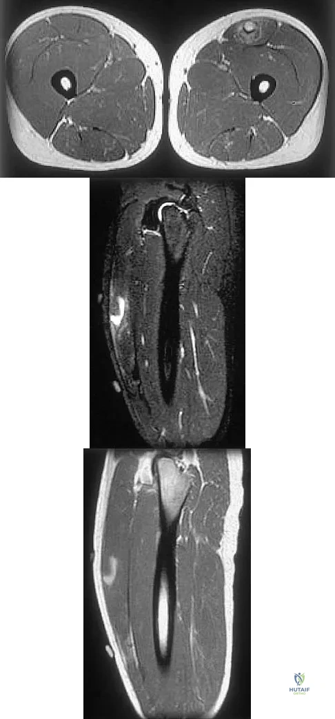


Explanation

The images reveal a region of increased signal within the rectus femoris muscle with mild, ill-defined surrounding edema. The presence of high intensity signal on the T1-weighted image favors acute blood, in this case associated with a rectus femoris muscle tear or fatty tissue. However, because of fat suppression, a fatty lesion or lipoma would be dark on STIR, rather than bright as in this image. Most foreign bodies are low intensity signal and if small, are difficult to evaluate with MRI. The lack of adjacent subcutaneous soft-tissue edema or surrounding fluid makes pyomyositis an unlikely diagnosis.

Question 15

The artery located within the substance of the coracoacromial ligament is a branch of what artery?


Explanation

The acromial branch of the thoracoacromial artery courses along the medial aspect of the coracoacromial ligament and may be encountered when performing an open or arthroscopic subacromial decompression. Bleeding can be controlled by ligation of its branch from the thoracoacromial artery. The other arteries may be injured in other surgical exposures of the shoulder. Esch JC, Baker CL: The shoulder and elbow, in Whipple TL (ed): Arthroscopic Surgery. Philadelphia, PA, JB Lippincott, 1993, pp 65-66.

Question 16

Figures 35a and 35b show the axial T2-weighted and coronal T1-weighted MRI scans of a patient who has enlargement of the right thigh. What is the most likely diagnosis?
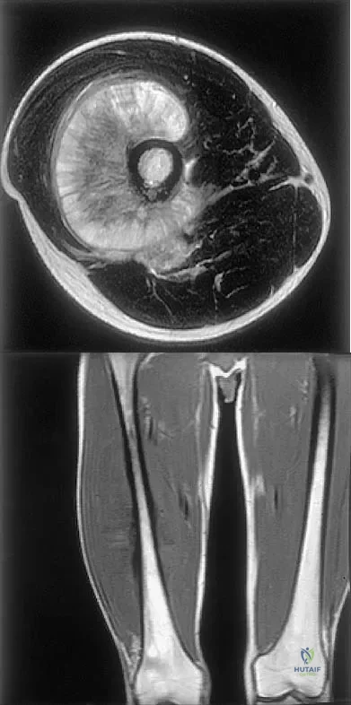


Explanation

The images show a large, almost circumferential, mass surrounding the diaphysis of the femur. The intramedullary signal is normal with minimal cortical destruction, both findings that should be abnormal in conventional osteosarcoma and Ewing's sarcoma. There are very low-signal striations representing osteoid formation that would have a sunburst radiographic pattern. This indicates an osteogenic lesion. Myositis ossificans is not indicated because studies would reveal zonal ossification starting in the periphery rather than the more central pattern seen in this patient. This appearance is typical for periosteal osteosarcoma.

Question 17

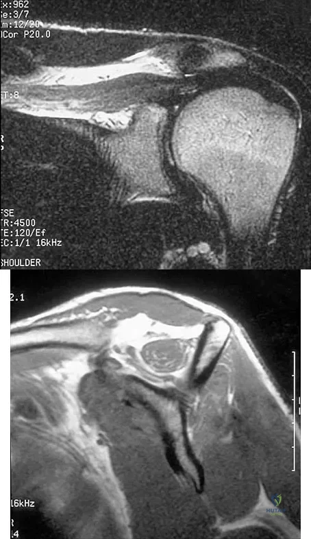
Figures 36a and 36b show the MRI scans of a patient who has shoulder weakness. What is the most likely diagnosis?


Explanation

The sagittal image reveals increased signal and decreased size of the supraspinatus and infraspinatus muscles, indicating muscle atrophy. The rotator cuff tendon signal is normal. The subscapularis and teres minor muscles are unaffected. Muscular dystrophy and thoracic outlet syndrome would be expected to have a more global effect. Although muscular atrophy can occur in the setting of a rotator cuff tear, the coronal image shows an intact supraspinatus. The suprascapular nerve supplies the supraspinatus and infraspinatus muscles. Therefore, suprascapular nerve entrapment would result in atrophy of these muscles with sparing of the surrounding musculature. Any lesion within the suprascapular notch, including neoplastic disease, a venous varix, or neuroma, can place pressure on the suprascapular nerve. Suprascapular nerve entrapment most commonly results from extension of a paralabral cyst or ganglion, often with associated labral pathology. Spinal accessory nerve disruption would show trapezius muscle atrophy. Resnick D, Kang HS (eds): Internal Derangement of Joints: Emphasis on MR Imaging. Philadelphia, PA, WB Saunders, 1997, pp 308-317.

Question 18

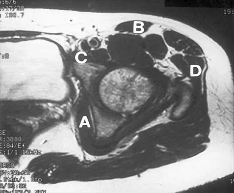
Figure 37 shows the T2-weighted MRI scan of the hip joint. What structure is labeled A?

Anatomy 2002 Practice Questions: Set 3 (Solved) - Figure 19


Explanation

The obturator internus originates on the obturator membrane and adjacent bone, including the quadrilateral plate, and exits the lesser sciatic notch to insert on the posterior medial greater trochanter. The structure labeled C is the pectineus, B is the sartorius, and D is the gluteus medius. Last RJ: Anatomy: Regional and Applied, ed 6. London, England, Churchill Livingstone, 1978, pp 145-150, 324.

Question 19

The great medullary artery, also known as the Adamkiewicz artery, originates from which of the following arteries?


Explanation

The great medullary artery originates as a direct or indirect branch of the left posterior intercostal artery, usually between T8 and T12. It becomes intradural and crosses over one to three disk spaces before turning to the midline where it anastomoses with the anterior spinal artery. Injury to this artery can result in devastating ischemia of the lower spinal cord. Lu J, Ebraheim NA, Biyani A, Brown JA, Yeasting RA: Vulnerability of great medullary artery. Spine 1996;21:1852-1855.

Question 20

A patient who underwent total knee arthroplasty now reports a loss of sensation in the area circled in Figure 38. This area is innervated by which of the following nerves?

Anatomy 2002 Practice Questions: Set 3 (Solved) - Figure 20


Explanation

The saphenous nerve follows the saphenous vein, giving off the infrapatellar branch that crosses the knee anteriorly to supply the peripatellar skin. A longitudinal incision can interrupt the nerve, leaving the terminal distribution without sensation.

Question 21

During an anterior approach to the shoulder, excessive traction on the conjoined tendon is most likely to result in loss of


Explanation

The musculocutaneous nerve travels through the conjoined tendon approximately 8 cm distal to the tip of the acromion. The musculocutaneous nerve innervates the biceps muscle and the bracialis muscle, both of which are responsible for elbow flexion. Shoulder flexion is facilitated by the anterior fibers of the deltoid muscle (axillary nerve) and the supraspinatus muscle (suprascapular nerve). The subscapular muscle facilitates internal rotation of the shoulder (upper and lower subscapularis nerve). Shoulder abduction is performed by the deltoid muscle (axillary nerve), and forearm pronation is facilitated by the pronator teres (median nerve). Hollinshead WH: Anatomy for Surgeons: The Back and Limbs, ed 3. Philadelphia, PA, Harper and Row, 1982, pp 391-393.

Question 22

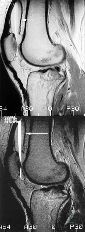
Figure 39 shows the sagittal T1-weighted MRI scan of a 27-year-old man who twisted his knee 2 weeks ago. The arrow is pointing to

Anatomy 2002 Practice Questions: Set 3 (Solved) - Figure 21


Explanation

The arrow identifies a transverse dark line that represents primary trabeculae of the physeal scar. A similar finding is seen in the proximal tibia. These lines may persist indefinitely. They do not represent ongoing growth, an abnormally open physeal plate, a stress fracture, or Looser's line (fatigue fracture in osteomalacia).

Question 23

Figure 40 shows the AP radiograph of a 55-year-old man who reports left knee pain. Which of the following conditions is least likely to produce this radiographic presentation?

Anatomy 2002 Practice Questions: Set 3 (Solved) - Figure 22


Explanation

The radiograph reveals densities within the articular cartilage of the knee commonly referred to as chondrocalcinosis. The term chondrocalcinosis refers to the presence of calcium-containing crystals detected as radiodensities in cartilage. Calcium-containing crystals other than calcium pyrophosphate dihydrate may also deposit in articular cartilage and menisci, producing both radiographically detectable densities in cartilage and joint inflammation or degeneration. Hemochromatosis, alkaptonuria (ochronosis), and Wilson's disease are characterized by cellular deposition of iron, calcium, and copper ions, respectively, into various tissues including articular cartilage and can give this appearance. Septic arthritis does not usually cause chondrocalcinosis. Klippel JH (ed): Primer on the Rheumatic Diseases, ed 11. Atlanta, GA, Arthritis Foundation, 1997, pp 226-229 and 328-331.

Question 24

Figure 41 shows the MRI scan of a 39-year-old man who has severe left groin and anterior thigh pain. What is the most likely diagnosis?

Anatomy 2002 Practice Questions: Set 3 (Solved) - Figure 23


Explanation

The MRI scan shows near complete involvement of the femoral head with bone marrow changes and some collapse of the necrotic segment. This is most suggestive of osteonecrosis.

Question 25

Iliosacral screws placed for stabilization of posterior pelvic ring injuries (eg, sacroiliac dislocation) that exit the sacrum anteriorly are most likely to injure which of the following structures?


Explanation

Iliosacral screws have gained popularity for posterior stabilization of pelvic ring disruptions, but complications attributed to incorrect placement are a clinical problem. The L5 nerve root is at greatest risk and is in closest proximity to a malpositioned screw (exiting the sacrum). The L4 root is more anterior at this level. The S1 root is still intraosseous at this level and is at risk but not from the screw exiting anteriorly at this level. The arteries are at risk but are more anterior and are at less risk than the L5 nerve root.

Question 26

A 28-year-old male sustains a displaced talar neck fracture (Hawkins Type III). Which of the following arteries provides the primary blood supply to the body of the talus, placing it at high risk for avascular necrosis in this injury?


Explanation

The artery of the tarsal canal, a branch of the posterior tibial artery, provides the majority of the blood supply to the talar body. Displaced neck fractures disrupt this supply, leading to a high rate of avascular necrosis.

Question 27

During a posterior lumbar decompression for spinal stenosis, the surgeon removes a thickened ligament that bridges the laminae of adjacent vertebrae. Which of the following tissues is most abundant in this structure?


Explanation

The ligamentum flavum connects the laminae of adjacent vertebrae and is composed primarily of elastin (up to 80%). This gives it a yellow appearance and allows it to stretch and contract during spinal flexion and extension.

Question 28

A 24-year-old baseball pitcher presents with vague posterior shoulder pain and fatigue. MRI reveals isolated atrophy of the teres minor muscle. Compression of a nerve within which of the following anatomic spaces is the most likely cause?


Explanation

The axillary nerve and posterior humeral circumflex artery pass through the quadrilateral space. Compression here (Quadrilateral Space Syndrome) typically causes teres minor atrophy and poorly localized shoulder pain.

Question 29

A 45-year-old woman develops a spontaneous rupture of the extensor pollicis longus (EPL) tendon following a nondisplaced distal radius fracture treated in a cast. Around which bony prominence does this tendon normally pivot, making it susceptible to attrition?


Explanation

The EPL tendon travels in the 3rd dorsal extensor compartment and uses Lister's tubercle as a fulcrum to change direction toward the thumb. Callus formation or undisplaced fracture lines in this area can lead to attrition and rupture of the EPL.

Question 30

A 22-year-old football player sustains a midfoot injury. Imaging reveals a widening of the space between the first and second metatarsals. The primary stabilizing ligament of this articulation connects the base of the second metatarsal to which of the following structures?


Explanation

The Lisfranc ligament is a strong interosseous ligament that connects the lateral aspect of the medial cuneiform to the medial base of the second metatarsal. It is critical for the stability of the tarsometatarsal joint complex.

Question 31

In a surgical approach to the hip, protecting the medial femoral circumflex artery (MFCA) is critical to prevent iatrogenic avascular necrosis of the femoral head. The deep branch of the MFCA passes between which two muscles?


Explanation

The deep branch of the MFCA consistently travels anterior to the quadratus femoris and posterior to the obturator externus. Recognizing this interval is essential to protect the blood supply to the femoral head during posterior hip approaches.

Question 32

A 30-year-old man presents with an inability to cross his fingers and numbness over the volar aspect of his small finger following a deep laceration to his proximal medial forearm. Which cord of the brachial plexus gives rise to the nerve injured in this scenario?


Explanation

The patient has an ulnar nerve injury, evidenced by intrinsic muscle weakness and small finger numbness. The ulnar nerve is a terminal branch of the medial cord of the brachial plexus (C8, T1).

Question 33

During knee arthroscopy, a peripheral, longitudinal tear of the medial meniscus is identified in the "red-red" zone. Which of the following arteries provides the primary blood supply to this peripheral portion of the meniscus?


Explanation

The peripheral 10% to 30% of the medial meniscus is vascularized primarily by the inferior medial genicular artery. This rich blood supply enables the healing of peripheral meniscal tears following surgical repair.

Question 34

A patient develops acute compartment syndrome of the anterior leg following a tibia fracture. If left untreated, which of the following functional deficits is most likely to occur?


Explanation

The anterior compartment of the leg contains the deep peroneal nerve, tibialis anterior, extensor hallucis longus, and extensor digitorum longus. Ischemic injury to these structures leads to a foot drop and inability to extend the toes.

Question 35

An 8-year-old boy sustains a Salter-Harris Type II fracture of the distal radius. The fracture line passes through the growth plate and exits through the metaphysis. Through which histologic zone of the physis does the fracture line primarily propagate?


Explanation

Physeal fractures classically propagate through the zone of hypertrophy, specifically the layer of degenerating chondrocytes. This zone lacks both the abundant extracellular matrix of the proliferative zone and the structural reinforcement found in the zone of calcification.

Question 36

A patient presents with a "lumbrical plus" finger after a flexor digitorum profundus (FDP) tendon laceration in zone 1. What is the anatomic origin and insertion of the lumbrical muscle that contributes to this phenomenon?


Explanation

Lumbricals originate from the FDP tendons and insert on the radial lateral bands of the extensor hood. If the FDP is severed distally, proximal pull of the FDP transmits force through the lumbrical, causing paradoxical PIP and DIP extension during attempted flexion.

Question 37

A 25-year-old runner suffers an external rotation injury to his right ankle, resulting in a high ankle sprain. Which of the following ligaments is typically the first to tear in a syndesmotic injury?


Explanation

The anterior inferior tibiofibular ligament (AITFL) is the weakest of the syndesmotic ligaments. It is typically the first structure to rupture during an external rotation force applied to the dorsiflexed ankle.

Question 38

A 45-year-old male sustains a Jefferson burst fracture of C1. The stability of the C1-C2 complex depends significantly on the transverse ligament. This ligament attaches to which of the following osseous structures?


Explanation

The transverse ligament of the atlas runs behind the dens, holding it tightly against the anterior arch of C1. It attaches to a small tubercle on the medial surface of each lateral mass of the atlas.

Question 39

A postmenopausal woman is prescribed a bisphosphonate for osteoporosis. This medication primarily targets osteoclasts. Which of the following describes the microscopic feature by which actively resorbing osteoclasts attach to the bone surface?


Explanation

Osteoclasts sit in resorption pits called Howship lacunae. They attach to the bone matrix via podosomes and create a "ruffled border" to increase surface area for the secretion of acid and proteolytic enzymes during bone resorption.

Question 40

During a SLAP (Superior Labrum Anterior to Posterior) repair, the surgeon focuses on the attachment of the long head of the biceps tendon. The long head of the biceps tendon originates from the supraglenoid tubercle and which of the following structures?


Explanation

The long head of the biceps tendon originates from the supraglenoid tubercle of the scapula and blends extensively with the superior glenoid labrum. This structural connection is why injuries to the biceps anchor often involve SLAP tears.

Question 41

A patient demonstrates a positive Trendelenburg sign on the right side during the stance phase of gait. This indicates weakness of the right gluteus medius and minimus muscles. Which nerve innervates these muscles?


Explanation

The superior gluteal nerve (L4, L5, S1) innervates the gluteus medius, gluteus minimus, and tensor fasciae latae. Injury to this nerve leads to abductor weakness and a contralateral pelvic drop (positive Trendelenburg sign).

Question 42

A patient has isolated weakness in flexing the interphalangeal joint of the thumb and the distal interphalangeal joint of the index finger. There is no sensory loss. Compression of the involved nerve most commonly occurs at which of the following anatomical structures?


Explanation

The anterior interosseous nerve (AIN) is a purely motor nerve innervating the FPL, FDP to the index/long fingers, and pronator quadratus. It is most commonly entrapped by the tendinous edge of the deep head of the pronator teres or the FDS arcade.

Question 43

A 40-year-old male is involved in a high-speed motor vehicle collision and sustains an unstable vertical shear pelvic ring injury. Which of the following ligaments is the strongest and provides the most stability to the posterior pelvic ring?


Explanation

The posterior interosseous sacroiliac ligament is the thickest and strongest ligament in the pelvis. It serves as the primary restraint against vertical and anterior-posterior translation of the sacroiliac joint.

Question 44

The anterior (Smith-Petersen) surgical approach to the hip utilizes a superficial internervous plane. Which two muscles define this plane?


Explanation

The Smith-Petersen approach utilizes a true internervous plane between the sartorius (femoral nerve) and the tensor fasciae latae (superior gluteal nerve). The deep plane is between the rectus femoris (femoral nerve) and gluteus medius (superior gluteal nerve).

Question 45

During anterior cervical spine surgery, care must be taken to avoid injury to the vertebral artery. At which cervical level does the vertebral artery typically first enter the foramen transversarium?


Explanation

The vertebral artery arises from the subclavian artery and typically enters the transverse foramen at C6. It then travels superiorly through the transverse foramina of C6 to C1.

Question 46

The axillary nerve and posterior circumflex humeral artery exit the axilla through the quadrangular space. Which of the following accurately describes the borders of this space?


Explanation

The quadrangular space is bordered superiorly by the teres minor, inferiorly by the teres major, medially by the long head of the triceps, and laterally by the surgical neck of the humerus. It transmits the axillary nerve and the posterior circumflex humeral artery.

Question 47

Surgical dissection in the plantar midfoot requires navigating the Master Knot of Henry. Which of the following describes the correct anatomic relationship at this intersection?


Explanation

At the Master Knot of Henry, the flexor hallucis longus (FHL) crosses dorsal (deep) to the flexor digitorum longus (FDL). This is a critical landmark when harvesting the FDL or FHL tendons.

Question 48

Salter-Harris fractures typically occur through the weakest portion of the pediatric physis. Which zone of the physis is most frequently implicated as the site of failure?


Explanation

Fractures through the growth plate typically propagate through the zone of hypertrophy. This zone lacks substantial collagen and has a high ratio of chondrocyte volume to matrix, making it the weakest layer biomechanically.

Question 49

During an ilioinguinal or modified Stoppa approach for a pelvic ring injury, the surgeon encounters significant bleeding from the "corona mortis". This structure is an anastomosis between which two vascular systems?


Explanation

The corona mortis (crown of death) is a vascular anastomosis between the external iliac (or deep inferior epigastric) and obturator vessels. It typically courses over the superior pubic ramus and is at risk during anterior pelvic approaches.

Question 50

Which of the following structures is innervated by the anterior interosseous nerve (AIN)?


Explanation

The AIN is a motor branch of the median nerve that innervates the flexor pollicis longus, the pronator quadratus, and the radial half (index and middle fingers) of the flexor digitorum profundus.

Question 51

The primary blood supply to the adult femoral head is the medial circumflex femoral artery (MCFA). The deep branch of the MCFA consistently courses between which two muscles prior to ascending to the trochanteric fossa?


Explanation

The deep branch of the MCFA reliably courses posterior to the obturator externus and anterior to the quadratus femoris. Protecting this region during posterior approaches to the hip is essential to preserve femoral head vascularity.

Question 52

Within the deep posterior compartment of the lower leg, the structures pass behind the medial malleolus. Which structure is located most anteriorly and medially in this space?


Explanation

The order of structures from anterior/medial to posterior/lateral behind the medial malleolus is: Tibialis posterior, Flexor digitorum longus, posterior tibial Artery, tibial Vein, tibial Nerve, Flexor hallucis longus (Tom, Dick, And Very Nervous Harry).

Question 53

The arcade of Frohse is the most common site for entrapment of the posterior interosseous nerve (PIN). Which anatomical structure forms the proximal border of this arcade?


Explanation

The arcade of Frohse is a fibrous arch formed by the proximal edge of the superficial head of the supinator muscle. The PIN passes beneath this arch, making it a primary site of compression.

Question 54

Which of the following statements accurately describes the Martin-Gruber anastomosis?


Explanation

The Martin-Gruber anastomosis is a common anatomical variant involving a motor nerve connection passing from the median nerve to the ulnar nerve in the proximal forearm. It can cause confusing electromyographic findings in nerve entrapments.

Question 55

A patient presents with a winged scapula, and physical examination demonstrates isolated weakness of the serratus anterior muscle. Injury to the long thoracic nerve is suspected. Which nerve roots contribute to the formation of this nerve?


Explanation

The long thoracic nerve innervates the serratus anterior muscle and is formed by branches from the C5, C6, and C7 nerve roots. Injury leads to medial winging of the scapula.

Question 56

Microscopic analysis of cortical bone demonstrates the presence of Volkmann's canals. What is the primary function of these structures?


Explanation

Volkmann's canals are small channels in bone that transmit blood vessels from the periosteum into the bone and lie perpendicular to the Haversian canals. They connect adjacent Haversian systems to ensure adequate cortical vascularity.

Question 57

During an ulnar nerve decompression at the wrist, the surgeon releases the volar carpal ligament to open Guyon's canal. Which structures represent the medial and lateral bony borders of this canal?


Explanation

Guyon's canal is a fibro-osseous tunnel bordered medially by the pisiform and laterally by the hook of the hamate. The floor is the flexor retinaculum, and the roof is the volar carpal ligament.

Question 58

On an MRI of the knee, an intact meniscofemoral ligament is identified. The ligament of Wrisberg is characterized by its anatomic relationship to the posterior cruciate ligament (PCL). Which description is correct?


Explanation

The meniscofemoral ligaments connect the posterior horn of the lateral meniscus to the lateral aspect of the medial femoral condyle. The ligament of Humphrey passes anterior to the PCL, while the ligament of Wrisberg passes posterior to the PCL.

Question 59

The volar (Henry) approach to the radius uses an internervous plane between the brachioradialis and the pronator teres proximally. What is the respective nerve supply to these two muscles?


Explanation

The proximal internervous plane of the Henry approach lies between the brachioradialis, which is innervated by the radial nerve, and the pronator teres, which is innervated by the median nerve.

Question 60

Fractures of the scaphoid proximal pole are prone to nonunion and avascular necrosis due to its precarious blood supply. From which vessel does the scaphoid receive its primary blood supply, and where does it enter the bone?


Explanation

Approximately 80% of the scaphoid's blood supply comes from the dorsal carpal branch of the radial artery. These vessels enter the scaphoid distal to the waist and provide retrograde perfusion to the proximal pole.

Question 61

Reconstruction of the medial patellofemoral ligament (MPFL) requires accurate femoral tunnel placement. Where is the anatomic origin of the MPFL on the medial femur?


Explanation

The anatomic femoral footprint of the MPFL is located in a saddle-shaped depression between the adductor tubercle (superiorly) and the medial epicondyle (inferiorly). Proper isometric placement of the femoral graft is critical for successful reconstruction.

Question 62

During a physical examination, the knee demonstrates increased varus laxity at 30 degrees of flexion but is stable in full extension. Which structure provides the primary restraint to varus stress at 30 degrees of knee flexion?


Explanation

The fibular collateral ligament (LCL) is the primary static restraint to varus stress at all angles of knee flexion, but its isolation is tested best at 30 degrees of flexion, where the posterolateral corner and cruciate ligaments are relaxed.

Question 63

In an unstable syndesmotic injury of the ankle, multiple ligamentous structures are disrupted. Which structure provides the greatest resistance to lateral displacement (diastasis) of the distal fibula relative to the tibia?


Explanation

Biomechanical studies have shown that the posterior inferior tibiofibular ligament (PITFL) provides the strongest restraint to diastasis of the distal tibiofibular syndesmosis, accounting for approximately 42% of the resistance.

Question 64

During a surgical approach to the anterior shoulder, the musculocutaneous nerve is at risk of iatrogenic injury. At what average distance distal to the coracoid process does this nerve typically pierce its designated muscle?


Explanation

The musculocutaneous nerve typically pierces the coracobrachialis muscle 5 to 8 cm distal to the coracoid process. Retractors placed too far distally during anterior shoulder approaches, such as the Latarjet procedure, can result in nerve injury.

Question 65

The main blood supply to the adult femoral head is derived from the deep branch of the medial femoral circumflex artery. Which anatomic landmarks describe the course of this vessel before it enters the hip capsule?


Explanation

The deep branch of the medial femoral circumflex artery courses posterior to the obturator externus and anterior to the quadratus femoris. It then perforates the capsule to supply the femoral head via the retinacular vessels.

Question 66

The anterior cruciate ligament (ACL) is composed of two primary bundles that exhibit unique tension patterns throughout the arc of knee motion. Which of the following statements correctly describes the biomechanics of these bundles?


Explanation

The anteromedial (AM) bundle tightens in flexion and acts as the primary restraint to anterior tibial translation. The posterolateral (PL) bundle tightens in extension and is the primary restraint to rotatory instability.

Question 67

The flexor tendon pulley system of the hand prevents bowstringing of the tendons during digital flexion. Which two pulleys are considered the most critical to the biomechanical function of the digit and must be preserved or reconstructed?


Explanation

The A2 and A4 pulleys are the most mechanically critical for preventing bowstringing and maintaining functional flexor tendon excursion. They arise directly from the periosteum of the proximal and middle phalanges, respectively.

Question 68

The scapholunate interosseous ligament (SLIL) provides primary stability to the scapholunate joint. Which anatomic portion of the SLIL is the thickest and most biomechanically critical for preventing diastasis?


Explanation

The scapholunate interosseous ligament consists of volar, dorsal, and proximal membranous regions. The dorsal portion is the thickest, strongest, and most critical for transmitting forces and maintaining carpal stability.

Question 69

Injury to the posterolateral corner of the knee can result in significant rotatory and varus instability. Which of the following structures is the primary static stabilizer against external tibial rotation at 30 degrees of knee flexion?


Explanation

The popliteofibular ligament is the primary static restraint to external tibial rotation. The LCL primarily restrains varus stress, whereas the popliteus tendon acts as a dynamic stabilizer.

Question 70

The rotator interval is a critical anatomic space in the anterior shoulder involved in glenohumeral stability. Which of the following structures is NOT found within or bordering the rotator interval?


Explanation

The rotator interval is bordered by the subscapularis and supraspinatus tendons and contains the coracohumeral ligament, superior glenohumeral ligament, and the long head of the biceps tendon. The inferior glenohumeral ligament is located lower in the capsule and is not part of this interval.

Question 71

During a posterior suboccipital approach to the cervical spine, the surgeon encounters the suboccipital triangle. Which of the following muscles forms the medial border of this anatomic space?


Explanation

The suboccipital triangle is bordered medially by the rectus capitis posterior major, laterally by the obliquus capitis superior, and inferiorly by the obliquus capitis inferior. The vertebral artery and suboccipital nerve traverse this triangle.

Question 72

During an ilioinguinal approach to the acetabulum, severe hemorrhage can occur from tearing the "corona mortis". This structure represents a vascular anastomosis between which two major systems?


Explanation

The corona mortis is an anastomosis between the obturator vessels (internal iliac system) and the external iliac (or inferior epigastric) vessels. It runs over the superior pubic ramus and is highly vulnerable during anterior pelvic ring surgery.

Question 73

Posterolateral rotatory instability (PLRI) of the elbow presents with a clunk during extension and supination. This condition is primarily caused by insufficiency of which ligamentous structure?


Explanation

PLRI occurs due to deficiency of the lateral ulnar collateral ligament (LUCL). The LUCL serves as a primary restraint preventing posterior subluxation of the radial head relative to the capitellum.

Question 74

A 45-year-old overhead athlete presents with isolated weakness in external rotation of the shoulder. Electromyography reveals denervation of the infraspinatus with a normal supraspinatus. Where is the most likely anatomic site of nerve entrapment?


Explanation

The suprascapular nerve innervates the supraspinatus and infraspinatus. Entrapment at the spinoglenoid notch (often due to a paralabral cyst) affects only the infraspinatus branch, preserving supraspinatus function.

Question 75

The Lisfranc ligament is critical for maintaining the structural integrity of the midfoot. Between which two osseous structures does this ligament course?


Explanation

The Lisfranc ligament is an oblique interosseous ligament connecting the lateral aspect of the medial cuneiform to the medial base of the second metatarsal. Disruption of this ligament leads to tarsometatarsal instability.

Question 76

Articular cartilage is structurally divided into several distinct zones to handle different mechanical loads. Which zone is characterized by the highest concentration of proteoglycans and the lowest concentration of water?


Explanation

The deep zone of articular cartilage has the highest proteoglycan content and the lowest water content. Its collagen fibers run perpendicular to the joint surface to provide maximum resistance against compressive forces.

Question 77

The anterior interosseous nerve (AIN) is a purely motor branch that courses through the volar forearm. Which of the following muscles is NOT innervated by the AIN?


Explanation

The flexor digitorum superficialis is innervated directly by the main trunk of the median nerve. The AIN innervates the flexor pollicis longus, pronator quadratus, and the radial half of the flexor digitorum profundus.

Question 78

In a Bennett fracture-dislocation of the thumb base, a small volar-ulnar fragment remains anatomically reduced while the shaft displaces. Which structure maintains this fragment in its anatomic position?


Explanation

The anterior oblique ligament (AOL), also known as the beak ligament, firmly anchors the volar-ulnar fragment of the first metacarpal to the trapezium. The shaft displaces proximally and radially due to the pull of the abductor pollicis longus.

Question 79

During a four-compartment fasciotomy for acute compartment syndrome of the lower leg, the deep posterior compartment must be completely decompressed. Which of the following muscles is contained within this compartment?


Explanation

The deep posterior compartment contains the flexor hallucis longus, flexor digitorum longus, and tibialis posterior, along with the posterior tibial neurovascular bundle. The gastrocnemius and soleus belong to the superficial posterior compartment.

Question 80

Talar neck fractures are notorious for carrying a high risk of avascular necrosis of the talar body. The primary blood supply to the talar body enters through the tarsal canal and is typically a branch of which vessel?


Explanation

The artery of the tarsal canal provides the dominant blood supply to the talar body. It typically arises as a branch of the posterior tibial artery and courses superiorly into the talus.

Question 81

The medial meniscus of the knee is injured more frequently than the lateral meniscus, largely due to its restricted mobility. Which anatomic attachment primarily limits the translation of the medial meniscus during knee motion?


Explanation

The medial meniscus is firmly attached to the joint capsule and the deep medial collateral ligament (meniscofemoral and meniscotibial components). This rigid peripheral fixation prevents it from displacing to avoid shear forces, unlike the more mobile lateral meniscus.

Question 82

The volar (Henry) approach to the radius provides safe access to the diaphyseal bone without denervating the overlying musculature. This internervous plane is developed between muscles supplied by which two nerves?


Explanation

The Henry approach utilizes the true internervous plane between the flexor carpi radialis (innervated by the median nerve) and the brachioradialis (innervated by the radial nerve). This avoids injury to the major motor branches of the forearm.

Question 83

Osteoblasts regulate bone remodeling by modulating osteoclast activity through the secretion of specific cytokines. Which local factor, produced by osteoblasts, functions as a decoy receptor for RANKL to inhibit osteoclastogenesis?


Explanation

Osteoprotegerin (OPG) is a glycoprotein produced by osteoblasts that binds to RANKL, preventing it from activating RANK on osteoclast precursors. This competitive inhibition effectively downregulates osteoclast differentiation and bone resorption.

Question 84

A 25-year-old male sustains a direct blow to the anteromedial aspect of his knee, resulting in a posterolateral corner (PLC) injury. During surgical reconstruction, the surgeon must identify the femoral attachments of the LCL and the popliteus tendon. What is the correct anatomical relationship of the popliteus tendon origin relative to the LCL femoral footprint?


Explanation

The popliteus tendon inserts on the lateral femoral condyle consistently anterior and inferior to the femoral attachment of the lateral collateral ligament (LCL). Recognizing this anatomic relationship is critical during anatomic posterolateral corner reconstructions.

Question 85

A 22-year-old collegiate baseball pitcher presents with poorly localized posterior shoulder pain and paresthesias over the lateral deltoid. MRI reveals isolated atrophy of the teres minor muscle. The nerve affected in this syndrome passes through a specific anatomic space. Which of the following structures forms the superior boundary of this space?


Explanation

The patient has quadrilateral space syndrome affecting the axillary nerve. The quadrilateral space is bounded superiorly by the teres minor, inferiorly by the teres major, medially by the long head of the triceps, and laterally by the humeral shaft.

Question 86

During a posterior approach to the hip for a displaced femoral neck fracture, the surgeon carefully dissects the short external rotators. To avoid avascular necrosis of the femoral head in a joint-preserving procedure, the deep branch of the medial femoral circumflex artery (MFCA) must be protected. This vessel consistently runs between which two structures?


Explanation

The primary blood supply to the femoral head is the deep branch of the MFCA. It consistently travels posterior to the obturator externus and anterior to the quadratus femoris before ascending along the posterior femoral neck.

Question 87

A 45-year-old woman is undergoing shoulder arthroscopy for adhesive capsulitis. The surgeon plans a release of the rotator interval. Which of the following structures are the primary contents of the rotator interval?


Explanation

The rotator interval is a triangular space bounded by the supraspinatus superiorly, subscapularis inferiorly, and the coracoid base medially. Its primary contents are the coracohumeral ligament, the superior glenohumeral ligament (SGHL), and the intra-articular portion of the long head of the biceps tendon.

Question 88

A 35-year-old avid cyclist presents with profound weakness of finger abduction and adduction but normal sensation over the volar small finger. A lesion in Zone II of Guyon's canal is suspected. Which of the following structures forms the floor of Guyon's canal?


Explanation

The floor of Guyon's canal is formed by the transverse carpal ligament (flexor retinaculum) and the pisohamate ligament. The roof is formed by the volar carpal ligament, while the boundaries are the pisiform (ulnarly) and the hook of the hamate (radially).

Question 89

A patient sustained a midshaft humerus fracture and later developed an inability to flex the interphalangeal joint of the thumb and the distal interphalangeal joint of the index finger. The affected nerve typically branches from its parent nerve at which of the following anatomic locations?


Explanation

The anterior interosseous nerve (AIN) branches from the median nerve approximately 4 to 6 cm distal to the medial epicondyle, passing deep to the fibrous arch of the flexor digitorum superficialis. It innervates the FPL, FDP to the index/long fingers, and the pronator quadratus.

Question 90

During surgical repair of a bi-malleolar ankle fracture equivalent, the surgeon evaluates the deltoid ligament complex. Which component of the deltoid ligament is the thickest and serves as the primary restraint against lateral translation of the talus?


Explanation

The deep deltoid ligament, specifically the deep posterior tibiotalar ligament, is the strongest and thickest component of the medial ankle ligamentous complex. It acts as the primary mechanical restraint to lateral talar excursion and external rotation.

Question 91

A 10-year-old boy falls on his outstretched hand. Radiographs of the elbow reveal a displaced fracture of a secondary ossification center. Based on the typical sequence of elbow ossification, which of the following centers is the last to fuse to the adjacent bone?


Explanation

While the medial epicondyle is the third ossification center to appear (following the mnemonic CRITOE: Capitellum, Radius, Internal/Medial epicondyle, Trochlea, Olecranon, External/Lateral epicondyle), it is the last ossification center to fuse, typically doing so around age 15-18.

Question 92

A 40-year-old male sustains a knife wound to the spine resulting in Brown-Séquard syndrome. He exhibits ipsilateral loss of motor function and contralateral loss of pain and temperature sensation. The loss of pain and temperature sensation is due to injury to which spinal cord tract?


Explanation

The lateral spinothalamic tract carries pain and temperature fibers, which decussate near the level of entry in the anterior white commissure. Therefore, damage to this tract results in a contralateral deficit of pain and temperature sensation below the lesion.

Question 93

During a primary flexor tendon repair in Zone II, the surgeon must preserve the critical pulley system to prevent flexor tendon bowstringing. Anatomically, where does the A2 pulley originate?


Explanation

The A2 and A4 pulleys are the most mechanically critical for preventing bowstringing. The A2 pulley arises from the periosteum of the proximal and middle thirds of the proximal phalanx.

Question 94

Salter-Harris fractures typically propagate through the physis, affecting bone growth depending on the severity of the injury. Through which histologic zone of the physis do these fractures classically propagate?


Explanation

Fractures through the growth plate (physis) classically propagate through the zone of hypertrophy. This zone is mechanically the weakest due to the lack of collagen and large volume of intracellular fluid within the hypertrophic chondrocytes.

Question 95

A retroperitoneal approach to the anterior lumbar spine (L4-L5) is being performed. The surgeon identifies the psoas major muscle. Which of the following describes the correct anatomic relationship of the major nerves of the lumbar plexus relative to the psoas major?


Explanation

The femoral nerve emerges from the lateral border of the psoas major and descends in the groove between the psoas and the iliacus. The obturator nerve emerges medial to the psoas, and the genitofemoral nerve pierces the anterior surface of the psoas.

Question 96

A 21-year-old man sustains a scaphoid waist fracture. The surgeon opts for percutaneous screw fixation. Which artery provides the primary blood supply to the proximal pole of the scaphoid, making it susceptible to avascular necrosis following this fracture?


Explanation

The primary blood supply to the scaphoid is from the dorsal carpal branch of the radial artery, which enters at the distal pole and flows in a retrograde fashion to the proximal pole. This retrograde blood flow makes proximal pole fractures highly susceptible to avascular necrosis.

Question 97

During arthroscopy for a suspected meniscus tear, the surgeon evaluates the peripheral blood supply to determine the healing potential of a repair. What is the primary arterial supply to the peripheral aspects of the medial and lateral menisci?


Explanation

The peripheral 10% to 30% of the menisci (the red-red zone) is vascularized by a perimeniscal capillary plexus originating from the superior and inferior, medial and lateral genicular arteries. The central portions rely on diffusion from synovial fluid.

Question 98

A patient presents with tarsal tunnel syndrome. During the surgical release, the flexor retinaculum is divided. What is the most posterior/lateral structure passing beneath the flexor retinaculum behind the medial malleolus?


Explanation

The structures passing through the tarsal tunnel from anterior-medial to posterior-lateral follow the mnemonic 'Tom, Dick, AND Very Nervous Harry': Tibialis posterior, flexor Digitorum longus, Artery, Vein, Nerve, flexor Hallucis longus. Thus, the FHL tendon is the most posterior structure.

None

Clinic OS
Medically Verified Content by
Prof. Clinic OS
Consultant Orthopedic & Spine Surgeon
Chapter Index