العربية
Part of the Master Guide

AAOS & ABOS Basic Science MCQs (Set 3): Bone Biology, Biomechanics & Anatomy Review

Orthopedic Anatomy 2026 MCQs: Board Review Questions & Answers (Part 2)

23 Apr 2026 55 min read 92 Views
Figure for Anatomy 2000 MCQs - Part 2 - Question 26

Key Takeaway

For anyone wondering about Orthopedic Anatomy 2026 MCQs: Board Review Questions & Answers (Part 2), Top-rated Orthopedic Anatomy 2026 MCQs bank. Practice with clinical case questions, orthopedic surgery board review, and evidence-based answers updated for 2026.

Orthopedic Anatomy 2026 MCQs: Board Review Questions & Answers (Part 2)

Comprehensive 100-Question Exam


00:00

Start Quiz

Question 1

Figure 14 shows a lateral radiograph of a knee joint. The bony structure indicated by the arrow is a sesamoid bone that resides in what tendon?


Explanation

The radiograph shows a fabella, a sesamoid bone that is usually found within the tendon of the lateral head of the gastrocnemius. It can be confused with a loose body on radiographs. It occurs in 18% of patients and is often bilateral. Anderson JE (ed): Grant's Atlas of Anatomy, ed 7. Baltimore, MD, Williams & Wilkins, 1978, pp 4-69.

Question 2

Talar compression syndrome in ballet dancers typically involves injury to which of the following structures?


Explanation

Talar compression syndrome is also known as os trigonum syndrome or posterior ankle impingement syndrome and occurs in activities involving extreme ankle plantar flexion. It involves pinching of the posterior talus (os trigonum or posterior process of the talus) between the calcaneus and tibia. The flexor hallucis longus also may be impinged. The other structures are not commonly injured in this syndrome. Brodsky AE, Khalil MA: Talar compression syndrome. Am J Sports Med 1986;14:472-476. Wredmark T, Carlstedt CA, Bauer H, Saartok T: Os trigonum syndrome: A clinical entity in ballet dancers. Foot Ankle 1991;11:404-406.

Question 3

The sartorius muscle is innervated by which of the following nerves?


Explanation

The femoral nerve enters the thigh behind the inguinal ligament, lying on the surface of the iliopsoas muscle lateral to the femoral artery and vein. The nerve divides into numerous muscular and cutaneous branches in the femoral triangle. The first motor branch (sometimes two branches) is to the sartorius. There is a variable branch to the pectineus. Subsequent branches go to the rectus femoris and then the vastus muscles in variable order. The last motor branch is to the articularis genu. The muscular branches can be injured in anterior approaches to the hip, especially the middle window of the ilioinguinal approach. Hollinshead WH: Textbook of Anatomy, ed 3. Hagerstown, MD, Harper and Row, 1974, p 404.

Question 4

Pacinian corpuscles are lamellated nerve endings that are responsible for providing the perception of


Explanation

Pacinian corpuscles are nerve endings that provide the perception of pressure.

Question 5

An elite gymnast injured her ankle in an awkward dismount 36 hours ago. Examination reveals weakness on single leg step-up. A clinical photograph of the medial ankle is shown in Figure 15. Plain radiographs are normal. To help confirm the diagnosis, the next step in evaluation should consist of


Explanation

Ecchymosis on the medial side of the ankle is distributed in the posterior tibialis tendon sheath location, posterior to the medial malleolus, and extending inferiorly to the tendon's attachment on the navicular. MRI is the imaging study of choice to determine the extent of tendon damage. MRI will also help assess the deltoid ligament. Bone scans and CT are helpful in identifying osteochondral fractures and occult fractures; however, these studies are not indicated for this patient. Peroneal tendons are located lateral on the ankle. Arthroscopy of the ankle joint would not be helpful in assessing the posterior tibial tendons. Lutter LD, Mizel MS, Pfeffer GB (eds): Orthopaedic Knowledge Update: Foot and Ankle. Rosemont, IL, American Academy of Orthopaedic Surgeons, 1994, pp 307-317.

Question 6

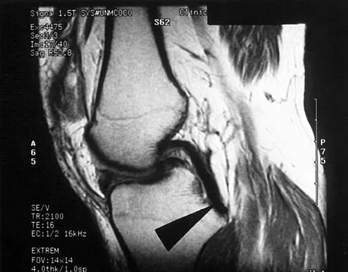
Figure 16 shows an axial MRI scan through the knee joint. What structure is identified by the arrow?


Explanation

The anterior cruciate ligament can be visualized on an axial MRI scan as a low-signal structure lying in the lateral aspect of the intercondylar notch. Visualization in multiple planes increases the accuracy of MRI to view the anterior cruciate ligament. The posterior cruciate ligament and ligament of Wrisberg are located on the medial wall of the notch. The ligamentum mucosum is anterior to the notch, and the popliteus tendon is posterior to the lateral femoral condyle. Resnick D, Kang HS (eds): Internal Derangements of Joints: Emphasis on MR Imaging. Philadelphia, PA, WB Saunders, 1997, pp 675-699.

Question 7

Which of the following nerves is most commonly injured when obtaining a bone graft from the posterior ilium?


Explanation

Cutaneous sensation to the buttock is provided by the superior, middle, and inferior cluneal nerves. The superior cluneal nerves are the lateral branches of the dorsal rami of the upper three lumbar nerves and penetrate deep fascia just proximal to the iliac crest. They pass distally to the skin of the buttock and will be injured if the exposure extends more than 8 cm anterolateral to the posterior superior iliac spine. The lateral femoral cutaneous nerve can be injured in an anterior ilium bone graft. The superior gluteal nerve or even the sciatic nerve can be injured if bone is removed from the sciatic notch or dissection is not kept subperiosteal; however, the rate of injury is far less than cluneal nerve injury. The L5 and S1 nerve roots are anterior and can be injured if the inner table bone is harvested and the dissection is not kept subperiosteal or is too medial; however, the rate of injury still is far less than cluneal nerve injury. Hoppenfeld S, deBoer P: Surgical Exposures in Orthopaedics: The Anatomic Approach. Philadelphia, PA, JB Lippincott, 1984, pp 295-297. Hollinshead WH: Textbook of Anatomy, ed 3. Hagerstown, MD, Harper and Row, 1974, p 379. Last RJ: Anatomy: Regional and Applied, ed 6. London, England, Churchill Livingstone, 1978, p 23.

Question 8

Based on the findings seen in the posteroanterior radiograph of the wrist shown in Figure 17, which of the following structures is torn?


Explanation

The radiograph shows widening between the scaphoid and lunate. The normal variance is up to 5 mm. Although several ligaments may be torn, the scapholunate interosseous ligament must be torn for this widening to occur. Cooney WP, Linscheid RL, Dobyns JH: The Wrist: Diagnosis and Operative Treatment. St Louis, MO, Mosby-Year Book, 1998, vol 1, pp 503-506.

Question 9

What tendon has an intra-articular (instrasynovial) location in the knee joint?


Explanation

The popliteal tendon arises from the posterior aspect of the tibia and courses through the knee joint through the popliteus hiatus of the lateral meniscus before attaching on the lateral femur anterior to the lateral collateral ligament. It is the only tendon in the knee joint that can be viewed directly on arthroscopy. Kimura M, Shirakura K, Hasegawa A, Kobayashi Y, Udagawa E: Anatomy and pathophysiology of the popliteal tendon area in the lateral meniscus: 1. Arthroscopic and anatomical investigation. Arthroscopy 1992;8:419-423.

Question 10

A patient undergoes hip arthroscopy, and the pathology is seen in Figure 18. What is the most likely diagnosis?


Explanation

The motorized shaver is adjacent to the acetabular labrum, which is torn. The femoral head and acetabulum are normal in appearance. Neither the fat pad nor a loose body is identified.

Question 11

Figure 19 shows an arthroscopic view from the anterior lateral portal of the knee looking into the suprapatella pouch. The use of an electrothermal device during this procedure most commonly causes significant postoperative complications by damaging which of the following structures?


Explanation

While it is possible to damage any of these structures, unrecognized intraoperative laceration without adequate coagulation of the superior lateral geniculate artery is common. This can result in significant postoperative hemarthrosis and a return to surgery when bleeding cannot be controlled. Cash JD, Hughston JC: Treatment of acute patella dislocation. Am J Sports Med 1988;16:244-249.

Question 12

Figure 20 shows the resting and stress radiographs of a patient who has had pain and feelings of instability after undergoing a total knee arthroplasty 1 year ago. Which of the following ligaments is not functional and is therefore responsible for the patient's symptoms?


Explanation

The radiographs show posterior instability caused by an absent posterior cruciate ligament. The tibia is significantly displaced posteriorly with respect to the femur. This can be demonstrated with a lateral radiograph obtained with the knee in flexion. The anterior cruciate ligament has been resected but is not responsible for the instability shown.

Question 13

On MRI, a nonsanguinous effusion has what appearance?


Explanation

Nonbloody effusions that are greater than 1 mL are readily detected by MRI. They appear black on T1-weighted images and white on T2-weighted images. A sanguinous effusion is seen as white on T1-weighted images and black on T2-weighted images. Resnick D, Kang HS: Synovial joints, in Resnick D, Kang HS (eds): Internal Derangements of Joints: Emphasis on MR Imaging. Philadelphia, PA, WB Saunders, 1997, pp 49-53.

Question 14

Which of the following illustrations shown in Figures 21a through 21e correctly shows the projection of the sacroiliac joint on the outer table of the ilium?


Explanation

21b 21c 21d 21e The projection of the sacroiliac joint on the outer surface of the ilium should be well understood to avoid violation of the joint during bone graft harvesting and to help in insertion of the screw across the joint. The sacroiliac joint has superior and inferior limbs. The average lengths of the superior and inferior limbs are 4.4 cm and 5.6 cm, respectively. The average width of each limb is 2.0 cm. The average distance from the longitudinal axis of the superior limb to the posterior superior iliac spine is 5.5 cm. The average longitudinal axis of the inferior limb is 1.2 cm superior to the inferior margin of the posterior inferior iliac spine. The average angle between the two axes is 93 degrees. Figure 21c most closely shows the projection of the sacroiliac joint on the outer table of the ilium. Waldrop JT, Ebraheim NA, Yeasting RA, Jackson WT: The location of the sacroiliac joint on the outer table of the posterior ilium. J Orthop Trauma 1993;7:510-513.

Question 15

In children between the ages of 4 and 8 years, the major blood supply to the femoral head comes from the


Explanation

From birth until the age of 4 years, the primary blood supply to the femoral head is from the medial and lateral circumflex arteries that traverse the femoral neck. After the age of 4 years, the contribution of the lateral femoral circumflex artery, which traverses the anterior portion of the femoral neck, becomes negligible. The posterosuperior and posteroinferior retinacular vessels, branches of the medial femoral circumflex artery, become the primary blood supply to the epiphysis. The contribution of the artery of the ligamentum teres is minimal after the age of 4 years. Hughes LO, Beaty JH: Fractures of the head and neck of the femur in children. J Bone Joint Surg Am 1994;76:283-292.

Question 16

What is the most anatomic location for placement of the femoral tunnel in anterior cruciate ligament reconstruction?


Explanation

It is critical for graft isometry and knee stability that the femoral tunnel be placed as far posterior as possible on the lateral femoral condyle. Superiorly, the graft should be at the one o'clock position on the left knee. Resident's ridge is a false posterior shelf that often seems like the extreme posterior cortex. Abnormal tunnel placement results in a variety of complications, including an unstable knee, early graft failure, and joint stiffness. Johnson RJ, Beynnon BD, Nichols CE, Renstrom PA: The treatment of injuries of the anterior cruciate ligament. J Bone Joint Surg Am 1992;74:140-151.

Question 17

In the anterior approach (Smith-Petersen) to the hip, dissection is carried out between muscles innervated by the


Explanation

In the Smith-Petersen approach to the hip, dissection is carried out between the tensor fascia lata laterally (supplied by the superior gluteal nerve) and the sartorius and rectus femoris medially (both supplied by the femoral nerve). Crenshaw AH Jr: Surgical techniques and approaches, in Canale ST (ed): Campbell's Operative Orthopaedics, ed 9. St Louis, MO, Mosby-Year Book, 1998, vol 1, pp 82-85.

Question 18

What structure is marked Q in the diagram of the brachial plexus shown in Figure 22?


Explanation

From proximal to distal, the brachial plexus is divided into roots, trunks, divisions, and cords before forming specific peripheral nerve branches. The structure marked Q is called the posterior cord because it lies posterior to the axially artery at the level of the cords. Its terminal branches are the upper subscapular (V), thoracodorsal (W), lower subscapular (X), axillary (F), and radial (G) nerves. Anderson JE (ed): Grant's Atlas of Anatomy, ed 7. Baltimore, MD, Williams and Wilkins, 1978, pp 6-24.

Question 19

A sagittal T1-weighted MRI scan of the knee joint is shown in Figure 23. What structure is identified by the arrow?


Explanation

On T1-weighted images, the posterior cruciate ligament is a low-signal (black) structure that courses from the posterior aspect of the tibia to the medial femoral condyle. The posterior cruciate ligament can appear as arcuate, U-shaped, or kinked. The other structures have similar signal but different anatomic locations. Gross ML, Grover JS, Bassett LW, Seeger LL, Finerman GA: Magnetic resonance imaging of the posterior cruciate ligament: Clinical use to improve diagnostic accuracy. Am J Sports Med 1992;20:732-737.

Question 20

Figure 24 shows the arthroscopic view of a patient with ankle impingement syndrome. This is commonly seen after high ankle sprains and represents fibrotic granulation thickening of what structure?


Explanation

Chronic anterior inferior tibiofibular ligament sprains can lead to thickening and synovitis that catches or impinges dorsiflexion; patients often note painful clicking with dorsiflexion eversion. The other structures are not affected by this injury. Hopkinson WJ, St Pierre P, Ryan JB, Wheeler JH: Syndesmosis sprains of the ankle. Foot Ankle 1990;10:325-330. Amendola A: Controversies in diagnosis and management of syndesmosis injuries of the ankle. Foot Ankle 1992;13:44-50. Baxter DE: The Foot and Ankle in Sports. St Louis, MO, Mosby-Year Book, 1995, p 30.

Question 21

The modified Brostrom lateral ankle ligamentous reconstruction uses which of the following structures to provide supplementary stabilization?


Explanation

The modified Brostrom lateral ankle ligament stabilization procedure uses the remnants of the anterior talofibular and the calcaneofibular ligaments, supplemented by the inferior retinaculum and the transferred talocalcaneal ligament to stabilize the lateral ankle. Chrisman and associates described the use of one half of the peroneus brevis. Watson-Jones and Evans used the entire peroneus brevis. The peroneus longus has been taken by mistake. The plantaris has been used in triligamentous reconstruction. Gould N, Seligson D, Gassman J: Early and late repair of lateral ligament of the ankle. Foot Ankle 1980;1:84-89. Hamilton WG, Thompson FM, Snow SW: The modified Brostrom procedure for lateral ankle instability. Foot Ankle 1993;14:1-7. Chrisman OD, Snook GA: Reconstruction of lateral ligament tears of the ankle: An experimental study and clinical evaluation of seven patients treated by a new modification of the Elmslie procedure. J Bone Joint Surg Am 1969;51:904-912. Evans DL: Recurrent instability of the ankle: My method of surgical treatment. Proc R Soc Med 1953;46:343. Watson-Jones R: Fractures and Joint Injuries, ed 3. Baltimore, MD, Williams and Wilkins, 1946, p 234.

Question 22

Figure 25 shows an arthroscopic thermal capsular shrinkage device being used in the anterior inferior quadrant of a patient with a subluxating shoulder. Which of the following neurologic complications is most frequently reported with this technique?


Explanation

The axillary nerve lies within millimeters of the anterior inferior capsule. The inferior capsule is of varying thickness, and thermal energy used in shortening the ligament can cause damage to the sensory fibers of the axillary nerve. Clinically, this is manifested as a burnt skin sensation in the axillary nerve distribution area. The motor branch of the axillary nerve is usually spared. The suprascapular nerve and the radial nerve are far from the shrinkage zone. The musculocutaneous nerve, frequently at risk with open procedures, lies well anterior. Fanton GS: Arthroscopic electrothermal surgery of the shoulder. Op Tech Sports Med 1998;6:157-160.

Question 23

A coronal MRI scan through the shoulder joint is shown in Figure 26. The cyst indicated by the arrow will most likely cause compression of what nerve?


Explanation

The MRI scan shows a ganglion cyst in the region of the spinoglenoid notch. These are difficult to diagnose clinically but are readily apparent on MRI. They usually cause compression of the suprascapular nerve and weakness of the infraspinatus and supraspinatus muscles. Resnick D, Kang HS (eds): Internal Derangements of Joints: Emphasis on MR Imaging. Philadelphia, PA, WB Saunders, 1997, pp 306-309.

Question 24

Which of the following ligaments is most commonly involved in posterolateral rotatory instability of the elbow?


Explanation

Recurrent posterolateral rotatory instability of the elbow is difficult to diagnose. Such instability can be demonstrated only by the lateral pivot-shift test. The cause for this condition is laxity of the ulnar part of the lateral collateral ligament, which allows a transient rotatory subluxation of the ulnohumeral joint and a secondary dislocation of the radiohumeral joint. The annular ligament remains intact, so the radioulnar joint does not dislocate. Treatment consists of surgical reconstruction of the lax ulnar part of the lateral collateral ligament. The anterior band is the most important part of the medial collateral which is lax in valgus instability of the elbow. Morrey BF: Acute and chronic instability of the elbow. J Am Acad Orthop Surg 1996;4:117-128.

Question 25

Which of the following anatomic structures is labeled 6 in Figure 27?


Explanation

The line labeled 6 points to the A2 pulley. This structure is the condensation of the digital flexor tendon sheath corresponding to the proximal aspect of the proximal phalanx. Grayson's ligament is volar to the digital nerve and artery. Cleland's ligament is dorsal to the digital nerve and artery. The sagittal band anchors the extensor tendons over the metacarpophalangeal joints. The triangular ligament connects the lateral bands just proximal to the terminal tendon inserting onto the base of the distal phalanx. Hollinshead WH: Anatomy for Surgeons: The Back and Limbs, ed 3. Philadelphia, PA, Harper and Row, 1982, p 467.

Question 26

A 45-year-old mechanic presents with weakness in extending the fingers and thumb, but normal wrist extension. Sensory examination is completely normal. Compression of the involved nerve most commonly occurs at the proximal tendinous edge of which of the following muscles?


Explanation

This patient has Posterior Interosseous Nerve (PIN) syndrome. The most common site of PIN compression is the Arcade of Frohse, which is the proximal aponeurotic edge of the supinator muscle.

Question 27

During a posterolateral approach to the tibial plateau, the common peroneal nerve must be carefully identified and protected. As it wraps around the fibular neck, it passes between the two heads of which muscle?


Explanation

The common peroneal nerve wraps around the fibular neck and enters the anterior/lateral leg by passing between the superficial and deep heads of the peroneus longus muscle.

Question 28

An orthopedic surgeon is evaluating a volleyball player with an isolated suprascapular nerve entrapment at the spinoglenoid notch. Which of the following physical exam findings is most expected?


Explanation

Entrapment at the spinoglenoid notch affects the suprascapular nerve after it has already innervated the supraspinatus. Therefore, it causes isolated denervation and weakness of the infraspinatus (external rotation).

Question 29

A 25-year-old male sustains a midshaft humerus fracture and presents with an inability to extend his wrist and fingers. The injured nerve normally pierces the lateral intermuscular septum to enter the anterior compartment of the arm at approximately what distance proximal to the lateral epicondyle?


Explanation

The radial nerve passes from the posterior to the anterior compartment by piercing the lateral intermuscular septum approximately 10 cm proximal to the lateral epicondyle.

Question 30

During a posterior approach to the shoulder, the surgeon must avoid injury to the axillary nerve as it passes through the quadrangular space. Which of the following accurately describes the boundaries of the quadrangular space?


Explanation

The quadrangular space is bordered superiorly by the teres minor, inferiorly by the teres major, medially by the long head of the triceps, and laterally by the surgical neck of the humerus.

Question 31

The direct anterior (Smith-Petersen) approach to the hip utilizes both superficial and deep internervous planes. The deep internervous plane lies between which two muscles?


Explanation

The superficial plane is between the sartorius (femoral n.) and TFL (superior gluteal n.). The deep plane is between the rectus femoris (femoral n.) and gluteus medius (superior gluteal n.).

Question 32

To avoid iatrogenic injury during percutaneous pinning of a fifth metacarpal neck fracture, the surgeon must be mindful of the dorsal sensory branch of the ulnar nerve. This branch typically diverges from the main ulnar nerve how far proximal to the ulnar styloid?


Explanation

The dorsal sensory branch of the ulnar nerve typically arises 5 to 8 cm proximal to the ulnar styloid and passes dorsally deep to the flexor carpi ulnaris.

Question 33

A 30-year-old runner develops chronic medial heel pain. Entrapment of the first branch of the lateral plantar nerve (Baxter's nerve) is suspected. This nerve is most commonly compressed between the deep fascia of the abductor hallucis and which other muscle?


Explanation

Baxter's nerve (first branch of the lateral plantar nerve) courses between the deep fascia of the abductor hallucis and the medial aspect of the quadratus plantae, where it is frequently entrapped.

Question 34

During a posterior approach to the shoulder, the axillary nerve is at risk when dissecting inferior to the teres minor. Which of the following structures forms the medial boundary of the quadrangular space through which the axillary nerve passes?


Explanation

The quadrangular space is bounded medially by the long head of the triceps, laterally by the surgical neck of the humerus, superiorly by the teres minor, and inferiorly by the teres major. It transmits the axillary nerve and posterior circumflex humeral artery.

Question 35

A 24-year-old man sustains a scaphoid waist fracture. He is at high risk for avascular necrosis of the proximal pole. What is the primary arterial supply to the proximal pole of the scaphoid?


Explanation

The primary blood supply to the scaphoid enters distally via the dorsal carpal branch of the radial artery. This supplies the proximal pole in a retrograde fashion, making it highly susceptible to avascular necrosis following a waist or proximal pole fracture.

Question 36

A 30-year-old patient presents with posterolateral corner (PLC) instability of the knee. During surgical reconstruction, the popliteofibular ligament must be addressed. What are the anatomic attachments of the popliteofibular ligament?


Explanation

The popliteofibular ligament originates from the popliteus tendon and inserts onto the posteromedial aspect of the fibular styloid. It is a critical static stabilizer of the posterolateral corner against varus and external rotation forces.

Question 37

Following a lymph node biopsy in the posterior triangle of the neck, a patient develops shoulder weakness and lateral scapular winging. Which of the following muscles is primarily affected due to the injured nerve?


Explanation

The spinal accessory nerve (CN XI) courses through the posterior triangle of the neck and innervates the trapezius. Injury results in lateral scapular winging, contrasting with medial winging seen in serratus anterior paralysis from long thoracic nerve injury.

Question 38

During an open cubital tunnel release, the surgeon must be careful to protect the medial antebrachial cutaneous nerve. What is the typical anatomic relationship of this nerve to the basilic vein in the distal arm?


Explanation

The medial antebrachial cutaneous nerve typically courses superficial and anterior to the basilic vein in the distal arm. Surgeons must carefully protect its branches to avoid painful postoperative neuromas during cubital tunnel surgery.

Question 39

A 45-year-old man sustains a displaced talar neck fracture (Hawkins type III). The artery of the tarsal canal, which provides the dominant blood supply to the talar body, is a branch of which vessel?


Explanation

The artery of the tarsal canal arises from the posterior tibial artery and provides the dominant blood supply to the body of the talus. It enters the talus inferiorly and forms an anastomotic sling with the artery of the sinus tarsi.

Question 40

In a patient with ulnar tunnel syndrome (Guyon's canal syndrome), surgical release is planned. Which of the following structures forms the floor of Guyon's canal?


Explanation

The floor of Guyon's canal is formed by the transverse carpal ligament (flexor retinaculum) and pisohamate ligament. The roof is formed by the volar carpal ligament.

Question 41

A 40-year-old bodybuilder sustains a distal biceps tendon rupture. During surgical repair through a single anterior incision, the surgeon must be mindful of a nerve that crosses the surgical field deep to the brachioradialis. Which nerve is most at risk?


Explanation

The posterior interosseous nerve (PIN) supinates around the radial neck and passes through the supinator muscle. It is at significant risk during distal biceps repair, especially with excessive lateral retraction or incorrect placement of retractors.

Question 42

A patient with acquired adult flatfoot deformity has attenuation of the spring ligament. What are the specific bony attachments of the spring ligament?


Explanation

The spring ligament (plantar calcaneonavicular ligament) attaches from the sustentaculum tali of the calcaneus to the plantar surface of the navicular. It is a key static stabilizer of the medial longitudinal arch, supporting the talar head.

Question 43

A 28-year-old elite volleyball player presents with isolated weakness in external rotation of the shoulder. Atrophy of the infraspinatus is noted without supraspinatus involvement. Entrapment of the suprascapular nerve is most likely occurring at which location?


Explanation

Entrapment of the suprascapular nerve at the spinoglenoid notch results in isolated infraspinatus weakness and atrophy. Entrapment at the suprascapular notch, which is more proximal, would affect both the supraspinatus and infraspinatus.

Question 44

A 35-year-old patient with a pelvic ring injury requires open reduction and internal fixation via an ilioinguinal approach. The corona mortis, an important vascular anastomosis, is located on the posterior aspect of the superior pubic ramus. It connects which two vascular systems?


Explanation

The corona mortis (crown of death) is an anastomosis between the external iliac (or inferior epigastric) vessels and the obturator vessels. It crosses the superior pubic ramus and can cause life-threatening hemorrhage if disrupted during pelvic surgery.

Question 45

A patient undergoes total hip arthroplasty via a posterior approach. To preserve the primary blood supply to the femoral head in joint-preserving surgery, the surgeon must protect the deep branch of the medial femoral circumflex artery (MFCA). Where does this vessel anatomically course?


Explanation

The deep branch of the MFCA runs deep to the quadratus femoris and posterior to the obturator externus. Protecting the obturator externus tendon during posterior hip approaches helps protect this critical vessel.

Question 46

The anterior interosseous nerve (AIN) is a motor branch of the median nerve. Which of the following muscles is NOT innervated by the AIN?


Explanation

The AIN innervates the flexor pollicis longus, the flexor digitorum profundus to the index and long fingers, and the pronator quadratus. The flexor digitorum superficialis is innervated by the main branch of the median nerve.

Question 47

A 19-year-old athlete sustains a knee injury with a positive pivot shift test. An MRI reveals a torn ACL and an injury to the anterolateral ligament (ALL). The ALL primarily originates from which structure?


Explanation

The anterolateral ligament (ALL) originates on the lateral femoral epicondyle, slightly anterior and distal to the fibular collateral ligament (FCL) origin. It inserts on the proximal tibia midway between Gerdy's tubercle and the fibular head.

Question 48

During an extensile lateral approach to the calcaneus, the sural nerve is at risk. What is the typical sensory distribution of the sural nerve?


Explanation

The sural nerve provides sensory innervation to the lateral and posterolateral aspect of the hindfoot and midfoot. It is closely associated with the small saphenous vein and must be protected during the lateral extensile approach to the calcaneus.

Question 49

A 55-year-old man presents with an unstable proximal humerus fracture. During the deltopectoral approach, the cephalic vein is identified. The cephalic vein lies in the deltopectoral groove between the deltoid and pectoralis major. To which structure should the vein be retracted to minimize the risk of tearing its primary tributary branches?


Explanation

The cephalic vein should ideally be retracted laterally with the deltoid during a deltopectoral approach. Most of its venous tributaries drain from the deltoid muscle, so lateral retraction minimizes the risk of tearing these branches.

Question 50

A patient with an impending pathologic fracture of the distal femur undergoes prophylactic fixation. The femoral artery becomes the popliteal artery as it passes through the adductor hiatus. The adductor hiatus is formed by the tendinous insertions of which muscle?


Explanation

The adductor hiatus is an opening in the aponeurotic insertion of the adductor magnus muscle. It marks the transition of the femoral vessels into the popliteal vessels as they pass from the anterior compartment of the thigh into the popliteal fossa.

Question 51

A surgeon is performing a volar approach (Henry) to the radius. In the proximal forearm, the radial artery must be protected. Which muscle serves as the primary anatomic landmark covering the radial artery and superficial radial nerve in this region?


Explanation

In the proximal forearm, the brachioradialis serves as the key landmark. The radial artery and the superficial sensory branch of the radial nerve lie deep to the brachioradialis muscle belly.

Question 52

Fasciotomy of the leg is planned for a patient with compartment syndrome. When releasing the lateral compartment, which nerve is most at risk of injury as it exits the deep fascia to become subcutaneous in the distal third of the leg?


Explanation

The superficial peroneal nerve exits the deep fascia of the lateral compartment approximately 10 to 12 cm proximal to the lateral malleolus. It is highly susceptible to injury during lateral compartment fasciotomy or placement of anterolateral arthroscopy portals.

Question 53

A hand surgeon is evaluating a patient with a lumbrical plus finger following an amputation. The lumbrical muscles of the hand originate from the tendons of the flexor digitorum profundus (FDP). What is the anatomical arrangement of the lumbricals?


Explanation

The first and second lumbricals are unipennate and originate from the radial sides of the FDP tendons to the index and long fingers. The third and fourth lumbricals are bipennate, originating from the adjacent sides of the FDP tendons of the long, ring, and small fingers.

Question 54

During a posterior approach to the shoulder, the axillary nerve is at risk in the quadrangular space. Which of the following vessels directly accompanies the axillary nerve in this space?


Explanation

The quadrangular space transmits the axillary nerve and the posterior circumflex humeral artery. It is bounded by the teres minor (superior), teres major (inferior), long head of triceps (medial), and surgical neck of humerus (lateral).

Question 55

A patient develops anterolateral thigh numbness following a direct anterior approach to the hip. The injured nerve typically exits the pelvis in which relation to the ASIS?


Explanation

The lateral femoral cutaneous nerve typically exits the pelvis deep to the inguinal ligament, just medial to the anterior superior iliac spine (ASIS). Identifying and protecting this nerve is critical during the anterior approach to the hip.

Question 56

In a displaced femoral neck fracture, the main blood supply to the adult femoral head is disrupted. This critical vascular supply predominantly arises from which of the following?


Explanation

The predominant blood supply to the adult femoral head comes from the lateral epiphyseal artery, a terminal branch of the medial femoral circumflex artery (MFCA). The MFCA is routinely protected during posterior hip approaches by identifying the quadratus femoris.

Question 57

In the most common anatomical variant, the sciatic nerve exits the pelvis in what relation to the piriformis muscle?


Explanation

In approximately 80-85% of individuals, the entire sciatic nerve exits the greater sciatic foramen inferior to the piriformis muscle. Variations include the common peroneal division piercing the muscle or passing superior to it.

Question 58

During a plantar approach to the midfoot, a surgeon identifies the "Master Knot of Henry." Which of the following structures cross at this precise anatomical landmark?


Explanation

The Master Knot of Henry is the chiasm in the plantar midfoot where the flexor digitorum longus (FDL) tendon crosses superficial (plantar) to the flexor hallucis longus (FHL) tendon. It is a critical landmark for tendon transfers in the foot.

Question 59

A patient presents with a distal third humeral shaft fracture (Holstein-Lewis). Which nerve is most at risk as it passes through the lateral intermuscular septum?


Explanation

The radial nerve transitions from the posterior to the anterior compartment of the arm by piercing the lateral intermuscular septum approximately 10 cm proximal to the lateral epicondyle. It is vulnerable to tethering and injury in distal third humeral shaft fractures.

Question 60

During an in situ ulnar nerve decompression at the cubital tunnel, the nerve is traced proximally. Which structure can compress the ulnar nerve as it pierces the medial intermuscular septum?


Explanation

The Arcade of Struthers is a fascial band extending from the medial head of the triceps to the medial intermuscular septum, located about 8 cm proximal to the medial epicondyle. It is a potential site of ulnar nerve compression, distinct from the ligament of Struthers (median nerve).

Question 61

A professional volleyball player presents with isolated weakness of the infraspinatus and normal supraspinatus strength. Entrapment of the suprascapular nerve is most likely occurring at which location?


Explanation

The suprascapular nerve innervates the supraspinatus before passing through the spinoglenoid notch to innervate the infraspinatus. Entrapment at the spinoglenoid notch (commonly by a paralabral cyst) causes isolated infraspinatus weakness.

Question 62

The brachialis muscle is the primary flexor of the elbow. It receives dual innervation from the musculocutaneous nerve and which other nerve?


Explanation

The brachialis muscle receives its primary innervation from the musculocutaneous nerve, but its lateral portion is innervated by a branch of the radial nerve. This dual innervation must be considered during anterior approaches to the humerus.

Question 63

During an extensile lateral approach to the calcaneus, the sural nerve is at risk. From which two nerves does the sural nerve typically derive its medial and lateral contributing branches?


Explanation

The sural nerve is formed by the union of the medial sural cutaneous nerve (a branch of the tibial nerve) and the sural communicating branch (a branch of the common peroneal nerve). It provides sensation to the posterolateral lower leg and lateral foot.

Question 64

The pectineus muscle aids in hip flexion and adduction. It typically receives dual innervation from which of the following nerves?


Explanation

The pectineus receives its primary innervation from the femoral nerve, but also typically receives a branch from the anterior division of the obturator nerve. This reflects its anatomical position between the anterior and medial compartments of the thigh.

Question 65

A patient cannot make an "OK" sign with their thumb and index finger. Which muscle is unaffected in isolated Anterior Interosseous Nerve (AIN) syndrome?


Explanation

The AIN innervates the flexor pollicis longus, the radial half of the flexor digitorum profundus, and the pronator quadratus. The flexor carpi radialis is innervated by the main branch of the median nerve proximal to the AIN origin.

Question 66

The common peroneal nerve is susceptible to compression at the fibular neck. Which of the following muscles is innervated by the superficial peroneal nerve?


Explanation

The superficial peroneal nerve innervates the lateral compartment of the leg, which includes the peroneus longus and peroneus brevis. The tibialis anterior, EHL, and peroneus tertius are innervated by the deep peroneal nerve.

Question 67

The posterior interosseous nerve (PIN) passes between the two heads of the supinator muscle. Which of the following muscles is innervated by the radial nerve proximal to its division into the PIN and superficial sensory branch?


Explanation

The extensor carpi radialis longus (ECRL) and brachioradialis are innervated by the radial nerve proper proximal to its bifurcation. The ECU, EDC, EIP, and APL are innervated by the posterior interosseous nerve (PIN).

Question 68

The posterior meniscofemoral ligament (Ligament of Wrisberg) connects the posterior horn of the lateral meniscus to which structure?


Explanation

The posterior meniscofemoral ligament (Wrisberg) passes posterior to the PCL, attaching the posterior horn of the lateral meniscus to the lateral aspect of the medial femoral condyle. The anterior meniscofemoral ligament (Humphrey) passes anterior to the PCL.

Question 69

The adductor canal (Hunter's canal) transmits the superficial femoral artery and vein. Which nerve exits the adductor canal by piercing the vastoadductor fascia?


Explanation

The saphenous nerve travels within the adductor canal and exits by piercing the vastoadductor fascia. It then travels superficially to provide sensation to the medial aspect of the leg and foot.

Question 70

During surgical release of Guyon's canal, a surgeon identifies the anatomical borders. The floor of Guyon's canal is primarily formed by which structure?


Explanation

The floor of Guyon's canal is formed by the flexor retinaculum (transverse carpal ligament) and the pisohamate ligament. The roof is formed by the volar carpal ligament.

Question 71

The syndesmotic ligament complex of the ankle provides crucial stability to the distal tibiofibular joint. Which of the following ligaments is NOT part of this complex?


Explanation

The syndesmotic complex consists of the anterior inferior tibiofibular ligament (AITFL), posterior inferior tibiofibular ligament (PITFL), interosseous ligament, and the transverse tibiofibular ligament. The calcaneofibular ligament is part of the lateral collateral complex.

Question 72

The deltoid ligament complex of the medial ankle has superficial and deep components. The superficial deltoid ligament primarily resists which motion of the talus?


Explanation

The superficial deltoid ligament (comprising tibionavicular, tibiocalcaneal, and superficial tibiotalar fibers) primarily restricts valgus tilting of the talus. The deep deltoid ligament is the primary restraint to external rotation and lateral translation.

Question 73

The recurrent motor branch of the median nerve (the "million dollar nerve") classically branches from the median nerve at which location relative to the transverse carpal ligament?


Explanation

In the most common anatomical configuration (approx 50-80%), the recurrent motor branch is extraligamentous and recurrent, branching distal to the transverse carpal ligament and curving back to innervate the thenar muscles. Surgeons must be aware of subligamentous and transligamentous variations.

Question 74

During the posterior approach to the hip for a total hip arthroplasty, the short external rotators are divided. To protect the primary blood supply to the femoral head in a joint-preserving procedure (e.g., surgical hip dislocation), the surgeon must understand the course of the medial circumflex femoral artery (MCFA). The deep branch of the MCFA passes consistently between which two muscles?


Explanation

The deep branch of the MCFA is the primary blood supply to the femoral head. It passes posteriorly between the obturator externus and quadratus femoris, making preservation of the obturator externus critical during surgical dislocation.

Question 75

A patient presents with isolated weakness of the abductor digiti minimi, interossei, and the two ulnar lumbricals, but has normal sensation over the entire little finger and the ulnar half of the ring finger. Based on the zones of Guyon's canal, a compressive lesion is most likely located in which zone, and bounded radially by which structure?


Explanation

Zone 2 of Guyon's canal contains only the deep motor branch of the ulnar nerve. It is bounded radially by the hook of the hamate and ulnarly by the pisiform, where compression causes isolated motor deficits.

Question 76

During reconstruction of the medial patellofemoral ligament (MPFL), identifying the anatomic femoral attachment is crucial to restore normal patellofemoral kinematics. Where is the femoral origin of the MPFL located in relation to the bony landmarks of the medial femur?


Explanation

The anatomic femoral origin of the MPFL is located in the saddle or sulcus between the adductor tubercle proximally and the medial epicondyle distally. Non-anatomic placement leads to abnormal graft tension throughout flexion.

Question 77

A surgeon performing an extensile lateral approach to the calcaneus for a highly comminuted intra-articular fracture must be cautious to avoid devascularizing the lateral skin flap. The primary blood supply to this surgical flap is derived from which of the following arteries?


Explanation

The lateral calcaneal artery, a terminal branch of the peroneal artery, provides the primary vascular supply to the L-shaped lateral flap used in calcaneus fracture fixation. Careful full-thickness handling is required to prevent flap necrosis.

Question 78

In performing a transfer of the latissimus dorsi for a massive, irreparable posterosuperior rotator cuff tear, the nerve supplying the transferred muscle must be protected. This nerve arises from which portion of the brachial plexus?


Explanation

The latissimus dorsi is innervated by the thoracodorsal nerve. This nerve is a branch of the posterior cord of the brachial plexus, carrying fibers from C6, C7, and C8.

Question 79

An orthopedic surgeon is utilizing the ilioinguinal approach to fix a transverse acetabular fracture. During the dissection, a significant vascular anastomosis, the corona mortis, is encountered and must be ligated. This structure connects the external iliac system to the internal iliac system via which vessels?


Explanation

The corona mortis is an anastomotic connection between the external iliac (or inferior epigastric) and internal iliac (obturator) vascular systems. It is located over the superior pubic ramus and is at high risk during anterior pelvic approaches.

Question 80

During a posterior cervical foraminotomy at the C5-C6 level for radiculopathy, aggressive lateral dissection with a burr anterior to the neural foramen places a major arterial structure at risk. This artery typically enters the transverse foramen at which cervical level?


Explanation

The vertebral artery most commonly enters the cervical spine at the C6 transverse foramen. It ascends anterior to the exiting cervical nerve roots, making it vulnerable during overly aggressive anterior and lateral foraminotomies.

Question 81

A patient presents with an inability to actively extend the proximal interphalangeal (PIP) and distal interphalangeal (DIP) joints of the index and middle fingers. Active flexion of the metacarpophalangeal (MCP) joints is also weakened. Which of the following accurately describes the origin and innervation of the primarily affected intrinsic muscles?


Explanation

The first and second lumbricals flex the MCP joints and extend the PIP and DIP joints of the index and middle fingers. They originate from the radial sides of the corresponding flexor digitorum profundus tendons and are innervated by the median nerve.

Question 82

A 45-year-old male requires a subtrochanteric fracture fixation using a cephalomedullary nail. During the lateral approach for distal locking screw placement, a branch of the profunda femoris artery is at risk. The first perforating artery typically pierces which of the following structures?


Explanation

The perforating branches of the profunda femoris artery wrap around the posterior femur. The first perforating artery pierces the adductor magnus and the lateral intermuscular septum to supply the posterior thigh, placing it at risk during lateral femoral exposures.

Question 83

An adult patient presents with a severe posterior interosseous nerve (PIN) syndrome. On examination, the patient demonstrates weakness in thumb and finger extension. When asked to extend the wrist, the hand deviates radially. This radial deviation occurs because the PIN lesion spares the innervation to the:


Explanation

The ECRL is innervated by the radial nerve proper before it bifurcates into the PIN and superficial radial nerve. In PIN syndrome, ECRL function is preserved while ECU function is lost, resulting in radial deviation during wrist extension.

Question 84

During flexor tendon repair in Zone II of the hand, preserving the critical pulley system is essential to prevent bowstringing of the tendon. Which two pulleys are considered the most biomechanically critical for normal finger flexion?


Explanation

The A2 and A4 pulleys are located over the proximal and middle phalanges, respectively. They are the most crucial annular pulleys for preventing tendon bowstringing and preserving the mechanical advantage of the flexor tendons.

Question 85

A 28-year-old athlete presents with deltoid weakness and an area of numbness over the lateral shoulder following an anterior glenohumeral dislocation. The affected nerve normally passes through a quadrilateral space. Which of the following structures forms the superior boundary of this space?


Explanation

The quadrangular space transmits the axillary nerve and posterior circumflex humeral artery. It is bounded superiorly by the teres minor, inferiorly by the teres major, medially by the long head of the triceps, and laterally by the surgical neck of the humerus.

Question 86

A 45-year-old patient sustains a displaced femoral neck fracture. To counsel the patient regarding the risk of avascular necrosis, the surgeon considers the vascular anatomy. Which artery provides the primary blood supply to the weight-bearing portion of the adult femoral head?


Explanation

The medial circumflex femoral artery gives rise to the lateral epiphyseal artery branches, which provide the majority of the blood supply to the weight-bearing, posterosuperior aspect of the adult femoral head.

Question 87

During the open reduction of a tarsometatarsal fracture-dislocation, the surgeon plans to reconstruct the Lisfranc ligament to restore midfoot stability. What are the true anatomical attachments of this ligament?


Explanation

The Lisfranc ligament is an interosseous ligament that connects the lateral aspect of the medial cuneiform to the medial aspect of the base of the second metatarsal. There is no direct ligamentous connection between the first and second metatarsal bases.

Question 88

An avid cyclist presents with isolated weakness of the intrinsic hand muscles, resulting in a weak pinch grip, but maintains normal sensation over the volar and dorsal little finger. Compression of the ulnar nerve is most likely occurring in which anatomical region?


Explanation

Zone 2 of Guyon's canal contains only the deep motor branch of the ulnar nerve. Compression here causes isolated intrinsic muscle weakness without sensory deficits.

Question 89

During a posterolateral corner reconstruction of the knee, the surgeon must accurately place the femoral graft tunnels. What is the correct anatomical footprint of the fibular collateral ligament relative to the popliteus tendon insertion on the lateral femur?


Explanation

On the lateral femoral epicondyle, the attachment of the fibular collateral ligament (FCL) is located proximal and posterior to the insertion of the popliteus tendon.

Question 90

A surgeon is navigating the rotator interval during a shoulder arthroscopy for a patient with adhesive capsulitis. Which of the following accurately describes a true boundary or content of this anatomical space?


Explanation

The rotator interval is bounded superiorly by the supraspinatus and inferiorly by the subscapularis. It contains the long head of the biceps tendon, the coracohumeral ligament, and the superior glenohumeral ligament.

Question 91

An orthopedic trauma surgeon is using the modified Stoppa approach for anterior ring fixation of an acetabular fracture. Life-threatening hemorrhage can occur if the corona mortis is inadvertently injured. This vascular structure is typically an anastomosis between the external iliac or inferior epigastric vessels and which other system?


Explanation

The corona mortis is an anatomical variant anastomosis between the obturator vessels and the external iliac or inferior epigastric vessels, crossing over the superior pubic ramus.

Question 92

A 45-year-old mechanic presents with an inability to actively extend his fingers at the metacarpophalangeal joints, but his wrist extension is preserved with slight radial deviation. Examination reveals no sensory deficits. The most likely site of nerve compression is between the two heads of which muscle?


Explanation

The posterior interosseous nerve (PIN) is commonly entrapped at the arcade of Frohse, a fibrous band at the proximal edge of the superficial head of the supinator muscle. This causes motor deficits in finger and thumb extension but spares radial-sided wrist extension.

Question 93

A patient with a displaced proximal pole scaphoid fracture is counseled regarding the high risk of nonunion and avascular necrosis. This risk is primarily due to the retrograde blood supply of the scaphoid. The major vascular contribution enters the scaphoid at which specific location?


Explanation

Approximately 70 to 80 percent of the scaphoid's blood supply comes from branches of the radial artery that enter at the dorsal ridge near the waist and perfuse the proximal pole in a retrograde fashion.

Question 94

During an anterior approach to the thoracolumbar spine for a T11 corpectomy, the surgeon must avoid ligating segmental vessels unnecessarily to prevent spinal cord ischemia. The Artery of Adamkiewicz most commonly enters the spinal canal on the left side between which vertebral levels?


Explanation

The Artery of Adamkiewicz (arteria radicularis magna) is the major blood supply to the lower two-thirds of the spinal cord. In the majority of individuals, it arises on the left side between the T9 and L1 vertebral levels.

Question 95

A patient undergoes a regional block in the adductor canal (Hunter's canal) for postoperative pain control following a total knee arthroplasty. Which of the following muscles forms the anterolateral boundary of this anatomical canal?


Explanation

The adductor canal is bounded anterolaterally by the vastus medialis, posteriorly by the adductor longus and magnus, and its anteromedial roof is formed by the sartorius muscle.

Question 96

A patient develops acute compartment syndrome of the leg after a high-energy tibia fracture. A four-compartment fasciotomy is planned. Which of the following muscles is located within the deep posterior compartment of the lower leg?


Explanation

The deep posterior compartment of the leg contains the flexor hallucis longus, flexor digitorum longus, tibialis posterior, and popliteus muscles, along with the posterior tibial and peroneal vessels and tibial nerve.

Question 97

A patient with persistent buttock and posterior thigh pain is diagnosed with piriformis syndrome due to an anomalous relationship between the sciatic nerve and the piriformis muscle. What is the most common anatomical variant responsible for this condition?


Explanation

In the most common anatomical variation of the sciatic nerve (occurring in 10-15% of the population), the nerve divides prematurely, with the common peroneal division passing through the piriformis muscle and the tibial division passing inferior to it.

Question 98

During the anterior (volar) approach to the forearm for fixation of a midshaft radius fracture, the surgeon develops the proximal internervous plane. This plane is located between two muscles that are supplied by which respective nerves?


Explanation

The proximal internervous plane in the volar approach to the forearm (Henry's approach) is between the brachioradialis (radial nerve) and the pronator teres (median nerve).

Question 99

A 35-year-old sustains an Essex-Lopresti injury, characterized by a radial head fracture, distal radioulnar joint dislocation, and rupture of the interosseous membrane (IOM). In the anatomical position, what is the correct orientation of the fibers of the central band of the forearm IOM?


Explanation

The central band of the interosseous membrane is the primary stabilizer against longitudinal migration of the radius. Its fibers run obliquely from proximal on the radius to distal on the ulna.

Question 100

The anterior cruciate ligament (ACL) is composed of anteromedial and posterolateral bundles that function synergistically to provide stability throughout the knee's range of motion. When the knee is in terminal extension, what is the relative tension state of these bundles?


Explanation

In terminal knee extension, the posterolateral bundle of the ACL is taut, providing primary rotational stability, while the anteromedial bundle is relatively lax. In flexion, the tension states reverse.

None

Clinic OS
Medically Verified Content by
Prof. Clinic OS
Consultant Orthopedic & Spine Surgeon
Chapter Index