العربية
Part of the Master Guide

AAOS & ABOS Basic Science MCQs (Set 3): Bone Biology, Biomechanics & Anatomy Review

Anatomy Board Review (Set 2): Musculoskeletal & Neurovascular MCQs | AAOS, ABOS Prep

23 Apr 2026 55 min read 81 Views
Anatomy 2000 MCQs - Part 2

Key Takeaway

This high-yield anatomy question set for AAOS, ABOS, and OITE exams (Set 2) focuses on essential musculoskeletal structures, neurovascular pathways, and regional anatomical landmarks critical for orthopedic practice. Prepare for board success with these foundational concepts.

Anatomy Board Review (Set 2): Musculoskeletal & Neurovascular MCQs | AAOS, ABOS Prep

Comprehensive 100-Question Exam


00:00

Start Quiz

Question 1

Figure 14 shows a lateral radiograph of a knee joint. The bony structure indicated by the arrow is a sesamoid bone that resides in what tendon?


Explanation

The radiograph shows a fabella, a sesamoid bone that is usually found within the tendon of the lateral head of the gastrocnemius. It can be confused with a loose body on radiographs. It occurs in 18% of patients and is often bilateral. Anderson JE (ed): Grant's Atlas of Anatomy, ed 7. Baltimore, MD, Williams & Wilkins, 1978, pp 4-69.

Question 2

Talar compression syndrome in ballet dancers typically involves injury to which of the following structures?


Explanation

Talar compression syndrome is also known as os trigonum syndrome or posterior ankle impingement syndrome and occurs in activities involving extreme ankle plantar flexion. It involves pinching of the posterior talus (os trigonum or posterior process of the talus) between the calcaneus and tibia. The flexor hallucis longus also may be impinged. The other structures are not commonly injured in this syndrome. Brodsky AE, Khalil MA: Talar compression syndrome. Am J Sports Med 1986;14:472-476. Wredmark T, Carlstedt CA, Bauer H, Saartok T: Os trigonum syndrome: A clinical entity in ballet dancers. Foot Ankle 1991;11:404-406.


Question 3

The sartorius muscle is innervated by which of the following nerves?


Explanation

The femoral nerve enters the thigh behind the inguinal ligament, lying on the surface of the iliopsoas muscle lateral to the femoral artery and vein. The nerve divides into numerous muscular and cutaneous branches in the femoral triangle. The first motor branch (sometimes two branches) is to the sartorius. There is a variable branch to the pectineus. Subsequent branches go to the rectus femoris and then the vastus muscles in variable order. The last motor branch is to the articularis genu. The muscular branches can be injured in anterior approaches to the hip, especially the middle window of the ilioinguinal approach. Hollinshead WH: Textbook of Anatomy, ed 3. Hagerstown, MD, Harper and Row, 1974, p 404.


Question 4

Pacinian corpuscles are lamellated nerve endings that are responsible for providing the perception of


Explanation

Pacinian corpuscles are nerve endings that provide the perception of pressure.


Question 5

An elite gymnast injured her ankle in an awkward dismount 36 hours ago. Examination reveals weakness on single leg step-up. A clinical photograph of the medial ankle is shown in Figure 15. Plain radiographs are normal. To help confirm the diagnosis, the next step in evaluation should consist of


Explanation

Ecchymosis on the medial side of the ankle is distributed in the posterior tibialis tendon sheath location, posterior to the medial malleolus, and extending inferiorly to the tendon's attachment on the navicular. MRI is the imaging study of choice to determine the extent of tendon damage. MRI will also help assess the deltoid ligament. Bone scans and CT are helpful in identifying osteochondral fractures and occult fractures; however, these studies are not indicated for this patient. Peroneal tendons are located lateral on the ankle. Arthroscopy of the ankle joint would not be helpful in assessing the posterior tibial tendons. Lutter LD, Mizel MS, Pfeffer GB (eds): Orthopaedic Knowledge Update: Foot and Ankle. Rosemont, IL, American Academy of Orthopaedic Surgeons, 1994, pp 307-317.


Question 6

Figure 16 shows an axial MRI scan through the knee joint. What structure is identified by the arrow?


Explanation

The anterior cruciate ligament can be visualized on an axial MRI scan as a low-signal structure lying in the lateral aspect of the intercondylar notch. Visualization in multiple planes increases the accuracy of MRI to view the anterior cruciate ligament. The posterior cruciate ligament and ligament of Wrisberg are located on the medial wall of the notch. The ligamentum mucosum is anterior to the notch, and the popliteus tendon is posterior to the lateral femoral condyle. Resnick D, Kang HS (eds): Internal Derangements of Joints: Emphasis on MR Imaging. Philadelphia, PA, WB Saunders, 1997, pp 675-699.


Question 7

Which of the following nerves is most commonly injured when obtaining a bone graft from the posterior ilium?


Explanation

Cutaneous sensation to the buttock is provided by the superior, middle, and inferior cluneal nerves. The superior cluneal nerves are the lateral branches of the dorsal rami of the upper three lumbar nerves and penetrate deep fascia just proximal to the iliac crest. They pass distally to the skin of the buttock and will be injured if the exposure extends more than 8 cm anterolateral to the posterior superior iliac spine. The lateral femoral cutaneous nerve can be injured in an anterior ilium bone graft. The superior gluteal nerve or even the sciatic nerve can be injured if bone is removed from the sciatic notch or dissection is not kept subperiosteal; however, the rate of injury is far less than cluneal nerve injury. The L5 and S1 nerve roots are anterior and can be injured if the inner table bone is harvested and the dissection is not kept subperiosteal or is too medial; however, the rate of injury still is far less than cluneal nerve injury. Hoppenfeld S, deBoer P: Surgical Exposures in Orthopaedics: The Anatomic Approach. Philadelphia, PA, JB Lippincott, 1984, pp 295-297. Hollinshead WH: Textbook of Anatomy, ed 3. Hagerstown, MD, Harper and Row, 1974, p 379. Last RJ: Anatomy: Regional and Applied, ed 6. London, England, Churchill Livingstone, 1978, p 23.


Question 8

Based on the findings seen in the posteroanterior radiograph of the wrist shown in Figure 17, which of the following structures is torn?


Explanation

The radiograph shows widening between the scaphoid and lunate. The normal variance is up to 5 mm. Although several ligaments may be torn, the scapholunate interosseous ligament must be torn for this widening to occur. Cooney WP, Linscheid RL, Dobyns JH: The Wrist: Diagnosis and Operative Treatment. St Louis, MO, Mosby-Year Book, 1998, vol 1, pp 503-506.


Question 9

What tendon has an intra-articular (instrasynovial) location in the knee joint?


Explanation

The popliteal tendon arises from the posterior aspect of the tibia and courses through the knee joint through the popliteus hiatus of the lateral meniscus before attaching on the lateral femur anterior to the lateral collateral ligament. It is the only tendon in the knee joint that can be viewed directly on arthroscopy. Kimura M, Shirakura K, Hasegawa A, Kobayashi Y, Udagawa E: Anatomy and pathophysiology of the popliteal tendon area in the lateral meniscus: 1. Arthroscopic and anatomical investigation. Arthroscopy 1992;8:419-423.


Question 10

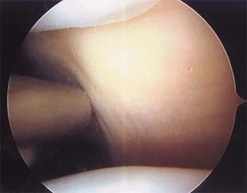
A patient undergoes hip arthroscopy, and the pathology is seen in Figure 18. What is the most likely diagnosis?


Explanation

The motorized shaver is adjacent to the acetabular labrum, which is torn. The femoral head and acetabulum are normal in appearance. Neither the fat pad nor a loose body is identified.


Question 11

Figure 19 shows an arthroscopic view from the anterior lateral portal of the knee looking into the suprapatella pouch. The use of an electrothermal device during this procedure most commonly causes significant postoperative complications by damaging which of the following structures?


Explanation

While it is possible to damage any of these structures, unrecognized intraoperative laceration without adequate coagulation of the superior lateral geniculate artery is common. This can result in significant postoperative hemarthrosis and a return to surgery when bleeding cannot be controlled. Cash JD, Hughston JC: Treatment of acute patella dislocation. Am J Sports Med 1988;16:244-249.


Question 12

Figure 20 shows the resting and stress radiographs of a patient who has had pain and feelings of instability after undergoing a total knee arthroplasty 1 year ago. Which of the following ligaments is not functional and is therefore responsible for the patient's symptoms?


Explanation

The radiographs show posterior instability caused by an absent posterior cruciate ligament. The tibia is significantly displaced posteriorly with respect to the femur. This can be demonstrated with a lateral radiograph obtained with the knee in flexion. The anterior cruciate ligament has been resected but is not responsible for the instability shown.


Question 13

On MRI, a nonsanguinous effusion has what appearance?


Explanation

Nonbloody effusions that are greater than 1 mL are readily detected by MRI. They appear black on T1-weighted images and white on T2-weighted images. A sanguinous effusion is seen as white on T1-weighted images and black on T2-weighted images. Resnick D, Kang HS: Synovial joints, in Resnick D, Kang HS (eds): Internal Derangements of Joints: Emphasis on MR Imaging. Philadelphia, PA, WB Saunders, 1997, pp 49-53.


Question 14

Which of the following illustrations shown in Figures 21a through 21e correctly shows the projection of the sacroiliac joint on the outer table of the ilium?


Explanation

The projection of the sacroiliac joint on the outer surface of the ilium should be well understood to avoid violation of the joint during bone graft harvesting and to help in insertion of the screw across the joint. The sacroiliac joint has superior and inferior limbs. The average lengths of the superior and inferior limbs are 4.4 cm and 5.6 cm, respectively. The average width of each limb is 2.0 cm. The average distance from the longitudinal axis of the superior limb to the posterior superior iliac spine is 5.5 cm. The average longitudinal axis of the inferior limb is 1.2 cm superior to the inferior margin of the posterior inferior iliac spine. The average angle between the two axes is 93 degrees. Figure 21c most closely shows the projection of the sacroiliac joint on the outer table of the ilium. Waldrop JT, Ebraheim NA, Yeasting RA, Jackson WT: The location of the sacroiliac joint on the outer table of the posterior ilium. J Orthop Trauma 1993;7:510-513.


Question 15

In children between the ages of 4 and 8 years, the major blood supply to the femoral head comes from the


Explanation

From birth until the age of 4 years, the primary blood supply to the femoral head is from the medial and lateral circumflex arteries that traverse the femoral neck. After the age of 4 years, the contribution of the lateral femoral circumflex artery, which traverses the anterior portion of the femoral neck, becomes negligible. The posterosuperior and posteroinferior retinacular vessels, branches of the medial femoral circumflex artery, become the primary blood supply to the epiphysis. The contribution of the artery of the ligamentum teres is minimal after the age of 4 years. Hughes LO, Beaty JH: Fractures of the head and neck of the femur in children. J Bone Joint Surg Am 1994;76:283-292.


Question 16

What is the most anatomic location for placement of the femoral tunnel in anterior cruciate ligament reconstruction?


Explanation

It is critical for graft isometry and knee stability that the femoral tunnel be placed as far posterior as possible on the lateral femoral condyle. Superiorly, the graft should be at the one o'clock position on the left knee. Resident's ridge is a false posterior shelf that often seems like the extreme posterior cortex. Abnormal tunnel placement results in a variety of complications, including an unstable knee, early graft failure, and joint stiffness. Johnson RJ, Beynnon BD, Nichols CE, Renstrom PA: The treatment of injuries of the anterior cruciate ligament. J Bone Joint Surg Am 1992;74:140-151.


Question 17

In the anterior approach (Smith-Petersen) to the hip, dissection is carried out between muscles innervated by the


Explanation

In the Smith-Petersen approach to the hip, dissection is carried out between the tensor fascia lata laterally (supplied by the superior gluteal nerve) and the sartorius and rectus femoris medially (both supplied by the femoral nerve). Crenshaw AH Jr: Surgical techniques and approaches, in Canale ST (ed): Campbell's Operative Orthopaedics, ed 9. St Louis, MO, Mosby-Year Book, 1998, vol 1, pp 82-85.


Question 18

What structure is marked Q in the diagram of the brachial plexus shown in Figure 22?


Explanation

From proximal to distal, the brachial plexus is divided into roots, trunks, divisions, and cords before forming specific peripheral nerve branches. The structure marked Q is called the posterior cord because it lies posterior to the axially artery at the level of the cords. Its terminal branches are the upper subscapular (V), thoracodorsal (W), lower subscapular (X), axillary (F), and radial (G) nerves. Anderson JE (ed): Grant's Atlas of Anatomy, ed 7. Baltimore, MD, Williams and Wilkins, 1978, pp 6-24.


Question 19

A sagittal T1-weighted MRI scan of the knee joint is shown in Figure 23. What structure is identified by the arrow?


Explanation

On T1-weighted images, the posterior cruciate ligament is a low-signal (black) structure that courses from the posterior aspect of the tibia to the medial femoral condyle. The posterior cruciate ligament can appear as arcuate, U-shaped, or kinked. The other structures have similar signal but different anatomic locations. Gross ML, Grover JS, Bassett LW, Seeger LL, Finerman GA: Magnetic resonance imaging of the posterior cruciate ligament: Clinical use to improve diagnostic accuracy. Am J Sports Med 1992;20:732-737.


Question 20

Figure 24 shows the arthroscopic view of a patient with ankle impingement syndrome. This is commonly seen after high ankle sprains and represents fibrotic granulation thickening of what structure?


Explanation

Chronic anterior inferior tibiofibular ligament sprains can lead to thickening and synovitis that catches or impinges dorsiflexion; patients often note painful clicking with dorsiflexion eversion. The other structures are not affected by this injury. Hopkinson WJ, St Pierre P, Ryan JB, Wheeler JH: Syndesmosis sprains of the ankle. Foot Ankle 1990;10:325-330. Amendola A: Controversies in diagnosis and management of syndesmosis injuries of the ankle. Foot Ankle 1992;13:44-50. Baxter DE: The Foot and Ankle in Sports. St Louis, MO, Mosby-Year Book, 1995, p 30.


Question 21

The modified Brostrom lateral ankle ligamentous reconstruction uses which of the following structures to provide supplementary stabilization?


Explanation

The modified Brostrom lateral ankle ligament stabilization procedure uses the remnants of the anterior talofibular and the calcaneofibular ligaments, supplemented by the inferior retinaculum and the transferred talocalcaneal ligament to stabilize the lateral ankle. Chrisman and associates described the use of one half of the peroneus brevis. Watson-Jones and Evans used the entire peroneus brevis. The peroneus longus has been taken by mistake. The plantaris has been used in triligamentous reconstruction. Gould N, Seligson D, Gassman J: Early and late repair of lateral ligament of the ankle. Foot Ankle 1980;1:84-89. Hamilton WG, Thompson FM, Snow SW: The modified Brostrom procedure for lateral ankle instability. Foot Ankle 1993;14:1-7. Chrisman OD, Snook GA: Reconstruction of lateral ligament tears of the ankle: An experimental study and clinical evaluation of seven patients treated by a new modification of the Elmslie procedure. J Bone Joint Surg Am 1969;51:904-912. Evans DL: Recurrent instability of the ankle: My method of surgical treatment. Proc R Soc Med 1953;46:343. Watson-Jones R: Fractures and Joint Injuries, ed 3. Baltimore, MD, Williams and Wilkins, 1946, p 234.


Question 22

Figure 25 shows an arthroscopic thermal capsular shrinkage device being used in the anterior inferior quadrant of a patient with a subluxating shoulder. Which of the following neurologic complications is most frequently reported with this technique?


Explanation

The axillary nerve lies within millimeters of the anterior inferior capsule. The inferior capsule is of varying thickness, and thermal energy used in shortening the ligament can cause damage to the sensory fibers of the axillary nerve. Clinically, this is manifested as a burnt skin sensation in the axillary nerve distribution area. The motor branch of the axillary nerve is usually spared. The suprascapular nerve and the radial nerve are far from the shrinkage zone. The musculocutaneous nerve, frequently at risk with open procedures, lies well anterior. Fanton GS: Arthroscopic electrothermal surgery of the shoulder. Op Tech Sports Med 1998;6:157-160.


Question 23

A coronal MRI scan through the shoulder joint is shown in Figure 26. The cyst indicated by the arrow will most likely cause compression of what nerve?


Explanation

The MRI scan shows a ganglion cyst in the region of the spinoglenoid notch. These are difficult to diagnose clinically but are readily apparent on MRI. They usually cause compression of the suprascapular nerve and weakness of the infraspinatus and supraspinatus muscles. Resnick D, Kang HS (eds): Internal Derangements of Joints: Emphasis on MR Imaging. Philadelphia, PA, WB Saunders, 1997, pp 306-309.


Question 24

Which of the following ligaments is most commonly involved in posterolateral rotatory instability of the elbow?


Explanation

Recurrent posterolateral rotatory instability of the elbow is difficult to diagnose. Such instability can be demonstrated only by the lateral pivot-shift test. The cause for this condition is laxity of the ulnar part of the lateral collateral ligament, which allows a transient rotatory subluxation of the ulnohumeral joint and a secondary dislocation of the radiohumeral joint. The annular ligament remains intact, so the radioulnar joint does not dislocate. Treatment consists of surgical reconstruction of the lax ulnar part of the lateral collateral ligament. The anterior band is the most important part of the medial collateral which is lax in valgus instability of the elbow. Morrey BF: Acute and chronic instability of the elbow. J Am Acad Orthop Surg 1996;4:117-128.


Question 25

Which of the following anatomic structures is labeled 6 in Figure 27?


Explanation

The line labeled 6 points to the A2 pulley. This structure is the condensation of the digital flexor tendon sheath corresponding to the proximal aspect of the proximal phalanx. Grayson's ligament is volar to the digital nerve and artery. Cleland's ligament is dorsal to the digital nerve and artery. The sagittal band anchors the extensor tendons over the metacarpophalangeal joints. The triangular ligament connects the lateral bands just proximal to the terminal tendon inserting onto the base of the distal phalanx. Hollinshead WH: Anatomy for Surgeons: The Back and Limbs, ed 3. Philadelphia, PA, Harper and Row, 1982, p 467.


Question 26

A 24-year-old baseball pitcher presents with right upper extremity numbness and fatigue. Examination reveals a diminished radial pulse with shoulder hyperabduction. The compression is clinically suspected to occur within the interscalene triangle. Which of the following structures is most likely NOT compressed in this specific space?


Explanation

The subclavian vein runs anterior to the anterior scalene muscle and therefore does not pass through the interscalene triangle. The subclavian artery and the roots/trunks of the brachial plexus pass between the anterior and middle scalene muscles, making them susceptible to compression in thoracic outlet syndrome.

Question 27

A patient presents with the inability to form an "OK" sign, demonstrating a loss of flexion at the thumb interphalangeal joint and index finger distal interphalangeal joint. The affected nerve normally travels distally in the forearm in the interval between which two muscles?


Explanation

The anterior interosseous nerve (AIN) is a pure motor branch of the median nerve that innervates the FPL, FDP to the index/middle fingers, and pronator quadratus. It travels distally in the forearm strictly in the interval between the FDP and FPL muscle bellies.

Question 28

A 35-year-old male sustains a severe proximal humerus fracture. Follow-up electromyography reveals isolated denervation of the teres minor and deltoid muscles. The affected nerve passes through an anatomic space bordered superiorly by which of the following structures?


Explanation

The axillary nerve travels through the quadrangular space, which is bordered superiorly by the teres minor, inferiorly by the teres major, medially by the long head of the triceps, and laterally by the surgical neck of the humerus. It contains the axillary nerve and posterior circumflex humeral artery.

Question 29

During a posterior approach to the knee for a displaced medial tibial plateau fracture, the surgeon dissects meticulously through the popliteal fossa. What is the correct anatomical sequence of the primary neurovascular structures encountered from superficial (posterior) to deep (anterior)?


Explanation

In the popliteal fossa, the structures from superficial to deep are the tibial nerve, popliteal vein, and popliteal artery. The popliteal artery is the deepest structure, lying directly against the posterior joint capsule and femur.

Question 30

A surgeon is performing a Smith-Petersen approach for an open reduction of a developmental dysplasia of the hip. This anterior approach utilizes a true internervous plane. Which of the following nerve combinations supplies the muscles that define this superficial plane?


Explanation

The Smith-Petersen approach utilizes the internervous plane between the sartorius (innervated by the femoral nerve) and the tensor fasciae latae (innervated by the superior gluteal nerve). This protects the regional neurovascular supply while providing excellent access to the anterior hip.

Question 31

During an inside-out medial meniscus repair, a pure sensory nerve is inadvertently injured. This nerve normally travels through Hunter's canal in the thigh alongside which of the following vessels?


Explanation

The saphenous nerve is a sensory branch of the femoral nerve that travels through Hunter's canal alongside the superficial femoral artery. It exits the canal by piercing the vasoadductor membrane and is at significant risk during medial knee exposures.

Question 32

The superficial peroneal nerve is at risk during a lateral surgical approach to the fibula for fracture fixation. At what average distance proximal to the tip of the lateral malleolus does this nerve typically pierce the crural fascia to become subcutaneous?


Explanation

The superficial peroneal nerve provides motor innervation to the lateral compartment before piercing the crural fascia to become subcutaneous. This fascial penetration reliably occurs approximately 10 to 12 cm proximal to the tip of the lateral malleolus.

Question 33

A 28-year-old professional volleyball player presents with isolated weakness in external rotation of the shoulder. Her abduction strength is completely normal (5/5). An MRI is likely to demonstrate a paralabral ganglion cyst compressing a nerve at which of the following locations?


Explanation

Compression of the suprascapular nerve at the spinoglenoid notch affects only the infraspinatus, causing isolated external rotation weakness. Compression more proximally at the suprascapular notch would affect both the supraspinatus (abduction) and infraspinatus.

Question 34

A 32-year-old male sustains a highly comminuted talar neck fracture. The primary blood supply to the body of the talus, which is now critically disrupted, is derived from the artery of the tarsal canal. This vessel is an anatomic branch of which artery?


Explanation

The artery of the tarsal canal is a branch of the posterior tibial artery and provides the dominant blood supply to the body of the talus. It enters inferiorly and forms an anastomosis with the artery of the sinus tarsi within the tarsal canal.

Question 35

During surgical decompression for recalcitrant intersection syndrome, the surgeon identifies intense tenosynovitis at the crossing point of two muscle bellies over two underlying tendons. The muscle bellies involved in this pathology belong to the:


Explanation

Intersection syndrome is a painful tenosynovitis occurring where the muscle bellies of the first dorsal compartment (APL and EPB) cross over the tendons of the second dorsal compartment (ECRL and ECRB). It typically presents with pain and swelling approximately 4 to 6 cm proximal to Lister's tubercle.

Question 36

A patient is evaluated for an inability to actively extend the fingers at the metacarpophalangeal joints, though wrist extension is maintained with slight radial deviation. The compressed nerve normally enters the posterior forearm by passing between the two heads of which muscle?


Explanation

The posterior interosseous nerve (PIN) is a pure motor branch of the radial nerve that enters the posterior forearm by passing between the superficial and deep heads of the supinator muscle. The proximal edge of the superficial head is known as the arcade of Frohse, the most common site of PIN compression.

Question 37

A professional cyclist presents with severe intrinsic muscle weakness of the hand but normal sensation on the palmar aspect of the small finger. The ulnar nerve is diagnosed as compressed selectively in Zone 2 of Guyon's canal. Which of the following structures forms the floor of this specific zone?


Explanation

Zone 2 of Guyon's canal contains only the deep motor branch of the ulnar nerve. Its floor is strictly formed by the pisohamate and pisometacarpal ligaments, while the roof is formed by the palmaris brevis and fibrous connective tissue.

Question 38

A patient sustains a midshaft humerus fracture. Upon examination, they are unable to extend their wrist or digits. Assuming the lesion is distal to the spiral groove, which of the following muscles is typically the first to regain function during spontaneous nerve recovery?


Explanation

The brachioradialis is typically the first muscle innervated by the radial nerve distal to the fracture site in the spiral groove. Recovery progresses distal to this, followed by the extensor carpi radialis longus.

Question 39

During an anterior approach (Smith-Petersen) to the hip, the superficial internervous plane is between the sartorius and the tensor fasciae latae. What are the respective nerve supplies of these muscles?


Explanation

The sartorius is innervated by the femoral nerve, and the tensor fasciae latae is innervated by the superior gluteal nerve. This creates a true internervous plane for safe superficial dissection.

Question 40

During the Henry approach to the proximal radius, the deep dissection requires managing the supinator to expose the bone. To minimize the risk of injury to the posterior interosseous nerve (PIN), how should the supinator be managed?


Explanation

The supinator should be detached from its insertion on the radius and reflected laterally. This protects the PIN, which runs within the substance of the muscle, from iatrogenic injury.

Question 41

Which of the following neurovascular structures pass through the quadrangular space of the shoulder?


Explanation

The quadrangular space is bound by the teres minor, teres major, long head of the triceps, and the surgical neck of the humerus. It transmits the axillary nerve and the posterior humeral circumflex artery.

Question 42

A patient with a severely displaced fibular neck fracture presents with a new foot drop. Physical examination reveals weakness in both ankle dorsiflexion and eversion, as well as numbness over the dorsum of the foot. Which nerve is most likely injured?


Explanation

The common peroneal nerve wraps around the fibular neck and is highly susceptible to injury here. Injury results in deficits of both its deep (dorsiflexion) and superficial (eversion, dorsal sensation) branches.

Question 43

During a posterior approach to the hip (Kocher-Langenbeck), the blood supply to the femoral head via the deep branch of the medial femoral circumflex artery (MFCA) is at risk. Which of the following structures acts as the primary anatomic barrier protecting the MFCA and should generally be preserved?


Explanation

The deep branch of the MFCA runs anterior to the short external rotators and posterior to the obturator externus tendon. Preserving the obturator externus helps protect the MFCA from iatrogenic transection.

Question 44

A 25-year-old male receives a stab wound to the volar wrist, lacerating the median nerve 2 cm proximal to the carpal tunnel. Assuming no other injuries, which of the following clinical deficits is expected?


Explanation

A median nerve injury at the wrist spares the anterior interosseous nerve (which innervates FDP to the index) but denervates the thenar muscles, resulting in loss of thumb opposition.

Question 45

During a lateral extensile approach for open reduction and internal fixation of a calcaneus fracture, the sural nerve is at risk. What is the usual anatomic course of the sural nerve at the level of the lateral malleolus?


Explanation

The sural nerve courses posterior to the lateral malleolus in close association with the small (short) saphenous vein. It provides sensation to the lateral aspect of the hindfoot and midfoot.

Question 46

Which muscle is primarily responsible for internal rotation of the tibia on the femur, a motion necessary to 'unlock' the knee from terminal extension?


Explanation

The popliteus muscle acts to internally rotate the tibia on the femur (or externally rotate the femur on the tibia in closed-chain), unlocking the knee from full extension to allow flexion.

Question 47

A patient sustains a traumatic anterior shoulder dislocation. The most commonly injured nerve in this scenario arises primarily from which of the following roots of the brachial plexus?


Explanation

The axillary nerve is the most commonly injured nerve in anterior shoulder dislocations. It originates from the posterior cord and receives its primary contributions from the C5 and C6 nerve roots.

Question 48

During surgical release of the first dorsal compartment for de Quervain's tenosynovitis, care must be taken to ensure all tendon slips are decompressed. Which of the following best describes the anatomy of this compartment?


Explanation

The APL tendon frequently consists of two to four slips, while the EPB usually has only one slip. Additionally, the EPB may be contained within its own separate subsheath.

Question 49

A distal radius fracture is approached via the standard volar flexor carpi radialis (FCR) approach. After incising the FCR sheath and retracting the FCR tendon ulnarly, what is the immediate deep structure that must be identified and mobilized to expose the pronator quadratus?


Explanation

Once the FCR sheath is incised and the tendon is retracted, the flexor pollicis longus (FPL) tendon is encountered deep to it. The FPL is retracted ulnarly to expose the underlying pronator quadratus.

Question 50

A 24-year-old weightlifter presents with right posterior shoulder pain and selective weakness in external rotation. MRI reveals an isolated paralabral cyst located strictly within the spinoglenoid notch. Which of the following muscles is most likely denervated?


Explanation

The suprascapular nerve passes through the suprascapular notch (innervating supraspinatus) and then the spinoglenoid notch (innervating infraspinatus). A compressive lesion exclusively at the spinoglenoid notch results in isolated infraspinatus denervation.

Question 51

During a posterior approach to the shoulder, the axillary nerve must be identified to prevent iatrogenic injury. Which of the following sets of structures forms the borders of the quadrangular space, through which the axillary nerve exits?


Explanation

The quadrangular space is bordered superiorly by the teres minor, inferiorly by the teres major, medially by the long head of the triceps, and laterally by the humeral shaft. It transmits the axillary nerve and the posterior circumflex humeral artery.

Question 52

During an anterior intrapelvic (modified Stoppa) approach for an acetabular fracture, massive bleeding is encountered directly posterior to the superior pubic ramus. This is most likely due to injury to the corona mortis, which represents an anastomosis between which two vascular systems?


Explanation

The corona mortis is a critical vascular anastomosis connecting the obturator (internal iliac) and external iliac (or inferior epigastric) systems. It lies on the posterior aspect of the superior pubic ramus, roughly 5-6 cm from the pubic symphysis.

Question 53

A 65-year-old female sustains a displaced femoral neck fracture. The primary blood supply to the weight-bearing dome of the adult femoral head is at high risk of disruption. Which of the following vessels provides this dominant vascular supply?


Explanation

The medial circumflex femoral artery (MFCA) supplies the majority of the blood to the adult femoral head via its lateral epiphyseal branches. Disruption of these retinacular vessels significantly increases the risk of avascular necrosis.

Question 54

Surgical treatment of a highly comminuted talar neck fracture requires extensive dissection, increasing the risk of avascular necrosis. The dominant blood supply to the body of the talus arises from which of the following arteries?


Explanation

The artery of the tarsal canal, a branch of the posterior tibial artery, is the dominant blood supply to the body of the talus. It forms an anastomotic sling with the artery of the tarsal sinus beneath the talar neck.

Question 55

The triangular fibrocartilage complex (TFCC) acts as the primary stabilizer of the distal radioulnar joint (DRUJ). The TFCC takes its origin from the ulnar styloid and inserts primarily into which of the following structures?


Explanation

The TFCC originates from the base and fovea of the ulnar styloid and inserts radially into the hyaline cartilage of the sigmoid notch. Tears at its radial insertion (Palmer 1D) frequently cause DRUJ instability.

Question 56

A patient is evaluated for posterolateral rotatory instability (PLRI) of the elbow following a dislocation. This condition is primarily associated with incompetence of the lateral ulnar collateral ligament (LUCL). What is the exact distal insertion site of the LUCL?


Explanation

The LUCL originates on the lateral epicondyle and passes posterior to the radial head to insert on the supinator crest of the proximal ulna. It acts as the primary restraint against posterolateral subluxation of the radial head.

Question 57

A patient presents with high median nerve neuropathy causing weakness in wrist flexion, forearm pronation, and thumb IP flexion. If the site of compression is the ligament of Struthers, this anatomic structure connects the medial epicondyle to what landmark?


Explanation

The ligament of Struthers is an anomalous band present in about 1% of the population, connecting the supracondylar process to the medial epicondyle. Compression here causes high median neuropathy, unlike compression at the Arcade of Struthers, which affects the ulnar nerve.

Question 58

A 22-year-old male sustains a scaphoid waist fracture. The proximal pole is highly susceptible to avascular necrosis due to its retrograde blood flow. Which vessel is the primary source of blood supply to the scaphoid?


Explanation

The dorsal carpal branch of the radial artery provides the dominant blood supply to the scaphoid, entering at the distal pole and flowing in a retrograde fashion. This anatomy explains the high risk of nonunion and avascular necrosis in proximal pole fractures.

Question 59

During a plantar approach for an excision of a midfoot mass, dissection proceeds deep near the Master Knot of Henry. Which two tendons cross at this specific anatomic decussation?


Explanation

The Master Knot of Henry is the location in the plantar midfoot where the flexor hallucis longus (FHL) crosses dorsal to the flexor digitorum longus (FDL). The medial plantar nerve runs in close proximity to this intersection.

Question 60

During surgical release of a trigger finger, maximum preservation of the digital flexor sheath is required to prevent tendon bowstringing. Which two annular pulleys are biomechanically the most critical to preserve?


Explanation

The A2 pulley (over the proximal phalanx) and the A4 pulley (over the middle phalanx) are the most critical annular pulleys for preventing bowstringing. Trigger finger release involves dividing the A1 pulley, which resides over the MCP joint.

Question 61

To safely access the posterior hip joint, an understanding of the relationship between the sciatic nerve and the piriformis muscle is essential. In the normal and most common anatomic arrangement, where does the sciatic nerve pass?


Explanation

In the majority of the population (greater than 80%), the undivided sciatic nerve passes entirely inferior to the piriformis muscle through the greater sciatic foramen. The most common variant is the peroneal division piercing the piriformis.

Question 62

During anterior cervical corpectomy and fusion, aggressive lateral dissection places the vertebral artery at risk. Ascending from the subclavian artery, the vertebral artery classically enters the foramen transversarium at which cervical level?


Explanation

The vertebral artery typically arises from the subclavian artery and enters the transverse foramen at C6. It does not pass through the transverse foramen of C7, making this an important surgical landmark.

Question 63

During an anterolateral (Henry) approach to the distal humerus, the brachialis muscle is split to expose the humeral shaft. Which of the following describes the innervation of the medial and lateral portions of the brachialis muscle?


Explanation

The brachialis muscle has a dual innervation. The medial portion is innervated by the musculocutaneous nerve, while the lateral portion is innervated by the radial nerve. Splitting the muscle between these two distinct nerve territories protects its function.

Question 64

During an ilioinguinal approach for an acetabular fracture, significant hemorrhage occurs while dissecting near the superior pubic ramus. This is most likely due to a variant anastomotic vessel connecting the external iliac system to which of the following arteries?


Explanation

The corona mortis is a critical vascular anastomosis between the external iliac and the obturator (internal iliac) vessels. It is classically located traversing the superior pubic ramus and is highly susceptible to injury during the ilioinguinal approach.

Question 65

Which of the following arteries typically provides the major blood supply to the anterior lower two-thirds of the spinal cord and most commonly arises on the left side between T8 and L1?


Explanation

The Artery of Adamkiewicz is the largest anterior segmental medullary artery. It typically arises from the left side of the aorta between T8 and L1, providing crucial blood supply to the anterior lower two-thirds of the spinal cord.

Question 66

The deep peroneal nerve supplies the motor innervation to the anterior compartment of the leg. Which of the following muscles is also innervated by the deep peroneal nerve but resides anatomically outside the anterior compartment of the leg?


Explanation

The deep peroneal nerve innervates the anterior compartment of the leg and then continues onto the dorsum of the foot. In the foot, its lateral terminal branch innervates the extensor digitorum brevis and extensor hallucis brevis muscles.

Question 67

A patient sustains a laceration to the deep motor branch of the ulnar nerve at the wrist. Which of the following deficits is expected regarding the lumbrical muscles?


Explanation

The ulnar nerve innervates the 3rd and 4th lumbricals, which function to flex the metacarpophalangeal (MCP) joints and extend the interphalangeal (IP) joints of the ring and small fingers. Loss of this motor branch results in a claw hand deformity due to unopposed IP flexion and MCP extension.

Question 68

A 28-year-old overhead athlete presents with posterior shoulder pain and deltoid weakness. An MRI demonstrates isolated atrophy of the teres minor. Entrapment of a nerve in which of the following spaces is most likely responsible?


Explanation

Quadrilateral space syndrome involves entrapment of the axillary nerve and posterior circumflex humeral artery. It presents with axillary nerve distribution deficits, notably isolated teres minor or deltoid atrophy visible on MRI.

Question 69

At the anatomically critical "Master Knot of Henry" in the plantar midfoot, which of the following relationships is correct?


Explanation

The Master Knot of Henry is located in the medial plantar midfoot. At this intersection, the FHL tendon courses medially from its lateral fibular origin, passing deep (dorsal) to the medially originating FDL tendon.

Question 70

During a surgical approach to the medial thigh, the boundaries of the femoral triangle must be respected to avoid neurovascular injury. What structure forms the medial border of the femoral triangle?


Explanation

The femoral triangle is bounded superiorly by the inguinal ligament, laterally by the medial border of the sartorius, and medially by the medial border of the adductor longus. The floor is primarily formed by the iliopsoas and pectineus muscles.

Question 71

The posterior interosseous nerve (PIN) is most commonly entrapped at the Arcade of Frohse. This anatomic structure represents the proximal fibrous edge of which muscle?


Explanation

The Arcade of Frohse is a fibrous arch forming the proximal edge of the superficial layer of the supinator muscle. It is anatomically the most common site of posterior interosseous nerve (PIN) entrapment.

Question 72

During an anterior cervical discectomy and fusion (ACDF), lateral dissection carries the risk of injuring the vertebral artery. At which cervical level does the vertebral artery typically first enter the transverse foramen?


Explanation

The vertebral artery arises from the subclavian artery and typically enters the transverse foramen at the level of C6. It then ascends through the transverse foramina of the upper cervical vertebrae before entering the foramen magnum.

Question 73

The anterior cruciate ligament (ACL) is composed of two primary bundles that function synergistically throughout the knee's range of motion. In which position of the knee is the anteromedial (AM) bundle most taut?


Explanation

The ACL has two distinct bundles: the anteromedial (AM) and posterolateral (PL). The AM bundle tightens in flexion and is the primary restraint to anterior tibial translation at 90 degrees of flexion, whereas the PL bundle is tightest in extension.

Question 74

The scaphoid is at high risk for avascular necrosis following fracture due to its retrograde blood supply. Which vessel provides the primary blood supply to the proximal pole of the scaphoid?


Explanation

The scaphoid receives 70-80% of its blood supply via retrograde flow from branches of the dorsal carpal branch of the radial artery. These vessels enter at the dorsal ridge near the distal pole and run proximally, placing proximal pole fractures at high risk for avascular necrosis.

Question 75

During percutaneous pinning of a slipped capital femoral epiphysis (SCFE), the surgeon must avoid the posterosuperior retinacular vessels to prevent osteonecrosis. These vessels are terminal branches of which artery?


Explanation

The medial femoral circumflex artery (MFCA) provides the primary blood supply to the femoral head via the posterosuperior retinacular vessels. Injury to these vessels during SCFE pinning or femoral neck fracture can lead directly to avascular necrosis.

Question 76

In a severe external rotation ankle injury, the distal tibiofibular syndesmosis may be completely disrupted. Which of the following syndesmotic ligaments provides the greatest mechanical resistance to lateral translation of the fibula?


Explanation

The posterior inferior tibiofibular ligament (PITFL) is the strongest component of the syndesmosis. Biomechanical studies indicate it provides approximately 42% of the resistance to lateral fibular displacement, more than any other syndesmotic structure.

Question 77

The standard deltopectoral approach to the shoulder utilizes a true internervous plane. This plane exists between muscles innervated by which of the following pairs of nerves?


Explanation

The deltopectoral approach utilizes the internervous plane between the deltoid (innervated by the axillary nerve) and the pectoralis major (innervated by the medial and lateral pectoral nerves). This allows for extensile exposure without denervating the overlying musculature.

Question 78

A patient presents with an inability to form the 'OK' sign, demonstrating extended interphalangeal joints of the thumb and index finger. This deficit localizes to the anterior interosseous nerve (AIN). Which of the following muscles is innervated by this nerve?


Explanation

The AIN is a motor branch of the median nerve that innervates the flexor pollicis longus (FPL), the pronator quadratus, and the flexor digitorum profundus (FDP) to the index and middle fingers.

Question 79

Entrapment of the deep peroneal nerve beneath the inferior extensor retinaculum (anterior tarsal tunnel syndrome) typically results in sensory loss in which of the following distributions?


Explanation

The deep peroneal nerve provides motor innervation to the short toe extensors and sensory innervation strictly to the first dorsal web space of the foot.

Question 80

A 28-year-old overhead throwing athlete presents with posterior shoulder pain and isolated teres minor atrophy on MRI. Compression of the axillary nerve is suspected in the quadrilateral space. Which structure forms the superior border of this space?


Explanation

The quadrilateral space is bounded superiorly by the teres minor, inferiorly by the teres major, medially by the long head of the triceps, and laterally by the humeral shaft.

Question 81

The main blood supply to the adult femoral head is derived from the medial circumflex femoral artery (MCFA). The deep branch of the MCFA travels to the hip capsule in the interval between which two muscles?


Explanation

The deep branch of the MCFA consistently runs between the quadratus femoris posteriorly and the obturator externus anteriorly before reaching the trochanteric fossa to supply the femoral head.

Question 82

During a lateral approach to the distal humerus, the radial nerve is identified and mobilized. At what approximate distance proximal to the lateral epicondyle does the radial nerve typically pierce the lateral intermuscular septum to enter the anterior compartment of the arm?


Explanation

The radial nerve pierces the lateral intermuscular septum to transition from the posterior compartment to the anterior compartment approximately 10 cm proximal to the lateral epicondyle.

Question 83

Which of the following structures is NOT a border or content of the rotator interval?


Explanation

The rotator interval is bordered by the supraspinatus (superiorly) and subscapularis (inferiorly). It contains the coracohumeral ligament, superior glenohumeral ligament, and the long head of the biceps tendon.

Question 84

The 'master knot of Henry' is a surgically important anatomical landmark in the plantar aspect of the foot. It is characterized by the crossing of which two tendons?


Explanation

The master knot of Henry is located at the level of the navicular bone, where the flexor hallucis longus (FHL) crosses dorsal (deep) to the flexor digitorum longus (FDL) tendon.

Question 85

A patient presents with ulnar nerve palsy secondary to a ganglion cyst compressing Guyon's canal. What structure forms the floor of Guyon's canal?


Explanation

The floor of Guyon's canal is formed by the flexor retinaculum (transverse carpal ligament) and the pisohamate ligament. The roof is formed by the volar carpal ligament.

Question 86

The pectoralis major tendon inserts onto the lateral lip of the bicipital groove. Which of the following best describes the anatomical arrangement of its fibers at the insertion site?


Explanation

The pectoralis major tendon twists 90 degrees before insertion such that the sternal (inferior) fibers insert proximal and deep to the clavicular (superior) fibers.

Question 87

The 'corona mortis' is a vascular anastomosis that is at high risk of injury during anterior intrapelvic approaches. It typically connects the obturator system with which of the following vessel systems?


Explanation

The corona mortis is a vascular connection between the external iliac (or inferior epigastric) vessels and the obturator vessels. It courses over the posterior aspect of the superior pubic ramus.

Question 88

The suprascapular nerve is at risk of entrapment at both the suprascapular notch and the spinoglenoid notch. An isolated lesion at the spinoglenoid notch will typically result in which of the following clinical findings?


Explanation

Entrapment at the spinoglenoid notch affects only the branch to the infraspinatus, leading to isolated external rotation weakness. The supraspinatus (abduction) is spared because its branches originate more proximally.

Question 89

Which of the following describes the correct origin and innervation of the 3rd lumbrical muscle in the hand?


Explanation

The 1st and 2nd lumbricals are unipennate and innervated by the median nerve. The 3rd and 4th lumbricals are bipennate (originating from adjacent FDP tendons) and are innervated by the deep branch of the ulnar nerve.

Question 90

A 45-year-old man presents with neck pain, numbness in the thumb, and weakness in wrist extension. MRI reveals a posterolateral cervical disc herniation at the C5-C6 level. Which nerve root is most likely compressed?


Explanation

In the cervical spine, nerve roots exit above the pedicle of their corresponding vertebra (e.g., C6 root exits between C5 and C6). A posterolateral disc herniation at C5-C6 will compress the exiting C6 nerve root.

Question 91

During a lateral extensile approach to the calcaneus for fracture fixation, the sural nerve is at risk. What is its typical anatomical location relative to the lateral malleolus?


Explanation

The sural nerve travels posterior and inferior to the lateral malleolus alongside the short saphenous vein. It provides sensation to the lateral aspect of the foot and ankle.

Question 92

The Arcade of Frohse is a frequent site of compression for the posterior interosseous nerve (PIN). This structure represents the proximal fibrous edge of which muscle?


Explanation

The Arcade of Frohse is the thickened proximal aponeurotic edge of the superficial head of the supinator muscle. It is the most common site of PIN entrapment in radial tunnel syndrome.

Question 93

In normal pelvic anatomy, the sciatic nerve typically exits the greater sciatic foramen in what positional relationship to the piriformis muscle?


Explanation

In approximately 80-85% of individuals, the entire sciatic nerve exits the greater sciatic foramen inferior to the piriformis muscle belly.

Question 94

Which of the following tendons does NOT pass through the primary compartment of the carpal tunnel?


Explanation

The flexor carpi radialis (FCR) tendon runs in its own separate fibro-osseous tunnel enclosed by a split in the transverse carpal ligament, rather than within the main carpal tunnel.

Question 95

A 28-year-old overhead athlete presents with insidious onset of poorly localized posterior shoulder pain and paresthesias over the lateral deltoid. MRI reveals isolated atrophy of the teres minor. The neurovascular structures affected are compressed within an anatomical space. What are the precise borders of this space?


Explanation

Quadrangular space syndrome involves compression of the axillary nerve and posterior humeral circumflex artery. The anatomical borders of the quadrangular space are the teres minor (superiorly), teres major (inferiorly), long head of the triceps (medially), and humeral shaft (laterally).

Question 96

During an open reconstruction of the posterolateral corner (PLC) of the knee, the surgeon develops an interval between the biceps femoris and the iliotibial band. Which of the following neurovascular structures is at greatest risk during this approach, and what is its correct anatomical relationship?


Explanation

The common peroneal nerve is highly vulnerable during surgical approaches to the posterolateral corner. It courses distally on the posterior aspect of the biceps femoris, then wraps anteriorly around the fibular neck deep to the fascia of the peroneus longus muscle.

Question 97

A 45-year-old woman undergoes an open carpal tunnel release. Postoperatively, she reports a new inability to palmar abduct her thumb, despite intact sensation over the thenar eminence and volar digits. An iatrogenic injury to the recurrent motor branch of the median nerve is suspected. According to the most common anatomical variation (extraligamentous type), how does this branch course to innervate the thenar muscles?


Explanation

According to Lanz's classification, the extraligamentous type is the most common anatomical variation of the recurrent motor branch of the median nerve (seen in over 50% of cases). It branches distal to the transverse carpal ligament and hooks back recurrently to innervate the thenar musculature.

None

Clinic OS
Medically Verified Content by
Prof. Clinic OS
Consultant Orthopedic & Spine Surgeon
Chapter Index