العربية
Part of the Master Guide

AAOS & ABOS Basic Science MCQs (Set 3): Bone Biology, Biomechanics & Anatomy Review

Orthopedic Anatomy 2026 MCQs: Board Review Questions & Answers (Part 3)

23 Apr 2026 75 min read 86 Views
Figure for Anatomy 2008 MCQs - Part 3 - Question 51

Key Takeaway

This topic focuses on Orthopedic Anatomy 2026 MCQs: Board Review Questions & Answers (Part 3), Top-rated Orthopedic Anatomy 2026 MCQs bank. Practice with clinical case questions, orthopedic surgery board review, and evidence-based answers updated for 2026.

Orthopedic Anatomy 2026 MCQs: Board Review Questions & Answers (Part 3)

Comprehensive 100-Question Exam


00:00

Start Quiz

Question 1

In the most common condition causing a winged scapula, which of the following nerves is affected?


Explanation

A winged scapula is most often associated with Parsonage-Turner syndrome, a condition thought to be due to an inflammatory or immune-mediated mechanism. Certain muscles are predisposed, particularly the serratus anterior muscle innervated by the long thoracic nerve. Other less common nerve lesions (eg, the spinal accessory and dorsal scapular nerves) may also cause winged scapulae. Kline DG, Hudson AR: Nerve Injuries: Operative Results for Major Nerve Injuries, Entrapments and Tumors. Philadelphia, PA, WB Saunders, 1995.

Question 2

A 17-year-old woman seen in the emergency department reports right knee pain and swelling that has progressively worsened over the past several weeks. Radiographs are shown in Figures 31a and 31b. What is the most likely diagnosis?


Explanation

31b The radiographs reveal a blastic lesion of the proximal tibial metaphysis with cortical destruction, mineralization extending into the soft tissue laterally, indistinct margins, and destruction of the normal trabecular pattern. In this age group, with this aggressive appearance, osteosarcoma is the most likely diagnosis. Chondroblastoma and giant cell tumor are generally geographic and lytic. Chondrosarcoma is rare in this age group and would likely be a secondary lesion from an underlying chondroid tumor that is not present here. Whereas infection can have a wide variety of appearances, it tends to be more lytic in the acute presentation. Sanders TG, Parsons TW: Radiographic imaging of musculoskeletal neoplasia. Cancer Control 2001;8:221-231.

Question 3

A 26-year-old man has had hand pain and progressive swelling in the knuckle for the past several months. He denies any trauma to the hand. The ring finger metacarpophalangeal joint is tender, and there is loss of motion in the digit. Figure 32a shows the radiograph and Figures 32b through 32d show the T1-weighted, T2-weighted, and gadolinium MRI scans, respectively. What is the most likely diagnosis?


Explanation

32b 32c 32d The radiograph reveals a subchondral lesion in the metacarpophalangeal joint that is lytic and expansile. The MRI scans show a mass that is moderate in intensity on the T2-weighted image and has some gadolinium uptake. There are no cystic components in this lesion. The subchondral location and expansile nature are highly suggestive of giant cell tumor of bone. A lesion with this appearance might also represent an aneurysmal bone cyst, given the amount of expansion present. Menendez LR (ed): Orthopaedic Knowledge Update: Musculoskeletal Tumors. Rosemont, IL, American Academy of Orthopaedic Surgeons, 2002, pp 113-118.

Question 4

Which of the following best describes the relationship of the median nerve to the flexor carpi radialis tendon just proximal to the carpal canal?


Explanation

The median nerve has an intimate association with the palmaris longus and the flexor carpi radialis at the proximal aspect of the carpal canal. The median nerve lies just ulnar and dorsal to the flexor carpi radialis tendon. Hoppenfeld S, deBoer P: Surgical Exposures in Orthopaedics, ed 2. Philadelphia, PA, Lippincott-Raven, 1994, pp 118-131.

Question 5

Which of the following muscles has dual innervation?


Explanation

The brachialis muscle typically receives dual innervation. The major portion is innervated by the musculocutaneous nerve. Its inferolateral portion is innervated by the radial nerve. The others listed have single innervation. The anterior approach to the humerus, which requires splitting of the brachialis, capitalizes on this dual innervation.

Question 6

Figure 33a shows a line drawing of a normal hemipelvis. The anterior acetabular rim is bold. Figure 33b illustrates a hemipelvis with a crossover sign, which is indicative of what acetabular pathology?


Explanation

33b In a normal AP pelvis radiograph, the anterior rim of the acetabulum runs medially and distally, diverging from the posterior rim which runs much more vertically. In excessive acetabular retroversion, the anterior rim (bold line in Figure 33b) and posterior rim start laterally, and as these lines progress medially and distally, the anterior line crosses the posterior line. This predisposes to femoral acetabular impingement. Reynolds D, Lucas J, Klaue K: Retroversion of the acetabulum: A cause of hip pain. J Bone Joint Surg Br 1999;81:281-288.

Question 7

Which of the following structures is most vulnerable during a medial sesamoidectomy of the hallux?


Explanation

The plantar-medial cutaneous nerve is at risk with the surgical approach to the medial sesamoid. It is found directly underlying an incision made at the junction of the glabrous skin of the hallux and must be identified before the approach can proceed. Transection will result in a painful neuroma that impinges on the plantar-medial surface of the toe and cause problems with shoe wear. The only other structure that lies near the surgical field is the abductor hallucis tendon which lies dorsal to the incision.

Question 8

What structure is most at risk for injury from a retractor against the tracheoesophageal junction during an anterior approach to the cervical spine?


Explanation

Although any of these structures can be injured by pressure from the medial blade of a self-retaining retractor, the recurrent laryngeal nerve runs cephalad in the interval between the esophagus and trachea and is vulnerable to pressure if caught between the retractor and an inflated endotracheal tube balloon. Ebraheim NA, Lu J, Skie M, et al: Vulnerability of the recurrent laryngeal nerve in the anterior approach to the lower cervical spine. Spine 1997;22:2664-2667.

Question 9

A 40-year-old man has had hip pain with increased activity over the past year. Examination reveals restriction of motion and tenderness with combined hip flexion, adduction, and internal rotation. An AP radiograph is shown in Figure 34. What is the most likely diagnosis?


Explanation

Femoral acetabular impingement (FAI) is a pathologic entity leading to pain, reduced range of motion in flexion and internal rotation, and development of secondary arthritis of the hip. There are two types of FAI: cam impingement and pincher impingement. Cam impingement is seen when a nonspherical femoral head produces a cam effect when the prominent portion to the femoral head rotates into the joint. This mechanism produces shear forces that damage articular cartilage. Radiographs reveal early joint degeneration and flattening of the head neck junction (the so-called "pistol grip deformity") as seen in this image. The pincher type of impingement involves abnormal contact between the femoral head neck junction and the acetabulum, in the presence of a spherical femoral head. Beall DP, Sweet CF, Martin HD, et al: Imaging findings of femoraoacetabular impingement syndrome. Skeletal Radiol 2005;34:691-701.

Question 10

Figure 35 shows the radiograph of a 44-year-old woman with rheumatoid arthritis who reports neck pain. Below what threshold number is surgical stabilization warranted for the interval shown by the arrow?


Explanation

The posterior atlanto-dens interval represents the space available for the spinal cord and a distance of less than 14 mm is predictive of neurologic progression, thus warranting consideration for fusion, even in the absence of symptoms.

Question 11

An axillary nerve lesion may cause weakness in the deltoid and the


Explanation

While the most prominent functional deficit from axillary nerve lesions occurs from denervation of the deltoid, denervation of the teres minor also occurs.

Question 12

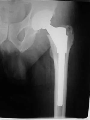
Figure 36 shows an AP radiograph of a 65-year-old man who reports activity-related groin pain. History reveals that he underwent total hip arthroplasty 12 years ago. What is the most likely diagnosis?


Explanation

The AP radiograph demonstrates extensive periacetabular osteolysis. The central hole eliminator has dissociated from the shell and migrated into a lytic defect in the ischium. In a retrieval study, most periacetabular osteolytic lesions had a clear communication pathway with the joint space. Lesions with communication to the joint via several pathways or through a central dome hole (as in this patient) were larger and more likely to be associated with cortical erosion. Although periprosthetic tumors have been described, they are rare and particle-induced inflammation around a prosthesis does not seem to increase the risk for carcinogenesis. Visuri T, Pulkkinen P, Paavolainen P: Malignant tumors at the site of total hip prosthesis: Analytic review of 46 cases. J Arthroplasty 2006;21:311-323. Bezwada HP, Shah AR, Zambito K, et al: Distal femoral allograft reconstruction for massive osteolytic bone loss in revision total knee arthroplasty. J Arthroplasty 2006;21:242-248.

Question 13

A 21-year-old man who was injured in a snowboarding accident 18 months ago now reports wrist pain. An MRI scan is shown in Figure 37. Based on the image findings, what is the most likely diagnosis?


Explanation

The coronal MRI scan of the wrist shows the scaphoid. There is a subtle fracture line with a step-off at the radial surface consistent with a nonunion. The signal intensity is markedly different between the two fragments of the scaphoid. This strongly suggests osteonecrosis. Preiser's disease is osteonecrosis typically involving most or all of the scaphoid. Kienbock's disease involves the lunate. Intraosseous ganglia are easily diagnosed on MRI but typically have a fluid-filled area surrounded by denser bone in the periphery. Scapholunate dissociation can be seen on MRI as an injury to the scapholunate ligament and widening of the scapholunate interval, neither of which is seen on this image.

Question 14

An 82-year-old woman reports activity-related knee pain. History reveals that she underwent total knee arthroplasty 16 years ago. AP and lateral radiographs and a bone scan are shown in Figures 38a through 38c. What is the most likely diagnosis?


Explanation

38b 38c The radiographs reveal a large femoral metaphyseal lytic lesion with well-defined borders. Joint space narrowing medially is consistent with polyethylene wear. The most likely diagnosis is particle-mediated osteolysis. Metastatic tumors and primary sarcomas adjacent to an arthroplasty are extremely rare. In addition, malignant tumors and infection would more likely reveal a destructive lesion with poorly defined borders and increased uptake on a bone scan. Stress shielding with massive bone loss has not been described in knee arthroplasty literature, although this entity has been observed in fully porous-coated femoral implants in total hip arthroplasty. Robinson EJ, Mulliken BD, Bourne RB, et al: Catastrophic osteolysis in total knee replacement: A report of 17 cases. Clin Orthop Relat Res 1995;321:98-105. Archibeck MJ, Jacobs JJ, Roebuck KA, et al: The basic science of periprosthetic osteolysis. Instr Course Lect 2001;50:185-195.

Question 15

Which of the following tendons is found in the same dorsal compartment of the wrist as the posterior interosseous nerve?


Explanation

The terminal branch of the posterior interosseous nerve is contained in the fourth dorsal compartment. The contents of the various dorsal wrist compartments are as follows: 1st Compartment: Abductor pollicis longus, extensor pollis brevis; 2nd Compartment: Extensor carpi radialis brevis, extensor carpi radialis longus; 3rd Compartment: Extensor pollicis longus; 4th Compartment: Extensor digitorum comminus, extensor indicus proprius, posterior interosseous nerve; 5th Compartment: Extensor digiti minimi; 6th Compartment: Extensor carpi ulnaris. The extensor indicis proprius is also contained in the fourth dorsal compartment. The extensor digiti minimi is located in the fifth dorsal compartment. The extensor carpi radialis brevis is located in the second dorsal compartment. The extensor pollicis longus is located in the third dorsal compartment, and the abductor pollicis longus is located in the first dorsal compartment. Hoppenfeld S, deBoer P: Surgical Exposures in Orthopaedics, ed 2. Philadelphia, PA, Lippincott-Raven, 1994, pp 150-151.

Question 16

Figures 39a and 39b show the MRI scans of a 25-year-old man with right shoulder pain. Figure 39c shows the arthroscopic view from a posterior portal in the beach chair position. What is the most likely diagnosis?


Explanation

39b 39c The MRI scans show coronal oblique and sagittal oblique views of a partial articular surface supraspinatus tear or tendon avulsion (PASTA lesion). The arthroscopic view is a posterior portal of the glenohumeral joint viewing the articular surface of the supraspinatus. These tears are a common source of shoulder pain and are often amenable to transtendon arthroscopic repair without detachment of the intact bursal surface. Sanders TG, Miller MD: A systematic approach to magnetic resonance imaging interpretation of sports medicine injuries of the shoulder. Am J Sports Med 2005;33:1088-1105. McConville OR, Iannotti JP: Partial-thickness tears of the rotator cuff: Evaluation and management. J Am Acad Orthop Surg 1999;7:32-43.

Question 17

The posterior horn of the medial meniscus receives its primary blood supply from what artery?


Explanation

The middle genicular artery supplies the posterior capsule and intracapsular structures (anterior cruciate ligament, posterior cruciate ligament, posterior horns of the meniscus). The medial and lateral inferior geniculates anastomose anteriorly to form a capillary network to supply the fat pad, synovial cavity, and patellar tendon. The lateral superior and inferior genicular arteries supply the lateral retinaculum. Insall J, Scott WN: Anatomy, in Surgery of the Knee, ed 3. Philadelphia, PA, Churchill Livingstone, 2001, pp 64-70.

Question 18

In recurrent posterior shoulder instability, what is the recommended approach to the posterior capsule?


Explanation

Using an infraspinatus-splitting incision allows for excellent exposure of the posterior capsule and minimizes the risk of injury to the axillary nerve which lies inferior to the teres minor in the quadrilateral space. Dreese J, D'Alessandro D: Posterior capsulorrhaphy through infraspinatus split for posterior instability. Tech Shoulder Elbow Surg 2005;6:199-207. Shaffer BS, Conway J, Jobe FW, et al: Infraspinatus muscle-splitting incision in posterior shoulder surgery: An anatomic and electromyographic study. Am J Sports Med 1994;22:113-120.

Question 19

Following ankle arthroscopy performed through a posterolateral portal, a patient notes numbness on the lateral half of the heel pad of the foot. What is the most likely injured structure?


Explanation

The lateral calcaneal nerve is a branch of the sural nerve that runs along the lateral border of the Achilles tendon to innervate the lateral heel pad. Ankle arthroscopy involves posterior portals that hug the Achilles tendon to avoid the main trunks of the sural nerve and tibial nerve; however, the lateral calcaneal branch remains potentially vulnerable. The first branch of the lateral plantar nerve is actually a medial structure that partially innervates the plantar fascia and the abductor digiti quinti. The deep peroneal nerve is anterior to the ankle. Sitler DF, Amendola A, Bailey CS, et al: Posterior ankle arthroscopy: An anatomic study. J Bone Joint Surg Am 2002;84:763-769.

Question 20

Figure 40 shows the MRI scan of a 23-year-old man with a history of recurrent anterior shoulder instability. What is the most likely diagnosis?


Explanation

The MRI scan shows an ALPSA lesion. This is also known as a medialized Bankart with medial displacement of the torn anterior labrum. During surgical stabilization, the labrum and periosteal sleeve must be mobilized and repaired laterally to reduce recurrent instability. A Perthes lesion is a nondisplaced labral tear. A GLAD lesion represents a nondisplaced anterior labral tear with an associated articular cartilage injury. Neviaser TJ: The anterior labroligamentous periosteal sleeve avulsion lesion: A cause of anterior instability of the shoulder. Arthroscopy 1993;9:17-21.

Question 21

Figure 41 shows the MRI scan of a 38-year-old weightlifter. What does the arrow on the MRI scan indicate?


Explanation

Pectoralis major ruptures typically occur in avid weightlifters (often on supplements) and typically while bench-pressing. Clinically there is significant discoloration/bruising over the pectoralis and into the axilla. MRI helps confirm the diagnosis and may help determine if the tear is in the muscle belly or at the bone-tendon junction. Bal GK, Basamania CJ: Pectoralis major tendon ruptures: Diagnosis and treatment. Tech Shoulder Elbow Surg 2005;6:128-134.

Question 22

Which of the following describes the correct proximal to distal progression of the annular and cruciform pulleys of the digits?


Explanation

The correct progression of the annular and cruciform pulley in the digits is A1, A2, C1, A3, C2, A4, C3. The two cruciform pulleys are collapsible elements adjacent to the more rigid annular pulleys of the flexor tendon sheath. This arrangement enables unrestricted flexion of the proximal interphalangeal joint. Hoppenfeld S, deBoer P: Surgical Exposures in Orthopaedics, ed 2. Philadelphia, PA, Lippincott-Raven, 1994, pp 176-186.

Question 23

A 75-year-old woman began a walking program 2 months after undergoing right total knee arthroplasty. She had to stop the program after 4 weeks because of hindfoot pain and ankle swelling. Radiographs are shown in Figures 42a and 42b. What is the most likely diagnosis?


Explanation

42b It is often tempting to assign a diagnosis of plantar fasciitis in patients with hindfoot pain. In this patient, the radiographs confirm a diagnosis of a calcaneal insufficiency fracture. The dense condensation of bone on the lateral view confirms the diagnosis. There is no radiographic evidence of a heel spur, osteochondral lesions, or chondrocalinosis. Resnick D: Diagnosis of Bone and Joint Disorders, ed 3. Philadelphia, PA, WB Saunders, 1995, p 2591. Kearon C: Natural history of venous thromboembolism. Semin Vasc Med 2001;1:27-37.

Question 24

Figure 43 shows an arthroscopic view of a right shoulder through a lateral portal in the beach chair position. The arrow is pointing to what structure?


Explanation

This view from the lateral portal shows a full-thickness rotator cuff tear. The glenohumeral joint can be visualized through this tear. The glenoid, labrum, and biceps tendon attaching to the superior aspect of the glenoid are easily viewed from this portal, and the arrow is pointing to the biceps tendon. Arthroscopic rotator cuff repair can be performed while visualizing from this portal and using anterior and posterior working portals. Mazzocca AD, Noerdlinger M, Cole B, et al: Arthroscopy of the shoulder: Indications and general principles of techniques, in McGinty JB (ed): Operative Arthroscopy, ed 3. Philadelphia, PA, Lippincott Williams & Wilkins, 2003, pp 412-427.

Question 25

In Charcot-Marie-Tooth disease a progressive deformity develops in the foot. Which functional muscles predominate in deformity formation?


Explanation

In Charcot-Marie-Tooth disease, the posterior tibialis and peroneus longus tendons remain strong, serving to invert the hindfoot and depress the first metatarsal head thus causing the cavovarus foot associated with this disease. In contrast, the tibialis anterior and peroneus brevis are less functional and therefore cannot dorsiflex the ankle, elevate the first metatarsal, or evert the foot, contributing to the deformity.

Question 26

A 35-year-old man undergoes an ilioinguinal approach for an anterior column acetabular fracture. During the surgical exposure along the superior pubic ramus, severe bleeding is encountered from an injured vascular anastomosis. This structure represents an anastomosis between which of the following vascular systems?


Explanation

The corona mortis (crown of death) is an arterial or venous anastomosis between the obturator vessels (internal iliac system) and the external iliac or inferior epigastric vessels. It crosses the superior pubic ramus and is at significant risk of iatrogenic injury during the ilioinguinal approach and anterior pelvic ring surgery.

Question 27

A 28-year-old man sustains a closed anterior shoulder dislocation. After a successful closed reduction, he has persistent numbness over the lateral aspect of the shoulder and weakness in shoulder abduction. The nerve injured in this patient passes through a defined anatomic space in the posterior shoulder. Which of the following forms the inferior border of this space?


Explanation

The patient has an axillary nerve injury, a common complication of anterior shoulder dislocations. The axillary nerve passes through the quadrangular space, the borders of which are: superiorly the teres minor (posteriorly) and subscapularis (anteriorly), inferiorly the teres major, medially the long head of the triceps, and laterally the surgical neck of the humerus.

Question 28

During posterior spinal fusion for scoliosis, a surgeon places a thoracic pedicle screw at the T7 level. The surgeon must be acutely aware of the anatomic relationship of the exiting nerve root to the pedicle to prevent neurologic injury. The T7 nerve root exits the neural foramen in which position relative to the T7 pedicle?


Explanation

In the thoracic and lumbar spine, the nerve roots exit the neural foramen inferior to their corresponding named pedicles. For example, the T7 nerve root exits the foramen inferior to the T7 pedicle. Conversely, in the cervical spine (C1-C7), the nerve roots exit superior to their respective pedicles (with the C8 nerve root exiting inferior to C7).

Question 29

A 24-year-old football player sustains a multi-ligament knee injury. MRI demonstrates complete disruption of the posterolateral corner structures. For anatomical reconstruction, the surgeon identifies the femoral footprints of the lateral collateral ligament (LCL) and the popliteus tendon. What is the anatomic location of the LCL origin relative to the popliteus tendon insertion on the lateral femoral epicondyle?


Explanation

On the lateral femoral epicondyle, the origin of the fibular collateral ligament (LCL) is located proximal and posterior to the popliteus tendon insertion. This is a highly tested anatomical relationship crucial for anatomic reconstruction of the posterolateral corner of the knee.

Question 30

A 45-year-old woman presents with progressive, painful adult-acquired flatfoot deformity. Intraoperatively, the plantar calcaneonavicular ligament is found to be significantly attenuated and stretched. This ligament provides critical static support to prevent plantarflexion of which of the following structures?


Explanation

The plantar calcaneonavicular ligament (commonly known as the spring ligament) originates on the sustentaculum tali of the calcaneus and inserts on the plantar surface of the navicular. It forms a critical sling that provides primary static support to the talar head, preventing it from plantarflexing into a pes planovalgus deformity.

Question 31

During an anterior (Henry) approach to the radius for internal fixation of a midshaft fracture, the surgeon develops the interval between the brachioradialis and flexor carpi radialis. To expose the proximal radius, the supinator muscle must be elevated. What nerve must be protected, and how is it anatomically safeguarded during this specific step?


Explanation

The posterior interosseous nerve (PIN) passes through the two heads of the supinator muscle. During the anterior (Henry) approach to the proximal radius, the forearm should be supinated. Supination shifts the insertion of the supinator anteriorly and moves the PIN laterally and posteriorly, protecting it as the supinator is elevated sharply off the radius.

Question 32

A 30-year-old construction worker undergoes surgical exploration and debridement for a deep space infection of the hand. Purulent fluid is drained exclusively from the midpalmar space. The midpalmar space is anatomically separated from the thenar space by which of the following structures?


Explanation

The midpalmar septum, which typically extends from the deep surface of the palmar aponeurosis to the third metacarpal, forms a barrier that separates the thenar space (lateral) from the midpalmar space (medial). Infections in these distinct fascial spaces are contained by this septum unless it is violated.

Question 33

A 32-year-old marathon runner presents with chronic exertional compartment syndrome of the lower leg. Intracompartmental pressure testing reveals isolated elevation within the deep posterior compartment. Which of the following major structures courses through this specific compartment?


Explanation

The deep posterior compartment of the leg contains the tibialis posterior, flexor digitorum longus, and flexor hallucis longus muscles. The neurovascular bundle running through this compartment includes the tibial nerve and the posterior tibial artery and vein.

Question 34

A 12-year-old obese boy presents with acute-on-chronic hip pain and an externally rotated lower extremity, consistent with a slipped capital femoral epiphysis (SCFE). In situ pinning is planned. The surgeon must employ careful technique to avoid injury to the primary blood supply of the capital femoral epiphysis, which is a terminal branch derived mainly from the:


Explanation

The medial femoral circumflex artery (MFCA) provides the predominant blood supply to the femoral head and capital femoral epiphysis in children (after age 3-4) and adults. Specifically, the lateral epiphyseal artery, a terminal branch of the deep branch of the MFCA, is the most critical vessel, and injury to it significantly increases the risk of avascular necrosis.

Question 35

A 45-year-old man sustains a closed midshaft humerus fracture. Upon presentation, he is unable to extend his wrist or fingers and has numbness over the dorsal first web space. The nerve responsible for this deficit pierces the lateral intermuscular septum of the arm to cross from the posterior to the anterior compartment. At approximately what distance proximal to the lateral epicondyle does this nerve pierce the septum?


Explanation

The radial nerve runs in the spiral groove on the posterior humerus and pierces the lateral intermuscular septum to enter the anterior compartment of the arm at an average distance of approximately 10 cm (range, 7.5 to 12.5 cm) proximal to the lateral epicondyle. This anatomical landmark is critical when performing surgical approaches to the distal humerus.

Question 36

A 45-year-old male undergoes an open reduction and internal fixation for a proximal humerus fracture via a standard deltopectoral approach. To properly develop the internervous plane, the surgeon identifies the cephalic vein. Which of the following nerve pairs innervates the muscles that define the boundaries of this specific surgical interval?


Explanation

The deltopectoral approach exploits the internervous plane between the deltoid (innervated by the axillary nerve) and the pectoralis major (innervated by the medial and lateral pectoral nerves). The cephalic vein lies within this interval and is usually retracted laterally with the deltoid to protect its venous drainage, although medial retraction is an accepted alternative depending on the branching pattern.

Question 37

A surgeon is performing an anterior approach to the hip (Smith-Petersen) for an uncemented total hip arthroplasty in a 65-year-old patient. The approach requires careful dissection through both a superficial and a deep internervous plane. Which of the following correctly identifies the muscles and corresponding innervations of the superficial internervous plane?


Explanation

The Smith-Petersen (anterior) approach to the hip utilizes a superficial internervous plane between the sartorius (innervated by the femoral nerve) and the tensor fasciae latae (innervated by the superior gluteal nerve). The deep internervous plane lies between the rectus femoris (femoral nerve) and the gluteus medius (superior gluteal nerve). Recognizing these planes helps minimize denervation of the hip abductors.

Question 38

A 50-year-old man requires an excision of a symptomatic plantar fibromatosis. The surgeon utilizes a medial approach to the midfoot. Deep dissection reveals a critical intersection of tendons known as the Master Knot of Henry. Which of the following accurately describes the relationship of structures at this location?


Explanation

The Master Knot of Henry is an anatomical landmark located in the medial plantar midfoot where the flexor hallucis longus (FHL) tendon crosses dorsal (deep) to the flexor digitorum longus (FDL) tendon. The FHL originates laterally in the posterior leg but inserts medially on the great toe, whereas the FDL originates medially and inserts laterally on the lesser toes, necessitating this crossing.

Question 39

A 32-year-old man sustains a deep laceration at the medial aspect of the elbow, resulting in a complete transection of the ulnar nerve. On physical examination weeks later, he surprisingly demonstrates preserved motor function of the first dorsal interosseous, adductor pollicis, and the deep head of the flexor pollicis brevis, despite absolute loss of ulnar sensation. Which of the following anatomical variants is most likely responsible for this preserved motor function?


Explanation

A Martin-Gruber anastomosis is a communicating nerve branch extending from the median nerve to the ulnar nerve in the proximal forearm. It carries motor fibers. In the setting of a high ulnar nerve injury (proximal to the anastomosis), median nerve fibers cross over to the ulnar nerve distal to the injury site, thereby preserving function in the ulnar-innervated intrinsic hand muscles. Riche-Cannieu anastomosis occurs in the hand between the deep branch of the ulnar nerve and recurrent branch of the median nerve.

Question 40

A 60-year-old female is undergoing an L4-L5 posterior spinal fusion with instrumentation for degenerative spondylolisthesis. During the placement of L4 pedicle screws using a freehand technique, the surgeon must identify the correct starting point. Which of the following best describes the anatomical landmark for the ideal starting point for an L4 pedicle screw?


Explanation

The classic anatomic starting point for a lumbar pedicle screw is located at the intersection of a horizontal line bisecting the transverse process and a vertical line extending along the lateral border of the superior articular process (at the base of the superior articular process). This point aligns directly with the central axis of the lumbar pedicle.

Question 41

A 25-year-old male undergoes a four-compartment fasciotomy of the leg for acute compartment syndrome following a high-energy tibial plateau fracture. During the procedure, the deep posterior compartment is carefully released. Which of the following muscles is NOT located within the deep posterior compartment of the leg?


Explanation

The deep posterior compartment of the lower leg contains the flexor hallucis longus, flexor digitorum longus, tibialis posterior, and popliteus muscles. The peroneus brevis is located in the lateral compartment, alongside the peroneus longus muscle and the superficial peroneal nerve.

Question 42

During an ilioinguinal approach to the pelvis for acetabular fracture fixation, significant hemorrhage occurs while dissecting over the superior pubic ramus. The bleeding is most likely from an anomalous anastomotic vessel connecting the external iliac system to the internal iliac system. This vessel, known as the 'corona mortis,' typically connects which of the following?


Explanation

The corona mortis ('crown of death') is a vascular anastomosis between the obturator artery or vein (internal iliac system) and the inferior epigastric artery or vein (external iliac system). It traverses over the superior pubic ramus at a distance of roughly 4-6 cm from the symphysis pubis and is highly susceptible to iatrogenic injury during anterior pelvic approaches such as the ilioinguinal or Stoppa approach.

Question 43

During surgical exploration for a complex flexor tendon laceration in Zone II of the index finger, a surgeon plans to vent portions of the flexor sheath to facilitate tendon glide. To prevent bowstringing of the flexor tendons, which of the following annular pulleys are considered the most biomechanically critical to preserve?


Explanation

The A2 and A4 pulleys are the major annular pulleys in the digital flexor tendon sheath. They arise rigidly from the periosteum of the proximal and middle phalanges, respectively. Their preservation or reconstruction is absolutely essential to prevent bowstringing of the flexor tendons, which otherwise drastically reduces the moment arm, resulting in a loss of active flexion and mechanical disadvantage.

Question 44

A 22-year-old collegiate football player sustains a complex multi-ligament knee injury. Physical examination using the dial test reveals 15 degrees of increased external rotation of the tibia compared to the uninjured side at 30 degrees of knee flexion, but symmetrical external rotation at 90 degrees of flexion. This isolated physical examination finding is most indicative of an injury to which of the following structures?


Explanation

The dial test assesses the integrity of the posterolateral corner (PLC) and the posterior cruciate ligament (PCL). Increased external rotation (>10 degrees compared to the contralateral normal knee) exclusively at 30 degrees of knee flexion implies an isolated PLC injury. If external rotation is increased at both 30 and 90 degrees, it indicates a combined injury involving both the PLC and the PCL.

Question 45

A 28-year-old male bodybuilder presents with right posterior shoulder pain, as well as distinct weakness in shoulder abduction and external rotation. An MRI of the shoulder reveals an isolated paralabral cyst compressing the structures within the quadrangular space. Which of the following neurovascular structures are most likely being compressed?


Explanation

The quadrangular space in the posterior shoulder transmits the axillary nerve and the posterior circumflex humeral artery. It is bounded by the teres minor (superiorly), teres major (inferiorly), long head of the triceps (medially), and the surgical neck of the humerus (laterally). Compression in this space leads to Quadrilateral Space Syndrome, causing dysfunction of the axillary nerve, which manifests as weakness of the deltoid (abduction) and teres minor (external rotation), along with posterior shoulder pain.

Question 46

A surgeon is utilizing the anterior (Smith-Petersen) approach to the hip for an open reduction of a displaced femoral neck fracture. Which of the following best describes the deep internervous plane of this surgical approach?


Explanation

The anterior approach to the hip (Smith-Petersen) utilizes both a superficial and a deep internervous plane. The superficial plane is between the sartorius (innervated by the femoral nerve) and the tensor fasciae latae (innervated by the superior gluteal nerve). The deep internervous plane lies between the rectus femoris (femoral nerve) and the gluteus medius (superior gluteal nerve). Recognizing these planes prevents denervation of the surrounding hip musculature during deep dissection.

Question 47

A 35-year-old man requires open reduction and internal fixation of a middle-third humeral shaft fracture via a posterior approach. The surgeon must identify and protect the radial nerve as it passes through the intermuscular septum. At what average distance proximal to the lateral epicondyle does the radial nerve pierce the lateral intermuscular septum?


Explanation

The radial nerve spirals around the posterior humerus in the spiral groove and pieces the lateral intermuscular septum to enter the anterior compartment of the arm. On average, this crossing occurs approximately 10 cm (range, 7.5 to 12 cm) proximal to the lateral epicondyle. Knowledge of this landmark is highly critical to avoiding iatrogenic injury to the radial nerve during posterior and lateral approaches to the humerus.

Question 48

During an ilioinguinal approach to the pelvis for an anterior column acetabular fracture, severe hemorrhage is encountered superior to the superior pubic ramus while dissecting near the posterior aspect of the symphysis pubis. This bleeding is most likely originating from an anastomosis between which two vascular systems?


Explanation

The corona mortis ('crown of death') is a vascular anastomosis between the external iliac system (or its inferior epigastric branch) and the obturator system. It is found posterior to the superior pubic ramus at a variable distance (average 5-6 cm) from the symphysis pubis. Uncontrolled bleeding from this vascular ring can be life-threatening if it retracts into the true pelvis or external iliac system.

Question 49

A surgeon is plating a proximal radius fracture utilizing the volar (Henry) approach to the forearm. In the proximal portion of the incision, what is the primary internervous plane being developed?


Explanation

The proximal portion of the volar (Henry) approach to the forearm utilizes the internervous plane between the brachioradialis (innervated by the radial nerve) and the pronator teres (innervated by the median nerve). In the distal half of the forearm, the plane is between the brachioradialis and the flexor carpi radialis (which is also innervated by the median nerve).

Question 50

A 45-year-old woman is undergoing excision of a symptomatic accessory navicular with concurrent tendon advancement. During the medial approach to the midfoot, the posterior tibial tendon (PTT) is mobilized. Which of the following statements accurately describes the anatomy of the PTT?


Explanation

The posterior tibial tendon (PTT) is the primary dynamic stabilizer of the medial longitudinal arch of the foot. At the level of the medial malleolus, the PTT is the most anterior of the deep structures (anterior to the FDL tendon). It passes superficial (plantar) to the spring ligament, providing critical support, and is innervated by the tibial nerve.

Question 51

A 60-year-old man is undergoing an open reduction and internal fixation of a proximal humerus fracture using a minimally invasive deltoid-splitting approach. To avoid injury to the axillary nerve, the surgeon must be aware of its location. The axillary nerve typically crosses the humerus at what distance distal to the lateral edge of the acromion?


Explanation

The axillary nerve runs transversely from posterior to anterior along the deep surface of the deltoid muscle. It crosses the proximal humerus at an average distance of 5 to 7 cm distal to the lateral edge of the acromion. Deltoid-splitting approaches must remain proximal to this landmark (commonly limiting the split to 3-5 cm) to prevent denervation of the anterior deltoid.

Question 52

A 28-year-old carpenter suffers a deep laceration to the volar aspect of the wrist, immediately proximal to the wrist crease and directly overlying the flexor carpi radialis (FCR) tendon. Exploration confirms a complete transection of the FCR. Which neural structure is at the highest risk of concomitant injury due to its intimate anatomical relationship with the FCR tendon at this level?


Explanation

The palmar cutaneous branch of the median nerve arises approximately 5 cm proximal to the wrist joint. It travels distally between the tendons of the flexor carpi radialis (FCR) and the palmaris longus (PL), eventually crossing superficial to the flexor retinaculum. Due to its location just ulnar or directly adjacent to the FCR tendon, it is highly susceptible to injury during volar wrist lacerations or surgical approaches involving the FCR sheath.

Question 53

A spine surgeon is performing a posterolateral lumbar fusion at L4-L5 with pedicle screw fixation. When placing a pedicle screw into the right L4 pedicle, the surgeon must be cautious of the surrounding neural elements. What is the normal anatomical relationship of the L4 nerve root to the L4 pedicle?


Explanation

In the lumbar spine, the exiting nerve root is numbered according to the pedicle under which it passes. Therefore, the L4 nerve root exits the spinal canal through the intervertebral foramen directly inferior to the L4 pedicle. When placing pedicle screws, a breach of the inferior pedicle wall places the exiting nerve root at immediate risk of injury.

Question 54

A 24-year-old professional soccer player undergoes surgical reconstruction of the posterolateral corner (PLC) of the knee following a multiligamentous injury. During the exposure, the femoral footprints of both the fibular collateral ligament (FCL) and the popliteus tendon are identified. What is the location of the popliteus tendon femoral footprint relative to the FCL footprint?


Explanation

Anatomical reconstruction of the posterolateral corner requires precise knowledge of femoral footprints. The popliteus tendon footprint is located an average of 18.5 mm distal and anterior to the footprint of the fibular collateral ligament (FCL, also known as the lateral collateral ligament) on the lateral femoral condyle.

Question 55

A 30-year-old man requires open reduction and internal fixation of a displaced medial malleolus fracture. The surgeon plans a longitudinal incision centered directly over the medial malleolus. If the dissection strays too far anteriorly over the medial malleolus, which structures are at greatest risk of iatrogenic injury?


Explanation

The saphenous nerve and the great saphenous vein run together anterior to the medial malleolus before continuing along the medial aspect of the foot. Anteriorly malpositioned incisions or excessive anterior retraction during a medial malleolar approach can easily injure these structures, resulting in medial foot numbness and potential neuroma formation. The structures posterior to the medial malleolus include the posterior tibial tendon, FDL, posterior tibial artery, tibial nerve, and FHL (Tom, Dick, AND Harry).

Question 56

A surgeon is performing a posterolateral approach to the tibial plateau for a complex bicondylar fracture. To safely expose the posterior aspect of the lateral tibial plateau, the popliteus muscle must be carefully mobilized. Which of the following structures is most at risk of iatrogenic injury during the deep dissection when elevating the popliteus muscle off the posterior tibia?


Explanation

The inferior lateral genicular artery courses along the lateral joint line deep to the lateral collateral ligament and runs transversely over the popliteus tendon. When elevating or retracting the popliteus to expose the posterolateral corner of the tibial plateau, this artery is placed at significant risk and must be identified and protected, or safely ligated.

Question 57

During an anterior approach (Smith-Petersen) to the hip for a core decompression, the superficial internervous plane is developed between the sartorius and the tensor fasciae latae. What is the deep internervous plane utilized in this surgical approach?


Explanation

The Smith-Petersen (anterior) approach uses a superficial internervous plane between the sartorius (innervated by the femoral nerve) and the tensor fasciae latae (innervated by the superior gluteal nerve). The deep internervous plane lies between the rectus femoris (femoral nerve) and the gluteus medius (superior gluteal nerve). This strict internervous approach minimizes denervation to the hip musculature.

Question 58

A 45-year-old man undergoes open reduction and internal fixation of a diaphyseal radius fracture via a volar (Henry) approach. The surgeon begins the exposure in the proximal forearm. Which of the following describes the correct internervous plane for the proximal third of this approach?


Explanation

The volar approach to the radius (Henry approach) utilizes the plane between muscles innervated by the radial and median nerves. Proximally, this plane is between the brachioradialis (radial nerve) and the pronator teres (median nerve). Distally, the plane transitions to run between the brachioradialis and the flexor carpi radialis (median nerve).

Question 59

A 55-year-old woman is noted to have a weakness in the internal rotation and adduction of her shoulder following an extensive axillary node dissection. Injury to the thoracodorsal nerve is suspected. Which of the following best describes the anatomic origin of the thoracodorsal nerve?


Explanation

The thoracodorsal nerve (also known as the middle subscapular nerve) arises directly from the posterior cord of the brachial plexus. It carries fibers from C6, C7, and C8 nerve roots. It innervates the latissimus dorsi muscle, which functions to extend, adduct, and internally rotate the humerus.

Question 60

An orthopedic trauma surgeon is preparing to plate a displaced midshaft clavicle fracture. To avoid catastrophic injury to the neurovascular bundle during drilling, the surgeon must be aware of the local anatomy. Which muscular structure serves as the primary anatomical landmark separating the subclavian vein anteriorly from the subclavian artery posteriorly?


Explanation

The anterior scalene muscle is the critical anatomical landmark in the base of the neck. The subclavian vein passes anterior to the anterior scalene muscle, while the subclavian artery and the roots/trunks of the brachial plexus pass posterior to it, emerging through the interscalene triangle (between the anterior and middle scalene muscles).

Question 61

A 28-year-old male sustains an isolated, displaced fracture of the sustentaculum tali. During open reduction and internal fixation through a medial approach to the calcaneus, a specific tendon running directly inferior to the sustentaculum tali must be identified and protected to visualize the inferior margin of the fracture. Which tendon is this?


Explanation

The flexor hallucis longus (FHL) tendon runs in a groove directly inferior to the sustentaculum tali on the medial aspect of the calcaneus. The tibialis posterior and flexor digitorum longus tendons run superior and medial to the sustentaculum tali. The anatomical relationship 'Tom, Dick, and Harry' refers to the order of these structures from anterior/superior to posterior/inferior behind the medial malleolus.

Question 62

A patient presents with isolated weakness in shoulder abduction and external rotation, as well as numbness over the lateral deltoid, following a forceful posterior shoulder dislocation. An MRI reveals soft tissue entrapment in the quadrilateral space. Which of the following anatomical structures forms the superior boundary of the quadrilateral space?


Explanation

The quadrilateral space is bounded superiorly by the teres minor (in the posterior view; or subscapularis anteriorly), inferiorly by the teres major, medially by the long head of the triceps brachii, and laterally by the surgical neck of the humerus. It transmits the axillary nerve and the posterior circumflex humeral artery.

Question 63

A 35-year-old male undergoes a modified Stoppa approach for a displaced acetabular fracture involving the anterior column. During the dissection along the pelvic brim, the surgeon carefully ligates a vascular structure known as the 'corona mortis'. This structure represents an anastomotic connection between which two vascular systems?


Explanation

The corona mortis ('crown of death') is a significant vascular anastomosis between the external iliac vascular system (typically the inferior epigastric vessels) and the obturator vascular system (a branch of the internal iliac system). It is located over the superior pubic ramus and is highly susceptible to iatrogenic injury during anterior pelvic surgical approaches.

Question 64

A 22-year-old collegiate runner develops acute anterior leg pain and numbness in the first dorsal web space after a marathon, consistent with acute compartment syndrome. A decompressive fasciotomy is performed. To fully release the anterior compartment, the surgeon must ensure all structures within it are decompressed. Which of the following muscles is NOT located in the anterior compartment of the leg?


Explanation

The peroneus brevis muscle is located in the lateral compartment of the leg, along with the peroneus longus, both innervated by the superficial peroneal nerve. The anterior compartment contains the tibialis anterior, extensor hallucis longus, extensor digitorum longus, and peroneus tertius, all innervated by the deep peroneal nerve.

Question 65

During surgical exploration for a compressive neuropathy in the proximal forearm, the radial nerve is identified. The superficial branch of the radial nerve and the deep branch (posterior interosseous nerve, PIN) are visualized at their bifurcation. The PIN classically passes beneath the arcade of Frohse and dives between the two heads of which muscle?


Explanation

The posterior interosseous nerve (PIN), which is the deep motor branch of the radial nerve, passes between the superficial and deep heads of the supinator muscle. The proximal fibrous edge of the superficial head of the supinator is known as the arcade of Frohse, which is the most common site of PIN entrapment (radial tunnel syndrome).

Question 66

A 45-year-old man is undergoing an open reduction and internal fixation of an anterior column acetabular fracture via an ilioinguinal approach. During the deep dissection near the posterior aspect of the superior pubic ramus, brisk arterial bleeding is suddenly encountered. Which of the following vascular anastomoses was most likely injured in this location?


Explanation

The patient has sustained an injury to the corona mortis (crown of death), which is an anatomic variant anastomosis between the obturator system (internal iliac) and the external iliac or inferior epigastric system. It is situated on the posterior aspect of the superior pubic ramus, typically 4 to 7 cm lateral to the pubic symphysis. If lacerated and not properly addressed, it retracts into the pelvis, leading to catastrophic hemorrhage during anterior acetabular approaches (e.g., ilioinguinal or Stoppa approaches).

Question 67

A 32-year-old male sustains a Monteggia fracture-dislocation. He undergoes open reduction and internal fixation of the ulna with closed reduction of the radial head. Postoperatively, he is unable to actively extend his thumb and fingers at the metacarpophalangeal joints. Wrist extension is preserved but occurs with radial deviation. Compression or injury to the affected nerve most commonly occurs at which of the following anatomic structures?


Explanation

The patient is exhibiting symptoms of a posterior interosseous nerve (PIN) palsy. The PIN innervates the finger and thumb extensors and the extensor carpi ulnaris (ECU). Because the extensor carpi radialis longus (ECRL) and brevis (ECRB) are often innervated by the radial nerve proximal to the PIN branch, wrist extension is preserved but deviates radially. The most common site of PIN compression is the Arcade of Frohse, which is the thickened proximal tendinous edge of the superficial head of the supinator muscle.

Question 68

A 28-year-old professional volleyball player presents with vague posterior shoulder pain and progressive weakness in external rotation. An MRI reveals isolated fatty infiltration and atrophy of the teres minor muscle. Compression of the nerve responsible for this clinical finding most likely occurs within an anatomic space defined by which of the following sets of boundaries?


Explanation

The patient has Quadrilateral Space Syndrome, characterized by compression of the axillary nerve and the posterior circumflex humeral artery. The anatomic boundaries of the quadrilateral space are the teres minor (superior), teres major (inferior), long head of the triceps (medial), and surgical neck of the humerus (lateral). Compression here typically results in isolated denervation and atrophy of the teres minor, as the deltoid branch of the axillary nerve may remain unaffected or symptomatic earlier.

Question 69

During a surgical dislocation of the hip to address femoroacetabular impingement (Ganz approach), the surgeon meticulously preserves the obturator externus muscle. Preservation of this muscle directly protects which of the following vital structures that provides the primary blood supply to the native femoral head?


Explanation

The deep branch of the medial femoral circumflex artery (MFCA) provides the primary blood supply to the femoral head. During a surgical hip dislocation (Ganz approach), the obturator externus muscle is left intact to protect the MFCA. The artery courses posterior to the obturator externus tendon and anterior to the short external rotators (superior to the quadratus femoris and inferior to the inferior gemellus). Preserving the obturator externus physically shields the vessel from traction and direct injury.

Question 70

A 42-year-old marathon runner undergoes surgical exploration of the plantar aspect of the midfoot for chronic tendinopathy. The surgeon identifies the 'Master Knot of Henry.' Which of the following accurately describes the tendinous anatomic relationships at this precise anatomic node?


Explanation

The Master Knot of Henry is an anatomic landmark located in the plantar aspect of the midfoot at the level of the navicular bone. Here, the flexor hallucis longus (FHL) tendon crosses the flexor digitorum longus (FDL) tendon. The anatomically critical relationship is that the FHL courses dorsal (deep, closer to the osseous structures) to the FDL tendon. Fibrous slip connections between the two tendons are frequently found at this intersection.

Question 71

A 22-year-old male sustains a proximal pole fracture of the scaphoid. He is educated regarding his high risk of avascular necrosis and nonunion. This risk is primarily due to the precarious retrograde blood supply of the scaphoid. The primary blood supply to the proximal pole of the scaphoid enters at which location and originates from which artery?


Explanation

The scaphoid receives 70% to 80% of its blood supply from the dorsal carpal branch of the radial artery. These vessels enter the scaphoid distally (at the dorsal ridge and distal pole) and course in a retrograde fashion to supply the proximal pole. Consequently, fractures occurring at the waist or proximal pole disrupt this delicate intraosseous supply, leading to a high rate of avascular necrosis and nonunion of the proximal fragment.

Question 72

A surgeon is performing a lateral lumbar interbody fusion (LLIF) at the L4-L5 level utilizing a transpsoas approach. Postoperatively, the patient reports significant new-onset weakness in hip flexion and knee extension, along with numbness over the anterior thigh. Injury to which of the following neural structures within the psoas major muscle is the most likely cause?


Explanation

The transpsoas approach places the lumbar plexus at risk, particularly at the L4-L5 level where the plexus tends to migrate more anteriorly within the psoas muscle. The femoral nerve (L2-L4 roots) forms within the psoas major and supplies the major hip flexors (iliacus, pectineus) and knee extensors (quadriceps), while providing sensation to the anterior thigh. Weakness in hip flexion and knee extension following this procedure classicly indicates a femoral nerve injury. The lateral femoral cutaneous nerve causes strictly sensory deficits, and the obturator nerve innervates hip adductors.

Question 73

During posterior cervical instrumentation, a spine surgeon prepares to place C1 lateral mass screws. To avoid catastrophic vascular injury, the surgeon must be acutely aware of the course of the vertebral artery in this region. Immediately after exiting the C1 transverse foramen, the vertebral artery typically lies in which position before entering the foramen magnum?


Explanation

After exiting the transverse foramen of C1 (the atlas), the vertebral artery (V3 segment) curves posteriorly and medially to lie in the vertebral artery groove located on the superior surface of the posterior arch of C1. It then pierces the posterior atlanto-occipital membrane to enter the foramen magnum. This horizontal segment is at high risk during posterior exposures of C1; dissection should generally not extend more than 1.5 cm lateral to the midline on the superior aspect of the C1 arch.

Question 74

A 25-year-old athlete sustains a multi-ligament knee injury. Physical examination reveals an asymmetric, increased external tibial rotation at both 30 degrees and 90 degrees of knee flexion compared to the contralateral side. The primary static stabilizing structures of the posterolateral corner (PLC) are ruptured. Which of the following correctly lists the three major static stabilizers of the PLC?


Explanation

The primary static stabilizers of the posterolateral corner (PLC) of the knee are the lateral collateral ligament (LCL), the popliteus tendon, and the popliteofibular ligament. An injury to these structures results in posterolateral rotatory instability. An isolated PLC injury typically demonstrates increased external rotation on the Dial test at 30 degrees, which decreases at 90 degrees. If external rotation is increased at both 30 and 90 degrees, it suggests a combined injury of the PLC and the posterior cruciate ligament (PCL).

Question 75

A 35-year-old carpenter sustained a zone III volar forearm laceration that resulted in a complete transaction of his flexor digitorum profundus (FDP) tendons, which were subsequently repaired. Several weeks later, when he forcefully attempts to make a fist, he demonstrates paradoxical extension of the proximal interphalangeal (PIP) and distal interphalangeal (DIP) joints of the affected digits instead of flexion. What is the anatomic basis for this specific paradoxical movement?


Explanation

The patient is demonstrating the 'lumbrical plus' phenomenon. The lumbrical muscles uniquely originate from the flexor digitorum profundus (FDP) tendons in the palm and insert into the lateral bands of the extensor hood mechanism. If the FDP tendon is severed distal to the lumbrical origin, or if a tendon graft is too long, the proximal muscular pull of the FDP is transmitted directly through the lumbricals. This results in paradoxical extension of the PIP and DIP joints through the extensor mechanism when the patient actively attempts to flex the digits.

Question 76

A 45-year-old man undergoes an open reduction and internal fixation of a femoral head fracture via a Smith-Petersen (anterior) approach to the hip. The superficial internervous plane used in this approach lies between muscles innervated by which of the following pairs of nerves?


Explanation

The Smith-Petersen (anterior) approach utilizes the internervous plane between the sartorius, which is innervated by the femoral nerve, and the tensor fasciae latae (TFL), which is innervated by the superior gluteal nerve. The lateral femoral cutaneous nerve is highly at risk during the superficial dissection of this approach.

Question 77

A 62-year-old man presents with severe radicular leg pain. Magnetic resonance imaging reveals a far lateral (extraforaminal) disc herniation at the L4-L5 level. Which of the following nerve roots is most likely to be compressed by this specific herniation?


Explanation

In the lumbar spine, a far lateral (or extraforaminal) disc herniation impinges the exiting nerve root at the level of the herniation. Therefore, an L4-L5 far lateral disc herniation compresses the L4 nerve root. In contrast, a typical paracentral disc herniation at L4-L5 would impinge upon the descending (traversing) L5 nerve root.

Question 78

A surgeon is performing an open reduction and internal fixation of a severely displaced proximal humerus fracture. To identify and protect the axillary nerve, the surgeon explores the quadrangular space. Which of the following correctly describes the anatomical boundaries of this space?


Explanation

The quadrangular space transmits the axillary nerve and the posterior humeral circumflex artery. Its boundaries are the teres minor (superiorly), the teres major (inferiorly), the long head of the triceps brachii (medially), and the surgical neck of the humerus (laterally).

Question 79

A 35-year-old new mother undergoes surgical release for recalcitrant De Quervain's tenosynovitis. During the release of the first dorsal compartment, the surgeon notes anatomical variations. Which of the following tendon structures most commonly has multiple distal slips that must be completely visualized to prevent surgical failure?


Explanation

The first dorsal compartment of the wrist contains the abductor pollicis longus (APL) and extensor pollicis brevis (EPB) tendons. Anatomical variations are extremely common, with multiple tendon slips of the APL present in up to 75-80% of individuals. The EPB may also lie in its own separate fibro-osseous subsheath, which can be a cause of persistent symptoms if left unreleased.

Question 80

A 28-year-old male is involved in a motor vehicle collision and sustains a Hawkins Type III talar neck fracture. He is counseled on the high risk of avascular necrosis of the talar body. Which of the following arteries provides the primary, dominant blood supply to the body of the talus?


Explanation

The body of the talus has a precarious blood supply. The dominant blood supply to the talar body is the artery of the tarsal canal, which is a branch of the posterior tibial artery. It enters the talar neck inferiorly and supplies the majority of the talar body. The artery of the sinus tarsi is formed by the perforating peroneal and anterior lateral malleolar arteries and provides a secondary supply.

Question 81

A 40-year-old carpenter complains of a progressive inability to cross his index and middle fingers, associated with numbness over the volar aspect of his small finger. Symptoms are exacerbated with prolonged elbow flexion. The nerve responsible for these findings passes between which two muscle heads in the proximal forearm?


Explanation

The patient's symptoms (interosseous weakness resulting in inability to cross fingers, ulnar-sided numbness) indicate an ulnar neuropathy. At the elbow, the ulnar nerve frequently becomes entrapped at the cubital tunnel, where it passes between the humeral and ulnar heads of the flexor carpi ulnaris (FCU) muscle, beneath Osborne's ligament.

Question 82

An orthopedic trauma surgeon is stabilizing an anteroposterior compression type III (APC-III) pelvic ring injury. Complete disruption of the posterior sacroiliac complex is noted. Which specific ligamentous structure in this complex is primarily responsible for resisting vertical shear/translation of the hemipelvis?


Explanation

The posterior sacroiliac complex is the strongest ligamentous complex in the body. The robust interosseous sacroiliac ligaments are the primary stabilizers against vertical translation (shear) of the hemipelvis. The anterior sacroiliac ligaments resist external rotation, while the sacrotuberous and sacrospinous ligaments also primarily limit external rotation and secondary vertical translation.

Question 83

During an anterior cervical discectomy and fusion (ACDF), the surgeon dissects laterally toward the uncinate processes. Extreme lateral dissection poses a risk of catastrophic injury to the vertebral artery. In the majority of the population, at what cervical level does the vertebral artery enter the foramen transversarium as it ascends toward the brain?


Explanation

The vertebral artery typically arises from the subclavian artery and enters the foramen transversarium at the C6 level in approximately 90% of individuals. It then ascends through the upper cervical foramina transversaria. Working lateral to the uncinate process at or above the C6 level puts the vertebral artery directly at risk.

Question 84

A 22-year-old motorcyclist presents with a traumatic brachial plexus injury after landing on his shoulder. Physical examination reveals a complete loss of active shoulder abduction, external rotation, and elbow flexion. Hand and wrist functions are fully preserved, and there is no Horner syndrome. This clinical picture is most consistent with an injury to which of the following nerve roots?


Explanation

This classic presentation represents Erb's palsy, an upper trunk brachial plexus injury involving the C5 and C6 nerve roots. The deficit includes loss of function in the deltoid and supraspinatus (shoulder abduction), infraspinatus (external rotation), and biceps/brachialis (elbow flexion). The preservation of wrist and hand function (innervated by lower roots C8-T1) rules out pan-plexus or lower trunk involvement.

Question 85

A 26-year-old marathon runner undergoes a four-compartment fasciotomy for acute exertional compartment syndrome of the lower leg. During the release of the deep posterior compartment, the surgeon must carefully protect the neurovascular bundle that travels intimately with these deep muscles. Which structures make up this specific neurovascular bundle?


Explanation

The deep posterior compartment of the lower leg contains the tibialis posterior, flexor digitorum longus, and flexor hallucis longus muscles. It also houses the tibial nerve and the posterior tibial artery/veins. The deep peroneal nerve and anterior tibial artery are located in the anterior compartment, while the superficial peroneal nerve is in the lateral compartment.

Question 86

An orthopedic surgeon is performing a modified Stoppa approach for an acetabular fracture. Which of the following vascular structures represents the 'corona mortis,' a significant potential source of hemorrhage during this dissection?


Explanation

The corona mortis is a vascular anastomosis between the obturator vessels and the external iliac or inferior epigastric vessels. It crosses the superior pubic ramus and can be a source of significant, life-threatening hemorrhage during anterior approaches to the acetabulum, such as the ilioinguinal or modified Stoppa approaches.

Question 87

During an anterolateral approach to the distal tibia and ankle for pilon fracture fixation, the surgeon must be careful to protect a nerve that typically courses from medial to lateral across the surgical field. Injury to this nerve leads to what primary deficit?


Explanation

The anterolateral approach to the distal tibia places the superficial peroneal nerve at risk. The superficial peroneal nerve typically courses from medial to lateral across the anterolateral distal tibia and ankle. An iatrogenic injury at this distal level results in sensory loss over the dorsum of the foot and toes, except for the first web space (which is innervated by the deep peroneal nerve). Foot eversion is preserved because the motor branches to the peroneus longus and brevis arise much further proximal in the leg.

Question 88

During a posteromedial approach to the knee for open reduction and internal fixation of a medial tibial plateau fracture, the dissection involves the interval between the medial head of the gastrocnemius and the pes anserinus. Which of the following nerves is at greatest risk of iatrogenic injury during the superficial dissection?


Explanation

The saphenous nerve is the largest cutaneous branch of the femoral nerve. It exits the adductor canal and courses distally along the posteromedial aspect of the knee, posterior to the sartorius muscle, before giving off the infrapatellar branch and continuing down the medial leg. It is highly susceptible to injury during the superficial dissection of the posteromedial approach to the knee.

Question 89

Which of the following statements best describes the anatomical relationship of the posterior interosseous nerve (PIN) as it relates to surgical approaches of the proximal radius?


Explanation

The posterior interosseous nerve (PIN) originates from the radial nerve at the level of the radiocapitellar joint. It enters the arcade of Frohse and runs between the superficial and deep heads of the supinator muscle, wrapping around the radial neck and proximal shaft. Protection of the PIN by keeping the forearm in supination (to move the nerve away from the surgical field) is critical during volar approaches to the proximal radius (e.g., Henry approach).

Question 90

A 45-year-old patient undergoes an open reduction and internal fixation of a midshaft clavicle fracture. Postoperatively, the patient notes numbness over the anterolateral shoulder and anterior chest wall. Which of the following nerves was most likely injured during the procedure?


Explanation

The supraclavicular nerves arise from the superficial cervical plexus (C3, C4) and descend to provide sensation over the clavicle, anterolateral shoulder, and superior chest wall. They are frequently encountered crossing the clavicle and can be stretched, sacrificed, or injured during superior approaches, leading to the common postoperative complaint of numbness in this distribution.

Question 91

When performing a standard anterior approach to the cervical spine, the surgeon encounters the recurrent laryngeal nerve. Which of the following accurately describes its typical anatomical course?


Explanation

The right recurrent laryngeal nerve branches from the vagus nerve, loops under the right subclavian artery, and ascends in the tracheoesophageal groove at a more oblique angle. The left recurrent laryngeal nerve loops under the aortic arch and ascends in a more consistent vertical path within the tracheoesophageal groove. Due to the less predictable and sometimes anomalous course of the right nerve, left-sided surgical approaches to the lower cervical spine are frequently preferred to minimize iatrogenic injury.

Question 92

During the surgical exposure of the posterior column of the acetabulum via a Kocher-Langenbeck approach, the surgeon carefully identifies and protects the sciatic nerve. In what percentage of the general population does a portion of the sciatic nerve (usually the peroneal division) pass directly through the piriformis muscle?


Explanation

In the majority of the population (~85%), the entire sciatic nerve exits the greater sciatic foramen inferior to the piriformis muscle. However, in approximately 10% to 15% of people, the sciatic nerve bifurcates prematurely, and the common peroneal division passes directly through the belly of the piriformis muscle. Recognition of this anatomic variation is critical to avoid nerve laceration or excessive tension during retraction in posterior hip exposures.

Question 93

A surgeon is repairing a posterolateral corner (PLC) injury of the knee. The fibular collateral ligament (FCL), popliteus tendon (PT), and popliteofibular ligament (PFL) are the primary static stabilizers of the PLC. What is the typical femoral footprint insertion site of the FCL relative to the lateral epicondyle?


Explanation

Detailed anatomical studies (such as those by LaPrade et al.) have mapped the footprints of the PLC. The fibular collateral ligament (FCL) arises from the lateral femoral condyle approximately 1.4 mm proximal and 3.1 mm posterior to the lateral epicondyle. The popliteus tendon inserts on the femur 18.5 mm anterior and distal to the FCL origin. Accurate knowledge of these footprints is necessary for successful anatomic reconstruction of the PLC.

Question 94

The scaphoid bone is highly susceptible to avascular necrosis following a proximal pole fracture due to its retrograde blood supply. Which of the following arterial branches is the primary source of blood supply to the proximal pole of the scaphoid?


Explanation

The scaphoid receives 70-80% of its blood supply from branches of the radial artery, predominantly the dorsal carpal branch. These vessels enter the scaphoid at the dorsal ridge in its distal half and flow in a retrograde fashion to perfuse the proximal pole. The volar carpal branches supply only the distal 20-30% of the bone. This unique vascular anatomy explains the high rate of nonunion and avascular necrosis in proximal scaphoid fractures.

Question 95

The recurrent motor branch of the median nerve innervates the thenar musculature. When performing an open carpal tunnel release, the surgeon must be aware of anatomical variations of this branch. What is the most common anatomical variation (occurring in >50% of the population) of the recurrent motor branch relative to the transverse carpal ligament?


Explanation

According to the Poisel classification, the extraligamentous type is the most common anatomical variant (seen in approximately 46% to 90% of individuals). In this variant, the recurrent motor branch arises from the median nerve distal to the transverse carpal ligament and curls back to innervate the thenar muscles. Subligamentous (branching under the ligament) and transligamentous (piercing the ligament) variants are less frequent but pose a significantly higher risk of inadvertent transection if the ligament is divided too radially.

Question 96

During a direct lateral approach to the distal fibula for an open reduction and internal fixation of an ankle fracture, the surgeon identifies a nerve piercing the crural fascia approximately 10 to 12 cm proximal to the tip of the lateral malleolus. If this nerve is inadvertently transected at this level, which of the following clinical deficits will the patient most likely experience postoperatively?


Explanation

The correct answer is loss of sensation over the anterolateral distal leg and dorsum of the foot. The superficial peroneal nerve (SPN) originates from the common peroneal nerve and provides motor innervation to the lateral compartment of the leg (peroneus longus and brevis). After giving off its motor branches in the proximal leg, the SPN pierces the crural fascia approximately 10 to 12 cm proximal to the tip of the lateral malleolus to become a purely sensory nerve. Therefore, iatrogenic transection at the level of the fascial penetration during an extensile lateral approach to the fibula will result in an isolated sensory deficit over the anterolateral distal leg and the dorsum of the foot. Sensation in the first web space is supplied by the deep peroneal nerve, which also provides motor innervation for ankle dorsiflexion. Active eversion would be spared because the motor branches to the peroneal muscles have already branched off proximally. Sensation to the plantar heel is supplied by the medial calcaneal branches of the tibial nerve.

Question 97

A 32-year-old male recreational volleyball player complains of vague posterior right shoulder pain and progressive weakness. On physical examination, he demonstrates 5/5 strength in shoulder abduction but 3/5 strength in external rotation. MRI reveals a large paralabral cyst. Based on the physical examination findings, at which of the following anatomic locations is the nerve most likely compressed?


Explanation

The correct answer is the spinoglenoid notch. The patient presents with isolated weakness of external rotation, which points to a deficit in the infraspinatus muscle. The suprascapular nerve originates from the upper trunk of the brachial plexus and passes through the suprascapular notch, where it gives off a motor branch to the supraspinatus (responsible for shoulder abduction initiation). It then travels distally through the spinoglenoid notch to innervate the infraspinatus (responsible for external rotation). Compression at the suprascapular notch (often due to a transverse scapular ligament cyst) typically results in weakness of both abduction and external rotation. In contrast, compression at the spinoglenoid notch (often due to a paralabral cyst associated with SLAP tears) selectively denervates the infraspinatus, leading to isolated external rotation weakness. The quadrilateral space transmits the axillary nerve, and its compression would present with teres minor and deltoid deficits.

Question 98

A 38-year-old man sustains an unstable anteroposterior compression (APC-III) pelvic ring injury. The surgeon proceeds with open reduction and internal fixation of the anterior ring via a modified Stoppa approach. During dissection along the superior pubic ramus, brisk arterial hemorrhage is encountered. This bleeding is most likely caused by injury to the 'corona mortis', which represents an anastomosis between the obturator artery and which of the following vascular systems?


Explanation

The correct answer is the external iliac artery. The 'corona mortis' (crown of death) is a significant anatomical variant consisting of a vascular anastomosis between the obturator system (internal iliac) and the external iliac or inferior epigastric systems. It courses over the posterior aspect of the superior pubic ramus at a variable distance (average 4-6 cm) from the pubic symphysis. This structure is highly susceptible to iatrogenic injury during anterior pelvic ring approaches, such as the ilioinguinal or modified Stoppa approach. If lacerated, it can retract into the true pelvis or retroperitoneal space, causing severe and potentially life-threatening hemorrhage.

Question 99

A 28-year-old professional rock climber presents with acute pain and a 'bowstringing' deformity over the volar aspect of his left ring finger after slipping on a hold. He is diagnosed with a severe flexor pulley rupture. Which of the following best describes the most biomechanically critical pulleys to prevent flexor tendon bowstringing, and their correct anatomical origins?


Explanation

The correct answer is the A2 pulley originating from the proximal phalanx, and the A4 pulley originating from the middle phalanx. The flexor tendon pulley system of the digits consists of five annular (A) and three cruciform (C) pulleys. The A2 and A4 pulleys are the most biomechanically critical for preventing bowstringing of the flexor tendons and preserving mechanical efficiency for digital flexion. They are unique in that they arise directly from the periosteum of the diaphysis of the proximal and middle phalanges, respectively. The A1, A3, and A5 pulleys are situated over the metacarpophalangeal, proximal interphalangeal, and distal interphalangeal joints, respectively, and originate from their respective volar plates.

Question 100

A 45-year-old woman presents with a 4-month history of vague proximal anterior forearm pain, weakness with pinching, and numbness in her thumb, index, and middle fingers. On examination, she has decreased sensation over both the palmar digits and the thenar eminence. Radiographs of the elbow reveal an osseous spur on the anteromedial aspect of the distal humerus. Neural compression is most likely occurring as the nerve passes deep to which of the following structures?


Explanation

The correct answer is the Ligament of Struthers. The patient has clinical signs of high median nerve compression (weakness of AIN-innervated muscles and sensory loss in both the median digits and thenar eminence). Sensation over the thenar eminence is supplied by the palmar cutaneous branch of the median nerve, which arises proximal to the carpal tunnel; thus, carpal tunnel syndrome is excluded. The presence of an osseous spur on the anteromedial distal humerus (supracondylar process) indicates the presence of the Ligament of Struthers, a fibrous band extending from the supracondylar process to the medial epicondyle. The median nerve and brachial artery pass deep to this ligament, and compression here leads to proximal median nerve neuropathy. The Arcade of Struthers is a fascial band in the distal medial arm that can compress the ulnar nerve. The lacertus fibrosus, FDS arch, and pronator teres can compress the median nerve but are not associated with a distal humerus osseous spur.

None

Clinic OS
Medically Verified Content by
Prof. Clinic OS
Consultant Orthopedic & Spine Surgeon
Chapter Index