العربية
Part of the Master Guide

AAOS & ABOS Basic Science MCQs (Set 3): Bone Biology, Biomechanics & Anatomy Review

AAOS & ABOS Anatomy MCQs (Set 3): Skeletal, Joint, and Muscle Anatomy | Orthopedic Board Prep

23 Apr 2026 52 min read 95 Views
Anatomy 2008 MCQs - Part 3

Key Takeaway

This high-yield question set (Set 3) for AAOS, ABOS, and OITE exams covers essential musculoskeletal anatomy. Questions focus on the detailed structures of bones, joints, muscles, and their neurovascular supply, crucial for understanding orthopedic principles and surgical approaches. Ideal for comprehensive board preparation.

AAOS & ABOS Anatomy MCQs (Set 3): Skeletal, Joint, and Muscle Anatomy | Orthopedic Board Prep

Comprehensive 100-Question Exam


00:00

Start Quiz

Question 1

In the most common condition causing a winged scapula, which of the following nerves is affected?


Explanation

A winged scapula is most often associated with Parsonage-Turner syndrome, a condition thought to be due to an inflammatory or immune-mediated mechanism. Certain muscles are predisposed, particularly the serratus anterior muscle innervated by the long thoracic nerve. Other less common nerve lesions (eg, the spinal accessory and dorsal scapular nerves) may also cause winged scapulae. Kline DG, Hudson AR: Nerve Injuries: Operative Results for Major Nerve Injuries, Entrapments and Tumors. Philadelphia, PA, WB Saunders, 1995.

Question 2

A 17-year-old woman seen in the emergency department reports right knee pain and swelling that has progressively worsened over the past several weeks. Radiographs are shown in Figures 31a and 31b. What is the most likely diagnosis?


Explanation

The radiographs reveal a blastic lesion of the proximal tibial metaphysis with cortical destruction, mineralization extending into the soft tissue laterally, indistinct margins, and destruction of the normal trabecular pattern. In this age group, with this aggressive appearance, osteosarcoma is the most likely diagnosis. Chondroblastoma and giant cell tumor are generally geographic and lytic. Chondrosarcoma is rare in this age group and would likely be a secondary lesion from an underlying chondroid tumor that is not present here. Whereas infection can have a wide variety of appearances, it tends to be more lytic in the acute presentation. Sanders TG, Parsons TW: Radiographic imaging of musculoskeletal neoplasia. Cancer Control 2001;8:221-231.


Question 3

A 26-year-old man has had hand pain and progressive swelling in the knuckle for the past several months. He denies any trauma to the hand. The ring finger metacarpophalangeal joint is tender, and there is loss of motion in the digit. Figure 32a shows the radiograph and Figures 32b through 32d show the T1-weighted, T2-weighted, and gadolinium MRI scans, respectively. What is the most likely diagnosis?


Explanation

The radiograph reveals a subchondral lesion in the metacarpophalangeal joint that is lytic and expansile. The MRI scans show a mass that is moderate in intensity on the T2-weighted image and has some gadolinium uptake. There are no cystic components in this lesion. The subchondral location and expansile nature are highly suggestive of giant cell tumor of bone. A lesion with this appearance might also represent an aneurysmal bone cyst, given the amount of expansion present. Menendez LR (ed): Orthopaedic Knowledge Update: Musculoskeletal Tumors. Rosemont, IL, American Academy of Orthopaedic Surgeons, 2002, pp 113-118.


Question 4

Which of the following best describes the relationship of the median nerve to the flexor carpi radialis tendon just proximal to the carpal canal?


Explanation

The median nerve has an intimate association with the palmaris longus and the flexor carpi radialis at the proximal aspect of the carpal canal. The median nerve lies just ulnar and dorsal to the flexor carpi radialis tendon. Hoppenfeld S, deBoer P: Surgical Exposures in Orthopaedics, ed 2. Philadelphia, PA, Lippincott-Raven, 1994, pp 118-131.


Question 5

Which of the following muscles has dual innervation?


Explanation

The brachialis muscle typically receives dual innervation. The major portion is innervated by the musculocutaneous nerve. Its inferolateral portion is innervated by the radial nerve. The others listed have single innervation. The anterior approach to the humerus, which requires splitting of the brachialis, capitalizes on this dual innervation.


Question 6

Figure 33a shows a line drawing of a normal hemipelvis. The anterior acetabular rim is bold. Figure 33b illustrates a hemipelvis with a crossover sign, which is indicative of what acetabular pathology?


Explanation

In a normal AP pelvis radiograph, the anterior rim of the acetabulum runs medially and distally, diverging from the posterior rim which runs much more vertically. In excessive acetabular retroversion, the anterior rim (bold line in Figure 33b) and posterior rim start laterally, and as these lines progress medially and distally, the anterior line crosses the posterior line. This predisposes to femoral acetabular impingement. Reynolds D, Lucas J, Klaue K: Retroversion of the acetabulum: A cause of hip pain. J Bone Joint Surg Br 1999;81:281-288.


Question 7

Which of the following structures is most vulnerable during a medial sesamoidectomy of the hallux?


Explanation

The plantar-medial cutaneous nerve is at risk with the surgical approach to the medial sesamoid. It is found directly underlying an incision made at the junction of the glabrous skin of the hallux and must be identified before the approach can proceed. Transection will result in a painful neuroma that impinges on the plantar-medial surface of the toe and cause problems with shoe wear. The only other structure that lies near the surgical field is the abductor hallucis tendon which lies dorsal to the incision.

Question 8

What structure is most at risk for injury from a retractor against the tracheoesophageal junction during an anterior approach to the cervical spine?


Explanation

Although any of these structures can be injured by pressure from the medial blade of a self-retaining retractor, the recurrent laryngeal nerve runs cephalad in the interval between the esophagus and trachea and is vulnerable to pressure if caught between the retractor and an inflated endotracheal tube balloon. Ebraheim NA, Lu J, Skie M, et al: Vulnerability of the recurrent laryngeal nerve in the anterior approach to the lower cervical spine. Spine 1997;22:2664-2667.

Question 9

A 40-year-old man has had hip pain with increased activity over the past year. Examination reveals restriction of motion and tenderness with combined hip flexion, adduction, and internal rotation. An AP radiograph is shown in Figure 34. What is the most likely diagnosis?


Explanation

Femoral acetabular impingement (FAI) is a pathologic entity leading to pain, reduced range of motion in flexion and internal rotation, and development of secondary arthritis of the hip. There are two types of FAI: cam impingement and pincher impingement. Cam impingement is seen when a nonspherical femoral head produces a cam effect when the prominent portion to the femoral head rotates into the joint. This mechanism produces shear forces that damage articular cartilage. Radiographs reveal early joint degeneration and flattening of the head neck junction (the so-called "pistol grip deformity") as seen in this image. The pincher type of impingement involves abnormal contact between the femoral head neck junction and the acetabulum, in the presence of a spherical femoral head. Beall DP, Sweet CF, Martin HD, et al: Imaging findings of femoraoacetabular impingement syndrome. Skeletal Radiol 2005;34:691-701.


Question 10

Figure 35 shows the radiograph of a 44-year-old woman with rheumatoid arthritis who reports neck pain. Below what threshold number is surgical stabilization warranted for the interval shown by the arrow?


Explanation

The posterior atlanto-dens interval represents the space available for the spinal cord and a distance of less than 14 mm is predictive of neurologic progression, thus warranting consideration for fusion, even in the absence of symptoms.


Question 11

An axillary nerve lesion may cause weakness in the deltoid and the


Explanation

While the most prominent functional deficit from axillary nerve lesions occurs from denervation of the deltoid, denervation of the teres minor also occurs.

Question 12

Figure 36 shows an AP radiograph of a 65-year-old man who reports activity-related groin pain. History reveals that he underwent total hip arthroplasty 12 years ago. What is the most likely diagnosis?


Explanation

The AP radiograph demonstrates extensive periacetabular osteolysis. The central hole eliminator has dissociated from the shell and migrated into a lytic defect in the ischium. In a retrieval study, most periacetabular osteolytic lesions had a clear communication pathway with the joint space. Lesions with communication to the joint via several pathways or through a central dome hole (as in this patient) were larger and more likely to be associated with cortical erosion. Although periprosthetic tumors have been described, they are rare and particle-induced inflammation around a prosthesis does not seem to increase the risk for carcinogenesis. Visuri T, Pulkkinen P, Paavolainen P: Malignant tumors at the site of total hip prosthesis: Analytic review of 46 cases. J Arthroplasty 2006;21:311-323. Bezwada HP, Shah AR, Zambito K, et al: Distal femoral allograft reconstruction for massive osteolytic bone loss in revision total knee arthroplasty. J Arthroplasty 2006;21:242-248.


Question 13

A 21-year-old man who was injured in a snowboarding accident 18 months ago now reports wrist pain. An MRI scan is shown in Figure 37. Based on the image findings, what is the most likely diagnosis?


Explanation

The coronal MRI scan of the wrist shows the scaphoid. There is a subtle fracture line with a step-off at the radial surface consistent with a nonunion. The signal intensity is markedly different between the two fragments of the scaphoid. This strongly suggests osteonecrosis. Preiser's disease is osteonecrosis typically involving most or all of the scaphoid. Kienbock's disease involves the lunate. Intraosseous ganglia are easily diagnosed on MRI but typically have a fluid-filled area surrounded by denser bone in the periphery. Scapholunate dissociation can be seen on MRI as an injury to the scapholunate ligament and widening of the scapholunate interval, neither of which is seen on this image.


Question 14

An 82-year-old woman reports activity-related knee pain. History reveals that she underwent total knee arthroplasty 16 years ago. AP and lateral radiographs and a bone scan are shown in Figures 38a through 38c. What is the most likely diagnosis?


Explanation

The radiographs reveal a large femoral metaphyseal lytic lesion with well-defined borders. Joint space narrowing medially is consistent with polyethylene wear. The most likely diagnosis is particle-mediated osteolysis. Metastatic tumors and primary sarcomas adjacent to an arthroplasty are extremely rare. In addition, malignant tumors and infection would more likely reveal a destructive lesion with poorly defined borders and increased uptake on a bone scan. Stress shielding with massive bone loss has not been described in knee arthroplasty literature, although this entity has been observed in fully porous-coated femoral implants in total hip arthroplasty. Robinson EJ, Mulliken BD, Bourne RB, et al: Catastrophic osteolysis in total knee replacement: A report of 17 cases. Clin Orthop Relat Res 1995;321:98-105. Archibeck MJ, Jacobs JJ, Roebuck KA, et al: The basic science of periprosthetic osteolysis. Instr Course Lect 2001;50:185-195.


Question 15

Which of the following tendons is found in the same dorsal compartment of the wrist as the posterior interosseous nerve?


Explanation

The terminal branch of the posterior interosseous nerve is contained in the fourth dorsal compartment. The contents of the various dorsal wrist compartments are as follows: 1st Compartment: Abductor pollicis longus, extensor pollis brevis; 2nd Compartment: Extensor carpi radialis brevis, extensor carpi radialis longus; 3rd Compartment: Extensor pollicis longus; 4th Compartment: Extensor digitorum comminus, extensor indicus proprius, posterior interosseous nerve; 5th Compartment: Extensor digiti minimi; 6th Compartment: Extensor carpi ulnaris. The extensor indicis proprius is also contained in the fourth dorsal compartment. The extensor digiti minimi is located in the fifth dorsal compartment. The extensor carpi radialis brevis is located in the second dorsal compartment. The extensor pollicis longus is located in the third dorsal compartment, and the abductor pollicis longus is located in the first dorsal compartment. Hoppenfeld S, deBoer P: Surgical Exposures in Orthopaedics, ed 2. Philadelphia, PA, Lippincott-Raven, 1994, pp 150-151.

Question 16

Figures 39a and 39b show the MRI scans of a 25-year-old man with right shoulder pain. Figure 39c shows the arthroscopic view from a posterior portal in the beach chair position. What is the most likely diagnosis?


Explanation

The MRI scans show coronal oblique and sagittal oblique views of a partial articular surface supraspinatus tear or tendon avulsion (PASTA lesion). The arthroscopic view is a posterior portal of the glenohumeral joint viewing the articular surface of the supraspinatus. These tears are a common source of shoulder pain and are often amenable to transtendon arthroscopic repair without detachment of the intact bursal surface. Sanders TG, Miller MD: A systematic approach to magnetic resonance imaging interpretation of sports medicine injuries of the shoulder. Am J Sports Med 2005;33:1088-1105. McConville OR, Iannotti JP: Partial-thickness tears of the rotator cuff: Evaluation and management. J Am Acad Orthop Surg 1999;7:32-43.


Question 17

The posterior horn of the medial meniscus receives its primary blood supply from what artery?


Explanation

The middle genicular artery supplies the posterior capsule and intracapsular structures (anterior cruciate ligament, posterior cruciate ligament, posterior horns of the meniscus). The medial and lateral inferior geniculates anastomose anteriorly to form a capillary network to supply the fat pad, synovial cavity, and patellar tendon. The lateral superior and inferior genicular arteries supply the lateral retinaculum. Insall J, Scott WN: Anatomy, in Surgery of the Knee, ed 3. Philadelphia, PA, Churchill Livingstone, 2001, pp 64-70.

Question 18

In recurrent posterior shoulder instability, what is the recommended approach to the posterior capsule?


Explanation

Using an infraspinatus-splitting incision allows for excellent exposure of the posterior capsule and minimizes the risk of injury to the axillary nerve which lies inferior to the teres minor in the quadrilateral space. Dreese J, D'Alessandro D: Posterior capsulorrhaphy through infraspinatus split for posterior instability. Tech Shoulder Elbow Surg 2005;6:199-207. Shaffer BS, Conway J, Jobe FW, et al: Infraspinatus muscle-splitting incision in posterior shoulder surgery: An anatomic and electromyographic study. Am J Sports Med 1994;22:113-120.

Question 19

Following ankle arthroscopy performed through a posterolateral portal, a patient notes numbness on the lateral half of the heel pad of the foot. What is the most likely injured structure?


Explanation

The lateral calcaneal nerve is a branch of the sural nerve that runs along the lateral border of the Achilles tendon to innervate the lateral heel pad. Ankle arthroscopy involves posterior portals that hug the Achilles tendon to avoid the main trunks of the sural nerve and tibial nerve; however, the lateral calcaneal branch remains potentially vulnerable. The first branch of the lateral plantar nerve is actually a medial structure that partially innervates the plantar fascia and the abductor digiti quinti. The deep peroneal nerve is anterior to the ankle. Sitler DF, Amendola A, Bailey CS, et al: Posterior ankle arthroscopy: An anatomic study. J Bone Joint Surg Am 2002;84:763-769.

Question 20

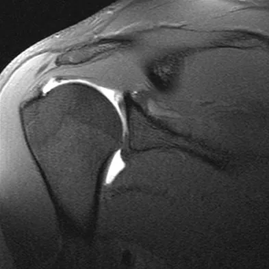
Figure 40 shows the MRI scan of a 23-year-old man with a history of recurrent anterior shoulder instability. What is the most likely diagnosis?


Explanation

The MRI scan shows an ALPSA lesion. This is also known as a medialized Bankart with medial displacement of the torn anterior labrum. During surgical stabilization, the labrum and periosteal sleeve must be mobilized and repaired laterally to reduce recurrent instability. A Perthes lesion is a nondisplaced labral tear. A GLAD lesion represents a nondisplaced anterior labral tear with an associated articular cartilage injury. Neviaser TJ: The anterior labroligamentous periosteal sleeve avulsion lesion: A cause of anterior instability of the shoulder. Arthroscopy 1993;9:17-21.


Question 21

Figure 41 shows the MRI scan of a 38-year-old weightlifter. What does the arrow on the MRI scan indicate?


Explanation

Pectoralis major ruptures typically occur in avid weightlifters (often on supplements) and typically while bench-pressing. Clinically there is significant discoloration/bruising over the pectoralis and into the axilla. MRI helps confirm the diagnosis and may help determine if the tear is in the muscle belly or at the bone-tendon junction. Bal GK, Basamania CJ: Pectoralis major tendon ruptures: Diagnosis and treatment. Tech Shoulder Elbow Surg 2005;6:128-134.


Question 22

Which of the following describes the correct proximal to distal progression of the annular and cruciform pulleys of the digits?


Explanation

The correct progression of the annular and cruciform pulley in the digits is A1, A2, C1, A3, C2, A4, C3. The two cruciform pulleys are collapsible elements adjacent to the more rigid annular pulleys of the flexor tendon sheath. This arrangement enables unrestricted flexion of the proximal interphalangeal joint. Hoppenfeld S, deBoer P: Surgical Exposures in Orthopaedics, ed 2. Philadelphia, PA, Lippincott-Raven, 1994, pp 176-186.

Question 23

A 75-year-old woman began a walking program 2 months after undergoing right total knee arthroplasty. She had to stop the program after 4 weeks because of hindfoot pain and ankle swelling. Radiographs are shown in Figures 42a and 42b. What is the most likely diagnosis?


Explanation

It is often tempting to assign a diagnosis of plantar fasciitis in patients with hindfoot pain. In this patient, the radiographs confirm a diagnosis of a calcaneal insufficiency fracture. The dense condensation of bone on the lateral view confirms the diagnosis. There is no radiographic evidence of a heel spur, osteochondral lesions, or chondrocalinosis. Resnick D: Diagnosis of Bone and Joint Disorders, ed 3. Philadelphia, PA, WB Saunders, 1995, p 2591. Kearon C: Natural history of venous thromboembolism. Semin Vasc Med 2001;1:27-37.


Question 24

Figure 43 shows an arthroscopic view of a right shoulder through a lateral portal in the beach chair position. The arrow is pointing to what structure?


Explanation

This view from the lateral portal shows a full-thickness rotator cuff tear. The glenohumeral joint can be visualized through this tear. The glenoid, labrum, and biceps tendon attaching to the superior aspect of the glenoid are easily viewed from this portal, and the arrow is pointing to the biceps tendon. Arthroscopic rotator cuff repair can be performed while visualizing from this portal and using anterior and posterior working portals. Mazzocca AD, Noerdlinger M, Cole B, et al: Arthroscopy of the shoulder: Indications and general principles of techniques, in McGinty JB (ed): Operative Arthroscopy, ed 3. Philadelphia, PA, Lippincott Williams & Wilkins, 2003, pp 412-427.


Question 25

In Charcot-Marie-Tooth disease a progressive deformity develops in the foot. Which functional muscles predominate in deformity formation?


Explanation

In Charcot-Marie-Tooth disease, the posterior tibialis and peroneus longus tendons remain strong, serving to invert the hindfoot and depress the first metatarsal head thus causing the cavovarus foot associated with this disease. In contrast, the tibialis anterior and peroneus brevis are less functional and therefore cannot dorsiflex the ankle, elevate the first metatarsal, or evert the foot, contributing to the deformity.

Question 26

During a surgical approach to the proximal radius, the surgeon plans to expose the radial shaft. To protect the posterior interosseous nerve (PIN), which muscle should be carefully retracted, considering the PIN passes between its two heads?


Explanation

The PIN passes between the superficial and deep heads of the supinator muscle within the arcade of Frohse. Careful identification and retraction of this muscle are essential to avoid iatrogenic injury.

Question 27

During a surgical dislocation of the hip for femoroacetabular impingement, the surgeon must preserve the primary blood supply to the adult femoral head. Which of the following vessels is the most critical to protect?


Explanation

The deep branch of the medial femoral circumflex artery (MFCA) is the primary blood supply to the adult femoral head. It courses posterior to the obturator externus and anterior to the short external rotators.

Question 28

The anteromedial (AM) bundle of the anterior cruciate ligament (ACL) is tightest in which of the following positions, and what is its primary function?


Explanation

The AM bundle of the ACL is tightest in knee flexion and primarily restrains anterior tibial translation. The posterolateral (PL) bundle is tightest in extension and provides rotational stability.

Question 29

A patient presents with the inability to form a perfect "OK" sign, resulting in a pinch with extended distal interphalangeal (DIP) and interphalangeal (IP) joints. Which nerve is most likely compressed, and which muscle is consequently affected?


Explanation

The anterior interosseous nerve (AIN) innervates the flexor pollicis longus and flexor digitorum profundus to the index finger. AIN palsy leads to the inability to flex the IP joint of the thumb and DIP of the index finger.

Question 30

During a direct lateral (Hardinge) approach to the hip, extending the proximal split in the gluteus medius more than 3 to 5 cm proximal to the greater trochanter places which of the following structures at highest risk of injury?


Explanation

The superior gluteal nerve runs approximately 3 to 5 cm proximal to the tip of the greater trochanter. Extending the gluteus medius split beyond this "safe zone" risks denervating the anterior portion of the gluteus medius and minimus.

Question 31

In the deltopectoral approach to the shoulder, the axillary nerve can be identified at the inferior border of the subscapularis. What is the approximate distance of the axillary nerve from the inferior glenoid labrum at the 6 o'clock position?


Explanation

The axillary nerve passes through the quadrangular space and is located approximately 10 to 15 mm inferior to the inferior glenoid rim. It is at significant risk during inferior capsular releases.

Question 32

The primary restraint to varus stress at the knee at 30 degrees of flexion is the fibular collateral ligament (FCL). What is the anatomical location of the FCL footprint on the femur relative to the lateral epicondyle?


Explanation

The fibular collateral ligament (FCL) originates on the lateral femoral condyle slightly proximal and posterior to the lateral epicondyle. It inserts on the lateral aspect of the fibular head.

Question 33

During a minimally invasive lateral transpsoas approach to the lumbar spine, which nerve is at greatest risk of iatrogenic injury when working at the L4-L5 disc space?


Explanation

The lumbar plexus lies within the posterior aspect of the psoas major muscle. At the L4-L5 level, the femoral nerve migrates anteriorly and is at the highest risk of injury during a transpsoas approach.

Question 34

In a patient with stage II acquired adult flatfoot deformity, the spring ligament is often attenuated. What are the primary bony attachments of the spring ligament?


Explanation

The spring ligament (plantar calcaneonavicular ligament) forms a critical sling supporting the talar head. It connects the sustentaculum tali of the calcaneus to the plantar surface of the navicular.

Question 35

During a posterior approach to the humerus for fracture plating, the radial nerve is identified in the spiral groove. At approximately what distance proximal to the olecranon fossa does the nerve cross the posterior humerus?


Explanation

The radial nerve lies in the spiral groove, crossing the posterior humerus from medial to lateral approximately 14 to 15 cm proximal to the olecranon fossa and 20 cm proximal to the medial epicondyle.

Question 36

Which of the following ligaments is the strongest and provides the greatest biomechanical resistance to diastasis of the distal tibiofibular syndesmosis?


Explanation

The posterior inferior tibiofibular ligament (PITFL) is the strongest component of the ankle syndesmosis. It contributes the most resistance to lateral displacement of the fibula.

Question 37

The distal biceps tendon normally inserts on the radial tuberosity. To maximize supination strength during a surgical repair of a ruptured tendon, it should be reattached to which specific footprint?


Explanation

The native distal biceps tendon inserts on the ulnar/posterior aspect of the radial tuberosity. Reattaching it as posterior as possible maximizes the supination moment arm and restores functional strength.

Question 38

A cyclist presents with numbness in the little finger and weakness of finger abduction. The ulnar nerve is compressed in Guyon's canal. Which of the following structures forms the floor of Guyon's canal?


Explanation

The floor of Guyon's canal is formed by the transverse carpal ligament and the pisohamate ligament. The roof is formed by the volar carpal ligament and the palmaris brevis muscle.

Question 39

During an ilioinguinal approach to the acetabulum, the "corona mortis" is encountered and must be carefully ligated to prevent catastrophic bleeding. What vessels are connected by the corona mortis?


Explanation

The corona mortis is an anastomotic vascular connection between the obturator vessels and the external iliac or inferior epigastric vessels. It crosses over the superior pubic ramus at an average distance of 5-6 cm from the symphysis.

Question 40

A patient presents with shoulder weakness and lateral winging of the scapula following a lymph node biopsy in the posterior triangle of the neck. Which nerve was injured, and which muscle is paralyzed?


Explanation

The spinal accessory nerve (CN XI) innervates the trapezius muscle and is highly susceptible to injury during procedures in the posterior triangle of the neck. Paralysis results in shoulder droop and lateral scapular winging.

Question 41

The anterior bundle of the medial collateral ligament (AMCL) of the elbow is the primary restraint to valgus stress. Where is its primary insertion on the proximal ulna?


Explanation

The anterior bundle of the elbow MCL originates on the anteroinferior aspect of the medial epicondyle and inserts on the sublime tubercle of the proximal ulna.

Question 42

The ulnar nerve passes through the cubital tunnel at the elbow. The roof of the cubital tunnel is formed by Osborne's ligament, which anatomically spans between which two structures?


Explanation

Osborne's ligament (the cubital tunnel retinaculum) forms the roof of the cubital tunnel. It stretches between the medial epicondyle and the tip of the olecranon.

Question 43

A professional volleyball player presents with isolated atrophy and weakness of the infraspinatus muscle, with normal supraspinatus strength and bulk. Where is the most likely site of nerve compression?


Explanation

Compression of the suprascapular nerve at the spinoglenoid notch affects only the infraspinatus muscle. Compression more proximally at the suprascapular notch would affect both the supraspinatus and infraspinatus.

Question 44

The popliteal artery is at risk of injury during a high tibial osteotomy (HTO) or total knee arthroplasty. At the level of the knee joint line, what is the anatomical relationship of the popliteal artery to the vein and nerve?


Explanation

From superficial to deep (posterior to anterior), the order is Nerve, Vein, Artery. At the joint line, the popliteal artery is the deepest and most medial structure, placing it at high risk from over-penetrating instruments.

Question 45

During an anterolateral approach to the distal tibia, the superficial peroneal nerve must be protected to prevent painful neuromas. Where does this nerve typically pierce the crural fascia to become subcutaneous?


Explanation

The superficial peroneal nerve typically pierces the crural fascia approximately 10 to 12 cm proximal to the tip of the lateral malleolus. It then divides into the intermediate and medial dorsal cutaneous nerves.

Question 46

A 45-year-old man undergoes a fasciotomy for chronic exertional compartment syndrome of the lateral leg. During the procedure, a nerve that pierces the deep fascia to become subcutaneous is at high risk of injury. Approximately where does this nerve typically exit the deep fascia?


Explanation

The superficial peroneal nerve typically pierces the deep fascia of the lateral compartment approximately 10 to 12 cm proximal to the tip of the lateral malleolus. It then divides into medial and intermediate dorsal cutaneous nerves to supply sensation to the dorsum of the foot.

Question 47

A patient presents with an inability to form a valid "OK" sign, instead demonstrating a pinch with extended interphalangeal and distal interphalangeal joints of the thumb and index finger. Assuming an isolated anterior interosseous nerve (AIN) palsy, which of the following muscles will maintain intact innervation?


Explanation

The AIN provides motor innervation to the flexor pollicis longus, pronator quadratus, and the flexor digitorum profundus of the index and middle fingers. The supinator is innervated by the posterior interosseous nerve (PIN), a branch of the radial nerve.

Question 48

A 32-year-old woman sustains a displaced talar neck fracture. She is at high risk for avascular necrosis of the talar body due to retrograde blood supply. Which of the following arteries provides the predominant blood supply to the talar body?


Explanation

The artery of the tarsal canal is a branch of the posterior tibial artery and provides the dominant blood supply to the talar body. It forms an anastomotic sling with the artery of the sinus tarsi.

Question 49

A 35-year-old weightlifter presents with vague posterior shoulder pain and weakness in external rotation. An MRI reveals an isolated cyst compressing a nerve within the quadrangular space. Which of the following muscles forms the superior border of this anatomic space?


Explanation

The quadrangular space is bordered superiorly by the teres minor, inferiorly by the teres major, medially by the long head of the triceps, and laterally by the surgical neck of the humerus. It contains the axillary nerve and the posterior circumflex humeral artery.

Question 50

During a posterior approach to the hip for a total hip arthroplasty, the surgeon must be careful to protect the primary blood supply to the femoral head. Which of the following vessels provides the predominant blood supply to the adult femoral head?


Explanation

The medial femoral circumflex artery (MFCA) provides the primary blood supply to the adult femoral head via the lateral epiphyseal artery. It courses posterior to the femoral neck and must be protected during posterior hip approaches.

Question 51

A 24-year-old football player sustains a direct blow to the anteromedial aspect of his knee, resulting in a posterolateral corner (PLC) injury. Which of the following structures is considered one of the three primary static stabilizers of the PLC?


Explanation

The three primary static stabilizers of the posterolateral corner (PLC) are the lateral collateral ligament (LCL), the popliteus tendon, and the popliteofibular ligament. The biceps femoris provides dynamic stability but is not a primary static stabilizer.

Question 52

A rock climber presents with pain and a "bowstringing" deformity of the index finger after feeling a pop. Rupture of which of the following annular pulleys is most biomechanically detrimental to normal finger flexion?


Explanation

The A2 and A4 pulleys are the most critical biomechanically for preventing bowstringing of the flexor tendons. The A2 pulley arises from the proximal phalanx, and its loss severely compromises flexor tendon mechanics.

Question 53

When placing a pedicle screw in the lumbar spine, the surgeon identifies the starting point at the intersection of the pars interarticularis, the midpoint of the transverse process, and the superior articular facet. Medial breach of the pedicle screw at the L4 vertebral body most directly endangers the dural sac and which of the following nerve roots?


Explanation

A medial pedicle breach at the L4 level endangers the dural sac and the traversing L5 nerve root. The exiting L4 root travels directly inferior to the L4 pedicle and is at greatest risk with an inferior breach.

Question 54

During a plantar approach for an excision of a plantar fibroma, the surgeon dissects near the Master Knot of Henry. Which two tendons cross at this specific anatomic landmark?


Explanation

The Master Knot of Henry is located in the midfoot where the flexor hallucis longus (FHL) tendon crosses dorsal to the flexor digitorum longus (FDL) tendon. This intersection is a critical anatomic landmark during midfoot dissections and tendon transfers.

Question 55

A 28-year-old gymnast sustains an elbow dislocation. After closed reduction, she has persistent posterolateral rotatory instability (PLRI). Deficiency of which of the following structures is the primary cause of this instability?


Explanation

The lateral ulnar collateral ligament (LUCL) is the primary stabilizer against posterolateral rotatory instability (PLRI) of the elbow. It originates from the lateral epicondyle and inserts on the supinator crest of the ulna.

Question 56

An orthopedic oncologist is performing a resection of a soft tissue sarcoma located within the femoral triangle. Which of the following defines the lateral border of this anatomic region?


Explanation

The femoral triangle is bordered superiorly by the inguinal ligament, medially by the medial border of the adductor longus, and laterally by the medial border of the sartorius muscle.

Question 57

A volar surgical approach to the proximal radius (Henry approach) requires developing an internervous plane. Which of the following muscles is a component of the "mobile wad of Henry" that is retracted laterally during this approach?


Explanation

The mobile wad of Henry consists of the brachioradialis, extensor carpi radialis longus, and extensor carpi radialis brevis. The volar Henry approach to the radius utilizes the internervous plane between the brachioradialis (radial n.) and the pronator teres (median n.).

Question 58

During an endoscopic carpal tunnel release, the surgeon must be aware of the motor branch of the median nerve. Through which of the following mechanisms does this recurrent motor branch most commonly exit the carpal tunnel?


Explanation

The recurrent motor branch of the median nerve most commonly follows an extraligamentous course (occurring in approximately 50-70% of individuals). It typically branches distal to the transverse carpal ligament and turns back to innervate the thenar muscles.

Question 59

During an ilioinguinal approach for an anterior column acetabular fracture, significant hemorrhage occurs just posterior to the superior pubic ramus. This is most likely due to an injury to the "corona mortis," which is an anastomosis between which two vascular systems?


Explanation

The corona mortis is a vascular anastomosis between the external iliac system (inferior epigastric vessels) and the obturator system. It is located on the posterior aspect of the superior pubic ramus, placing it at risk during anterior pelvic approaches.

Question 60

A 25-year-old soccer player is diagnosed with a high ankle sprain. Which of the following ligaments is the strongest and provides the most stability to the distal tibiofibular syndesmosis?


Explanation

The posterior inferior tibiofibular ligament (PITFL) is the strongest component of the syndesmotic ligament complex. It contributes approximately 42% of the overall strength of the syndesmosis.

Question 61

A patient presents with isolated weakness of the infraspinatus muscle but normal supraspinatus strength. The suprascapular nerve is most likely compressed at which of the following anatomic locations?


Explanation

The suprascapular nerve innervates the supraspinatus muscle before passing through the spinoglenoid notch to innervate the infraspinatus. Entrapment at the spinoglenoid notch bypasses the supraspinatus branch, resulting in isolated infraspinatus weakness.

Question 62

A patient sustains a midshaft humerus fracture and subsequently presents with an inability to extend the wrist and digits, but retains the ability to extend the elbow. Sensation is decreased over the dorsal web space. The injured nerve originates from which cord(s) of the brachial plexus?


Explanation

The clinical presentation is consistent with a radial nerve palsy distal to the triceps innervation. The radial nerve is the terminal continuation of the posterior cord of the brachial plexus.

Question 63

During an ilioinguinal approach to the acetabulum, massive bleeding occurs while dissecting over the superior pubic ramus. Which of the following represents the most likely anatomical source of this hemorrhage?


Explanation

The corona mortis is a vascular anastomosis between the obturator and external iliac (or inferior epigastric) vessels. It lies approximately 5 cm from the pubic symphysis over the superior pubic ramus and is at significant risk during the ilioinguinal approach.

Question 64

A patient sustains a posterior shoulder dislocation and subsequently demonstrates weakness in shoulder external rotation and a sensory deficit over the lateral deltoid. The affected nerve exits the axilla through a space bordered superiorly by which of the following structures?


Explanation

The axillary nerve passes through the quadrangular space, which is bordered superiorly by the teres minor, inferiorly by the teres major, medially by the long head of the triceps, and laterally by the surgical neck of the humerus.

Question 65

A 24-year-old athlete sustains a knee injury resulting in increased external tibial rotation at both 30 and 90 degrees of knee flexion. Which of the following anatomical structures are most likely completely ruptured?


Explanation

Isolated posterolateral corner (PLC) injuries typically present with increased external rotation at 30 degrees of flexion but normal rotation at 90 degrees. Increased external rotation at both 30 and 90 degrees of flexion indicates a combined PCL and PLC injury.

Question 66

During flatfoot reconstruction, the surgeon evaluates the plantar calcaneonavicular ligament. This structure, which is a primary static stabilizer of the longitudinal arch, is directly supported plantarly by which of the following tendons?


Explanation

The plantar calcaneonavicular (spring) ligament complex bridges the calcaneus and navicular, supporting the talar head. The tibialis posterior tendon courses directly plantar to the spring ligament, providing dynamic arch support.

Question 67

To prevent bowstringing of the flexor tendons following a tenolysis procedure in zone II of the hand, the surgeon must preserve which of the following critical annular pulleys?


Explanation

The A2 and A4 pulleys are the most mechanically critical for preventing bowstringing of the flexor tendons. The A2 pulley arises from the proximal phalanx, while the A4 pulley arises from the middle phalanx.

Question 68

A 35-year-old man sustains a severe fall, leading to an Essex-Lopresti lesion. The central band of the interosseous membrane is torn. Which of the following best describes the normal anatomical orientation of the fibers of this central band?


Explanation

The central band of the interosseous membrane is the primary stabilizer against longitudinal proximal migration of the radius. Its fibers run obliquely from proximal on the radius to distal on the ulna.

Question 69

During a posterior approach to the hip, protecting the primary blood supply to the femoral head is critical. The medial femoral circumflex artery (MFCA) courses deep to which of the following short external rotators?


Explanation

The deep branch of the MFCA passes deep (anterior) to the quadratus femoris. To protect the blood supply to the femoral head during a posterior approach, the quadratus femoris should be released with a cuff or spared, and the obturator externus must remain intact.

Question 70

A patient presents with weakness in wrist extension and altered sensation over the dorsal web space of the hand. An MRI reveals a herniated cervical disc. Between which two cervical vertebrae is the herniation most likely located?


Explanation

Weakness in wrist extension and triceps, combined with numbness in the dorsal web space (middle finger), is characteristic of a C7 radiculopathy. In the cervical spine, the C7 nerve root exits through the C6-C7 neural foramen.

Question 71

The anterior cruciate ligament (ACL) is composed of two primary bundles. In full knee extension, what is the anatomical status of the anteromedial (AM) bundle compared to the posterolateral (PL) bundle?


Explanation

The ACL consists of the anteromedial (AM) and posterolateral (PL) bundles. In full knee extension, the PL bundle is taut and the AM bundle is relatively lax, providing crucial rotational stability.

Question 72

A pitcher complains of right shoulder pain and weakness with overhead activities. Examination reveals isolated wasting of the supraspinatus and infraspinatus. Entrapment of the affected nerve is most likely to occur at which of the following anatomical sites?


Explanation

The suprascapular nerve innervates both the supraspinatus and infraspinatus muscles. Entrapment at the suprascapular notch affects both muscles, whereas entrapment further distal at the spinoglenoid notch typically causes isolated infraspinatus weakness.

Question 73

A surgeon is performing an open anterior shoulder stabilization and identifies the rotator interval. Which of the following structures is NOT considered a normal anatomical border or content of the rotator interval?


Explanation

The rotator interval is a triangular space bordered by the supraspinatus superiorly, subscapularis inferiorly, and the coracoid process medially. Its contents include the coracohumeral ligament, superior glenohumeral ligament, and the long head of the biceps tendon.

Question 74

During surgical release of the tarsal tunnel, the flexor retinaculum is divided. From anterior to posterior, what is the correct anatomical order of the structures passing behind the medial malleolus?


Explanation

The structures passing through the tarsal tunnel from anterior to posterior are remembered by the mnemonic 'Tom, Dick, And Very Nervous Harry': Tibialis posterior, flexor Digitorum longus, posterior tibial Artery, tibial Nerve, and flexor Hallucis longus.

Question 75

A patient presents with posterolateral rotatory instability (PLRI) of the elbow following a dislocation. This condition is primarily caused by insufficiency of the lateral ulnar collateral ligament (LUCL). Where does the LUCL anatomically insert?


Explanation

The lateral ulnar collateral ligament (LUCL) is the primary restraint to posterolateral rotatory instability of the elbow. It originates on the lateral epicondyle and inserts on the supinator crest of the proximal ulna.

Question 76

A patient undergoes a four-compartment fasciotomy of the leg. The deep posterior compartment is released to decompress its contents. Which of the following neurovascular structures is contained within the deep posterior compartment?


Explanation

The deep posterior compartment of the leg contains the tibialis posterior, flexor hallucis longus, and flexor digitorum longus muscles. It also encases the posterior tibial and peroneal arteries along with the tibial nerve.

Question 77

A 25-year-old gymnast sustains a wrist injury, and an MRI reveals an avulsion of the triangular fibrocartilage complex (TFCC) from its foveal attachment. The fovea of the ulna lies anatomically between which two structures?


Explanation

The fovea of the ulna is the primary attachment site for the deep fibers of the TFCC (ligamentum subcruentum). It is located at the base of the ulnar styloid, between the styloid itself and the articular surface of the ulnar dome (head).

Question 78

During a deltopectoral approach to the shoulder, the cephalic vein is typically retracted. Which of the following describes the correct internervous plane for this approach?


Explanation

The deltopectoral approach utilizes an internervous plane between the deltoid (axillary nerve) and pectoralis major (medial and lateral pectoral nerves). The cephalic vein marks this interval and is usually retracted laterally to preserve its primary venous drainage.

Question 79

A surgeon is performing an anterior (Smith-Petersen) approach to the hip. The superficial surgical interval is between the sartorius and the tensor fasciae latae. What is the internervous plane of this superficial interval?


Explanation

The superficial interval in the Smith-Petersen approach is between the sartorius (femoral nerve) and the tensor fasciae latae (superior gluteal nerve). The deep interval continues between the rectus femoris (femoral nerve) and gluteus medius (superior gluteal nerve).

Question 80

During a posterolateral approach to the ankle for fixation of a posterior malleolus fracture, the sural nerve is at risk of iatrogenic injury. What two nerves typically join to form the sural nerve?


Explanation

The sural nerve is typically formed by the confluence of the medial sural cutaneous nerve (a branch of the tibial nerve) and the peroneal communicating branch (from the lateral sural cutaneous nerve). It supplies sensation to the posterolateral aspect of the distal leg and lateral foot.

Question 81

An orthopedic resident is dissecting the posterior shoulder. The quadrangular space transmits the axillary nerve and posterior circumflex humeral artery. Which of the following forms the superior border of this space in a normal anatomic specimen?


Explanation

The quadrangular space is bordered superiorly by the teres minor, inferiorly by the teres major, medially by the long head of the triceps, and laterally by the surgical neck of the humerus.

Question 82

The Thompson (posterolateral) approach to the proximal radius utilizes an internervous plane between which two muscles?


Explanation

The Thompson approach utilizes the internervous plane between the extensor carpi radialis brevis (radial nerve) and the extensor digitorum communis (posterior interosseous nerve). It exposes the proximal and middle thirds of the radius while allowing the PIN to be protected within the supinator.

Question 83

A 32-year-old man sustains a displaced talar neck fracture (Hawkins Type III). Which of the following provides the primary blood supply to the talar body, placing it at high risk for avascular necrosis?


Explanation

The artery of the tarsal canal, a branch of the posterior tibial artery, provides the dominant blood supply to the talar body. Disruption of this artery during a displaced talar neck fracture significantly increases the risk of avascular necrosis.

Question 84

During knee arthroscopy, the surgeon visualizes a ligamentous structure originating from the posterior horn of the lateral meniscus and inserting into the lateral aspect of the medial femoral condyle, passing anterior to the PCL. What is this structure?


Explanation

The meniscofemoral ligament of Humphrey passes anterior to the posterior cruciate ligament (PCL). The ligament of Wrisberg passes posterior to the PCL, but both originate from the posterior horn of the lateral meniscus.

Question 85

Compression of the posterior interosseous nerve (PIN) at the elbow frequently occurs at the Arcade of Frohse. This fibrous arch is formed by the proximal edge of which muscle?


Explanation

The Arcade of Frohse is a fibrous band formed by the proximal edge of the superficial head of the supinator muscle. It is the most common site of compression for the posterior interosseous nerve in radial tunnel syndrome.

Question 86

The blood supply to the scaphoid predominantly enters at which location, making proximal pole fractures particularly susceptible to nonunion?


Explanation

Approximately 70% to 80% of the scaphoid's blood supply is retrograde, entering through the dorsal ridge at the waist via branches of the radial artery. Proximal pole fractures disrupt this flow, leading to a high rate of avascular necrosis and nonunion.

Question 87

During a medial approach to the knee for a medial collateral ligament repair, the infrapatellar branch of the saphenous nerve is transected. What clinical deficit will the patient experience?


Explanation

The infrapatellar branch of the saphenous nerve provides sensation to the anterior and anterolateral skin of the proximal tibia. Iatrogenic transection during anteromedial knee approaches results in a characteristic patch of numbness in this area.

Question 88

An anterior approach to the humeral shaft utilizes the plane between the biceps brachii and brachialis. During deep dissection, the brachialis muscle is split longitudinally. Which aspect of the split brachialis is retracted laterally to protect the radial nerve?


Explanation

The brachialis has dual innervation; the medial portion is innervated by the musculocutaneous nerve, while the lateral portion is innervated by the radial nerve. Splitting the brachialis longitudinally and retracting the lateral half laterally cushions and protects the radial nerve.

Question 89

During the ilioinguinal approach for an anterior column acetabular fracture, the surgeon develops three surgical "windows." What anatomical structure forms the boundary between the lateral and middle windows?


Explanation

The iliopectineal fascia divides the lateral and middle windows in the ilioinguinal approach. The middle window lies between this fascia (laterally) and the external iliac vessels (medially).

Question 90

A patient sustains a high-energy distal tibia fracture. During a minimally invasive percutaneous plate osteosynthesis (MIPPO) via an anterolateral approach, which nerve is most at risk of iatrogenic injury as it pierces the deep fascia of the leg?


Explanation

The superficial peroneal nerve pierces the deep fascia of the lateral compartment approximately 10 to 12 cm proximal to the lateral malleolus. It crosses the anterolateral distal tibia, making it highly vulnerable during percutaneous anterolateral plating.

Question 91

During the proximal portion of the volar (Henry) approach to the forearm, supination of the forearm is performed to protect a major nerve. Which muscle's insertion is stripped and elevated to expose the proximal radius, and what is the anatomical relationship of the at-risk nerve to this muscle?


Explanation

In the volar (Henry) approach to the proximal radius, the supinator muscle is elevated to expose the bone. Supinating the forearm protects the posterior interosseous nerve (PIN), which safely moves laterally away from the surgical field as it courses between the superficial and deep heads of the supinator.

Question 92

A 28-year-old runner presents with chronic exertional compartment syndrome requiring fasciotomy of the lateral leg compartment. During the fascial release, which nerve is at greatest risk of iatrogenic injury as it exits the deep fascia to become subcutaneous in the distal third of the leg?


Explanation

The superficial peroneal nerve courses within the lateral compartment of the leg and pierces the crural fascia to become subcutaneous approximately 10-12 cm proximal to the lateral malleolus. It is at high risk of iatrogenic injury during lateral compartment fasciotomies or the placement of anterolateral ankle arthroscopy portals.

Question 93

A spine surgeon is performing a lateral transpsoas approach (LLIF) for an L4-L5 interbody fusion. Which of the following nerves emerges on the anterior surface of the psoas major muscle and runs longitudinally, making it particularly vulnerable to injury during initial retractor placement?


Explanation

The genitofemoral nerve pierces the anterior surface of the psoas major muscle and travels longitudinally down its anterior aspect. Due to this anterior course, it is highly vulnerable during the lateral transpsoas approach, with iatrogenic injury typically leading to groin and anterior thigh paresthesia.

None

Clinic OS
Medically Verified Content by
Prof. Clinic OS
Consultant Orthopedic & Spine Surgeon
Chapter Index