العربية
Part of the Master Guide

AAOS & ABOS Basic Science MCQs (Set 3): Bone Biology, Biomechanics & Anatomy Review

Orthopedic Anatomy 2026 MCQs: Board Review Questions & Answers (Part 3)

23 Apr 2026 56 min read 72 Views
Figure for Anatomy 2000 MCQs - Part 3 - Question 51

Key Takeaway

Looking for accurate information on Orthopedic Anatomy 2026 MCQs: Board Review Questions & Answers (Part 3)? Top-rated Orthopedic Anatomy 2026 MCQs bank. Practice with clinical case questions, orthopedic surgery board review, and evidence-based answers updated for 2026.

Orthopedic Anatomy 2026 MCQs: Board Review Questions & Answers (Part 3)

Comprehensive 100-Question Exam


00:00

Start Quiz

Question 1

A 41-year-old man who plays golf regularly has had ulnar-sided wrist pain for the past several days after striking a tree root with a golf club. Examination reveals significant pain with resisted flexion of the ring and small fingers and tenderness over the hook of the hamate. Which of the following radiographic views would be most helpful in identifying the pathology of this injury?


Explanation

The history and examination findings suggest an acute fracture of the hook of the hamate. The radiographic study considered most helpful in identifying this type of fracture is the carpal tunnel view. PA and lateral views of the wrist will not adequately visualize the hook of the hamate. Bruerton's view is intended for the assessment of the metacarpophalangeal joints. Pathology would not be suspected in the scaphoid, metacarpals, or the phalanges, so the scaphoid view and the PA, lateral, and oblique views of the hand would not be helpful. Green DP, Hotchkiss RN, Pederson WC (eds): Operative Hand Surgery, ed 4. New York, NY, Churchill Livingstone, 1999, p 855.

Question 2

What is the primary limiting membrane and mechanical support for the periphery of the physis?


Explanation

The perichondrial fibrous ring of La Croix acts as a limiting membrane that provides mechanical support for the bone-cartilage junction of the growth plate. It is continuous with the ossification groove of Ranvier, which contributes chondrocytes for the increase in width of the growth plate. The zone of provisional calcification lies at the bottom of the hypertrophic zone and is the site of initial calcification of the matrix. It is quite weak and usually is the cleavage plane for fractures; therefore, it does not qualify as mechanical support. The last intact transverse septum separates the zone of provisional calcification from the primary spongiosa and provides no real support to the physis. The primary spongiosa is the part of the metaphysis nearest the physis. Netter FH: Growth plate, in Woodburne RT, Crelin ES, Kaplan FS, Dingle RV (eds): The Ciba Collection of Medical Illustrations. Summit, NJ, Ciba-Geigy Corporation, 1987, vol 8, pp 166-167.

Question 3

What normal tissue has a low signal intensity (appears black) on both T1- and T2-weighted images?


Explanation

Tendons, cortical bone, ligaments, menisci, and fibrous tissue will show low signal intensity (SI) on both T1- and T2-weighted images. Fat-containing tissues, such as subcutaneous fat and bone marrow, will show high SI on T1-weighted images and low SI on T2-weighted images. Tissues with high water content, such as joint fluid, intervertebral disk, and edema, will show low SI on T1-weighted images and high SI on T2-weighted images. Kasser JR (ed): Orthopaedic Knowledge Update 5. Rosemont, IL, American Academy of Orthopaedic Surgeons, 1996, pp 65-70.

Question 4

A positive Froment sign indicates weakness of which of the following muscles?


Explanation

Thumb adduction is powered by the adductor pollicis (ulnar nerve). Testing involves having the patient forcibly hold a piece of paper between the thumb and radial side of the index proximal phalanx. When this muscle is weak or nonfunctioning, the thumb interphalangeal joint flexes with this maneuver, resulting in a positive Froment sign. The paper is held by action of the thumb flexion (flexor pollicis longus and flexor pollicis brevis; median innervated).

Question 5

Figure 28 shows an AP radiograph of a 54-year-old woman who underwent lumbar laminectomy and fusion at the L4 and L5 levels with placement of a bone stimulator 8 years ago. She also underwent a left total hip arthroplasty 2 years ago; aspiration of that joint now reveals that it is infected with a gram-positive cocci organism. History is also significant for IV drug use and human immunodeficiency virus (HIV). The patient reports fever, chills, and left flank and abdominal pain. Examination reveals significant pain with resisted left hip flexion and passive hip extension. She also has lumbar hyperlordosis. Which of the following studies would best identify the underlying cause of her infection?


Explanation

The patient's clinical signs (fever and flank, hip, and abdominal pain) suggest a primary iliopsoas abscess. With an increased patient population who abuse drugs and/or who are HIV-positive, iliopsoas abscess may be more prevalent because of systemic bacterial seeding and may be potentially unrecognized. Diagnostic imaging studies provide a better understanding of the anatomic magnitude of the infection, give concrete confirmation of the diagnosis, and may suggest an underlying cause. Neither standard abdominal radiographs nor ultrasound studies are sensitive enough to be diagnostic of this disease process. CT has been established as the standard study for identifying the underlying cause of this abscess. The hip infection has most likely developed as a result of hematogenous spread from an infected skin lesion from the patient's IV drug use. Santaella RO, Fishman EK, Lipsett PA: Primary vs secondary iliopsoas abscess: Presentation, microbiology, and treatment. Arch Surg 1995;130:1309-1313.

Question 6

A 55-year-old woman who underwent a left total hip arthroplasty 8 months ago using a modified Hardinger approach reports a persistent painless limp. Examination reveals that when she is not using a cane, she lurches to the left during weight bearing on the left lower extremity. An AP radiograph is shown in Figure 29. Which of the following hip muscle groups should be strengthened to improve the gait abnormality?


Explanation

The modified Hardinger approach includes a partial anterior trochanteric osteotomy creating a trochanteric wafer (as seen on the radiograph) that is displaced anterior and medial in continuity with the gluteus medius and vastus lateralis. Failure of abductor reattachment, migration of the trochanter, nonunion of the osteotomy site, and excessive splitting of the gluteus medius muscle causing injury to the inferior branch of the superior gluteal nerve can result in weakness of the abductor mechanism. Abductor strength should be evaluated with the patient lying on the opposite side and elevating the affected limb. Although slight weakness may manifest itself as a limp only after prolonged muscular activity, significant weakness results in a constant limp without associated discomfort. Morrey BF (ed): Joint Replacement Arthroplasty. New York, NY, Churchill Livingstone, 1991, pp 512-526.

Question 7

Figure 30 shows an axial cross section of extensor tendon anatomy in zone 7 of the wrist. What letter best depicts the location of the posterior interosseous nerve?


Explanation

The posterior interosseous nerve in contained in the floor of the fourth dorsal compartment of the wrist, which is labelled C in this diagram. Tubiana R, McCullough CJ, Masquelet AC: An Atlas of Surgical Exposures of the Upper Extremity. Philadelphia, PA, JB Lippincott, 1990, p 224.

Question 8

Figures 31a and 31b show the T1- and T2-weighted MRI scans of a patient's knee joint. What is the most likely diagnosis?


Explanation

31b The scans show a lipohemarthrosis. There is the characteristic layering of a superior zone containing fat (high signal intensity), a central zone containing serum (low signal intensity), and an inferior zone that contains red blood cells (low signal intensity). The most common cause of a lipohemarthrosis is an intra-articular fracture with leakage of marrow fat into the joint. Resnick D, Kang HS: Synovial joints, in Resnick D, Kang HS (eds): Internal Derangements of Joints: Emphasis on MR Imaging. Philadelphia, PA, WB Saunders, 1997, pp 49-53.

Question 9

A 15-year-old boy reports feeling a pop and notes sudden giving way of the left knee while playing basketball. He has immediate pain and swelling in the knee. An AP radiograph is shown in Figure 32. A small avulsion fragment from the lateral tibial margin is the only finding. What is the most likely diagnosis?


Explanation

An avulsion fracture from the lateral tibial margin carries the eponym Segond fracture and is pathognomonic for an anterior cruciate ligament (ACL) tear. The fragment is located posterior to Gerdy's tubercle and is superior and anterior to the fibular head. It represents an avulsion of the lateral capsular ligament of the knee and is caused by the same mechanism that causes the ACL tear. The pes anserinus is the insertion point of the medial hamstrings and would not be affected in a lateral avulsion injury. The posterior cruciate ligament may be seen on a lateral view if associated with an avulsion fragment, but a tear of the PCL generally cannot be diagnosed on an AP view. The insertion of the iliotibial band is broad and is unlikely to produce an avulsion injury such as that seen in the radiograph. This view is not consistent with the appearance of a lateral collateral ligament injury. Beaty JH (ed): Orthopaedic Knowledge Update 6. Rosemont, IL, American Academy of Orthopaedic Surgeons, 1999, pp 533-557.

Question 10

What neurovascular structure is most at risk when performing an inside-out repair of the posterior horn of the medial meniscus?


Explanation

The saphenous nerve is located on the posterior medial aspect of the knee and must be protected when performing an inside-out repair of the medial meniscus. The peroneal nerve is most at risk with lateral meniscal repairs. The other structures usually are not at risk with meniscal repair. Cannon WD Jr, Morgan CD: Meniscal repair: Arthroscopic repair techniques. Instr Course Lect 1994;43:77-96.

Question 11

Figure 33 shows the AP and lateral radiographs of an obese 58-year-old man who underwent a cementless total hip arthroplasty 6 years ago. He reports no pain, and examination reveals a normal gait and painless hip range of motion. What is the most likely diagnosis?


Explanation

Osteolysis of an otherwise well-functioning total hip arthroplasty is a recognized complication, and its radiographic appearance is typical, as shown here. Distal osteolysis, such as that shown here, is more prevalent when there is noncircumferential sealing of the proximal femoral canal. Pellicci PM, Tria AJ Jr, Garvin KL (eds): Orthopaedic Knowledge Update: Hip and Knee Reconstruction 2. Rosemont, IL, American Academy of Orthopaedic Surgeons, 2000, pp 175-180.

Question 12

Which of the following muscles attaches to the coracoid process of the scapula?


Explanation

The insertion of the pectoralis minor is on the base of the coracoid process. The coracoid helps define the interval between the subscapularis and supraspinatus muscles but neither attaches to it. The coracobrachialis and short head of biceps attach to the tip of the coracoid but are not listed as options. The long head of the biceps attaches to the supraglenoid tubercle. The serratus arises from the vertebral border of the scapula. Jobe CM: Anatomy and surgical approaches, in Jobe FW (ed): Operative Techniques in Upper Extremity Sports Injuries. St Louis, MO, Mosby, 1996, pp 140-142.

Question 13

Turf toe typically involves injury to which of the following structures of the great toe?


Explanation

The term turf toe includes a range of injuries of the capsuloligamentous complex of the first metatarsophalangeal joint with or without osteochondral fracture of the first metatarsal head or one of the sesamoids. The mechanism of injury is hyperextension. Clanton TO, Butler JE, Eggert A: Injuries to the metatarsophalangeal joints in athletes. Foot Ankle 1986;7:162-176.

Question 14

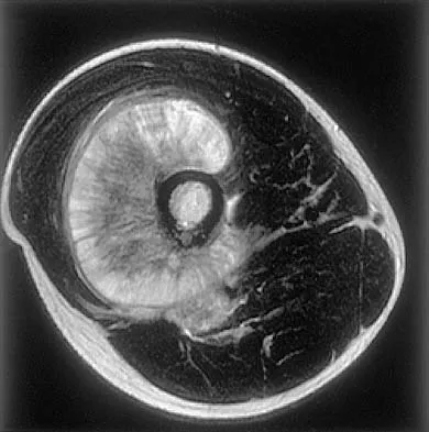
Figures 34a through 34c show an axial proton density (spin echo long TR, short TE) image, a sagittal inversion recovery (STIR) image, and a sagittal T1-weighted (short TR, short TE) image of the left thigh. What is the most likely diagnosis?


Explanation

34b 34c The images reveal a region of increased signal within the rectus femoris muscle with mild, ill-defined surrounding edema. The presence of high intensity signal on the T1-weighted image favors acute blood, in this case associated with a rectus femoris muscle tear or fatty tissue. However, because of fat suppression, a fatty lesion or lipoma would be dark on STIR, rather than bright as in this image. Most foreign bodies are low intensity signal and if small, are difficult to evaluate with MRI. The lack of adjacent subcutaneous soft-tissue edema or surrounding fluid makes pyomyositis an unlikely diagnosis.

Question 15

The artery located within the substance of the coracoacromial ligament is a branch of what artery?


Explanation

The acromial branch of the thoracoacromial artery courses along the medial aspect of the coracoacromial ligament and may be encountered when performing an open or arthroscopic subacromial decompression. Bleeding can be controlled by ligation of its branch from the thoracoacromial artery. The other arteries may be injured in other surgical exposures of the shoulder. Esch JC, Baker CL: The shoulder and elbow, in Whipple TL (ed): Arthroscopic Surgery. Philadelphia, PA, JB Lippincott, 1993, pp 65-66.

Question 16

Figures 35a and 35b show the axial T2-weighted and coronal T1-weighted MRI scans of a patient who has enlargement of the right thigh. What is the most likely diagnosis?


Explanation

35b The images show a large, almost circumferential, mass surrounding the diaphysis of the femur. The intramedullary signal is normal with minimal cortical destruction, both findings that should be abnormal in conventional osteosarcoma and Ewing's sarcoma. There are very low-signal striations representing osteoid formation that would have a sunburst radiographic pattern. This indicates an osteogenic lesion. Myositis ossificans is not indicated because studies would reveal zonal ossification starting in the periphery rather than the more central pattern seen in this patient. This appearance is typical for periosteal osteosarcoma.

Question 17

Figures 36a and 36b show the MRI scans of a patient who has shoulder weakness. What is the most likely diagnosis?


Explanation

36b The sagittal image reveals increased signal and decreased size of the supraspinatus and infraspinatus muscles, indicating muscle atrophy. The rotator cuff tendon signal is normal. The subscapularis and teres minor muscles are unaffected. Muscular dystrophy and thoracic outlet syndrome would be expected to have a more global effect. Although muscular atrophy can occur in the setting of a rotator cuff tear, the coronal image shows an intact supraspinatus. The suprascapular nerve supplies the supraspinatus and infraspinatus muscles. Therefore, suprascapular nerve entrapment would result in atrophy of these muscles with sparing of the surrounding musculature. Any lesion within the suprascapular notch, including neoplastic disease, a venous varix, or neuroma, can place pressure on the suprascapular nerve. Suprascapular nerve entrapment most commonly results from extension of a paralabral cyst or ganglion, often with associated labral pathology. Spinal accessory nerve disruption would show trapezius muscle atrophy. Resnick D, Kang HS (eds): Internal Derangement of Joints: Emphasis on MR Imaging. Philadelphia, PA, WB Saunders, 1997, pp 308-317.

Question 18

Figure 37 shows the T2-weighted MRI scan of the hip joint. What structure is labeled A?


Explanation

The obturator internus originates on the obturator membrane and adjacent bone, including the quadrilateral plate, and exits the lesser sciatic notch to insert on the posterior medial greater trochanter. The structure labeled C is the pectineus, B is the sartorius, and D is the gluteus medius. Last RJ: Anatomy: Regional and Applied, ed 6. London, England, Churchill Livingstone, 1978, pp 145-150, 324.

Question 19

The great medullary artery, also known as the Adamkiewicz artery, originates from which of the following arteries?


Explanation

The great medullary artery originates as a direct or indirect branch of the left posterior intercostal artery, usually between T8 and T12. It becomes intradural and crosses over one to three disk spaces before turning to the midline where it anastomoses with the anterior spinal artery. Injury to this artery can result in devastating ischemia of the lower spinal cord. Lu J, Ebraheim NA, Biyani A, Brown JA, Yeasting RA: Vulnerability of great medullary artery. Spine 1996;21:1852-1855.

Question 20

A patient who underwent total knee arthroplasty now reports a loss of sensation in the area circled in Figure 38. This area is innervated by which of the following nerves?


Explanation

The saphenous nerve follows the saphenous vein, giving off the infrapatellar branch that crosses the knee anteriorly to supply the peripatellar skin. A longitudinal incision can interrupt the nerve, leaving the terminal distribution without sensation.

Question 21

During an anterior approach to the shoulder, excessive traction on the conjoined tendon is most likely to result in loss of


Explanation

The musculocutaneous nerve travels through the conjoined tendon approximately 8 cm distal to the tip of the acromion. The musculocutaneous nerve innervates the biceps muscle and the bracialis muscle, both of which are responsible for elbow flexion. Shoulder flexion is facilitated by the anterior fibers of the deltoid muscle (axillary nerve) and the supraspinatus muscle (suprascapular nerve). The subscapular muscle facilitates internal rotation of the shoulder (upper and lower subscapularis nerve). Shoulder abduction is performed by the deltoid muscle (axillary nerve), and forearm pronation is facilitated by the pronator teres (median nerve). Hollinshead WH: Anatomy for Surgeons: The Back and Limbs, ed 3. Philadelphia, PA, Harper and Row, 1982, pp 391-393.

Question 22

Figure 39 shows the sagittal T1-weighted MRI scan of a 27-year-old man who twisted his knee 2 weeks ago. The arrow is pointing to


Explanation

The arrow identifies a transverse dark line that represents primary trabeculae of the physeal scar. A similar finding is seen in the proximal tibia. These lines may persist indefinitely. They do not represent ongoing growth, an abnormally open physeal plate, a stress fracture, or Looser's line (fatigue fracture in osteomalacia).

Question 23

Figure 40 shows the AP radiograph of a 55-year-old man who reports left knee pain. Which of the following conditions is least likely to produce this radiographic presentation?


Explanation

The radiograph reveals densities within the articular cartilage of the knee commonly referred to as chondrocalcinosis. The term chondrocalcinosis refers to the presence of calcium-containing crystals detected as radiodensities in cartilage. Calcium-containing crystals other than calcium pyrophosphate dihydrate may also deposit in articular cartilage and menisci, producing both radiographically detectable densities in cartilage and joint inflammation or degeneration. Hemochromatosis, alkaptonuria (ochronosis), and Wilson's disease are characterized by cellular deposition of iron, calcium, and copper ions, respectively, into various tissues including articular cartilage and can give this appearance. Septic arthritis does not usually cause chondrocalcinosis. Klippel JH (ed): Primer on the Rheumatic Diseases, ed 11. Atlanta, GA, Arthritis Foundation, 1997, pp 226-229 and 328-331.

Question 24

Figure 41 shows the MRI scan of a 39-year-old man who has severe left groin and anterior thigh pain. What is the most likely diagnosis?


Explanation

The MRI scan shows near complete involvement of the femoral head with bone marrow changes and some collapse of the necrotic segment. This is most suggestive of osteonecrosis.

Question 25

Iliosacral screws placed for stabilization of posterior pelvic ring injuries (eg, sacroiliac dislocation) that exit the sacrum anteriorly are most likely to injure which of the following structures?


Explanation

Iliosacral screws have gained popularity for posterior stabilization of pelvic ring disruptions, but complications attributed to incorrect placement are a clinical problem. The L5 nerve root is at greatest risk and is in closest proximity to a malpositioned screw (exiting the sacrum). The L4 root is more anterior at this level. The S1 root is still intraosseous at this level and is at risk but not from the screw exiting anteriorly at this level. The arteries are at risk but are more anterior and are at less risk than the L5 nerve root.

Question 26

A 28-year-old weightlifter presents with right shoulder pain and weakness in external rotation. An MRI shows an isolated cyst compressing the nerve in the quadrangular space. Which of the following muscles is most likely denervated?


Explanation

The axillary nerve passes through the quadrangular space and innervates the deltoid and teres minor. Compression in this space leads to weakness in shoulder abduction and external rotation, along with teres minor denervation.

Question 27

A 35-year-old sustains a displaced femoral neck fracture. Which of the following arteries provides the majority of the blood supply to the weight-bearing dome of the femoral head?


Explanation

The lateral epiphyseal artery, a terminal branch of the medial femoral circumflex artery (MFCA), provides the primary blood supply to the superolateral weight-bearing dome of the femoral head.

Question 28

A 35-year-old male sustains a displaced talar neck fracture following a motor vehicle accident. Which of the following arteries provides the primary blood supply to the body of the talus, placing it at high risk for avascular necrosis if disrupted?


Explanation

The artery of the tarsal canal, a branch of the posterior tibial artery, provides the dominant blood supply to the body of the talus. It forms an essential anastomosis with the artery of the tarsal sinus.

Question 29

A 28-year-old overhead athlete presents with posterior shoulder pain and weakness in external rotation. An MRI shows atrophy of the teres minor. Entrapment of the axillary nerve in the quadrilateral space is suspected. Which of the following structures forms the superior boundary of this space?


Explanation

The quadrilateral space is bounded superiorly by the teres minor, inferiorly by the teres major, medially by the long head of the triceps, and laterally by the humeral shaft. It contains the axillary nerve and posterior humeral circumflex artery.

Question 30

A 6-year-old child presents with a painful elbow after falling from monkey bars. On the true anteroposterior (AP) radiograph of the elbow, which of the following secondary ossification centers should typically be visible?


Explanation

Ossification centers appear in the order of CRITOE: Capitellum (1 yr), Radial head (3 yrs), Internal/medial epicondyle (5 yrs), Trochlea (7 yrs), Olecranon (9 yrs), External/lateral epicondyle (11 yrs). At 6 years old, the capitellum, radial head, and medial epicondyle are expected to be visible.

Question 31

During surgical repair of a zone II flexor tendon laceration in the index finger, preservation or reconstruction of specific pulleys is critical to prevent bowstringing. Which two pulleys are considered the most biomechanically essential?


Explanation

The A2 (located over the proximal phalanx) and A4 (over the middle phalanx) pulleys are the most biomechanically critical. They prevent tendon bowstringing and the subsequent loss of digit flexion excursion.

Question 32

A 22-year-old football player sustains a direct blow to the anteromedial tibia. Clinical examination reveals increased external tibial rotation at 30 degrees of knee flexion but symmetrical rotation at 90 degrees. Which of the following structures is the primary restraint to external tibial rotation at 30 degrees?


Explanation

The popliteofibular ligament (PFL) is a key component of the posterolateral corner. It acts as the primary restraint to external tibial rotation when the knee is flexed to 30 degrees.

Question 33

In a patient presenting with deep gluteal pain and suspected piriformis syndrome, anatomical variations in the sciatic nerve must be considered. What is the most common anatomical variant of the sciatic nerve relative to the piriformis muscle?


Explanation

The most common variant (Beaton and Anson type B, ~10%) involves the common peroneal nerve piercing the piriformis muscle while the tibial division exits inferiorly. In standard anatomy (~85%), the entire sciatic nerve passes inferior to the piriformis.

Question 34

A patient develops acute compartment syndrome in the anterior compartment of the leg following a high-energy tibial shaft fracture. Which of the following sensory deficits would most likely be observed if the nerve traversing this compartment becomes ischemic?


Explanation

The deep peroneal nerve runs within the anterior compartment of the leg. Its sensory distribution is uniquely isolated to the first dorsal web space between the great and second toes.

Question 35

A 30-year-old motorcyclist sustains a traction injury to the upper trunk of his brachial plexus (C5-C6). Which of the following terminal nerve branches is derived exclusively from the lateral cord?


Explanation

The musculocutaneous nerve is the terminal branch of the lateral cord (C5-C7). The axillary and radial nerves arise from the posterior cord, while the ulnar nerve arises from the medial cord.

Question 36

A 24-year-old man falls on an outstretched hand and sustains a displaced fracture through the proximal pole of the scaphoid. What anatomical feature of the scaphoid's blood supply predisposes this specific fracture pattern to nonunion and osteonecrosis?


Explanation

The scaphoid is supplied primarily by branches of the radial artery that enter the dorsal ridge distally and flow in a retrograde direction to supply the proximal pole. This unique vascular pattern leaves proximal pole fractures at high risk for avascular necrosis.

Question 37

The intervertebral disc relies on its complex ultrastructure to dissipate axial loads. What is the predominant collagen type found in the healthy nucleus pulposus?


Explanation

The nucleus pulposus primarily consists of water, proteoglycans, and Type II collagen, which efficiently resists compressive forces. In contrast, the annulus fibrosus is predominantly made of Type I collagen to resist tensile forces.

Question 38

A microsurgeon is planning to harvest a sural nerve graft for a brachial plexus reconstruction. To safely and efficiently locate the nerve in the distal third of the leg, the surgeon should identify which of the following accompanying structures?


Explanation

The sural nerve courses down the posterior aspect of the leg and runs posterior to the lateral malleolus. It closely accompanies the small saphenous vein in the distal third of the leg.

Question 39

During a pectoralis major tendon repair following a complete rupture at the musculotendinous junction, the surgeon mobilizes the sternocostal and clavicular heads. What is the normal anatomical insertion pattern of these two heads onto the humerus?


Explanation

The pectoralis major tendon undergoes a 180-degree twist before inserting onto the lateral lip of the bicipital groove. The sternocostal head twists so its inferior fibers insert superiorly and deep to the clavicular head fibers.

Question 40

A 65-year-old patient is undergoing a total hip arthroplasty via the anterior (Smith-Petersen) approach. Which of the following capsular ligaments is the strongest in the body and acts as the primary restraint to hip hyperextension?


Explanation

The iliofemoral ligament (Y ligament of Bigelow) is the strongest ligament in the human body. Located anteriorly, it serves as the primary restraint to hip hyperextension and external rotation.

Question 41

While performing a volar approach to the proximal radius (Henry approach), the surgeon must carefully identify and protect the median nerve. Between which two muscle bellies does the median nerve typically pass as it exits the cubital fossa?


Explanation

The median nerve enters the forearm by passing between the humeral and ulnar heads of the pronator teres muscle. It then travels distally between the flexor digitorum superficialis and profundus muscles.

Question 42

A 25-year-old motorcyclist sustains a traction injury to his right upper extremity. On examination, he has profound weakness of the intrinsic muscles of the hand and a noticeable ptosis and miosis on the right side of his face. Avulsion of which of the following nerve roots is most likely responsible for these facial findings?


Explanation

Horner syndrome (ptosis, miosis, anhydrosis) following a brachial plexus injury indicates an avulsion of the T1 nerve root. The T1 root contributes sympathetic fibers to the superior cervical ganglion.

Question 43

A 32-year-old man sustains a displaced talar neck fracture (Hawkins type III). Which of the following vessels provides the predominant blood supply to the talar body and is at highest risk of disruption in this injury?


Explanation

The artery of the tarsal canal, a branch of the posterior tibial artery, provides the dominant blood supply to the body of the talus. It forms an anastomotic sling with the artery of the tarsal sinus beneath the talar neck.

Question 44

A 24-year-old pitcher experiences posterior shoulder pain and isolated weakness in external rotation. An MRI reveals a paralabral cyst compressing a nerve within the quadrangular space. Which of the following structures forms the superior border of this space?


Explanation

The quadrangular space is bordered superiorly by the teres minor, inferiorly by the teres major, medially by the long head of the triceps, and laterally by the surgical neck of the humerus. It contains the axillary nerve and posterior humeral circumflex artery.

Question 45

During a knee reconstruction for a multi-ligamentous injury, the surgeon isolates the primary stabilizer to external tibial rotation at 30 degrees of knee flexion. Which of the following structures is being evaluated?


Explanation

The popliteofibular ligament is a primary stabilizer to external tibial rotation. The fibular collateral ligament (LCL) is the primary restraint to varus stress.

Question 46

A 28-year-old professional volleyball player presents with progressive right shoulder weakness. Examination reveals normal shoulder abduction and internal rotation but weakness in external rotation. Atrophy is noted over the posterior scapula, but the supraspinatus fossa appears normal. Compression of the suprascapular nerve is most likely occurring at which location?


Explanation

Isolated weakness of the infraspinatus with normal supraspinatus function indicates suprascapular nerve compression at the spinoglenoid notch. Compression at the suprascapular notch affects both the supraspinatus and infraspinatus.

Question 47

A 45-year-old mechanic presents with an inability to actively extend his fingers at the metacarpophalangeal joints, though he can extend his wrist with radial deviation. Sensation over the dorsum of the hand is completely intact. Where is the most likely site of neural compression?


Explanation

The patient has a posterior interosseous nerve (PIN) palsy, typically compressed at the Arcade of Frohse (proximal edge of the supinator). PIN palsy causes loss of finger extension and extensor carpi ulnaris function, while sensation is spared.

Question 48

In planning a surgical dislocation of the hip for femoroacetabular impingement, the surgeon must protect the primary blood supply to the femoral head. Which of the following represents the primary blood supply to the adult femoral head?


Explanation

The medial femoral circumflex artery (MFCA) provides the primary blood supply to the adult femoral head via its deep branch and lateral epiphyseal vessels. It must be carefully protected during posterior approaches and surgical dislocations.

Question 49

A patient cannot flex the interphalangeal joint of the thumb and the distal interphalangeal joint of the index finger. Which of the following muscles is also likely to be affected by this specific nerve palsy?


Explanation

Anterior interosseous nerve (AIN) palsy affects the flexor pollicis longus, flexor digitorum profundus (index/middle), and pronator quadratus. Other median-innervated muscles are spared, and there is no sensory deficit.

Question 50

During a medial meniscal repair, a surgeon places a retractor posteromedially. Postoperatively, the patient reports numbness over the anteromedial aspect of the lower leg. Which of the following nerves was most likely injured as it exited the adductor canal?


Explanation

The saphenous nerve provides sensation to the anteromedial lower leg and exits the adductor (Hunter's) canal. It is highly vulnerable to injury during posteromedial knee approaches and medial meniscus repairs.

Question 51

A 30-year-old male sustains a closed midshaft humerus fracture. Examination reveals an inability to extend the wrist and fingers, with decreased sensation over the dorsal web space. The injured nerve penetrates which structure as it transitions from the posterior to the anterior compartment of the arm?


Explanation

The radial nerve pierces the lateral intermuscular septum approximately 10 cm proximal to the radiocapitellar joint to pass from the posterior to the anterior compartment of the arm. It is frequently injured in Holstein-Lewis fractures.

Question 52

A patient undergoes fasciotomies for compartment syndrome of the foot following a crush injury. The surgeon plans to release the compartment that contains the quadratus plantae muscle. Which compartment is this?


Explanation

The calcaneal compartment contains the quadratus plantae muscle and the lateral plantar nerve. It communicates directly with the deep posterior compartment of the leg.

Question 53

In a patient presenting with buttock pain radiating down the posterior thigh, an MRI reveals an anatomic variant where the common peroneal division of the sciatic nerve passes directly through the piriformis muscle, while the tibial division passes below it. What is the approximate prevalence of this anatomic variant?


Explanation

The sciatic nerve typically exits below the piriformis muscle. In about 10-15% of the population, the common peroneal nerve pierces the piriformis, which can predispose patients to piriformis syndrome.

Question 54

A 22-year-old athlete undergoes an arthroscopic medial meniscal repair. Healing is most favorable in the peripheral third of the meniscus. The vascular supply to this region is primarily derived from which of the following?


Explanation

The peripheral 10-30% of the meniscus is vascularized by the perimeniscal capillary plexus, which arises from the medial and lateral inferior genicular arteries. The central avascular portion relies on diffusion from synovial fluid.

Question 55

A patient presents with proximal median nerve compression. Radiographs reveal a bony exostosis on the anteromedial aspect of the distal humerus. Compression is likely occurring beneath a ligament connecting this exostosis to the medial epicondyle. What is this structure?


Explanation

The Ligament of Struthers connects a supracondylar process to the medial epicondyle and can compress the median nerve and brachial artery. It is anatomically distinct from the Arcade of Struthers, which compresses the ulnar nerve.

Question 56

A 65-year-old man with cervical spondylosis sustains a hyperextension injury to his neck. He subsequently develops profound weakness in his upper extremities with relatively preserved motor function in his lower extremities. The pattern of weakness is due to the somatotopic organization of which of the following spinal cord tracts?


Explanation

Central cord syndrome preferentially affects the upper extremities because the motor fibers for the arms are located more centrally (medially) within the lateral corticospinal tract, whereas leg fibers are peripheral.

Question 57

A patient presents with an inability to actively extend the knee and numbness over the anterior thigh and medial calf following a retroperitoneal hematoma. The affected nerve is formed by the posterior divisions of which anterior rami?


Explanation

The femoral nerve is formed by the posterior divisions of the anterior rami of L2-L4. It innervates the quadriceps and provides sensation to the anterior thigh and medial leg (via the saphenous nerve).

Question 58

During a posterolateral approach to the distal humerus, the surgeon identifies the radial nerve as it pierces the lateral intermuscular septum. At approximately what distance proximal to the radiocapitellar joint does this occur?


Explanation

The radial nerve pierces the lateral intermuscular septum to pass from the posterior to the anterior compartment approximately 10 cm (range 8-12 cm) proximal to the radiocapitellar joint. It is crucial to identify and protect the nerve at this level during lateral exposures.

Question 59

Which of the following structures forms the medial border of the quadrangular space in the posterior shoulder?


Explanation

The quadrangular space transmits the axillary nerve and posterior circumflex humeral artery. It is bounded medially by the long head of the triceps, laterally by the humeral shaft, superiorly by the teres minor, and inferiorly by the teres major.

Question 60

In an anterior pelvic ring injury requiring internal fixation, the surgeon must be mindful of the "corona mortis". This vascular structure represents an anastomosis between the obturator vessels and branches of which of the following?


Explanation

The corona mortis is a potentially massive vascular anastomosis between the obturator vessels and the external iliac or inferior epigastric vessels. It frequently crosses the superior pubic ramus, placing it at risk during ilioinguinal or anterior intrapelvic approaches.

Question 61

When evaluating a patient with a suspected posterolateral corner (PLC) knee injury, understanding the femoral attachments is crucial for reconstruction. Where does the fibular collateral ligament (FCL) attach on the lateral femoral condyle relative to the popliteus tendon insertion?


Explanation

The FCL originates on the lateral femoral condyle proximal and posterior to the popliteus tendon insertion. Recognizing this anatomic relationship is critical for ensuring isometric graft placement during PLC reconstruction.

Question 62

A patient presents with midfoot pain and instability following a fall from a horse. Radiographs show a widened space between the first and second metatarsals. The primary ligament injured in this scenario originates from which bone?


Explanation

The Lisfranc ligament connects the lateral aspect of the medial cuneiform to the medial base of the second metatarsal. It is the strongest and most critical ligament for maintaining the stability of the tarsometatarsal articulation.

Question 63

A 25-year-old male sustains a laceration over the thenar eminence. He demonstrates an inability to oppose the thumb but maintains normal thumb adduction. Which of the following muscles is primarily innervated by the deep branch of the ulnar nerve and remains functional?


Explanation

The adductor pollicis and the deep head of the flexor pollicis brevis are innervated by the deep branch of the ulnar nerve. The median nerve supplies the remaining thenar intrinsic muscles, including the abductor pollicis brevis and opponens pollicis.

Question 64

During a far lateral approach to the L4-L5 lumbar spine for a foraminal disc herniation, the exiting nerve root is at risk. Where does the L4 nerve root travel in relation to the L4 pedicle?


Explanation

In the lumbar spine, the exiting nerve root travels inferior to the pedicle of the same numbered vertebra. Therefore, the L4 nerve root exits the neural foramen directly below the L4 pedicle.

Question 65

A fracture of the talar neck places the blood supply to the talar body at significant risk. Which artery provides the predominant blood supply to the talar body?


Explanation

The artery of the tarsal canal, a major branch of the posterior tibial artery, forms a sling around the talar neck and supplies the majority of the talar body. Disruption of this supply frequently leads to avascular necrosis.

Question 66

During a volar Henry approach to the forearm, the surgeon needs to expose the proximal radius. Pronation of the forearm protects the posterior interosseous nerve (PIN). The PIN passes between the two heads of which muscle?


Explanation

The posterior interosseous nerve (PIN) enters the posterior compartment of the forearm by passing between the superficial and deep heads of the supinator muscle. The superior edge of the superficial layer is known as the arcade of Frohse.

Question 67

The superficial palmar arch provides the primary blood supply to the digits and is formed predominantly by the ulnar artery. In which anatomical plane does the superficial palmar arch lie?


Explanation

The superficial palmar arch is located deep to the palmar aponeurosis and superficial to the flexor tendons and lumbrical muscles. It typically crosses the palm at the level of the distal border of the fully extended thumb.

Question 68

A surgeon is evaluating a patient with recurrent lateral patellar instability and plans to reconstruct the medial patellofemoral ligament (MPFL). Where is the anatomic femoral origin of the MPFL located?


Explanation

The femoral footprint of the MPFL is located in a saddle-like depression situated between the medial epicondyle and the adductor tubercle. Accurate identification of this point (Schöttle's point) is required to prevent non-isometric graft behavior.

Question 69

The alar ligaments play a critical role in stabilizing the craniocervical junction. What is their primary biomechanical function?


Explanation

The alar ligaments extend from the sides of the dens to the medial aspects of the occipital condyles. They act primarily to limit axial rotation and lateral bending at the atlanto-axial joint.

Question 70

When performing an extensile lateral approach to the calcaneus, the sural nerve is at risk of iatrogenic injury. The sural nerve typically courses parallel and adjacent to which vascular structure in the posterolateral hindfoot?


Explanation

The sural nerve travels down the posterior calf and curves posterior to the lateral malleolus, maintaining close proximity to the small saphenous vein. Care must be taken to elevate the full-thickness flap without dissecting out the nerve to preserve its blood supply.

Question 71

A 45-year-old female presents with a distal radius fracture and concurrent severe median nerve neuropathy. During open carpal tunnel release, the surgeon must identify the precise boundaries of the tunnel. Which of the following forms the radial border of the carpal tunnel?


Explanation

The carpal tunnel is bordered radially by the scaphoid tubercle and the ridge of the trapezium. The ulnar border consists of the pisiform and the hook of the hamate.

Question 72

A patient undergoes a total hip arthroplasty via a direct anterior approach. The surgeon utilizes the true internervous plane between the tensor fasciae latae and the sartorius. Which nerve supplies the tensor fasciae latae?


Explanation

The tensor fasciae latae is innervated by the superior gluteal nerve, which also supplies the gluteus medius and gluteus minimus. The sartorius is innervated by the femoral nerve, making this a true internervous plane.

Question 73

A surgeon uses the Smith-Petersen approach for a periacetabular osteotomy. During the deep dissection, the internervous plane is developed between which two muscles?


Explanation

The deep internervous plane of the Smith-Petersen (anterior) approach to the hip is between the rectus femoris (femoral nerve) and the gluteus medius (superior gluteal nerve). The superficial plane is between the sartorius and tensor fasciae latae.

Question 74

A 25-year-old man sustains a proximal pole scaphoid fracture. The risk of avascular necrosis is high due to the retrograde blood supply. Which of the following arteries provides the primary blood supply to the proximal pole of the scaphoid?


Explanation

The dorsal carpal branch of the radial artery provides 70-80% of the blood supply to the scaphoid, entering distally and flowing retrograde. This anatomical configuration places the proximal pole at high risk for avascular necrosis in fractures.

Question 75

A 32-year-old overhead throwing athlete presents with poorly localized posterior shoulder pain and deltoid weakness. MRI reveals a paralabral cyst in the quadrilateral space. What nerve and artery are most likely compressed?


Explanation

The quadrilateral space contains the axillary nerve and the posterior humeral circumflex artery. Compression here causes quadrilateral space syndrome, characterized by deltoid/teres minor weakness and posterior shoulder pain.

Question 76

Which of the following accurately describes the anatomy and biomechanical properties of the medial meniscus compared to the lateral meniscus?


Explanation

The medial meniscus is C-shaped, covers less articular surface area, and has firmer attachments to the deep medial collateral ligament and joint capsule. This decreased mobility makes it more prone to tearing than the O-shaped lateral meniscus.

Question 77

In a growing child, most Salter-Harris fractures typically propagate through which specific histologic zone of the physis?


Explanation

Salter-Harris fractures typically propagate through the zone of hypertrophy. This zone is the structurally weakest portion of the physis because it lacks both type II collagen and a mineralized matrix.

Question 78

A surgeon is performing an arthroscopic rotator interval closure for shoulder instability. Which of the following structures form the superior and inferior borders of the rotator interval, respectively?


Explanation

The rotator interval is an anatomical space in the anterosuperior shoulder bordered superiorly by the anterior margin of the supraspinatus and inferiorly by the superior margin of the subscapularis. It contains the long head of the biceps tendon and the coracohumeral ligament.

Question 79

A 24-year-old man develops compartment syndrome of the forearm following a crush injury. The surgeon plans a volar release. Which of the following muscles is included in the mobile wad of Henry?


Explanation

The mobile wad of Henry consists of three muscles: the brachioradialis, the extensor carpi radialis longus (ECRL), and the extensor carpi radialis brevis (ECRB). These are functionally and anatomically distinct from the purely volar or dorsal compartments.

Question 80

A 65-year-old woman sustains a displaced femoral neck fracture. Which of the following arteries provides the predominant blood supply to the weight-bearing dome of the femoral head in an adult?


Explanation

The medial femoral circumflex artery (MFCA) provides the predominant blood supply to the adult femoral head via the lateral epiphyseal artery branches. The lateral femoral circumflex and artery of the ligamentum teres provide significantly less supply.

Question 81

Which ligament is anatomically the strongest component of the distal tibiofibular syndesmotic complex?


Explanation

The posterior inferior tibiofibular ligament (PITFL) is the strongest ligament of the syndesmosis, contributing over 40% of the complex's resistance to lateral fibular displacement. The AITFL is the most commonly injured component but is structurally weaker.

Question 82

A baseball pitcher undergoes reconstruction of the ulnar collateral ligament (UCL) of the elbow. Which bundle of the UCL is the primary restraint to valgus stress at 90 degrees of elbow flexion?


Explanation

The anterior bundle of the UCL is the primary restraint to valgus stress at the elbow between 30 and 120 degrees of flexion. It originates from the anteroinferior medial epicondyle and inserts on the sublime tubercle of the ulna.

Question 83

During a pinch mechanism, the adductor pollicis muscle is tested (Froment's sign). This muscle is innervated by the deep branch of the ulnar nerve. Where does the transverse head of the adductor pollicis originate?


Explanation

The adductor pollicis has two heads: the oblique head originates from the capitate and bases of the 2nd and 3rd metacarpals, while the transverse head originates from the palmar aspect of the 3rd metacarpal shaft.

Question 84

An 18-year-old gymnast complains of chronic lower back pain. Radiographs reveal a pars interarticularis defect (spondylolysis). Anatomically, the pars interarticularis is a bony bridge located between which two structures?


Explanation

The pars interarticularis is the specific region of the vertebral arch situated between the superior and inferior articular processes. It is highly susceptible to fatigue fractures in athletes experiencing repetitive hyperextension.

Question 85

A 30-year-old man sustains a Hawkins Type III talar neck fracture. Which of the following arteries represents the major source of blood supply to the body of the talus?


Explanation

The artery of the tarsal canal, a branch of the posterior tibial artery, is the dominant blood supply to the body of the talus. Disruption of this vessel in displaced talar neck fractures significantly increases the risk of avascular necrosis.

Question 86

A patient sustains a midshaft humerus fracture and presents with a wrist drop. The radial nerve is at highest risk where it pierces the lateral intermuscular septum. At what approximate distance proximal to the lateral epicondyle does the radial nerve pierce the septum?


Explanation

The radial nerve passes from the posterior compartment to the anterior compartment by piercing the lateral intermuscular septum approximately 10 cm proximal to the lateral epicondyle. This anatomical tether point puts it at high risk in distal third diaphyseal fractures (Holstein-Lewis).

Question 87

A patient with De Quervain's tenosynovitis undergoes surgical release. The surgeon must be cautious of septal variations within the first dorsal extensor compartment. Which tendons reside in this compartment?


Explanation

The first dorsal compartment of the wrist contains the abductor pollicis longus (APL) and extensor pollicis brevis (EPB) tendons. Multiple slips of the APL or a separate subcompartment for the EPB are common variations that can lead to surgical failure if not fully released.

Question 88

During an anterior surgical approach to the acetabulum (ilioinguinal approach), severe bleeding is encountered behind the superior pubic ramus. This is most likely due to an injury to the 'corona mortis.' The corona mortis is an anastomosis between which two vascular systems?


Explanation

The corona mortis ('crown of death') is an arterial or venous anastomosis between the external iliac system (often the inferior epigastric) and the obturator system. It courses over the superior pubic ramus and is highly vulnerable during anterior pelvic and acetabular surgery.

Question 89

A 22-year-old soccer player ruptures her anterior cruciate ligament (ACL). The ACL is composed of two primary bundles. In which position is the anteromedial (AM) bundle of the ACL most taut?


Explanation

The anteromedial (AM) bundle is most taut in flexion (optimally tested at 90 degrees with the anterior drawer test), whereas the posterolateral (PL) bundle is most taut in extension. They are named for their tibial footprint insertions.

Question 90

The lumbrical muscles of the hand are unique because they originate from tendons and insert onto tendons. Which of the following accurately describes the typical innervation pattern of the lumbricals?


Explanation

The typical innervation of the lumbricals follows the deep flexors: the radial two (index, long) are innervated by the median nerve, and the ulnar two (ring, small) are innervated by the deep branch of the ulnar nerve.

Question 91

A 35-year-old runner sustains an inversion ankle injury resulting in an avulsion fracture of the base of the fifth metatarsal. The tendon responsible for this avulsion fragment belongs to which muscle?


Explanation

The peroneus brevis tendon inserts onto the tuberosity at the base of the fifth metatarsal. Forced inversion of the foot can cause the tendon to avulse the tuberosity, resulting in a 'pseudo-Jones' fracture.

Question 92

A 45-year-old mechanic presents with weakness in extending his fingers and thumb, but normal wrist extension (with radial deviation). Sensation is entirely intact. He is diagnosed with Posterior Interosseous Nerve (PIN) syndrome. What is the most common anatomical site of PIN compression?


Explanation

The Arcade of Frohse (the proximal aponeurotic edge of the superficial head of the supinator muscle) is the most common site of PIN compression. The PIN carries motor fibers to the digital and ulnar wrist extensors but no cutaneous sensory fibers.

Question 93

A 25-year-old professional rugby player undergoes posterolateral corner reconstruction of the knee. The surgeon identifies the popliteofibular ligament (PFL) as a critical structure to reconstruct for rotational stability. What is the correct anatomical origin and insertion of the native PFL?


Explanation

The popliteofibular ligament (PFL) originates from the musculotendinous junction of the popliteus and inserts on the posteromedial aspect of the fibular styloid. It acts as a primary static stabilizer against external tibial rotation in the posterolateral corner of the knee.

Question 94

During a dorsal approach to the wrist for a scaphoid nonunion repair, the surgeon uses Lister's tubercle as a landmark. Which of the following tendons utilizes this bony prominence as a mechanical pulley to redirect its line of pull?


Explanation

The extensor pollicis longus (EPL) tendon runs in the third dorsal extensor compartment. It uses Lister's tubercle on the dorsal distal radius as a mechanical pulley to change its trajectory by 45 degrees toward the thumb.

Question 95

A 30-year-old overhead throwing athlete presents with vague posterior shoulder pain. An MRI reveals isolated atrophy of the teres minor. Compression of the axillary nerve within the quadrilateral space is suspected. What are the true anatomical borders of this space?


Explanation

The quadrilateral space is bordered by the teres minor superiorly, the teres major inferiorly, the long head of the triceps medially, and the surgical neck of the humerus laterally. It transmits the axillary nerve and the posterior circumflex humeral artery.

Question 96

During an anterior intrapelvic (modified Stoppa) approach for an acetabular fracture, massive hemorrhage can occur if a vascular anastomosis crossing over the superior pubic ramus is injured. This structure, known as the corona mortis, connects which two vascular systems?


Explanation

The corona mortis is an arterial or venous anastomosis between the obturator vessels (internal iliac system) and the inferior epigastric vessels (external iliac system). It is located over the superior pubic ramus, on average 5-6 cm from the pubic symphysis.

Question 97

A spine surgeon is planning freehand placement of thoracic pedicle screws for a severe scoliosis deformity. Understanding pedicle morphometry is critical to avoid spinal cord or vascular injury. In the normal thoracic spine, which level typically has the narrowest pedicle diameter in the transverse (mediolateral) plane?


Explanation

In the thoracic spine, the pedicle diameter in the transverse plane is generally narrowest between T3 and T6, with T4 typically being the narrowest. This makes pedicle screw placement in the upper-mid thoracic spine particularly challenging and increases the risk of cortical breach.

Question 98

A 45-year-old woman with stage IIb posterior tibial tendon dysfunction is undergoing flatfoot reconstruction. The surgeon plans a spring ligament reconstruction. Which of the following components of the spring ligament complex is the thickest and most critical for supporting the head of the talus?


Explanation

The spring ligament complex connects the calcaneus and navicular to cradle the talar head. The superomedial calcaneonavicular ligament is its thickest, most mechanically vital component and is frequently attenuated in adult-acquired flatfoot deformity.

Question 99

A 28-year-old elite volleyball player presents with painless weakness in shoulder external rotation. Clinical examination demonstrates isolated atrophy of the infraspinatus, with normal bulk and strength of the supraspinatus. Entrapment of the suprascapular nerve is most likely occurring at which anatomical location?


Explanation

The suprascapular nerve innervates the supraspinatus before passing through the spinoglenoid notch to innervate the infraspinatus. Entrapment at the spinoglenoid notch, often due to a paralabral cyst from a SLAP tear, causes isolated infraspinatus atrophy.

Question 100

A 32-year-old man develops acute compartment syndrome of the leg following a high-energy tibial shaft fracture. A dual-incision, four-compartment fasciotomy is performed. During release of the deep posterior compartment, which specific muscle is most frequently missed and inadequately decompressed?


Explanation

The tibialis posterior is situated deep within the deep posterior compartment and is invested by its own distinct fascial sheath. Failure to specifically open this epimysial sheath is the most common cause of inadequate decompression and subsequent severe foot and ankle contractures.

None

Clinic OS
Medically Verified Content by
Prof. Clinic OS
Consultant Orthopedic & Spine Surgeon
Chapter Index