العربية
Part of the Master Guide

AAOS & ABOS Basic Science MCQs (Set 3): Bone Biology, Biomechanics & Anatomy Review

Orthopedic Anatomy 2026 MCQs: Board Review Questions & Answers (Part 2)

23 Apr 2026 55 min read 76 Views
Figure for Anatomy 2002 MCQs - Part 2 - Question 26

Key Takeaway

Discover the latest medical recommendations for Orthopedic Anatomy 2026 MCQs: Board Review Questions & Answers (Part 2). Top-rated Orthopedic Anatomy 2026 MCQs bank. Practice with clinical case questions, orthopedic surgery board review, and evidence-based answers updated for 2026.

Orthopedic Anatomy 2026 MCQs: Board Review Questions & Answers (Part 2)

Comprehensive 100-Question Exam


00:00

Start Quiz

Question 1

Figure 14 shows a lateral radiograph of a knee joint. The bony structure indicated by the arrow is a sesamoid bone that resides in what tendon?


Explanation

The radiograph shows a fabella, a sesamoid bone that is usually found within the tendon of the lateral head of the gastrocnemius. It can be confused with a loose body on radiographs. It occurs in 18% of patients and is often bilateral. Anderson JE (ed): Grant's Atlas of Anatomy, ed 7. Baltimore, MD, Williams & Wilkins, 1978, pp 4-69.

Question 2

Talar compression syndrome in ballet dancers typically involves injury to which of the following structures?


Explanation

Talar compression syndrome is also known as os trigonum syndrome or posterior ankle impingement syndrome and occurs in activities involving extreme ankle plantar flexion. It involves pinching of the posterior talus (os trigonum or posterior process of the talus) between the calcaneus and tibia. The flexor hallucis longus also may be impinged. The other structures are not commonly injured in this syndrome. Brodsky AE, Khalil MA: Talar compression syndrome. Am J Sports Med 1986;14:472-476. Wredmark T, Carlstedt CA, Bauer H, Saartok T: Os trigonum syndrome: A clinical entity in ballet dancers. Foot Ankle 1991;11:404-406.

Question 3

The sartorius muscle is innervated by which of the following nerves?


Explanation

The femoral nerve enters the thigh behind the inguinal ligament, lying on the surface of the iliopsoas muscle lateral to the femoral artery and vein. The nerve divides into numerous muscular and cutaneous branches in the femoral triangle. The first motor branch (sometimes two branches) is to the sartorius. There is a variable branch to the pectineus. Subsequent branches go to the rectus femoris and then the vastus muscles in variable order. The last motor branch is to the articularis genu. The muscular branches can be injured in anterior approaches to the hip, especially the middle window of the ilioinguinal approach. Hollinshead WH: Textbook of Anatomy, ed 3. Hagerstown, MD, Harper and Row, 1974, p 404.

Question 4

Pacinian corpuscles are lamellated nerve endings that are responsible for providing the perception of


Explanation

Pacinian corpuscles are nerve endings that provide the perception of pressure.

Question 5

An elite gymnast injured her ankle in an awkward dismount 36 hours ago. Examination reveals weakness on single leg step-up. A clinical photograph of the medial ankle is shown in Figure 15. Plain radiographs are normal. To help confirm the diagnosis, the next step in evaluation should consist of


Explanation

Ecchymosis on the medial side of the ankle is distributed in the posterior tibialis tendon sheath location, posterior to the medial malleolus, and extending inferiorly to the tendon's attachment on the navicular. MRI is the imaging study of choice to determine the extent of tendon damage. MRI will also help assess the deltoid ligament. Bone scans and CT are helpful in identifying osteochondral fractures and occult fractures; however, these studies are not indicated for this patient. Peroneal tendons are located lateral on the ankle. Arthroscopy of the ankle joint would not be helpful in assessing the posterior tibial tendons. Lutter LD, Mizel MS, Pfeffer GB (eds): Orthopaedic Knowledge Update: Foot and Ankle. Rosemont, IL, American Academy of Orthopaedic Surgeons, 1994, pp 307-317.

Question 6

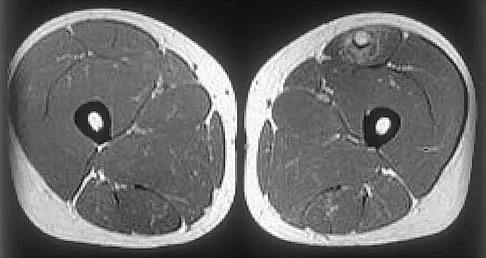
Figure 16 shows an axial MRI scan through the knee joint. What structure is identified by the arrow?


Explanation

The anterior cruciate ligament can be visualized on an axial MRI scan as a low-signal structure lying in the lateral aspect of the intercondylar notch. Visualization in multiple planes increases the accuracy of MRI to view the anterior cruciate ligament. The posterior cruciate ligament and ligament of Wrisberg are located on the medial wall of the notch. The ligamentum mucosum is anterior to the notch, and the popliteus tendon is posterior to the lateral femoral condyle. Resnick D, Kang HS (eds): Internal Derangements of Joints: Emphasis on MR Imaging. Philadelphia, PA, WB Saunders, 1997, pp 675-699.

Question 7

Which of the following nerves is most commonly injured when obtaining a bone graft from the posterior ilium?


Explanation

Cutaneous sensation to the buttock is provided by the superior, middle, and inferior cluneal nerves. The superior cluneal nerves are the lateral branches of the dorsal rami of the upper three lumbar nerves and penetrate deep fascia just proximal to the iliac crest. They pass distally to the skin of the buttock and will be injured if the exposure extends more than 8 cm anterolateral to the posterior superior iliac spine. The lateral femoral cutaneous nerve can be injured in an anterior ilium bone graft. The superior gluteal nerve or even the sciatic nerve can be injured if bone is removed from the sciatic notch or dissection is not kept subperiosteal; however, the rate of injury is far less than cluneal nerve injury. The L5 and S1 nerve roots are anterior and can be injured if the inner table bone is harvested and the dissection is not kept subperiosteal or is too medial; however, the rate of injury still is far less than cluneal nerve injury. Hoppenfeld S, deBoer P: Surgical Exposures in Orthopaedics: The Anatomic Approach. Philadelphia, PA, JB Lippincott, 1984, pp 295-297. Hollinshead WH: Textbook of Anatomy, ed 3. Hagerstown, MD, Harper and Row, 1974, p 379. Last RJ: Anatomy: Regional and Applied, ed 6. London, England, Churchill Livingstone, 1978, p 23.

Question 8

Based on the findings seen in the posteroanterior radiograph of the wrist shown in Figure 17, which of the following structures is torn?


Explanation

The radiograph shows widening between the scaphoid and lunate. The normal variance is up to 5 mm. Although several ligaments may be torn, the scapholunate interosseous ligament must be torn for this widening to occur. Cooney WP, Linscheid RL, Dobyns JH: The Wrist: Diagnosis and Operative Treatment. St Louis, MO, Mosby-Year Book, 1998, vol 1, pp 503-506.

Question 9

What tendon has an intra-articular (instrasynovial) location in the knee joint?


Explanation

The popliteal tendon arises from the posterior aspect of the tibia and courses through the knee joint through the popliteus hiatus of the lateral meniscus before attaching on the lateral femur anterior to the lateral collateral ligament. It is the only tendon in the knee joint that can be viewed directly on arthroscopy. Kimura M, Shirakura K, Hasegawa A, Kobayashi Y, Udagawa E: Anatomy and pathophysiology of the popliteal tendon area in the lateral meniscus: 1. Arthroscopic and anatomical investigation. Arthroscopy 1992;8:419-423.

Question 10

A patient undergoes hip arthroscopy, and the pathology is seen in Figure 18. What is the most likely diagnosis?


Explanation

The motorized shaver is adjacent to the acetabular labrum, which is torn. The femoral head and acetabulum are normal in appearance. Neither the fat pad nor a loose body is identified.

Question 11

Figure 19 shows an arthroscopic view from the anterior lateral portal of the knee looking into the suprapatella pouch. The use of an electrothermal device during this procedure most commonly causes significant postoperative complications by damaging which of the following structures?


Explanation

While it is possible to damage any of these structures, unrecognized intraoperative laceration without adequate coagulation of the superior lateral geniculate artery is common. This can result in significant postoperative hemarthrosis and a return to surgery when bleeding cannot be controlled. Cash JD, Hughston JC: Treatment of acute patella dislocation. Am J Sports Med 1988;16:244-249.

Question 12

Figure 20 shows the resting and stress radiographs of a patient who has had pain and feelings of instability after undergoing a total knee arthroplasty 1 year ago. Which of the following ligaments is not functional and is therefore responsible for the patient's symptoms?


Explanation

The radiographs show posterior instability caused by an absent posterior cruciate ligament. The tibia is significantly displaced posteriorly with respect to the femur. This can be demonstrated with a lateral radiograph obtained with the knee in flexion. The anterior cruciate ligament has been resected but is not responsible for the instability shown.

Question 13

On MRI, a nonsanguinous effusion has what appearance?


Explanation

Nonbloody effusions that are greater than 1 mL are readily detected by MRI. They appear black on T1-weighted images and white on T2-weighted images. A sanguinous effusion is seen as white on T1-weighted images and black on T2-weighted images. Resnick D, Kang HS: Synovial joints, in Resnick D, Kang HS (eds): Internal Derangements of Joints: Emphasis on MR Imaging. Philadelphia, PA, WB Saunders, 1997, pp 49-53.

Question 14

Which of the following illustrations shown in Figures 21a through 21e correctly shows the projection of the sacroiliac joint on the outer table of the ilium?


Explanation

21b 21c 21d 21e The projection of the sacroiliac joint on the outer surface of the ilium should be well understood to avoid violation of the joint during bone graft harvesting and to help in insertion of the screw across the joint. The sacroiliac joint has superior and inferior limbs. The average lengths of the superior and inferior limbs are 4.4 cm and 5.6 cm, respectively. The average width of each limb is 2.0 cm. The average distance from the longitudinal axis of the superior limb to the posterior superior iliac spine is 5.5 cm. The average longitudinal axis of the inferior limb is 1.2 cm superior to the inferior margin of the posterior inferior iliac spine. The average angle between the two axes is 93 degrees. Figure 21c most closely shows the projection of the sacroiliac joint on the outer table of the ilium. Waldrop JT, Ebraheim NA, Yeasting RA, Jackson WT: The location of the sacroiliac joint on the outer table of the posterior ilium. J Orthop Trauma 1993;7:510-513.

Question 15

In children between the ages of 4 and 8 years, the major blood supply to the femoral head comes from the


Explanation

From birth until the age of 4 years, the primary blood supply to the femoral head is from the medial and lateral circumflex arteries that traverse the femoral neck. After the age of 4 years, the contribution of the lateral femoral circumflex artery, which traverses the anterior portion of the femoral neck, becomes negligible. The posterosuperior and posteroinferior retinacular vessels, branches of the medial femoral circumflex artery, become the primary blood supply to the epiphysis. The contribution of the artery of the ligamentum teres is minimal after the age of 4 years. Hughes LO, Beaty JH: Fractures of the head and neck of the femur in children. J Bone Joint Surg Am 1994;76:283-292.

Question 16

What is the most anatomic location for placement of the femoral tunnel in anterior cruciate ligament reconstruction?


Explanation

It is critical for graft isometry and knee stability that the femoral tunnel be placed as far posterior as possible on the lateral femoral condyle. Superiorly, the graft should be at the one o'clock position on the left knee. Resident's ridge is a false posterior shelf that often seems like the extreme posterior cortex. Abnormal tunnel placement results in a variety of complications, including an unstable knee, early graft failure, and joint stiffness. Johnson RJ, Beynnon BD, Nichols CE, Renstrom PA: The treatment of injuries of the anterior cruciate ligament. J Bone Joint Surg Am 1992;74:140-151.

Question 17

In the anterior approach (Smith-Petersen) to the hip, dissection is carried out between muscles innervated by the


Explanation

In the Smith-Petersen approach to the hip, dissection is carried out between the tensor fascia lata laterally (supplied by the superior gluteal nerve) and the sartorius and rectus femoris medially (both supplied by the femoral nerve). Crenshaw AH Jr: Surgical techniques and approaches, in Canale ST (ed): Campbell's Operative Orthopaedics, ed 9. St Louis, MO, Mosby-Year Book, 1998, vol 1, pp 82-85.

Question 18

What structure is marked Q in the diagram of the brachial plexus shown in Figure 22?


Explanation

From proximal to distal, the brachial plexus is divided into roots, trunks, divisions, and cords before forming specific peripheral nerve branches. The structure marked Q is called the posterior cord because it lies posterior to the axially artery at the level of the cords. Its terminal branches are the upper subscapular (V), thoracodorsal (W), lower subscapular (X), axillary (F), and radial (G) nerves. Anderson JE (ed): Grant's Atlas of Anatomy, ed 7. Baltimore, MD, Williams and Wilkins, 1978, pp 6-24.

Question 19

A sagittal T1-weighted MRI scan of the knee joint is shown in Figure 23. What structure is identified by the arrow?


Explanation

On T1-weighted images, the posterior cruciate ligament is a low-signal (black) structure that courses from the posterior aspect of the tibia to the medial femoral condyle. The posterior cruciate ligament can appear as arcuate, U-shaped, or kinked. The other structures have similar signal but different anatomic locations. Gross ML, Grover JS, Bassett LW, Seeger LL, Finerman GA: Magnetic resonance imaging of the posterior cruciate ligament: Clinical use to improve diagnostic accuracy. Am J Sports Med 1992;20:732-737.

Question 20

Figure 24 shows the arthroscopic view of a patient with ankle impingement syndrome. This is commonly seen after high ankle sprains and represents fibrotic granulation thickening of what structure?


Explanation

Chronic anterior inferior tibiofibular ligament sprains can lead to thickening and synovitis that catches or impinges dorsiflexion; patients often note painful clicking with dorsiflexion eversion. The other structures are not affected by this injury. Hopkinson WJ, St Pierre P, Ryan JB, Wheeler JH: Syndesmosis sprains of the ankle. Foot Ankle 1990;10:325-330. Amendola A: Controversies in diagnosis and management of syndesmosis injuries of the ankle. Foot Ankle 1992;13:44-50. Baxter DE: The Foot and Ankle in Sports. St Louis, MO, Mosby-Year Book, 1995, p 30.

Question 21

The modified Brostrom lateral ankle ligamentous reconstruction uses which of the following structures to provide supplementary stabilization?


Explanation

The modified Brostrom lateral ankle ligament stabilization procedure uses the remnants of the anterior talofibular and the calcaneofibular ligaments, supplemented by the inferior retinaculum and the transferred talocalcaneal ligament to stabilize the lateral ankle. Chrisman and associates described the use of one half of the peroneus brevis. Watson-Jones and Evans used the entire peroneus brevis. The peroneus longus has been taken by mistake. The plantaris has been used in triligamentous reconstruction. Gould N, Seligson D, Gassman J: Early and late repair of lateral ligament of the ankle. Foot Ankle 1980;1:84-89. Hamilton WG, Thompson FM, Snow SW: The modified Brostrom procedure for lateral ankle instability. Foot Ankle 1993;14:1-7. Chrisman OD, Snook GA: Reconstruction of lateral ligament tears of the ankle: An experimental study and clinical evaluation of seven patients treated by a new modification of the Elmslie procedure. J Bone Joint Surg Am 1969;51:904-912. Evans DL: Recurrent instability of the ankle: My method of surgical treatment. Proc R Soc Med 1953;46:343. Watson-Jones R: Fractures and Joint Injuries, ed 3. Baltimore, MD, Williams and Wilkins, 1946, p 234.

Question 22

Figure 25 shows an arthroscopic thermal capsular shrinkage device being used in the anterior inferior quadrant of a patient with a subluxating shoulder. Which of the following neurologic complications is most frequently reported with this technique?


Explanation

The axillary nerve lies within millimeters of the anterior inferior capsule. The inferior capsule is of varying thickness, and thermal energy used in shortening the ligament can cause damage to the sensory fibers of the axillary nerve. Clinically, this is manifested as a burnt skin sensation in the axillary nerve distribution area. The motor branch of the axillary nerve is usually spared. The suprascapular nerve and the radial nerve are far from the shrinkage zone. The musculocutaneous nerve, frequently at risk with open procedures, lies well anterior. Fanton GS: Arthroscopic electrothermal surgery of the shoulder. Op Tech Sports Med 1998;6:157-160.

Question 23

A coronal MRI scan through the shoulder joint is shown in Figure 26. The cyst indicated by the arrow will most likely cause compression of what nerve?


Explanation

The MRI scan shows a ganglion cyst in the region of the spinoglenoid notch. These are difficult to diagnose clinically but are readily apparent on MRI. They usually cause compression of the suprascapular nerve and weakness of the infraspinatus and supraspinatus muscles. Resnick D, Kang HS (eds): Internal Derangements of Joints: Emphasis on MR Imaging. Philadelphia, PA, WB Saunders, 1997, pp 306-309.

Question 24

Which of the following ligaments is most commonly involved in posterolateral rotatory instability of the elbow?


Explanation

Recurrent posterolateral rotatory instability of the elbow is difficult to diagnose. Such instability can be demonstrated only by the lateral pivot-shift test. The cause for this condition is laxity of the ulnar part of the lateral collateral ligament, which allows a transient rotatory subluxation of the ulnohumeral joint and a secondary dislocation of the radiohumeral joint. The annular ligament remains intact, so the radioulnar joint does not dislocate. Treatment consists of surgical reconstruction of the lax ulnar part of the lateral collateral ligament. The anterior band is the most important part of the medial collateral which is lax in valgus instability of the elbow. Morrey BF: Acute and chronic instability of the elbow. J Am Acad Orthop Surg 1996;4:117-128.

Question 25

Which of the following anatomic structures is labeled 6 in Figure 27?


Explanation

The line labeled 6 points to the A2 pulley. This structure is the condensation of the digital flexor tendon sheath corresponding to the proximal aspect of the proximal phalanx. Grayson's ligament is volar to the digital nerve and artery. Cleland's ligament is dorsal to the digital nerve and artery. The sagittal band anchors the extensor tendons over the metacarpophalangeal joints. The triangular ligament connects the lateral bands just proximal to the terminal tendon inserting onto the base of the distal phalanx. Hollinshead WH: Anatomy for Surgeons: The Back and Limbs, ed 3. Philadelphia, PA, Harper and Row, 1982, p 467.

Question 26

A patient sustains a deep penetrating injury immediately anterior to the anterior scalene muscle in the lower neck. Which of the following structures is most likely injured in this specific location?


Explanation

The subclavian vein passes anterior to the anterior scalene muscle. In contrast, the subclavian artery and the roots/trunks of the brachial plexus pass through the interscalene triangle, which is located between the anterior and middle scalene muscles.

Question 27

Hypertrophy of the teres minor and the long head of the triceps brachii can lead to nerve compression within the quadrangular space. Which of the following clinical findings would most likely be observed in this scenario?


Explanation

The quadrangular space transmits the axillary nerve and the posterior circumflex humeral artery. Compression here causes axillary nerve dysfunction, leading to deltoid/teres minor weakness and decreased sensation over the lateral deltoid.

Question 28

During an ilioinguinal approach to the acetabulum, massive bleeding is encountered posterior to the superior pubic ramus. This bleeding is most likely originating from the 'corona mortis', which represents an anastomosis between which two vascular systems?


Explanation

The corona mortis is a vascular anastomosis between the external iliac system (usually via the inferior epigastric artery/vein) and the obturator system. It crosses the superior pubic ramus and is at high risk of iatrogenic injury during pelvic surgery.

Question 29

A surgeon is performing a complex midfoot reconstruction and explores the plantar aspect of the navicular. At the 'master knot of Henry', which of the following tendons crosses superior (dorsal) to the flexor digitorum longus (FDL) tendon?


Explanation

At the master knot of Henry in the plantar midfoot, the flexor digitorum longus (FDL) tendon crosses superficial (plantar) to the flexor hallucis longus (FHL) tendon. Therefore, the FHL is positioned superior (dorsal) to the FDL.

Question 30

A rock climber presents with acute bowstringing of the index finger flexor tendons following a sudden forceful grip. This condition typically results from rupture of the A2 and A4 pulleys. To what specific structures do these critical pulleys attach?


Explanation

The A2 and A4 pulleys are the most critical biomechanical pulleys to prevent flexor tendon bowstringing. They attach directly to the periosteum of the proximal and middle phalangeal shafts, respectively.

Question 31

A cyclist complains of persistent numbness in the ring and small fingers along with weakness in finger abduction. Compression of a nerve within Guyon's canal is suspected. Which of the following structures forms the floor of this anatomic canal?


Explanation

Guyon's canal contains the ulnar nerve and artery. The floor of the canal is formed by the transverse carpal ligament, pisohamate ligament, and pisometacarpal ligament, while the roof is formed by the volar carpal ligament.

Question 32

A professional volleyball player presents with isolated weakness of the infraspinatus muscle. MRI reveals a ganglion cyst compressing a nerve at the spinoglenoid notch. Which ligament forms the roof of this notch?


Explanation

The suprascapular nerve passes through the spinoglenoid notch, where it is roofed by the spinoglenoid ligament (inferior transverse scapular ligament). Entrapment here causes isolated infraspinatus weakness, sparring the supraspinatus.

Question 33

In an adult patient, which of the following branches of the medial femoral circumflex artery provides the primary blood supply to the weight-bearing dome of the femoral head?


Explanation

The lateral epiphyseal artery is a terminal branch of the medial femoral circumflex artery (MFCA). It supplies the majority of the blood to the weight-bearing superolateral portion of the adult femoral head.

Question 34

During surgical reconstruction of the posterolateral corner (PLC) of the knee, understanding the layered anatomy is essential. Which of the following structures are located in the superficial layer (Layer 1) of the lateral side of the knee?


Explanation

According to Seebacher's anatomical classification, the lateral aspect of the knee has three layers. Layer 1 (superficial) consists of the iliotibial band and the biceps femoris.

Question 35

A spine surgeon is placing pedicle screws in the L4 vertebra. Which anatomical relationship best describes the position of the exiting L4 nerve root relative to the L4 pedicle?


Explanation

In the lumbar spine, the exiting nerve root traverses the neuroforamen directly inferior to the pedicle of the same numeric level. Thus, the L4 nerve root exits inferior to the L4 pedicle.

Question 36

A patient presents with an inability to actively extend the fingers and thumb, but wrist extension is preserved with radial deviation. Posterior interosseous nerve (PIN) syndrome is suspected. What is the most common site of compression for this nerve?


Explanation

The Arcade of Frohse (the proximal fascial edge of the superficial head of the supinator muscle) is the most common site of compression for the posterior interosseous nerve. This leads to finger and thumb drop, while wrist extension is preserved via the ECRL (supplied proximal to the arcade).

Question 37

During an anterolateral approach to the distal tibia, a nerve is at risk of being injured where it transitions from deep to subcutaneous tissue. At what location does the superficial peroneal nerve typically pierce the deep crural fascia?


Explanation

The superficial peroneal nerve typically pierces the crural fascia to become subcutaneous at the junction of the middle and distal thirds of the lateral lower leg, making it highly vulnerable to injury during distal tibial surgical approaches.

Question 38

During a dorsal approach to the wrist, the surgeon uses Lister's tubercle as a landmark. The tendon that passes immediately adjacent and ulnar to this structure is innervated by which of the following nerves?


Explanation

The extensor pollicis longus (EPL) tendon passes immediately ulnar to Lister's tubercle and uses it as a pulley. The EPL is innervated by the posterior interosseous nerve (PIN).

Question 39

The volar approach to the proximal forearm (Henry approach) utilizes a safe internervous plane. Which of the following pairs of nerves supplies the two muscles that define this plane?


Explanation

The proximal Henry approach exploits the internervous plane between the brachioradialis (innervated by the radial nerve) and the pronator teres (innervated by the median nerve).

Question 40

While performing a lateral (Hardinge) approach to the hip, the surgeon must limit the proximal splitting of the gluteus medius muscle to no more than 3 to 5 cm above the greater trochanter. This precaution primarily protects which of the following structures?


Explanation

The superior gluteal nerve runs roughly 3-5 cm proximal to the tip of the greater trochanter. Extending the gluteus medius split beyond this point risks denervating the anterior portion of the gluteus medius and the entire tensor fasciae latae.

Question 41

A marathon runner with chronic, severe medial heel pain is diagnosed with entrapment of the first branch of the lateral plantar nerve (Baxter's nerve). Which of the following muscles receives its motor innervation from this specific nerve?


Explanation

Baxter's nerve is the first branch of the lateral plantar nerve. It provides sensory innervation to the calcaneal periosteum and motor innervation to the abductor digiti minimi muscle.

Question 42

A patient is undergoing a total knee arthroplasty and receives an ultrasound-guided saphenous nerve block within the adductor canal (Hunter's canal). Which muscle forms the anterolateral boundary of this canal?


Explanation

The adductor canal is bounded anterolaterally by the vastus medialis, posteromedially by the adductor longus and magnus, and is covered anteromedially (the roof) by the sartorius muscle.

Question 43

The coracoclavicular (CC) ligaments provide the primary vertical stability to the acromioclavicular joint. Which of the following describes the anatomical position of the conoid ligament relative to the trapezoid ligament?


Explanation

The coracoclavicular complex consists of the conoid and trapezoid ligaments. The conoid ligament is located posteromedial to the trapezoid ligament and inserts onto the conoid tubercle of the clavicle.

Question 44

In cases of thumb carpometacarpal (CMC) joint arthritis, progressive attenuation of the primary static stabilizing ligament is often noted. From which anatomical structure does this critical ligament, the anterior oblique ligament (beak ligament), originate?


Explanation

The anterior oblique ligament (often called the beak ligament) is the key primary static stabilizer of the thumb CMC joint. It originates from the volar tubercle of the trapezium and inserts on the volar base of the first metacarpal.

Question 45

During a sacrospinous ligament fixation procedure for pelvic organ prolapse, an orthopedic surgeon assisting the case notes a suture placement too close to the ischial spine. Which nerve loops around the ischial spine and is at greatest risk of iatrogenic entrapment here?


Explanation

The pudendal nerve exits the pelvis through the greater sciatic foramen, hooks around the ischial spine and sacrospinous ligament, and re-enters the pelvis via the lesser sciatic foramen. Sutures placed too close to the ischial spine can easily entrap it.

Question 46

The great anterior radiculomedullary artery (Artery of Adamkiewicz) provides major blood supply to the lower two-thirds of the spinal cord. It most commonly enters the spinal canal at which of the following levels?


Explanation

The artery of Adamkiewicz usually arises from a left posterior intercostal artery between the levels of T8 and L1. Injury to this vessel during anterior spinal surgery can lead to anterior spinal artery syndrome.

Question 47

During an ilioinguinal approach to the acetabulum, brisk bleeding is encountered just posterior to the superior pubic ramus. This bleeding is most likely originating from an anastomosis between which two vessels?


Explanation

The corona mortis is a vascular anastomosis between the obturator and external iliac (or inferior epigastric) vessels. It crosses the superior pubic ramus and is highly susceptible to iatrogenic injury during anterior pelvic exposures.

Question 48

Which of the following describes the most common configuration of a Martin-Gruber anastomosis?


Explanation

A Martin-Gruber anastomosis involves anomalous motor nerve fibers passing from the median nerve (or anterior interosseous nerve) to the ulnar nerve in the forearm. It can cause confusing electrodiagnostic findings in cases of carpal tunnel syndrome or ulnar neuropathy.

Question 49

A patient presents with isolated weakness of external rotation of the shoulder with preserved abduction. At which of the following anatomical locations is nerve entrapment most likely occurring?


Explanation

Entrapment of the suprascapular nerve at the spinoglenoid notch affects only the infraspinatus muscle, leading to isolated external rotation weakness. Entrapment at the suprascapular notch affects both the supraspinatus and infraspinatus.

Question 50

Which of the following is the primary anatomic footprint for the distal attachment of the lateral ulnar collateral ligament (LUCL)?


Explanation

The LUCL originates on the lateral epicondyle and inserts on the supinator crest of the proximal ulna. It acts as the primary restraint to posterolateral rotatory instability (PLRI) of the elbow.

Question 51

At the level of the "Master Knot of Henry" in the plantar aspect of the midfoot, what is the anatomical relationship between the flexor hallucis longus (FHL) and flexor digitorum longus (FDL) tendons?


Explanation

At the Master Knot of Henry, the FHL tendon crosses dorsal (superior) to the FDL tendon. This is a crucial anatomical landmark during plantar midfoot dissections and tendon transfers.

Question 52

The primary blood supply to the proximal pole of the scaphoid is derived from vessels entering at which anatomical location?


Explanation

The primary blood supply to the scaphoid enters distally along the dorsal ridge via the dorsal carpal branch of the radial artery. Because perfusion is retrograde, proximal pole fractures are at a high risk for avascular necrosis.

Question 53

During a posterior approach to the hip, protecting the medial circumflex femoral artery (MFCA) is critical. The main branch of the MFCA runs most consistently between which two muscles before piercing the hip capsule?


Explanation

The deep branch of the MFCA courses posteriorly between the superior border of the quadratus femoris and the inferior gemellus. A careless release of the quadratus femoris can compromise the main blood supply to the femoral head.

Question 54

Which of the following nerves exits the adductor (Hunter's) canal by piercing the vastoadductor fascia?


Explanation

The saphenous nerve and the descending genicular artery exit the adductor canal anteriorly by piercing the vastoadductor fascia. The femoral artery and vein continue distally to exit through the adductor hiatus.

Question 55

The vertebral artery typically enters the transverse foramen of the cervical spine at which vertebral level?


Explanation

The vertebral artery originates from the subclavian artery and typically enters the transverse foramen at the level of C6. It then travels cephalad through the foramina of C6 to C1.

Question 56

The posterior interosseous nerve (PIN) is most vulnerable to iatrogenic injury during a Henry approach to the proximal radius at which of the following anatomic structures?


Explanation

The PIN passes under the proximal fibrous edge of the superficial head of the supinator muscle, known as the Arcade of Frohse. Supination of the forearm during a volar approach moves the nerve laterally, protecting it from injury.

Question 57

The axillary nerve passes through the quadrangular space. What muscle forms the inferior border of this space?


Explanation

The quadrangular space is bounded superiorly by the teres minor, inferiorly by the teres major, medially by the long head of the triceps, and laterally by the surgical neck of the humerus.

Question 58

In approximately 10-15% of the population, a variation in the relationship between the sciatic nerve and the piriformis muscle exists. What is the most common anatomical variant?


Explanation

The most common variant (Beaton and Anson type B) features a bipartite piriformis where the common peroneal nerve pierces the muscle and the tibial nerve passes below it. This is frequently implicated in piriformis syndrome.

Question 59

The genicular arteries provide blood supply to the menisci of the knee. In an adult, vascular penetration extends to approximately what depth of the meniscus?


Explanation

In adults, only the peripheral 10% to 30% of the menisci (the red-red zone) receives direct blood supply from the perimeniscal capillary plexus. This limits the healing potential of more central, avascular meniscal tears.

Question 60

When utilizing the direct lateral (Hardinge) approach to the hip, what is the generally accepted "safe zone" for proximal splitting of the gluteus medius to avoid injury to the superior gluteal nerve?


Explanation

The superior gluteal nerve courses approximately 3 to 5 cm proximal to the tip of the greater trochanter. Splitting the gluteus medius beyond 5 cm places the nerve at significant risk, leading to an iatrogenic Trendelenburg gait.

Question 61

During trigger finger release of the ring finger, the A1 pulley is transected. Which pulleys are considered mechanically critical to prevent bowstringing of the flexor tendons and must be preserved?


Explanation

The A2 and A4 pulleys attach firmly to the periosteum of the proximal and middle phalanges, respectively. They are the most biomechanically critical structures for preventing flexor tendon bowstringing.

Question 62

The spring ligament complex is a critical static stabilizer of the longitudinal arch of the foot. Between which two osseous structures does it primarily attach?


Explanation

The spring ligament, also known as the plantar calcaneonavicular ligament, spans from the sustentaculum tali of the calcaneus to the plantar surface of the navicular. It forms a crucial sling supporting the talar head.

Question 63

Wartenberg's syndrome is characterized by pain and paresthesias over the dorsoradial hand. It is typically caused by compression of the superficial radial nerve between which two muscles during forearm pronation?


Explanation

The superficial branch of the radial nerve emerges between the brachioradialis and ECRL in the distal third of the forearm. Pronation creates a scissor-like compression between these muscles, leading to Wartenberg's syndrome.

Question 64

During reconstruction of the posterolateral corner (PLC) of the knee, the anatomic insertion of the popliteus tendon on the femur is located:


Explanation

The popliteus tendon inserts in the popliteal sulcus on the lateral femoral condyle. This location is distinctly distal and anterior to the lateral epicondyle.

Question 65

A Morton's neuroma most commonly occurs in the third web space of the foot. This is anatomically predisposed by the union of branches from which two nerves?


Explanation

The third common digital nerve is formed by anastomotic branches from both the medial and lateral plantar nerves. This makes the nerve thicker and more prone to tethering and compression deep to the transverse metatarsal ligament.

Question 66

A 45-year-old mechanic presents with weakness in extending his fingers and thumb, but normal wrist extension. Which of the following anatomical structures is the most common site of entrapment for the affected nerve?


Explanation

The posterior interosseous nerve is most commonly entrapped at the Arcade of Frohse, the proximal tendinous edge of the superficial head of the supinator muscle. It presents with weakness in finger and thumb extension while preserving radial wrist extension.

Question 67

During an operative fixation of a scaphoid waist fracture using a volar approach, the surgeon must be careful to preserve the primary blood supply to the proximal pole. This blood supply enters the scaphoid primarily through which of the following areas?


Explanation

The primary blood supply to the scaphoid is derived from the radial artery via branches that enter the dorsal ridge and supply the proximal pole in a retrograde fashion.

Question 68

The popliteofibular ligament is a crucial stabilizer of the posterolateral corner of the knee. From which structure does it originate and where does it insert?


Explanation

The popliteofibular ligament originates from the musculotendinous junction of the popliteus and inserts onto the posteromedial aspect of the fibular styloid. It acts as a primary static stabilizer against external rotation of the knee.

Question 69

Following a displaced femoral neck fracture in an adult, the principal blood supply to the femoral head is disrupted. Which of the following vessels provides the majority of the blood supply to the adult femoral head?


Explanation

The deep branch of the medial circumflex femoral artery provides the predominant blood supply to the adult femoral head. It courses posterior to the femoral neck and is at high risk of disruption in displaced neck fractures.

Question 70

A patient develops a severe horseshoe abscess of the hand extending from the thumb to the small finger. The infection spreads proximally into the distal forearm. In which anatomical space does this proximal extension reside?


Explanation

The space of Parona is a deep fascial space in the distal volar forearm located between the pronator quadratus and the deep flexor tendons. It serves as a conduit for proximal extension of infections from the radial and ulnar bursae.

Question 71

The anterolateral ligament (ALL) of the knee is increasingly recognized for its role in controlling rotatory laxity. What are its precise anatomical landmarks?


Explanation

The ALL originates on the lateral femoral epicondyle slightly anterior and distal to the LCL origin. It inserts on the proximal anterolateral tibia, midway between Gerdy's tubercle and the fibular head, helping to control internal tibial rotation.

Question 72

A marathon runner complains of medial heel pain and paresthesias radiating to the first three toes. The nerve involved in this condition runs beneath the flexor retinaculum. Which of the following muscles is innervated by the terminal branch of this nerve?


Explanation

The medial plantar nerve provides motor innervation to the abductor hallucis, flexor digitorum brevis, flexor hallucis brevis, and the first lumbrical. The other listed muscles are innervated by the lateral plantar nerve.

Question 73

A professional volleyball player presents with isolated weakness in external rotation of the shoulder. Atrophy is noted in the infraspinatus fossa, while the supraspinatus is normal. Entrapment of the suprascapular nerve is suspected at which location?


Explanation

Entrapment of the suprascapular nerve at the spinoglenoid notch affects only the infraspinatus, leading to isolated weakness in external rotation. Entrapment at the suprascapular notch would affect both the supraspinatus and infraspinatus.

Question 74

During reconstruction of an acromioclavicular joint separation, anatomic reduction of the coracoclavicular ligaments is desired. Which of the following statements accurately describes the anatomy of the conoid and trapezoid ligaments?


Explanation

The conoid ligament is the more medial of the two coracoclavicular ligaments and inserts onto the conoid tubercle on the posterior aspect of the clavicle. The trapezoid is located more laterally and inserts anterolaterally on the trapezoid line.

Question 75

The calcaneonavicular (spring) ligament complex is a critical stabilizer of the longitudinal arch. Which portion of the spring ligament is the strongest and most frequently torn in adult-acquired flatfoot deformity?


Explanation

The superomedial calcaneonavicular ligament is the thickest and strongest component of the spring ligament complex. It provides primary support to the talar head and is commonly attenuated or torn in posterior tibial tendon dysfunction.

Question 76

A cyclist presents with numbness in the ring and small fingers along with weakness in finger abduction and adduction. Sensory examination reveals normal sensation over the dorso-ulnar aspect of the hand. Where is the most likely site of compression?


Explanation

Compression in Zone 1 of Guyon's canal affects both the deep motor and superficial sensory branches of the ulnar nerve, sparing the dorsal ulnar cutaneous nerve which branches proximal to the wrist.

Question 77

An overhead throwing athlete sustains a tear of the ulnar collateral ligament (UCL) of the elbow. The anterior bundle of the UCL is the primary restraint to valgus stress. Where does the anterior bundle insert on the ulna?


Explanation

The anterior bundle of the ulnar collateral ligament originates on the anterior undersurface of the medial epicondyle and inserts on the sublime tubercle at the medial margin of the coronoid process.

Question 78

An anatomic repair of a distal biceps tendon rupture requires reattachment to its native footprint. Which of the following best describes the normal anatomical insertion of the distal biceps tendon?


Explanation

The distal biceps tendon inserts onto the posterior-ulnar aspect of the radial tuberosity. This specific anatomical location maximizes its mechanical advantage as a supinator of the forearm.

Question 79

A 25-year-old sustains a bucket-handle tear of the medial meniscus. The surgeon decides to repair the tear, relying on the vascularity of the peripheral meniscus. Which arteries provide the primary blood supply to the peripheral menisci?


Explanation

The lateral and medial, superior and inferior genicular arteries form a perimeniscal capillary plexus that supplies the peripheral 10% to 30% of the menisci. The middle genicular artery primarily supplies the cruciate ligaments.

Question 80

During a lateral approach to the calcaneus for an open reduction and internal fixation of a calcaneal fracture, the sural nerve is at risk. What is the typical course of the sural nerve at the level of the lateral malleolus?


Explanation

The sural nerve travels down the posterolateral leg, running posterior to the lateral malleolus and lateral to the Achilles tendon, supplying sensation to the lateral aspect of the foot.

Question 81

The central band of the interosseous membrane is critical for longitudinal radioulnar stability, particularly after a radial head excision. What is the anatomical orientation of the central band fibers?


Explanation

The central band of the interosseous membrane has strong fibers that run obliquely from proximal on the radius to distal on the ulna. This orientation effectively transfers axial compressive loads from the radius to the ulna.

Question 82

The Lisfranc ligament is crucial for midfoot stability. Between which two bones does the true Lisfranc ligament run?


Explanation

The true Lisfranc ligament is a strong interosseous ligament that connects the lateral aspect of the medial cuneiform to the medial base of the second metatarsal. It is the primary restraint to lateral translation of the lesser metatarsals.

Question 83

When performing an open reduction and internal fixation of a proximal humerus fracture via a deltopectoral approach, the surgeon must be aware of the axillary nerve. How far distal to the lateral edge of the acromion does the axillary nerve typically cross the humerus?


Explanation

The axillary nerve wraps around the surgical neck of the humerus and travels horizontally across the deep surface of the deltoid, typically 5 to 7 cm distal to the lateral edge of the acromion.

Question 84

The plantar fascia is a primary static stabilizer of the longitudinal arch of the foot. It originates from the calcaneal tuberosity and inserts distally. Which band of the plantar fascia is the most robust and most commonly involved in plantar fasciitis?


Explanation

The central band of the plantar fascia is the thickest and strongest component. It originates from the medial process of the calcaneal tuberosity and is the primary anatomical site of pathology in plantar fasciitis.

Question 85

A weightlifter sustains a complete rupture of the pectoralis major tendon at its insertion. To perform an anatomic repair, the surgeon must understand its bilaminar insertion. Which segment of the muscle forms the posterior lamina of the insertion?


Explanation

The pectoralis major tendon twists 180 degrees before inserting. The inferior sternocostal fibers form the posterior lamina and insert most proximally on the humerus, whereas the clavicular fibers form the anterior lamina.

Question 86

A 24-year-old male sustains a posterior shoulder dislocation. After reduction, he exhibits numbness over the lateral aspect of his shoulder and weak shoulder abduction. The injured nerve exits the axilla through a space bounded by which of the following structures?


Explanation

The axillary nerve exits through the quadrangular space, bounded superiorly by the teres minor, inferiorly by the teres major, medially by the long head of the triceps, and laterally by the surgical neck of the humerus.

Question 87

During an anterolateral (Watson-Jones) approach to the hip for a total hip arthroplasty, the surgical interval lies between the tensor fasciae latae and the gluteus medius. What is the innervation of the muscles defining this interval?


Explanation

The Watson-Jones approach uses the interval between the tensor fasciae latae and gluteus medius. Both muscles are innervated by the superior gluteal nerve, making this an intermuscular, rather than a true internervous, plane.

Question 88

A patient presents with a severe palmar laceration resulting in the loss of extension at the proximal and distal interphalangeal joints of the index and middle fingers. The lumbrical muscles to these specific digits are uniquely characterized by which of the following features?


Explanation

The first and second lumbricals (acting on the index and middle fingers) are unipennate and innervated by the median nerve. The third and fourth are bipennate and innervated by the deep branch of the ulnar nerve.

Question 89

A 22-year-old male sustains a minimally displaced scaphoid waist fracture. Which of the following best describes the primary arterial supply jeopardized by this injury, predisposing him to avascular necrosis of the proximal pole?


Explanation

The scaphoid receives 70-80% of its blood supply from the dorsal carpal branch of the radial artery, which enters at the distal pole and flows retrograde. This renders proximal fractures highly susceptible to avascular necrosis.

Question 90

A 45-year-old female presents with adult-acquired flatfoot deformity. The primary static stabilizer of the talonavicular joint is the spring ligament. Which of the following defines the exact anatomic attachments of this ligament?


Explanation

The spring ligament (plantar calcaneonavicular ligament) attaches from the sustentaculum tali of the calcaneus to the plantar and medial aspect of the navicular. It is a critical static stabilizer of the medial longitudinal arch.

Question 91

During an anterior cervical discectomy and fusion (ACDF) at C5-C6, the surgeon elevates the longus colli muscles. Injury to the sympathetic trunk in this region can cause Horner's syndrome. Where is the cervical sympathetic trunk typically located relative to the longus colli?


Explanation

The cervical sympathetic trunk runs vertically just lateral to the longus colli muscles, beneath the prevertebral fascia. Dissection should remain medial to the lateral borders of the longus colli to avoid Horner's syndrome.

Question 92

A 30-year-old athlete undergoes surgical reconstruction of the posterolateral corner (PLC) of the knee. The popliteus tendon must be anatomically secured. What is the normal anatomic insertion of the popliteus tendon on the femur?


Explanation

The popliteus tendon inserts on the lateral femoral condyle anterior and distal to the origin of the lateral collateral ligament (LCL) in the popliteal sulcus.

Question 93

During a modified Stoppa approach for an anterior column acetabular fracture, significant bleeding is encountered from the "corona mortis". This vascular structure represents an anastomosis between which two vascular systems?


Explanation

The corona mortis is a critical anastomotic connection between the obturator vessels (internal iliac system) and the inferior epigastric or external iliac vessels. It crosses over the superior pubic ramus and is vulnerable during intrapelvic approaches.

Question 94

A professional volleyball player presents with isolated atrophy and weakness of the infraspinatus muscle. Supraspinatus strength and muscle bulk are entirely normal. Where is the most likely site of nerve entrapment?


Explanation

The suprascapular nerve innervates the supraspinatus before passing through the spinoglenoid notch to innervate the infraspinatus. Entrapment at the spinoglenoid notch, often by a paralabral cyst, causes isolated infraspinatus weakness.

Question 95

A continuous saphenous nerve block is placed in the adductor (Hunter's) canal for postoperative analgesia after total knee arthroplasty. Which of the following muscles forms the anterolateral border of this anatomic canal?


Explanation

The adductor canal is bounded anterolaterally by the vastus medialis, posteromedially by the adductor longus and magnus, and anteriorly (the roof) by the sartorius.

Question 96

During a medial epicondylectomy for severe cubital tunnel syndrome, the surgeon releases the compressive structures overlying the ulnar nerve. Which of the following forms the primary roof of the cubital tunnel?


Explanation

The roof of the cubital tunnel is formed by Osborne's ligament (the cubital tunnel retinaculum), which spans between the medial epicondyle and the olecranon.

Question 97

During open reduction and internal fixation of a severe medial malleolus fracture, the surgeon dissects posterior to the medial malleolus. In what order, from anterior to posterior, do the structures normally lie in this region?


Explanation

From anterior to posterior behind the medial malleolus, the correct order is: Tibialis posterior, Flexor Digitorum longus, posterior tibial Artery, tibial Nerve, Flexor Hallucis longus (often remembered by the mnemonic 'Tom, Dick, AND Very Nervous Harry').

Question 98

The anterior cruciate ligament (ACL) is composed of two primary anatomic bundles. When the knee is in full extension, what is the relative tension state of these bundles?


Explanation

The posterolateral (PL) bundle of the ACL is tight in knee extension and provides rotational stability. Conversely, the anteromedial (AM) bundle is tight in flexion and provides primary anteroposterior stability.

Question 99

During a posterior triceps-splitting approach to the humerus for fracture fixation, the radial nerve is identified. At what approximate distance proximal to the lateral epicondyle does the radial nerve typically pierce the lateral intermuscular septum?


Explanation

The radial nerve pierces the lateral intermuscular septum approximately 10 to 12 cm proximal to the lateral epicondyle, transitioning from the posterior compartment to the anterior compartment of the arm.

Question 100

A 45-year-old male with an MRI-confirmed L4-L5 paracentral disc herniation is evaluated in the clinic. Based on typical neuroanatomy, this lesion will most likely produce which of the following distinct clinical findings?


Explanation

A paracentral disc herniation at L4-L5 typically impinges the traversing L5 nerve root. This results in weakness of the extensor hallucis longus (great toe extension) and altered sensation in the first dorsal web space, usually with normal deep tendon reflexes.

None

Clinic OS
Medically Verified Content by
Prof. Clinic OS
Consultant Orthopedic & Spine Surgeon
Chapter Index