العربية
Part of the Master Guide

AAOS & ABOS Basic Science MCQs (Set 3): Bone Biology, Biomechanics & Anatomy Review

Orthopedic Anatomy 2026 MCQs: Board Review Questions & Answers (Part 1)

23 Apr 2026 77 min read 73 Views
Figure for Anatomy 2005 MCQs - Part 1 - Question 1

Key Takeaway

Here are the crucial details you must know about Orthopedic Anatomy 2026 MCQs: Board Review Questions & Answers (Part 1). Top-rated Orthopedic Anatomy 2026 MCQs bank. Practice with clinical case questions, orthopedic surgery board review, and evidence-based answers updated for 2026.

Orthopedic Anatomy 2026 MCQs: Board Review Questions & Answers (Part 1)

Comprehensive 100-Question Exam


00:00

Start Quiz

Question 1

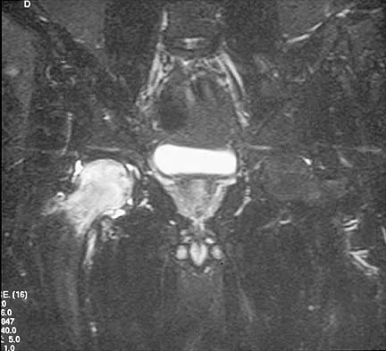
A 65-year-old man with ankylosing spondylitis sustains an extension injury to his cervical spine. Two days later, a progressive neurologic deficit develops at the C6 level. An MRI scan is shown in Figure 1. What is the most likely diagnosis?


Explanation

It is common for patients with ankylosing spondylitis to sustain extension-type fractures, typically near the cervicothoracic junction. These fractures can be minimally displaced, making them difficult to diagnose. In addition, the vertebral bodies are vascular and their canals are relatively enclosed, making them vulnerable to epidural bleeding. The MRI scan shows an epidural hematoma posteriorly compressing the cord. Bohlman HH: Acute fractures and dislocations of the cervical spine. J Bone Joint Surg Am 1979;61:1119-1142. Weinstein PR, Karpman RR, Gall EP, et al: Spinal cord injury, spine fracture and spinal stenosis in ankylosing spondylitis. J Neurosurg 1982;57:609-616.

Question 2

What are the most common portals for arthroscopic surgery of the ankle?


Explanation

The most commonly used portals are the anterolateral, anteromedial, and posterolateral portals. They have been shown to be the safest areas for portal placement, allowing no penetration of neurovascular structures. All the other portals involve placing another structure at risk. The anterocentral portal is close to the deep peroneal nerve and anterior tibular artery. The trans-Achilles portal is not recommended because of its limited utility and potential to injure the Achilles tendon. The posteromedial portal is too close to the posterotibial artery and nerve, the flexor hallucis longus and flexor digitorum longus tendons, and the branches of the calcaneal nerve. Stetson WB, Ferkel RD: Ankle arthroscopy: I. Technique and complications. J Am Acad Orthop Surg 1996;4:17-23.

Question 3

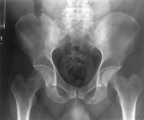
A patient who underwent primary total hip arthroplasty 7 years ago that resulted in excellent pain relief and a normal gait now reports pain and a limp. Postoperative and current AP radiographs are shown in Figures 2a and 2b. What is the most likely cause of the pathology seen?


Explanation

2b Osteolysis in the trochanteric bed can result in weakening of the bone and fracture. Nonsurgical management will provide reasonable clinical and radiographic results in patients with limited fracture displacement. Claus MC, Hopper RH, Engh CA: Fractures of the greater trochanter induced by osteolysis with the anatomic medullary locking prosthesis. J Arthroplasty 2002;17:706-712.

Question 4

The safest surgical approach to the insertion of the tibial posterior cruciate ligament uses the interval between which of the following muscles?


Explanation

Burks and Schaffer described an approach to the tibial insertion of the posterior cruciate ligament that uses the interval between the semimembranosus and the medial gastrocnemius. The medial gastrocnemius muscle is retracted laterally and protects the neurovascular bundle. This approach is used to repair an avulsion of the posterior cruciate ligament tibial attachment or for performing a posterior cruciate ligament tibial inlay reconstruction. Berg EE: Posterior cruciate ligament tibial inlay reconstruction. Arthroscopy 1995;8:95-99.

Question 5

A 50-year-old man with no history of trauma reports new-onset back pain after doing some yard work the previous day. He reports pain radiating down his leg posteriorly and into the first dorsal web space of his foot. MRI scans are shown in Figures 3a through 3c. What nerve root is affected?


Explanation

3b 3c The MRI scans clearly show an extruded L4-5 disk that is affecting the L5 nerve root on the left side. In addition, the L5 nerve root has a cutaneous distribution in the first dorsal web space. S1 affects the lateral foot. L4 affects the medial calf.

Question 6

A 19-year-old wrestler has numbness along the radial aspect of the forearm after undergoing an open Bankart repair through an anterior deltopectoral approach. Motor weakness would be expected along with what other finding?


Explanation

The musculocutaneous nerve may be injured by retracting the conjoined tendon medially. This nerve enters the coracobrachialis 5 cm distal to its origin. Its sensory distribution is the radial forearm, and its motor supply is to the biceps and brachialis. Bach BR, O'Brien SJ, Warren RF, et al: An unusual neurologic complication of the Bristow procedure. J Bone Joint Surg Am 1988;70:458-460.

Question 7

A 19-year-old man has had intermittent progressive knee pain with ambulation and pain at night following a rodeo accident 4 weeks ago. Figures 4a through 4e show the radiographs, a bone scan, CT scan, and T2-weighted MRI scan. What is the most likely diagnosis?


Explanation

4b 4c 4d 4e The imaging studies reveal a predominantly blastic lesion in the distal femur with posterolateral periosteal changes. The bone scan shows increased uptake in the distal femur, beyond that expected with radiography. Cross-sectional imaging confirms the presence of a soft-tissue mass extending from the lateral aspect of the femur, with diffuse intramedullary signal changes. This aggressive presentation, particularly in this location and in a patient of this age, is most consistent with osteosarcoma. The mineralization in the soft tissue strongly suggests neoplasm, not the reactive bony changes seen in an infectious process. Menendez LR (ed): Orthopaedic Knowledge Update: Musculoskeletal Tumors. Rosemont, IL, American Academy of Orthopaedic Surgeons, 2002, pp 175-186.

Question 8

Figures 5a and 5b show the radiographs of an active 52-year-old man who has increasing knee pain and progressive varus deformity after undergoing total knee arthroplasty 7 years ago. Examination reveals a small effusion, but he has good motion and stability. What is the most likely diagnosis?


Explanation

5b The radiographs show narrowing of the medial joint space, which indicates polyethylene wear and progressive varus alignment. Wear particles incite osteolytic lesions like the one seen on the lateral radiograph. O'Rourke MR, Callaghan JJ, Goetz DG, et al: Osteolysis associated with a cemented modular posterior-cruciate-substituting total knee design. J Bone Joint Surg Am 2002;84:1362-1371.

Question 9

Which of the following best describes the course of the median nerve at the elbow?


Explanation

The median nerve courses superficial to the ulnar artery, deep to the fibrous arch of the superficialis muscle, and deep to the superficial head of the pronator teres muscle. The median nerve lies within the interval between the flexor digitorum superficialis muscle and the flexor digitorium muscle as it progresses toward the wrist. Hoppenfeld S, deBoer P: Surgical Exposures in Orthopaedics, ed 2. Philadelphia, PA, Lippincott-Raven, 1994, pp 118-131.

Question 10

A 62-year-old woman with soft-tissue calcifications and telangiectasia has severe pain in the left index, middle, ring, and little fingers. History reveals that she does not smoke. The clinical history and arteriogram shown in Figure 6 are consistent with which of the following conditions?


Explanation

The arteriogram shows generalized disease of all vascular structures. Even though the image was obtained following an infusion of nitroglycerin, little flow is present to the fingers. Based on the history of soft-tissue calcifications and telangiectasia, the most likely diagnosis is CREST (chondrocalcinosis, Raynaud's phenomenon, esophageal dysmotility, sclerodactyly and telangiectasias). The arteriogram reveals Raynaud's phenomenon or the "R" component of CREST. Buerger's disease, or thromboangiitis obliterans, is strongly associated with a history of smoking. Hypothenar hammer syndrome involves repetitive trauma to the ulnar artery at the wrist, resulting in well-defined filling defects in the superficial palmar arch of the hand. Although not well visualized in this patient, the superficial arch is narrowed, showing no evidence of aneurysmal dilation. Koman LA, Ruch DS, Patterson Smith B, et al: Vascular disorders, in Green DP, Hotchkiss RN, Pederson WC (eds): Green's Operative Hand Surgery, ed 4. New York, NY, Churchill Livingstone, 1999, vol 2, pp 2254-2302.

Question 11

During excision of a Baker cyst, the base or stalk is usually found between the


Explanation

Although there are several bursae in the posterior portion of the knee, the most prevalent one with a connection to the knee joint is the one in the interval between the semimembranosus and the medial head of the gastrocnemius muscle. The popliteus muscle and posterior cruciate ligament, the posterior cruciate ligament and lateral gastrocnemius muscle, and the medial gastrocnemius muscle and posterior cruciate ligament are all too lateral and uncommon. The semitendinosus and medial head of the gastrocnemius muscles do not come in contact in the posterior aspect of the knee. Resnick D: Diagnosis of Bone and Joint Disorders, ed 3. Philadelphia, PA, WB Saunders, 1995, p 379.

Question 12

A direct lateral (Hardinge) approach is used during total hip arthroplasty. The structure labeled A in Figure 7 is the


Explanation

The superior gluteal nerve is located approximately 7.82 cm above the tip of the greater trochanter as it courses through the gluteus medius. This anatomic consideration is relevant during a Hardinge approach to the hip, where excessive proximal dissection or retraction could result in nerve injury. A split of the gluteus medius of no more than 4 cm above the greater trochanter is considered safe. Hoppenfeld S, deBoer P: Surgical Exposures in Orthopaedics: The Anatomic Approach. Philadelphia, PA, JB Lippincott, 1984, pp 333-335.

Question 13

The dorsal digital cutaneous nerve of the great toe shown in Figure 8 is a branch of what nerve?


Explanation

The dorsal digital cutaneous nerve of the great toe is a branch of the medial branch of the superficial peroneal nerve. The deep peroneal nerve supplies the first web space. McMinn RMH, Hutchings RT, Logan BM: Color Atlas of Foot and Ankle Anatomy. Weert, Netherlands, Wolfe Medical Publications, 1982, p 50.

Question 14

In hip arthroplasty, the location of the medial femoral circumflex artery is best described as


Explanation

The obturator artery lies closest to the transverse acetabular ligament. The femoral artery is closest to the anterior rim of the acetabulum. No named vessel lies within the substance of the gluteus minimus or superior to the piriformis tendon. The medial femoral circumflex artery lies medial or deep to the quadratus femoris muscle. Hoppenfeld S, deBoer P: Surgical Exposures in Orthopaedics: The Anatomic Approach, ed 1. Philadelphia, PA, JB Lippincott, 1984, Figure 7-53, p 346.

Question 15

A 47-year-old man has acute right shoulder pain after falling off a ladder. The MRI scan shown in Figure 9 reveals


Explanation

The MRI scan reveals a full-thickness rotator cuff tear with retraction and increased signal in the subacromial space indicating joint fluid. Herzog RJ: Magnetic resonance imaging of the shoulder. Instr Course Lect 1998;47:3-20.

Question 16

The oblique radiograph of the foot and the CT scan shown in Figures 10a and 10b show a patient whose symptoms have failed to respond to rest and non-steroidal anti-inflammatory drugs. What is the best course of action?


Explanation

10b The radiograph and MRI scan show elongation and fragmentation of the os peroneum. Although casting, orthoses, and steroid injection may relieve symptoms, excision of the os peroneum and primary repair when necessary, with or without tenodesis of the peroneus longus to the peroneus brevis, have been shown to produce excellent results. Haddad SL: Disorders of tendons: Peroneal tendon dysfunction, in Coughlin MJ, Mann RA (eds): Surgery of the Foot and Ankle, ed 7. St Louis, MO, Mosby, 1999, pp 812-817.

Question 17

What is the typical MRI signal intensity of bone marrow affected by acute osteomyelitis?


Explanation

The classic MRI findings of osteomyelitis are a decrease in the normally high signal intensity of marrow on T1-weighted images and normal or increased signal intensity on T2-weighted images. This is the result of replacement of marrow fat by inflammatory cells and edema, which causes lower signal intensity than fat on T1-weighted images and higher signal intensity than fat on T2-weighted images. The addition of gadolinium to a T1-weighted sequence reveals increased signal intensity in the hyperemic marrow. Unger E, Moldofsky P, Gatenby R, et al: Diagnosis of osteomyelitis by MR imaging. Am J Roentgenol 1988;150:605-610. Dormans JP, Drummond DS: Pediatric hematogenous osteomyelitis: New trends in presentation, diagnosis and treatment. J Am Acad Orthop Surg 1994;2:333-341.

Question 18

Based on the appearance of the imaging studies shown in Figures 11a through 11c, what structure has most likely been injured?


Explanation

11b 11c The radiographs reveal marked lateral subluxation of the patella in a patient who has recurrent patellar instability. The medial patellofemoral ligament is the main restraint to lateral subluxation of the patella. Boden BP, Pearsall AW: Patellofemoral instability: Evaluation and management. J Am Acad Orthop Surg 1997;5:47-57.

Question 19

In the anterior forearm approach to the distal radius (Henry approach), the radial artery is located between what two structures?


Explanation

The standard approach to the volar aspect of the distal radius is the Henry approach. Following incision of the skin and subcutaneous tissues, the forearm fascia is incised. The radial artery and venae comitantes lie in the interval between the tendons of the flexor carpi radialis muscle and the brachioradialis muscle. This interval is developed, and the radial artery and veins are retracted in a radial direction. Hoppenfeld S, deBoer P: Surgical Exposures in Orthopaedics, ed 2. Philadelphia, PA, Lippincott-Raven, 1994, pp 118-131.

Question 20

Following a radial nerve neurapraxia at or above the elbow, return of muscle function can be expected to start at the brachioradialis and return along which of the following progressions?


Explanation

Following a radial nerve neurapraxia above the elbow, muscle recovery can be expected in a predictable pattern. Although variations will occur, the return of function or reinnervation usually occurs in the following order: brachioradialis, extensor carpi radialis longus, supinator, extensor carpi radialis brevis, extensor carpi ulnaris, extensor digitorum comminus, extensor digiti minimi, extensor indicis proprious, extensor pollicis longus, abductor pollicis longus, and extensor pollicis brevis. Netter F: The Ciba Collection of Medical Illustrations: The Musculoskeletal System. Part 1, Anatomy, Physiology and Metabolic Disorders. West Caldwell, NJ, Ciba-Geigy, 1991, vol 8, p 53.

Question 21

To preserve blood supply to the fractured bone seen in Figures 12a and 12b, care should be taken when exposing which of the following areas?


Explanation

12b The blood supply to the adult capitellum and lateral trochlea comes from posterior vessels arising from the radial recurrent, radial collateral, and interosseous recurrent arteries. These arteries penetrate the distal humerus posterior and superior to the capitellum.

Question 22

An axial T1-weighted MRI scan of the pelvis is shown in Figure 13. The arrow is pointing to what muscle?


Explanation

The obturator internus muscle originates from the internal pelvic wall and passes laterally through the lesser sciatic foramen, banking around the ischium below the sacrospinous ligament before inserting on the medial aspect of the greater trochanter. Higuchi T: Normal anatomy and magnetic resonance appearance of the pelvis, in Takahashi HE, Morita T, Hotta T, Ogose A (eds): Operative Treatment of Pelvic Tumors. Tokyo, Japan, Springer-Verlag, 2003, pp 4-21.

Question 23

Which of the following radiographic views best depicts a Hill-Sachs defect?


Explanation

The Stryker notch view best shows this type of defect. An outlet view helps evaluate acromial shape, a true AP shows joint space narrowing, a serendipity view evaluates the sternoclavicular joint, and a Zanca view helps evaluate the acromioclavicular joint. An internal rotation AP may also depict a Hill-Sachs defect.

Question 24

What structure provides the major blood supply to the humeral head?


Explanation

The ascending branch of the anterior circumflex humeral artery provides the major blood supply to the humeral head. The posterior circumflex humeral artery supplies a much smaller portion of the proximal humerus. The nutrient humeral artery is the main blood supply for the humeral shaft. The thoracoacromial artery is primarily a muscular branch. The rotator cuff insertions contribute some blood supply to the tuberosities but not a major contribution. Beaty JH (ed): Orthopaedic Knowledge Update 6. Rosemont, IL, American Academy of Orthopaedic Surgeons, 1999, pp 271-286.

Question 25

Figure 14 shows an intra-articular gadolinium-enhanced MRI scan of a 52-year-old woman who has stopped playing tennis because of pain in her left shoulder while serving. What is the most likely diagnosis?


Explanation

The MRI scan shows increased signal intensity along the deep fibers of the supraspinatus near its insertion. This is typical of tendinosis and a probable partial-thickness rotator cuff tear. Herzog RJ: Magnetic resonance imaging of the shoulder. Instr Course Lect 1998;47:3-20.

Question 26

A 35-year-old male presents with an inability to actively extend his fingers at the metacarpophalangeal joints following a proximal radius fracture. Wrist extension is preserved but deviates radially. Sensation in the hand is completely normal. Where is the most likely site of nerve compression or injury?


Explanation

The Arcade of Frohse is the most common site for Posterior Interosseous Nerve (PIN) compression or injury. The PIN is a motor branch of the radial nerve. Injury leads to loss of extension of the digits at the MCP joints and thumb extension/abduction. Wrist extension is preserved (since extensor carpi radialis longus and brevis are innervated by the radial nerve proximal to the PIN branch) but deviates radially due to the loss of the extensor carpi ulnaris (innervated by the PIN). The ligament of Struthers and pronator teres are associated with median nerve compression.

Question 27

During an ilioinguinal approach for an anterior column acetabular fracture, the surgeon is exposing the posterior aspect of the superior pubic ramus. Massive hemorrhage is suddenly encountered. This bleeding is most likely due to an anomalous vascular connection between which two vessels?


Explanation

The hemorrhage is caused by injury to the corona mortis (crown of death), which is an anomalous vascular anastomosis between the external iliac vascular system (typically the inferior epigastric artery or vein) and the internal iliac system (obturator artery or vein). It is located on the posterior aspect of the superior pubic ramus, approximately 4-6 cm from the pubic symphysis, and is at high risk of injury during the ilioinguinal or Stoppa approaches.

Question 28

A 28-year-old overhead athlete presents with insidious onset of poorly localized posterior shoulder pain and paresthesias over the lateral aspect of the deltoid. Examination reveals isolated atrophy of the teres minor. The structure responsible for the patient's symptoms passes through a space bounded by which of the following structures?


Explanation

The patient has Quadrilateral Space Syndrome, characterized by compression of the axillary nerve and posterior circumflex humeral artery. The quadrilateral space is bounded superiorly by the teres minor, inferiorly by the teres major, medially by the long head of the triceps, and laterally by the surgical neck of the humerus. Compression typically presents with posterior shoulder pain, paresthesias over the lateral deltoid, and selective atrophy of the teres minor (and occasionally the deltoid).

Question 29

A 45-year-old female is undergoing an anterolateral approach to the distal tibia for a pilon fracture plating. To avoid iatrogenic injury to the superficial peroneal nerve, the surgeon must be aware of its typical anatomical course. At what approximate distance from the tip of the lateral malleolus does the superficial peroneal nerve typically pierce the crural fascia to become subcutaneous?


Explanation

The superficial peroneal nerve typically pierces the deep crural fascia to become subcutaneous in the distal third of the leg, approximately 10 to 12 cm (about 4.5 inches) proximal to the tip of the lateral malleolus. After piercing the fascia, it divides into the medial and intermediate dorsal cutaneous nerves to supply sensation to the dorsum of the foot. Awareness of this transition is crucial to prevent nerve injury during anterolateral approaches.

Question 30

A 32-year-old elite volleyball player complains of right shoulder pain and weakness. An MRI demonstrates a large paralabral cyst causing isolated compression at the spinoglenoid notch. Physical examination is most likely to demonstrate weakness in which of the following motions, and normal strength in which?


Explanation

The suprascapular nerve first passes through the suprascapular notch (innervating the supraspinatus) and then continues distally through the spinoglenoid notch to innervate the infraspinatus. A paralabral cyst at the spinoglenoid notch will compress only the distal portion of the nerve, resulting in isolated denervation of the infraspinatus (weakness in external rotation). Supraspinatus function (abduction) remains completely intact as its motor branches arise proximal to the cyst.

Question 31

Following a complete laceration of the median nerve at the level of the antecubital fossa, a patient retains some motor function of the intrinsic muscles of the hand normally innervated by the ulnar nerve. Electrodiagnostic testing confirms an anomalous nerve communication. The Martin-Gruber anastomosis most commonly involves nerve fibers crossing from the:


Explanation

The Martin-Gruber anastomosis is an anatomical variant present in approximately 15% to 20% of individuals. It is defined by nerve fibers crossing from the median nerve (or its anterior interosseous branch) to the ulnar nerve in the proximal forearm. This connection allows median nerve motor fibers to innervate intrinsic hand muscles (most commonly the first dorsal interosseous) that are traditionally supplied by the ulnar nerve.

Question 32

A surgeon is utilizing Schöttle's point on a true lateral fluoroscopic view of the knee to determine the precise femoral attachment for a medial patellofemoral ligament (MPFL) reconstruction graft. According to Schöttle's radiographic landmarks, the correct femoral attachment is located:


Explanation

Schöttle's point establishes the strict radiographic location of the femoral footprint of the MPFL. On a perfect lateral radiograph, it is found 1 mm anterior to a line extending the posterior femoral cortex, 2.5 mm distal to the posterior perpendicular line originating from the posterior-most point of the medial femoral condyle, and just proximal to Blumensaat's line. Placing the graft here ensures near-isometric behavior during knee flexion.

Question 33

A 42-year-old distance runner presents with chronic, recalcitrant midfoot pain and paresthesias radiating into the plantar aspect of the medial toes. You suspect entrapment of the medial plantar nerve at the Master Knot of Henry. Anatomically, which two tendons cross at this specific location?


Explanation

The Master Knot of Henry is a key anatomical landmark in the plantar midfoot where the flexor hallucis longus (FHL) tendon crosses dorsal to the flexor digitorum longus (FDL) tendon. The medial plantar nerve runs in close proximity to this intersection, and hypertrophy of the muscles/tendons or scar tissue can cause focal nerve entrapment known as medial plantar nerve entrapment (Jogger's foot).

Question 34

A 24-year-old rugby player sustains a severe contact injury to his knee, resulting in a grade 3 posterolateral corner (PLC) tear. The surgeon plans an anatomical reconstruction. Which of the following correctly describes the normal anatomical relationship of the fibular collateral ligament (FCL) and the popliteus tendon (PT) at their femoral insertions on the lateral epicondyle?


Explanation

According to the anatomical studies by LaPrade et al., on the lateral femoral condyle, the origin of the fibular collateral ligament (FCL) is situated 18.5 mm proximal and posterior to the origin of the popliteus tendon (PT). The popliteus inserts in the anterior portion of the popliteal sulcus. Understanding this spatial relationship is critical for accurate tunnel placement during anatomical posterolateral corner reconstructions.

Question 35

A 55-year-old woman undergoes a minimally invasive lateral transpsoas approach to the lumbar spine (LLIF) for an L4-L5 degenerative spondylolisthesis. Postoperatively, she experiences profound weakness in knee extension and numbness over the anterior aspect of her thigh. Which of the following nerves was most likely injured, and what is its normal anatomical location relative to the psoas major muscle at the L4-L5 disc level?


Explanation

The patient's clinical presentation (weakness in knee extension and anterior thigh numbness) indicates a femoral nerve injury. During a lateral transpsoas approach at the L4-L5 level, the lumbar plexus (specifically the femoral nerve) typically lies within the posterior third of the psoas major muscle. The plexus migrates progressively anterior as it descends from L2 to L5, making L4-L5 the highest-risk level for iatrogenic femoral nerve injury during psoas retraction.

Question 36

A 45-year-old female is undergoing a periacetabular osteotomy (PAO) via the anterior (Smith-Petersen) approach to the hip. The surgeon develops the superficial interval between the sartorius and the tensor fasciae latae. During this dissection, a significant vascular structure crossing the operative field must be identified and ligated to prevent hemorrhagic complications. Which of the following vessels is this?


Explanation

The anterior (Smith-Petersen) approach exploits the true internervous plane between the sartorius (femoral nerve) and the tensor fasciae latae (superior gluteal nerve). In the distal aspect of this superficial interval, the ascending branch of the lateral femoral circumflex artery crosses the field and must be identified and ligated to prevent significant bleeding and hematoma formation.

Question 37

A 25-year-old professional football player requires an anatomic posterolateral corner (PLC) reconstruction of the knee. During the preparation of the femoral tunnel for the popliteus tendon, the surgeon must be aware of its anatomic relationship to the origin of the lateral collateral ligament (LCL). What is the classic anatomic position of the popliteus insertion relative to the LCL femoral attachment?


Explanation

On the lateral femoral condyle, the insertion of the popliteus tendon is consistently located in the popliteus sulcus, which is anterior and distal (inferior) to the origin of the lateral collateral ligament (LCL). Recognizing this relationship is critical for accurate tunnel placement during posterolateral corner reconstruction.

Question 38

A 32-year-old male sustains a distal-third oblique humerus fracture (Holstein-Lewis type). A posterior approach to the humerus is chosen for open reduction and internal fixation. To safely mobilize the radial nerve, the surgeon tracks it from the posterior compartment to the anterior compartment. At what approximate distance proximal to the lateral epicondyle does the radial nerve pierce the lateral intermuscular septum?


Explanation

The radial nerve travels in the spiral groove of the humerus and pierces the lateral intermuscular septum to transition from the posterior to the anterior compartment of the arm. This predictably occurs approximately 10 cm proximal to the lateral epicondyle (radiocapitellar joint). Knowledge of this distance helps surgeons localize and protect the nerve during a posterior approach to the humerus.

Question 39

A 40-year-old male undergoes open reduction and internal fixation for a displaced anterior column acetabular fracture via the ilioinguinal approach. During dissection posterior to the superior pubic ramus, heavy arterial bleeding is encountered. This bleeding is most likely originating from an aberrant vascular connection between the external iliac system and which of the following arteries?


Explanation

The bleeding vessel is the Corona Mortis (crown of death), which is an aberrant vascular anastomosis between the external iliac system (or inferior epigastric vessels) and the obturator artery (internal iliac system). It traverses over the posterior aspect of the superior pubic ramus at a distance of roughly 5 to 7 cm from the pubic symphysis. It is a critical hazard during the ilioinguinal approach.

Question 40

A 28-year-old avid cyclist presents with significant weakness of the interosseous muscles and adductor pollicis, but normal sensation over both the volar and dorsal aspects of the little finger. Compression of the ulnar nerve is suspected. At which of the following anatomic locations is the compression most likely occurring?


Explanation

The patient exhibits an isolated motor deficit of the ulnar nerve with preserved sensation. Ulnar nerve compression at Guyon's canal is classified into three zones. Zone I compression affects both motor and sensory branches. Zone II contains only the deep motor branch, which passes between the hook of the hamate and the pisiform, and compression here yields isolated motor deficits. Zone III contains only the superficial sensory branch. A cubital tunnel syndrome would typically present with both sensory and motor deficits.

Question 41

A 45-year-old female marathon runner with recalcitrant heel pain that is worst with the first steps in the morning has failed 9 months of conservative management. Tenderness is distinctly maximal at the medial aspect of the calcaneal tuberosity, and she describes radiating burning pain. A release of the first branch of the lateral plantar nerve (Baxter's nerve) is planned. Between which two muscular structures does this nerve typically become entrapped?


Explanation

The first branch of the lateral plantar nerve, also known as Baxter's nerve, provides motor innervation to the abductor digiti minimi. It most commonly becomes entrapped as it travels vertically between the deep fascia of the abductor hallucis and the medial margin of the quadratus plantae muscle. Release of this fascial band is the basis of surgical decompression.

Question 42

A 24-year-old competitive weightlifter presents with vague posterior shoulder pain and weakness in external rotation. MRI reveals isolated denervation edema and early atrophy of the teres minor. Examination demonstrates diminished pinprick sensation over the lateral deltoid. Entrapment of the affected nerve is most likely occurring in an anatomic space bounded superiorly by which of the following structures?


Explanation

The clinical picture describes Quadrangular Space Syndrome, leading to axillary nerve compression (affecting the teres minor and lateral shoulder sensation). The quadrangular space is bounded superiorly by the teres minor (and capsule), inferiorly by the teres major, medially by the long head of the triceps, and laterally by the surgical neck of the humerus. The contents include the axillary nerve and the posterior circumflex humeral artery.

Question 43

A 30-year-old man sustains a severe hyperdorsiflexion injury of the ankle resulting in a displaced talar neck fracture (Hawkins Type III). Which of the following arterial vessels is the predominant blood supply to the body of the talus, placing it at the highest risk for avascular necrosis if disrupted?


Explanation

The primary blood supply to the body of the talus is the artery of the tarsal canal, which is a branch of the posterior tibial artery. It enters the talar body from the inferior surface. Disruption of this vessel, along with the dorsal network from the anterior tibial artery and the artery of the tarsal sinus, significantly increases the risk of avascular necrosis in displaced talar neck fractures.

Question 44

A 35-year-old woman requires a dorsal approach to the wrist for a proximal row carpectomy. The surgeon meticulously releases the extensor retinaculum over the third dorsal compartment to protect its contents. Which structure is contained entirely within this compartment, and around what bony landmark does it pivot?


Explanation

The third dorsal compartment of the wrist contains a single tendon: the extensor pollicis longus (EPL). The EPL tendon takes a sharp, angled turn around Lister's tubercle (the dorsal tubercle of the radius), using it as a fulcrum to direct its vector towards the thumb.

Question 45

A senior resident is utilizing the anterolateral (Watson-Jones) approach to the hip for a total hip arthroplasty. The superficial surgical interval is developed between the tensor fasciae latae and the gluteus medius. What is the true internervous plane utilized in this superficial dissection?


Explanation

The anterolateral (Watson-Jones) approach exploits an intermuscular plane, not a true internervous plane. The superficial interval is between the tensor fasciae latae (TFL) and the gluteus medius. Because both the TFL and the gluteus medius are innervated by the superior gluteal nerve, there is no true internervous plane, placing the nerve branches at theoretical risk during overly aggressive retraction or proximal extension.

Question 46

A 35-year-old male undergoes a Latarjet procedure for recurrent anterior shoulder instability. Postoperatively, he has weakness in elbow flexion and decreased sensation over the lateral forearm. Which of the following anatomic structures was most likely injured during the conjoint tendon retraction?


Explanation

The musculocutaneous nerve penetrates the coracobrachialis muscle typically 5-8 cm distal to the coracoid process, though variations exist. Retraction of the conjoint tendon during the Latarjet procedure places this nerve at high risk of neuropraxia or structural injury. It provides motor innervation to the biceps brachii and brachialis (elbow flexion) and continues as the lateral antebrachial cutaneous nerve, providing sensation to the lateral forearm.

Question 47

A 28-year-old male sustains a displaced talar neck fracture with subluxation of the subtalar joint (Hawkins type II). Which of the following arteries provides the predominant blood supply to the talar body, and is at greatest risk of disruption in this injury pattern?


Explanation

The artery of the tarsal canal, a branch of the posterior tibial artery, provides the dominant blood supply to the talar body. In displaced talar neck fractures (Hawkins II-IV), this major vascular supply is invariably disrupted or compromised, leading to a high rate of avascular necrosis. The artery of the tarsal sinus (formed by branches from the dorsalis pedis and peroneal arteries) chiefly supplies the head and neck, while the deltoid branch supplies the medial aspect of the talar body.

Question 48

A 40-year-old female presents with a deep space infection of the hand after a puncture wound to the palmar aspect of her index finger. The infection has spread proximally from the flexor tendon sheath. Which of the following anatomical structures serves as the primary conduit for this spread into the deep palmar spaces?


Explanation

The lumbrical canals act as potential anatomic spaces linking the digits to the deep palmar spaces. An infection in the flexor tendon sheath (purulent flexor tenosynovitis) can rupture proximally into the lumbrical canal. From there, infections from the index finger typically spread into the thenar space, whereas those from the middle, ring, and small fingers spread to the midpalmar space.

Question 49

During a posterolateral approach to the hip for a total hip arthroplasty, the surgeon splits the gluteus maximus in line with its fibers. To access the short external rotators, which of the following nerves must be identified and protected as it courses anterior to the piriformis in normal anatomy, but can pierce the piriformis muscle in up to 15% of patients?


Explanation

The sciatic nerve typically exits the greater sciatic foramen inferior to the piriformis muscle. However, in up to 15-20% of cases, anatomical variations exist where the common fibular branch or the entire sciatic nerve may pierce or pass superior to the piriformis. Given its proximity to the short external rotators, it must be carefully identified and protected during the posterolateral approach to the hip.

Question 50

A spine surgeon is placing L4 pedicle screws using a freehand technique. According to standard anatomical landmarks (the intersection technique), what is the optimal starting point for the L4 pedicle screw?


Explanation

The standard starting point for a lumbar pedicle screw is located at the intersection of a vertical line corresponding to the lateral border of the superior articular facet and a horizontal line bisecting the transverse process. An awl or burr is used at this junction to breach the outer cortex before advancing a pedicle probe.

Question 51

A 22-year-old football player sustains a direct blow to the anteromedial aspect of his knee, resulting in a posterolateral corner (PLC) injury. During surgical reconstruction of the PLC, the surgeon dissects near the fibular head. Where is the common peroneal nerve most vulnerable to iatrogenic injury in this region?


Explanation

The common peroneal nerve descends obliquely along the lateral side of the popliteal fossa to the head of the fibula. It lies posterior to the biceps femoris tendon, then winds around the lateral surface of the fibular neck, deep to the peroneus longus muscle. This subfascial course around the fibular neck makes it highly vulnerable to injury during procedures addressing the posterolateral corner or proximal fibula.

Question 52

A 31-year-old male presents with a suspected Essex-Lopresti injury after a fall on an outstretched hand. He has pain at the elbow and wrist. Which portion of the interosseous membrane of the forearm is the primary stabilizer against longitudinal radioulnar translation?


Explanation

The central band of the interosseous membrane (IOM) is the thickest and most critical portion for providing longitudinal stability to the forearm, transferring force from the radius to the ulna. In an Essex-Lopresti injury, which consists of a radial head fracture, disruption of the DRUJ, and a longitudinal tear of the IOM, the central band is disrupted, leading to proximal migration of the radius if the radial head is not reconstructed or replaced.

Question 53

A 50-year-old female presents with stage II adult-acquired flatfoot deformity secondary to posterior tibial tendon dysfunction. Which of the following ligamentous structures, critically important for supporting the talar head, is most commonly attenuated alongside the posterior tibial tendon in this condition?


Explanation

The plantar calcaneonavicular ligament, commonly known as the spring ligament, extends from the sustentaculum tali of the calcaneus to the navicular. It plays a crucial role in forming a 'sling' supporting the head of the talus and maintaining the medial longitudinal arch. In adult-acquired flatfoot deformity (AAFD), failure of the posterior tibial tendon shifts excessive load to the spring ligament, which frequently becomes stretched, attenuated, or torn.

Question 54

During an ilioinguinal approach for an anterior column acetabular fracture, significant hemorrhage is encountered behind the superior pubic ramus, approximately 5-6 cm from the pubic symphysis. Which of the following anatomical variants is the most likely source of the bleeding?


Explanation

The corona mortis ('crown of death') refers to an anatomical vascular communication (arterial, venous, or both) between the obturator system (internal iliac) and the external iliac or inferior epigastric systems. It typically crosses the posterior aspect of the superior pubic ramus. It is highly susceptible to iatrogenic injury and severe hemorrhage during anterior pelvic surgical approaches, such as the ilioinguinal or modified Stoppa approaches.

Question 55

A 45-year-old tennis player complains of vague posterior shoulder pain and weakness in external rotation. An MRI reveals a multilobulated paralabral cyst at the spinoglenoid notch. Which of the following muscles is most likely to exhibit isolated atrophy on physical examination or imaging?


Explanation

The suprascapular nerve passes through the suprascapular notch (where compression affects both the supraspinatus and infraspinatus) and continues laterally and inferiorly through the spinoglenoid notch to innervate the infraspinatus. A cyst located specifically at the spinoglenoid notch compresses the nerve distal to the motor branches supplying the supraspinatus, resulting in isolated denervation, weakness, and atrophy of the infraspinatus muscle.

Question 56

A 32-year-old bodybuilder presents with poorly localized posterior shoulder pain and numbness over the lateral aspect of the deltoid. Examination reveals weakness in external rotation and abduction. An MRI shows an isolated paralabral cyst compressing a nerve within a space bordered superiorly by the teres minor, inferiorly by the teres major, medially by the long head of the triceps, and laterally by the surgical neck of the humerus. Which of the following vessels accompanies the affected nerve in this space?


Explanation

The clinical scenario describes quadrangular space syndrome, characterized by compression of the axillary nerve. The quadrangular space is bordered by the teres minor (superiorly), teres major (inferiorly), long head of the triceps (medially), and surgical neck of the humerus (laterally). The axillary nerve and the posterior circumflex humeral artery pass through this space together. The circumflex scapular artery passes through the triangular space, and the profunda brachii artery passes through the triangular interval with the radial nerve.

Question 57

A 24-year-old man sustains a fall onto an outstretched hand. Radiographs demonstrate a fracture through the proximal pole of the scaphoid. He is informed that this specific fracture pattern is at a high risk for avascular necrosis due to the unique retrograde blood supply of the scaphoid. The predominant blood supply to the scaphoid enters at which location and originates from which artery?


Explanation

The primary blood supply to the scaphoid is retrograde. The major arterial supply (70-80%) comes from the dorsal carpal branch of the radial artery, which enters the scaphoid at the dorsal ridge (near the waist) and supplies the proximal pole in a retrograde fashion. A smaller volar branch supplies the distal 20-30%. Fractures through the proximal pole are highly prone to avascular necrosis due to interruption of this retrograde flow.

Question 58

A 28-year-old soccer player undergoes surgical reconstruction of the posterior cruciate ligament (PCL) after a dashboard injury. The surgeon plans a double-bundle reconstruction to restore the native biomechanics of the PCL. During graft tensioning, at which degree of knee flexion should the anterolateral (AL) bundle and posteromedial (PM) bundle be tensioned, respectively?


Explanation

The PCL is composed of two main bundles: the anterolateral (AL) bundle and the posteromedial (PM) bundle. Biomechanically, the AL bundle is the larger, primary restraint and is tightest in knee flexion (around 90 degrees). The PM bundle is smaller and is tightest in knee extension (0 degrees). Therefore, in a double-bundle PCL reconstruction, the AL bundle is typically tensioned in roughly 90 degrees of flexion, while the PM bundle is tensioned in full extension.

Question 59

A surgeon is performing a posterolateral approach (Kocher) to the radial head for a comminuted fracture in a 40-year-old woman. To minimize the risk of iatrogenic injury to the posterior interosseous nerve (PIN), the forearm should be held in which position, and the PIN is located within which muscle?


Explanation

During the Kocher approach to the radial head (which uses the internervous plane between the anconeus and extensor carpi ulnaris), the forearm should be placed in pronation. Pronation moves the posterior interosseous nerve (PIN) further anteriorly and medially, away from the surgical field, thus reducing the risk of iatrogenic injury. The PIN runs between the superficial and deep heads of the supinator muscle.

Question 60

A 45-year-old male laborer presents with a sudden pop and pain in his anterior elbow after lifting a heavy box. An MRI confirms a complete tear of the distal biceps tendon. The surgeon plans an anatomic repair. To properly recreate the native biomechanics for maximal supination strength, where should the biceps tendon be repaired on the radial tuberosity?


Explanation

The distal biceps tendon inserts onto the radial tuberosity. The native footprint is located on the posterior-ulnar aspect of the radial tuberosity. Reattaching the tendon to its anatomic posterior-ulnar position is crucial to restore the maximal moment arm for supination. Non-anatomic repair (too anteriorly) significantly decreases supination strength, although flexion strength is generally preserved.

Question 61

A 65-year-old woman sustains a displaced subcapital femoral neck fracture. The decision is made to perform a hemiarthroplasty due to the high risk of avascular necrosis. The predominant blood supply to the adult femoral head, which is disrupted in this injury, arises from the medial femoral circumflex artery (MFCA). The main branch of the MFCA supplying the femoral head is located between which two muscles before piercing the joint capsule?


Explanation

The predominant blood supply to the adult femoral head comes from the lateral epiphyseal artery, a branch of the medial femoral circumflex artery (MFCA). The MFCA travels posteriorly between the pectineus and iliopsoas, then its main terminal branch passes between the quadratus femoris and the obturator externus to reach the trochanteric fossa before piercing the joint capsule to become the retinacular vessels.

Question 62

A 25-year-old athlete is undergoing anterior ankle arthroscopy for an osteochondral lesion of the talus. The anteromedial portal is established first, followed by the anterolateral portal. Which structure is at greatest risk of iatrogenic injury during the establishment of the anterolateral portal?


Explanation

The anterolateral portal in ankle arthroscopy is typically placed just lateral to the peroneus tertius tendon. The superficial peroneal nerve is at significant risk during the creation of this portal. The intermediate dorsal cutaneous branch of the superficial peroneal nerve often crosses the ankle joint in this area. The anteromedial portal risks the saphenous nerve and vein. The deep peroneal nerve and anterior tibial artery are at risk if a central portal is used.

Question 63

A 55-year-old man undergoes a lateral transpsoas approach to the lumbar spine for interbody fusion at L4-L5. Postoperatively, he complains of profound weakness in extending the knee and numbness over the anteromedial thigh and medial calf. The injured nerve is formed by the ventral rami of which nerve roots, and where does it typically emerge in relation to the psoas major muscle?


Explanation

The clinical presentation is classic for a femoral nerve injury (weakness in knee extension, numbness over anteromedial thigh and medial calf via the saphenous nerve). The femoral nerve is formed by the posterior divisions of the ventral rami of L2-L4. Anatomically, it emerges from the lateral border of the psoas major muscle. The obturator nerve (anterior divisions of L2-L4) emerges medial to the psoas major. The genitofemoral nerve pierces the anterior surface of the psoas major.

Question 64

A 30-year-old rock climber presents with a 'bowstringing' deformity of his right ring finger following a sudden pop while gripping a small hold. Examination reveals pain and loss of mechanical advantage during finger flexion. Rupture of which two annular pulleys is most likely responsible for clinical bowstringing of the flexor tendons?


Explanation

The flexor tendon pulley system of the fingers consists of 5 annular (A1-A5) and 3 cruciate (C1-C3) pulleys. The A2 and A4 pulleys are the most critical biomechanically, as they arise from the periosteum of the proximal and middle phalanges, respectively. They prevent bowstringing of the flexor tendons during finger flexion. Rupture of either the A2 or both A2 and A4 pulleys (common in rock climbers) leads to significant clinical bowstringing and loss of mechanical advantage.

Question 65

A 42-year-old woman is evaluated for a 'frozen shoulder' (adhesive capsulitis). Surgical capsular release is considered after 6 months of failed conservative management. The surgeon plans to release the rotator cuff interval. Which of the following structures is NOT a boundary or content of the rotator cuff interval?


Explanation

The rotator cuff interval is a triangular anatomic space in the anterior shoulder. Its boundaries are the anterior margin of the supraspinatus tendon (superiorly), the superior margin of the subscapularis tendon (inferiorly), and the base of the coracoid process (medially). The contents of the interval include the long head of the biceps tendon, the coracohumeral ligament (CHL), and the superior glenohumeral ligament (SGHL). The middle glenohumeral ligament (MGHL) often blends with the inferior aspect of the interval. The teres minor is located posteriorly and is not associated with the rotator cuff interval.

Question 66

A 28-year-old overhead athlete presents with posterior shoulder pain and weakness in external rotation and abduction. An MRI shows an isolated paralabral cyst compressing the nerve within the quadrangular space. Which of the following describes the boundaries of the space where the compression occurs?


Explanation

The quadrangular space is bounded superiorly by the teres minor (posteriorly) and subscapularis (anteriorly), inferiorly by the teres major, medially by the long head of the triceps, and laterally by the surgical neck of the humerus. It transmits the axillary nerve and the posterior circumflex humeral artery. Compression here leads to Quadrangular Space Syndrome, presenting with weakness in the deltoid and teres minor.

Question 67

Following a closed reduction and percutaneous pinning of a displaced extension-type supracondylar humerus fracture, a 7-year-old child demonstrates an inability to flex the interphalangeal joint of the thumb and the distal interphalangeal joint of the index finger. Which of the following muscles is also most likely to be weakened due to this specific nerve injury?


Explanation

The scenario describes an anterior interosseous nerve (AIN) palsy, which is the most common nerve injury associated with extension-type supracondylar humerus fractures. The AIN is a pure motor branch of the median nerve that innervates the flexor pollicis longus (FPL), the flexor digitorum profundus (FDP) to the index and middle fingers, and the pronator quadratus. The flexor carpi radialis, FDS, and pronator teres are innervated by the median nerve proximal to the AIN origin, while the abductor pollicis brevis is supplied by the recurrent motor branch.

Question 68

A 25-year-old football player sustains a direct blow to the anteromedial aspect of the knee while hyperextended. Examination reveals increased external tibial rotation at 30 degrees of knee flexion but symmetrical external rotation at 90 degrees compared to the contralateral knee. Which of the following structures is most likely injured?


Explanation

The Dial test evaluates external tibial rotation to diagnose injuries to the posterolateral corner (PLC) and posterior cruciate ligament (PCL). Increased external rotation at 30 degrees of knee flexion, with symmetrical rotation at 90 degrees, indicates an isolated posterolateral corner injury. If increased external rotation is present at both 30 and 90 degrees, it suggests a combined PCL and PLC injury. The primary stabilizers of the PLC are the fibular collateral ligament, popliteus tendon, and popliteofibular ligament.

Question 69

A 32-year-old man sustains a displaced Hawkins type III talar neck fracture. Which of the following best describes the primary blood supply to the body of the talus, which is at the highest risk of disruption leading to avascular necrosis (AVN)?


Explanation

The primary blood supply to the talar body is the artery of the tarsal canal, which arises from the posterior tibial artery approximately 1 cm proximal to the bifurcation. A Hawkins III fracture involves a talar neck fracture with subtalar and tibiotalar dislocations, disrupting the artery of the tarsal canal, the artery of the tarsal sinus, and often the deltoid branch, leading to an extremely high risk of AVN.

Question 70

A surgeon is performing a minimally invasive percutaneous repair of an acute Achilles tendon rupture. To minimize the risk of iatrogenic injury to the sural nerve, the surgeon must be aware of its anatomical course. At what approximate level does the sural nerve typically cross the lateral border of the Achilles tendon?


Explanation

The sural nerve typically crosses the lateral border of the Achilles tendon approximately 10 to 12 cm proximal to its calcaneal insertion. It runs distally and laterally, eventually passing posterior to the lateral malleolus. In percutaneous or minimally invasive Achilles tendon repairs, the sural nerve is at the highest risk of entrapment or laceration when placing sutures in the proximal tendon stump on the lateral side.

Question 71

A 45-year-old woman is undergoing total hip arthroplasty via a posterior approach. The surgeon identifies the piriformis tendon and prepares to release it. According to the Beaton and Anson classification, what is the most common anatomical relationship between the sciatic nerve and the piriformis muscle?


Explanation

According to the classic anatomical study by Beaton and Anson, the most common relationship (Type I, seen in over 80% of individuals) is the entire sciatic nerve exiting the greater sciatic foramen inferior to the piriformis muscle. Type II (about 10%) occurs when the common peroneal nerve pierces the piriformis while the tibial nerve passes inferiorly. Understanding these variations is critical during posterior hip approaches to prevent iatrogenic nerve injury.

Question 72

A 24-year-old man presents with a closed distal third diaphyseal humerus fracture (Holstein-Lewis fracture) and an associated wrist drop on presentation. During surgical exploration via a posterior approach, the radial nerve is identified. In the posterior compartment of the arm, the radial nerve travels in the spiral groove between which two muscles?


Explanation

In the posterior aspect of the arm, the radial nerve travels in the spiral (radial) groove of the humerus. It lies directly on the periosteum and passes between the medial and lateral heads of the triceps before piercing the lateral intermuscular septum to enter the anterior compartment. The lateral head forms the roof of the groove.

Question 73

The semimembranosus tendon has a complex insertion at the posteromedial corner of the knee, providing dynamic stabilization. Which of the following is NOT a recognized major insertion arm of the semimembranosus tendon?


Explanation

The semimembranosus tendon has five primary insertions: (1) direct insertion into the posteromedial tibia, (2) an anterior arm extending deep to the superficial MCL, (3) the oblique popliteal ligament extending laterally across the posterior joint capsule, (4) an inferior arm inserting down the popliteal fascia, and (5) a capsular arm. The arcuate ligament is a key structure of the posterolateral corner (PLC) of the knee and is not an extension of the semimembranosus.

Question 74

A surgeon is applying a fine wire circular external fixator for a complex tibial plateau fracture. When placing transfixion wires in the proximal third of the tibia, which anatomic structures dictate the safe zone boundary posteromedially to avoid iatrogenic neurovascular injury?


Explanation

When placing pins and wires in the proximal tibia, the posteromedial safe zone is bounded posteriorly by the pes anserinus tendons, the saphenous nerve, and the great saphenous vein. Wires inserted from anterolateral to posteromedial must exit anterior to these structures to avoid injury. The common peroneal nerve is at risk laterally near the fibular neck.

Question 75

De Quervain's tenosynovitis involves the first dorsal extensor compartment of the wrist. During surgical release, the surgeon must carefully decompress all subcompartments. Which of the following describes the most common anatomical arrangement of the tendons within the first dorsal compartment?


Explanation

In the first dorsal compartment, the Abductor Pollicis Longus (APL) typically has multiple tendon slips (often 2 to 4), while the Extensor Pollicis Brevis (EPB) is generally a single tendon. A distinct intracompartmental septum separating the APL and EPB is found in about 40-60% of patients, and failure to recognize and release it is a primary cause of persistent symptoms postoperatively. The EPB inserts onto the base of the proximal phalanx, and the superficial branch of the radial nerve runs superficial to the compartment.

Question 76

A 24-year-old professional volleyball player presents with isolated weakness in external rotation of the shoulder. Electromyography reveals isolated denervation of the infraspinatus muscle with normal supraspinatus function. Entrapment of the affected nerve is most likely occurring at the spinoglenoid notch. Which of the following structures forms the roof of this anatomical space?


Explanation

The suprascapular nerve first passes through the suprascapular notch, under the superior transverse scapular ligament, where it innervates the supraspinatus. It then courses around the base of the scapular spine through the spinoglenoid notch, passing under the inferior transverse scapular ligament (also known as the spinoglenoid ligament), to innervate the infraspinatus. Compression at the spinoglenoid notch leads to isolated infraspinatus weakness, whereas compression at the suprascapular notch affects both the supraspinatus and infraspinatus.

Question 77

During an ilioinguinal approach for open reduction and internal fixation of an anterior column acetabular fracture, the surgeon dissects along the posterior aspect of the superior pubic ramus. Sudden, brisk arterial hemorrhage occurs. This is most likely due to an injury to the 'corona mortis', which is an anastomotic vessel connecting the obturator system with which of the following?


Explanation

The corona mortis ('crown of death') is a vascular anastomosis between the obturator vessels and the external iliac or inferior epigastric vessels. It is located on the posterior aspect of the superior pubic ramus, typically 4 to 9 cm from the pubic symphysis. Surgeons must identify and ligate this structure during the ilioinguinal or intrapelvic approaches to avoid catastrophic hemorrhage.

Question 78

A 30-year-old male undergoes surgical reconstruction of the posterolateral corner (PLC) of the knee following a multiligamentous knee injury. To achieve anatomic reconstruction, the surgeon must identify the exact femoral footprint of the popliteus tendon. Which of the following describes the correct location of the popliteus tendon insertion relative to the lateral collateral ligament (LCL) femoral attachment?


Explanation

On the lateral femoral condyle, the popliteus tendon inserts into the anterior portion of the popliteal sulcus. Anatomical studies consistently demonstrate that the femoral footprint of the popliteus tendon is situated distal and anterior (typically 18.5 mm anterior and distal) to the femoral origin of the lateral collateral ligament (LCL).

Question 79

A pediatric patient sustains a widely displaced supracondylar humerus fracture. Post-operatively, the patient presents with an inability to flex the interphalangeal joint of the thumb and the distal interphalangeal joint of the index finger.

The nerve responsible for this specific motor deficit branches from a major nerve trunk that classically travels between the two heads of which of the following muscles?


Explanation

The clinical presentation describes an anterior interosseous nerve (AIN) palsy, a recognized complication of supracondylar humerus fractures. The AIN is a motor branch of the median nerve. The median nerve classically travels between the humeral and ulnar heads of the pronator teres muscle in the proximal forearm before passing under the fibrous arch of the flexor digitorum superficialis (FDS). The AIN typically branches off the median nerve precisely as it exits the pronator teres.

Question 80

A foot and ankle surgeon is performing a plantar approach for the excision of a deep midfoot mass. During dissection, the surgeon encounters an anatomical landmark where the flexor digitorum longus (FDL) tendon crosses dorsal to the flexor hallucis longus (FHL) tendon. This intersection is known as the Master Knot of Henry. Where is this structure precisely located?


Explanation

The Master Knot of Henry is an important surgical landmark in the medial plantar aspect of the midfoot, situated directly plantar to the navicular bone. At this location, the tendon of the flexor digitorum longus (FDL) crosses over (dorsal/superior to) the tendon of the flexor hallucis longus (FHL). It is a critical site for tendon transfers, such as using the FDL for posterior tibial tendon dysfunction.

Question 81

A 45-year-old avid cyclist reports progressive numbness in the volar aspect of the small finger and ulnar half of the ring finger, along with weakness in finger abduction. Suspecting compression of the ulnar nerve at the wrist, the surgeon plans a release of Guyon's canal. Which of the following structures forms the true floor of Guyon's canal?


Explanation

Guyon's canal (the ulnar tunnel) contains the ulnar nerve and artery. Its boundaries are defined as follows: the roof is formed by the volar carpal ligament and the palmaris brevis muscle; the floor is formed by the transverse carpal ligament (flexor retinaculum) and the pisohamate ligament; the ulnar border is the pisiform; and the radial border is the hook of the hamate. Compression here typically affects ulnar nerve function without affecting the median nerve.

Question 82

A surgeon is utilizing a standard deltopectoral approach for open reduction and internal fixation of a 3-part proximal humerus fracture. If extending the incision laterally into the deltoid muscle is required, the surgeon must be highly cautious of the axillary nerve. The main trunk of the axillary nerve typically courses along the deep surface of the deltoid at approximately what distance distal to the lateral edge of the acromion?


Explanation

The axillary nerve wraps around the surgical neck of the humerus and travels horizontally on the deep surface of the deltoid muscle. Anatomical studies consistently show that the nerve lies approximately 5 to 7 cm distal to the lateral border of the acromion. A deltoid-splitting approach must remain proximal to this 'safe zone' limit to avoid denervating the anterior and middle heads of the deltoid.

Question 83

A 12-year-old overweight boy is diagnosed with a severe slipped capital femoral epiphysis (SCFE). The treating orthopedic surgeon counsels the parents on the risk of avascular necrosis. The primary blood supply to the capital femoral epiphysis in this age group is derived from the lateral epiphyseal vessels. These vessels are terminal branches of which of the following arteries?


Explanation

In children older than 3 to 4 years of age and adolescents, the primary blood supply to the femoral head is derived from the lateral epiphyseal artery. This artery is a terminal branch of the medial circumflex femoral artery (MCFA). The artery of the ligamentum teres (a branch of the obturator artery) provides a negligible amount of blood supply in this age group. Disruption of the MCFA or its terminal branches during a SCFE or its surgical treatment can lead to avascular necrosis.

Question 84

During a total hip arthroplasty via the anterior (Smith-Petersen) approach, the surgeon exploits an internervous plane to access the hip joint while minimizing denervation. Which two nerves supply the muscles that define the superficial surgical interval of this approach?


Explanation

The Smith-Petersen (anterior) approach to the hip utilizes a true internervous plane. The superficial interval is between the sartorius, which is innervated by the femoral nerve, and the tensor fasciae latae (TFL), which is innervated by the superior gluteal nerve. The deep interval is between the rectus femoris (femoral nerve) and the gluteus medius (superior gluteal nerve).

Question 85

A spine surgeon is performing an anterior cervical discectomy and fusion (ACDF) for C5-C6 myelopathy.

When aggressively decompressing the lateral aspect of the uncovertebral joint, there is a distinct risk of iatrogenic injury to the vertebral artery. Ascending from the subclavian artery, the vertebral artery classically first enters the transverse foramen at which cervical level in the vast majority of patients?


Explanation

The vertebral artery arises from the first part of the subclavian artery. In approximately 90-95% of individuals, it bypasses the transverse foramen of C7 and enters the cervical spine at the transverse foramen of C6. It then ascends through the transverse foramina of C6 up to C1 before entering the foramen magnum. While aberrant entry can occur (e.g., at C7 or C5), C6 is the classic and most common entry point.

Question 86

A 32-year-old competitive cyclist presents with intrinsic muscle weakness in his right hand. He has noticeable clawing of the ring and small fingers but maintains intact sensation over the hypothenar eminence and the palmar aspect of the ulnar digits. At which anatomical zone of Guyon's canal is the ulnar nerve most likely compressed?


Explanation

Guyon's canal is divided into three zones. Zone 1 contains both motor and sensory fibers; compression here causes mixed deficits. Zone 2 surrounds the deep motor branch; compression here causes isolated motor weakness of the ulnar-innervated intrinsic hand muscles. Zone 3 contains the superficial sensory branch; compression here yields isolated sensory deficits. The patient's intact sensation but isolated intrinsic weakness points to a Zone 2 compression.

Question 87

During an ilioinguinal approach for the internal fixation of an anterior column acetabular fracture, significant hemorrhage is encountered while dissecting over the posterior aspect of the superior pubic ramus. Which of the following is the most likely source of this bleeding?


Explanation

The 'corona mortis' (crown of death) is an anatomical variant representing a vascular anastomosis between the external iliac or inferior epigastric vessels and the obturator vessels. It typically lies on the posterior aspect of the superior pubic ramus, approximately 5-7 cm from the pubic symphysis. It is at significant risk for iatrogenic injury during anterior pelvic approaches, such as the ilioinguinal or Stoppa approach.

Question 88

When performing the volar (Henry) approach to the proximal radius for fracture fixation, the deep dissection requires careful positioning of the forearm to protect a major nerve. The forearm should be placed in full supination to protect which of the following structures?


Explanation

During the anterior (Henry) approach to the proximal radius, the forearm is supinated during the deep dissection. Supination rotates the bicipital tuberosity and the insertion of the supinator muscle laterally. This displaces the posterior interosseous nerve (PIN), which runs within the supinator muscle, further away from the medial surgical field, thus reducing the risk of iatrogenic injury.

Question 89

A 45-year-old man presents with severe lower back pain radiating down the right leg. Physical examination reveals weakness in right great toe extension (extensor hallucis longus) and diminished sensation over the dorsal first web space. Deep tendon reflexes are symmetrical and intact. An MRI reveals a far-lateral (extraforaminal) disc herniation at the L5-S1 level. Which nerve root is most likely compressed?


Explanation

In the lumbar spine, standard paracentral or posterolateral disc herniations typically compress the traversing nerve root (e.g., the S1 root at the L5-S1 level). However, a far-lateral (extraforaminal) disc herniation compresses the exiting nerve root at the same level. Therefore, a far-lateral herniation at L5-S1 compresses the exiting L5 nerve root, leading to an L5 radiculopathy (EHL weakness, numbness in the first dorsal web space).

Question 90

A 68-year-old woman sustains a displaced femoral neck fracture. Which of the following vessels is the primary contributor to the blood supply of the femoral head in an adult and is most at risk of disruption in this injury?


Explanation

The primary blood supply to the adult femoral head comes from the lateral epiphyseal (retinacular) branches of the medial femoral circumflex artery (MFCA). The MFCA courses posterior to the femoral neck and provides the dominant blood supply. The artery of the ligamentum teres (a branch of the obturator artery) supplies only a small, variable portion of the femoral head in adults.

Question 91

During a deltopectoral approach for a total shoulder arthroplasty, the cephalic vein is identified. Which of the following correctly describes its anatomical relationship and the recommended method of retraction during this approach?


Explanation

The cephalic vein lies in the internervous plane of the deltopectoral groove (between the deltoid and pectoralis major). During the deltopectoral approach, it is most commonly retracted laterally with the deltoid. This preserves its major venous tributaries, which predominantly originate from the deltoid muscle, decreasing the risk of bleeding and postoperative upper extremity edema.

Question 92

Anatomic reconstruction of the posterolateral corner (PLC) of the knee requires precise placement of tunnels. What is the correct anatomical attachment of the fibular collateral ligament (LCL) on the lateral femoral condyle?


Explanation

The fibular collateral ligament (LCL) originates slightly proximal and posterior to the lateral femoral epicondyle. The popliteus tendon inserts into the popliteal sulcus, which is located anterior and distal to the LCL attachment. Recognizing this anatomy is critical to avoid non-anatomic graft placement during PLC reconstruction, which can lead to early failure.

Question 93

A 28-year-old male is involved in a high-speed motor vehicle collision and sustains a displaced Hawkins Type III fracture of the talar neck. Which of the following arteries provides the dominant blood supply to the talar body and is most compromised in this injury?


Explanation

The talar body receives its dominant blood supply from the artery of the tarsal canal, a branch of the posterior tibial artery. It enters the talar body from the plantar surface. A displaced talar neck fracture disrupts this intraosseous blood supply, strongly predisposing the talar body to avascular necrosis (AVN). The artery of the tarsal sinus, formed by the dorsalis pedis and peroneal arteries, supplies the head and neck.

Question 94

The Triangular Fibrocartilage Complex (TFCC) is the major stabilizer of the distal radioulnar joint (DRUJ). Which specific component of the TFCC provides the primary restraint to dorsal translation of the distal radius relative to the ulna during active forearm pronation?


Explanation

The dorsal and volar radioulnar ligaments are the primary stabilizers of the DRUJ. During forearm pronation, the radius crosses over the ulna, and the dorsal radioulnar ligament stretches over the ulnar head and tightens. Thus, the dorsal radioulnar ligament prevents dorsal subluxation of the radius (or volar subluxation of the ulna) in pronation. Conversely, the volar radioulnar ligament tightens in supination.

Question 95

A 22-year-old football player sustains a hyperplantarflexion injury to his midfoot. Radiographs show a widening of the space between the first and second metatarsals. An MRI confirms a complete rupture of the Lisfranc ligament. Between which two specific osseous structures does the Lisfranc ligament course?


Explanation

The Lisfranc ligament is an oblique, interosseous ligament that runs from the lateral aspect of the medial cuneiform to the medial aspect of the base of the second metatarsal. It is the strongest and most important stabilizing structure of the tarsometatarsal joint complex, compensating for the lack of a transverse intermetatarsal ligament between the first and second metatarsal bases.

Question 96

A 45-year-old female undergoes a posterior cervical lymph node biopsy. Several weeks postoperatively, she presents with persistent shoulder aching, weakness with overhead activities, and prominent lateral winging of her scapula on physical examination. Damage to which of the following anatomical structures is most likely responsible for these findings?


Explanation

The clinical scenario describes a spinal accessory nerve (CN XI) injury, a well-known complication of procedures in the posterior triangle of the neck (such as a lymph node biopsy). CN XI innervates the trapezius muscle. Injury leads to trapezius palsy, presenting as shoulder drooping, weakness in active forward elevation and abduction beyond 90 degrees, and lateral winging of the scapula. Option A describes the long thoracic nerve, injury to which causes medial winging (serratus anterior palsy).

Question 97

During an ilioinguinal approach for an anterior column acetabular fracture, the surgeon encounters massive, pulsatile hemorrhage while exposing the superior pubic ramus. This bleeding most likely originates from an anomalous vascular connection (corona mortis) bridging which two vascular systems?


Explanation

The corona mortis ('crown of death') is an anatomical variant representing a vascular anastomosis between the external iliac system (usually the inferior epigastric artery or vein) and the internal iliac system (obturator artery or vein). It crosses the superior pubic ramus at an average distance of 5 to 6 cm from the symphysis pubis and is highly vulnerable to iatrogenic injury during anterior pelvic approaches (ilioinguinal or modified Stoppa) to the acetabulum or pelvic ring.

Question 98

A patient presents with recalcitrant tarsal tunnel syndrome. Surgical release of the flexor retinaculum is planned. From anteromedial to posterolateral, what is the anatomical arrangement of the contents within the tarsal tunnel posterior to the medial malleolus?


Explanation

The contents of the tarsal tunnel, arranged from anteromedial to posterolateral, can be recalled using the classic mnemonic 'Tom, Dick, And Very Nervous Harry': Tibialis posterior tendon, flexor digitorum longus (FDL) tendon, posterior tibial Artery, posterior tibial Vein, tibial Nerve, and flexor hallucis longus (FHL) tendon. Recognizing this exact anatomical sequence is essential for safe surgical decompression.

Question 99

In performing an inside-out repair of the posterior horn of the medial meniscus, a postero-medial approach is often utilized to retrieve needles safely. To prevent iatrogenic nerve injury, the surgeon must be aware of the anatomic course of the saphenous nerve. At the level of the medial joint line, the main trunk of the saphenous nerve is typically located between which two tendons?


Explanation

During an inside-out medial meniscus repair, the posterior medial approach requires careful dissection to retrieve the passing needles. The main trunk of the saphenous nerve is at high risk as it exits the adductor canal and courses posterior to the sartorius and anterior to the gracilis at the level of the joint line. Retractors must be meticulously placed anterior to the medial head of the gastrocnemius while protecting the superficial interval between the sartorius and gracilis.

Question 100

A 28-year-old carpenter sustains a volar laceration over his dominant index finger, resulting in complete transection of the flexor digitorum profundus (FDP) and flexor digitorum superficialis (FDS) tendons in Zone II. While retrieving the retracted FDP tendon, the surgeon visualizes the attached lumbrical muscle. Which of the following statements accurately describes the typical anatomy of the first lumbrical?


Explanation

The lumbrical muscles are unique in that they originate from tendons (the flexor digitorum profundus) and insert onto the extensor expansions of the digits. The first and second lumbricals (acting on the index and middle fingers) are unipennate, originate strictly from the radial sides of their respective FDP tendons, and are innervated by the median nerve. Conversely, the third and fourth lumbricals are bipennate, originate from the adjacent sides of the FDP tendons, and are innervated by the deep branch of the ulnar nerve.

None

Clinic OS
Medically Verified Content by
Prof. Clinic OS
Consultant Orthopedic & Spine Surgeon
Chapter Index