Subtalar Arthrodesis and Combined Surgical Approaches for Calcaneal Fractures
Key Takeaway
Subtalar arthrodesis is a definitive salvage or primary reconstructive procedure for severe calcaneal fractures and subtalar arthritis. This technique involves meticulous joint preparation, structural bone grafting to restore calcaneal height, and rigid internal fixation, typically using 6.5-mm cancellous screws. Utilizing a combined medial and lateral approach facilitates accurate reduction of the sustentacular fragment while minimizing wound complications, ensuring optimal biomechanical restoration of the hindfoot.
Comprehensive Introduction and Patho-Epidemiology
Subtalar arthrodesis represents a foundational and highly complex procedure in the realm of hindfoot reconstruction. It is frequently indicated as a definitive primary intervention for highly comminuted, high-energy intra-articular calcaneal fractures—most notably Sanders Type IV fracture patterns—or as a secondary, staged salvage procedure for the debilitating post-traumatic subtalar arthritis that invariably follows non-operative management or failed osteosynthesis of severe hindfoot trauma. The overarching primary objectives of this demanding procedure are the definitive alleviation of intractable axial and rotational hindfoot pain, the precise restoration of three-dimensional hindfoot alignment, the re-establishment of native calcaneal height and width, and the creation of a biomechanically stable, plantigrade foot capable of enduring the repetitive cyclical loading of the gait cycle.
The patho-epidemiology of intra-articular calcaneal fractures is intrinsically linked to high-energy axial loading mechanisms, such as falls from significant heights or high-velocity motor vehicle collisions. When the talus is driven vertically into the cancellous body of the calcaneus, the sheer force dissipates through the posterior facet, causing catastrophic articular comminution, lateral wall blowout, and severe depression of the articular surface. This mechanical failure results in a profound loss of calcaneal height, characterized radiographically by a flattened or reversed Böhler’s angle, and a widened heel that frequently leads to subfibular impingement of the peroneal tendons. Furthermore, the pull of the Achilles tendon on the avulsed or fractured tuberosity fragment typically drives the hindfoot into a severe varus deformity.
Historically, the management of severe calcaneal trauma has been a highly debated topic within orthopedic traumatology. While Open Reduction and Internal Fixation (ORIF) remains the gold standard for Sanders Type II and III fractures, the integration of ORIF with immediate primary subtalar arthrodesis for Sanders Type IV fractures has gained significant traction. This paradigm shift is driven by long-term outcome studies demonstrating that attempting to reconstruct a pulverized posterior facet often yields poor clinical results, necessitating a secondary salvage arthrodesis. By performing a primary fusion, the surgeon bypasses the inevitable post-traumatic arthritic phase, directly addressing the structural deformity while simultaneously eliminating the pain generator. However, executing this combined approach demands a profound, nuanced understanding of hindfoot biomechanics, meticulous soft tissue handling to prevent disastrous wound complications, and precise surgical execution to restore the critical anatomical parameters of the hindfoot.
Detailed Surgical Anatomy and Biomechanics
Osteology and Articular Anatomy
A masterful command of hindfoot anatomy is the absolute prerequisite for executing a successful subtalar arthrodesis. The subtalar (talocalcaneal) joint is a complex, multi-faceted articulation comprising three distinct facets: the anterior, middle, and posterior facets. The posterior facet is the largest and bears the majority of the axial load; it is convex on the calcaneal side and concave on the talar side. The anterior and middle facets are frequently continuous and are supported medially by the sustentaculum tali. The sustentaculum tali is a critical anatomical landmark in calcaneal fracture surgery; because it is tightly bound to the talus by the robust deltoid ligament complex and the interosseous talocalcaneal ligament, it rarely displaces from its anatomical position relative to the talus. Consequently, it is universally recognized as the "constant fragment" to which the displaced tuberosity and lateral articular fragments must be anatomically reduced.
Neurovascular and Soft Tissue Considerations
The soft tissue envelope surrounding the calcaneus is notoriously unforgiving, dictating the surgical approach and directly influencing the complication profile. Laterally, the skin is thin and supplied by a fragile angiosome derived from the lateral calcaneal artery, a branch of the peroneal artery. The sural nerve courses posterior to the fibula and along the lateral border of the foot, rendering it highly susceptible to iatrogenic injury during lateral approaches. Medially, the anatomy is vastly more complex. The posterior tibial neurovascular bundle (comprising the posterior tibial artery, tibial nerve, and their respective medial and lateral plantar branches) courses intimately behind the medial malleolus and beneath the abductor hallucis muscle. The flexor hallucis longus (FHL) tendon runs directly under the sustentaculum tali, serving as an important surgical landmark but also representing a structure at risk during medial dissection and screw placement.
Hindfoot Kinematics and Coupled Motion
Biomechanically, the subtalar joint functions as a complex, triplanar "mitered hinge," primarily responsible for the inversion and eversion of the hindfoot. The axis of rotation, often described by Henke, is directed approximately 42 degrees superiorly and 16 degrees medially relative to the longitudinal axis of the foot. Crucially, the subtalar joint operates in a highly coupled, interdependent manner with the transverse tarsal joint (comprising the talonavicular and calcaneocuboid joints), collectively referred to as the coxa pedis.
Arthrodesis of the subtalar joint has profound downstream biomechanical consequences. Fusing the subtalar joint eliminates approximately 70% of the native motion at the talonavicular joint and 30% of the motion at the calcaneocuboid joint. Because of this drastic reduction in midfoot compliance, achieving optimal spatial alignment during fusion is absolutely critical. The hindfoot must be fused in approximately 5 degrees of valgus. Fusing the calcaneus in varus is a catastrophic technical error; a varus hindfoot rigidly locks the transverse tarsal joint during the stance phase of gait, preventing the foot from accommodating uneven terrain and leading to severe lateral column overload, intractable fifth metatarsal pain, and rapid, progressive adjacent segment osteoarthritis.
Exhaustive Indications and Contraindications
The decision to proceed with a subtalar arthrodesis, whether as a primary procedure combined with ORIF or as a delayed salvage operation, requires a meticulous synthesis of patient-specific factors, fracture morphology, and soft tissue viability.
| Parameter | Primary Subtalar Arthrodesis | Secondary (Salvage) Subtalar Arthrodesis |
|---|---|---|
| Primary Indications | Sanders Type IV fractures (severe comminution of the posterior facet); Highly comminuted Sanders Type III fractures in manual laborers; Severe articular cartilage impaction not amenable to elevation; Delayed presentation of severe fractures (>4 weeks) where primary ORIF is no longer feasible. | Symptomatic post-traumatic subtalar arthritis following non-operative management or failed ORIF; Avascular necrosis (AVN) of the talus or calcaneus; Chronic subfibular impingement with lateral wall blowout; Unrecognized or untreated varus malunion of the calcaneal tuberosity. |
| Absolute Contraindications | Active deep soft tissue or bone infection (osteomyelitis); Critical limb ischemia or severe peripheral arterial disease (PAD); Inadequate soft tissue envelope (e.g., active fracture blisters, severe edema without wrinkle sign); Non-ambulatory patient status. | Active deep infection; Severe vascular compromise preventing wound healing; Charcot neuroarthropathy in the acute, inflammatory phase (requires stabilization first). |
| Relative Contraindications | Active smoking or nicotine use (high risk of nonunion and flap necrosis); Poorly controlled diabetes mellitus (HbA1c > 8.0%); Severe patient non-compliance; Advanced age with low functional demands (where non-operative management may suffice). | Active smoking; Poorly controlled diabetes; Concomitant ankle joint arthritis (may necessitate a tibiotalocalcaneal fusion instead); Severe osteopenia compromising hardware purchase. |
| Surgical Goals | Immediate restoration of calcaneal height, width, and alignment; Prevention of post-traumatic arthritis; Single-stage definitive management; Early mobilization compared to delayed salvage. | Alleviation of chronic arthritic pain; Correction of secondary deformities (varus/valgus); Decompression of subfibular space; Restoration of the Achilles tendon lever arm. |
Pre-Operative Planning, Templating, and Patient Positioning
Advanced Imaging Protocols
Thorough preoperative planning is the bedrock of a successful subtalar arthrodesis. Standard weight-bearing radiographs, including anteroposterior (AP), lateral, and Harris axial views of the heel, provide the initial assessment of the overall deformity, loss of calcaneal height (Böhler’s angle), and varus/valgus alignment of the tuberosity. Broden's views, obtained by internally rotating the leg 45 degrees and angling the beam cephalad at various degrees, can be utilized to visualize the posterior facet, though they have largely been superseded by advanced cross-sectional imaging.
A fine-cut Computed Tomography (CT) scan with multiplanar reconstructions (axial, coronal, and sagittal planes) is absolutely mandatory. The coronal cuts are critical for evaluating the degree of articular comminution of the posterior facet, assessing the integrity of the sustentaculum tali, and quantifying the extent of the lateral wall blowout. Sagittal reconstructions are invaluable for measuring the precise loss of calcaneal height and planning the dimensions of the required structural bone graft. Axial views allow for the assessment of the calcaneocuboid joint, which may be concomitantly injured and require inclusion in the fusion mass.
Pre-Operative Templating and Graft Planning
Templating involves calculating the exact dimensions of the structural bone void that will be created once the depressed calcaneal fragments are elevated to their native anatomical positions. In severe trauma, the elevation of the posterior facet inevitably creates a massive cancellous void within the body of the calcaneus. The surgeon must pre-plan the harvest of a tricortical iliac crest bone graft (ICBG). The graft must be precisely contoured to act as a structural pillar, preventing late collapse of the restored calcaneal height under the compressive forces of the internal fixation. Furthermore, the trajectory and length of the fixation screws (typically 6.5 mm or 7.0 mm fully threaded or partially threaded cancellous screws) must be templated to ensure maximal bony purchase in the dense bone of the talar neck and dome without violating the ankle joint.
Patient Positioning and Anesthetic Considerations
The procedure is typically performed under general anesthesia, often supplemented with a regional anesthetic technique, such as a continuous popliteal sciatic nerve block, to provide robust, opioid-sparing postoperative analgesia. A thigh or calf tourniquet is applied and inflated after meticulous exsanguination of the limb to ensure a pristine, bloodless surgical field.
Positioning is dictated by the chosen surgical approach. For an isolated extensile lateral approach, the patient is placed in the full lateral decubitus position. However, when utilizing the combined medial and lateral approach (Technique 88-2), a "sloppy lateral" or supine position with a large ipsilateral hip bump is vastly preferred. This positioning allows the surgeon to dynamically externally rotate the leg to access the medial hindfoot and internally rotate the leg to access the lateral hindfoot without requiring intraoperative repositioning. The fluoroscopy C-arm must be positioned to allow unhindered acquisition of lateral, AP, and Harris axial views throughout the procedure.
Step-by-Step Surgical Approach and Fixation Technique
The Combined Medial and Lateral Approach
Historically championed by institutions such as the Campbell Clinic, the combined medial and lateral approach offers a highly sophisticated, robust solution for complex calcaneal fractures requiring simultaneous ORIF and primary arthrodesis. By utilizing two strategically placed, smaller incisions rather than a single, massive extensile lateral approach, the risk of catastrophic lateral wound breakdown and flap necrosis is significantly mitigated. Furthermore, this dual-window technique provides unparalleled direct visualization of the medial sustentacular fragment, allowing for precise anatomical reduction of the entire calcaneal envelope.
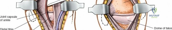
Medial Dissection and Sustentacular Exposure
The medial approach is initiated first. An incision is made starting approximately 2 cm inferior and 2 cm posterior to the tip of the medial malleolus, extending distally along the path of the abductor hallucis. The deep fascia is incised, and the abductor hallucis muscle belly is elevated and retracted inferiorly.
At this juncture, the surgeon must exercise profound respect for the neurovascular bundle (the posterior tibial artery and tibial nerve). The bundle is carefully identified, mobilized, and gently retracted superiorly using vessel loops. Vigorous or careless retraction in this anatomical zone will inevitably lead to iatrogenic neurapraxia, chronic regional pain syndrome, or catastrophic vascular compromise. Deep to the neurovascular bundle, the flexor hallucis longus (FHL) tendon is identified as it courses beneath the sustentaculum tali. The fracture line separating the medial sustentacular fragment (the constant fragment) from the displaced tuberosity is now directly visualized. The tuberosity is mobilized, the fracture hematoma is aggressively debrided, and the tuberosity is reduced to the sustentaculum tali, effectively correcting the varus deformity and restoring the medial column length. Provisional fixation is achieved with stout Kirschner wires (K-wires).
Lateral Dissection and Joint Preparation
Attention is then directed to the lateral aspect of the hindfoot. A limited lateral incision (often a sinus tarsi approach) is utilized, extending from the tip of the lateral malleolus toward the base of the fourth metatarsal. The sural nerve is identified and protected. The extensor digitorum brevis muscle belly is elevated from its origin to expose the subtalar joint and the lateral wall of the calcaneus.
Once the lateral wall blowout is mobilized, the severely comminuted posterior facet is exposed. Using a high-speed burr, curettes, and osteotomes, the surgeon must aggressively remove all remaining hyaline cartilage from both the posterior facet of the calcaneus and the corresponding posterior facet of the talus. Following cartilage denudation, the subchondral bone is systematically penetrated using a drill or a sharp osteotome. This "fish-scaling" or "feathering" technique is absolutely critical; it breaches the sclerotic subchondral plate, exposing healthy, bleeding cancellous bone and maximizing the surface area for osteogenesis and graft incorporation.
Structural Bone Grafting and Deformity Correction
With the joint prepared, the overall calcaneal morphology must be restored. A lamina spreader or a specialized distracter is inserted into the posterior subtalar joint to distract the talus from the calcaneus, restoring Böhler’s angle and native calcaneal height. This distraction creates a massive bone void.
A previously harvested tricortical block of iliac crest bone graft (ICBG) is meticulously shaped to precisely fit this defect. The structural graft is impacted into the void, acting as a biological pillar that maintains the restored height and prevents the talus from subsiding back into the calcaneal body. The remaining joint space, the sinus tarsi, and any residual cancellous defects are densely packed with the remaining autogenous cancellous bone graft. The osteogenic, osteoinductive, and osteoconductive properties of this autograft remain the gold standard for achieving a solid, rapid fusion. To further enhance biomechanical stability, the lateral aspect of the talus (lateral process) and the adjacent lateral wall of the calcaneus are denuded, and this lateral gutter is packed with graft to create a robust extra-articular fusion mass.
Rigid Internal Fixation and Compression
Rigid internal fixation is the final intraoperative step, ensuring absolute stability across the arthrodesis site to promote primary bone healing. The construct is typically secured with two or three large-diameter (6.5 mm or 7.0 mm) cannulated or non-cannulated cancellous screws.
The trajectory of these screws is paramount. They are generally introduced from the non-weight-bearing, posterior-inferior portion of the calcaneal tuberosity. The guide pins are directed anteriorly, medially, and superiorly, aiming for the dense, central cancellous bone of the talar body and the talar neck. The surgeon must utilize multi-planar intraoperative fluoroscopy to ensure that the pins do not penetrate the anterior ankle joint or the talonavicular joint.
A critical biomechanical distinction must be made regarding screw threading. While partially threaded lag screws are traditionally utilized to achieve interfragmentary compression across a fusion site, their use in the presence of a massive structural graft can be detrimental. Aggressive lagging can crush the tricortical graft, resulting in an immediate loss of the meticulously restored calcaneal height. Therefore, in cases utilizing large structural grafts, fully threaded screws are frequently employed as positional screws. These screws rigidly hold the distracted position and secure the graft without applying excessive compressive forces that could lead to structural collapse. Final fluoroscopic images (lateral, Harris axial, and AP ankle views) are obtained to confirm flawless hardware placement, absolute joint reduction, and the anatomical restoration of calcaneal height and alignment.
Complications, Incidence Rates, and Salvage Management
Despite flawless surgical technique and meticulous preoperative planning, subtalar arthrodesis in the setting of severe calcaneal trauma carries an inherent and significant risk profile. The orthopedic surgeon must be intimately familiar with these complications and possess the surgical armamentarium to manage them effectively.
| Complication | Estimated Incidence | Etiology and Risk Factors | Salvage and Management Strategies |
|---|---|---|---|
| Nonunion / Pseudarthrosis | 5% - 12% | Smoking (increases risk by 4x), poorly controlled diabetes, inadequate rigid fixation, failure to aggressively denude subchondral bone, avascular necrosis of the talus, inadequate bone grafting. | Symptomatic nonunions demand revision surgery. Requires complete hardware removal, aggressive debridement of the fibrous nonunion down to bleeding bone, application of massive autogenous bone graft (or orthobiologics like rhBMP-2), and revision internal fixation using a stiffer construct, such as a rigid hindfoot fusion nail or specialized locking plate systems. |
| Malunion (Varus Deformity) | 3% - 8% | Failure to anatomically reduce the tuberosity to the sustentaculum tali, inadequate intraoperative fluoroscopic assessment (Harris axial view), premature weight-bearing. | A catastrophic complication leading to a rigid transverse tarsal joint and lateral column overload. Requires a complex corrective calcaneal osteotomy (e.g., lateral closing wedge or sliding osteotomy) combined with revision arthrodesis to restore the mandatory 5 degrees of valgus. |
| Wound Breakdown / Deep Infection | 2% - 10% | Tenuous lateral soft tissue envelope, operating before the "wrinkle sign" appears, excessive retraction, hematoma formation, smoking. | Superficial necrosis is managed with aggressive local wound care and culture-directed oral antibiotics. Deep infections involving hardware or the structural bone graft are surgical emergencies requiring urgent, radical debridement, hardware removal (if stability is compromised), placement of antibiotic spacers, negative pressure wound therapy, and potentially free tissue transfer (e.g., anterolateral thigh flap). |
| Adjacent Segment Disease (ASD) | 15% - 30% (Long-term) | Altered hindfoot kinematics; subtalar fusion inherently places massively increased stress on the talonavicular, calcaneocuboid, and ankle joints over time. | Initial management is strictly conservative, utilizing custom orthotics (rigid Morton's extension, rocker-bottom shoe modifications) and targeted intra-articular corticosteroid injections. Refractory, debilitating cases necessitate surgical extension of the fusion mass, converting the subtalar fusion into a triple arthrodesis or a pantalar arthrodesis. |
| Sural Nerve Neurapraxia / Neuroma | 5% - 15% | Iatrogenic injury during the lateral approach, aggressive retraction, entrapment in scar tissue. | Prevention is paramount via meticulous dissection. Established neuromas may require gabapentinoids, targeted nerve blocks, or surgical excision of the neuroma with implantation of the proximal nerve stump into deep muscle or bone. |
Phased Post-Operative Rehabilitation Protocols
The ultimate clinical success of a subtalar arthrodesis relies as heavily on strict, unyielding adherence to postoperative rehabilitation protocols as it does on the initial surgical execution. The incorporation of a massive structural bone graft and the achievement of a solid arthrodesis is a prolonged biological process that cannot be rushed.
Phase 1: Immediate Postoperative Phase (Weeks 0 - 3)
Immediately following surgery, a closed suction drain is typically left in place to prevent hematoma formation—a primary catalyst for wound breakdown—and is removed on the first postoperative day. The limb is immobilized in a bulky, well-padded posterior splint with the ankle in neutral dorsiflexion. Strict, continuous elevation of the limb above the level of the heart is absolutely enforced to minimize edema, decrease venous congestion, and protect the fragile soft tissue envelope. The patient is strictly non-weight-bearing (NWB). Sutures or staples are removed at 2 to 3 weeks postoperatively, entirely contingent upon the clinical confirmation of complete, uncomplicated wound healing.
Phase 2: Intermediate Immobilization Phase (Weeks 3 - 6)
Following suture removal and confirmation of wound integrity, the patient is transitioned into a rigid, fiberglass short-leg cast. The patient must maintain strict NWB status. During this phase, the biological process of creeping substitution begins within the structural bone graft. While the hindfoot is immobilized, the patient is strongly encouraged to perform active range of motion exercises for the toes and the knee to prevent distal and proximal joint contractures and to promote venous return.
Phase 3: Clinical and Radiographic Monitoring Phase (Weeks 6 - 12)
At the 6-week mark, the cast is removed, and the first set of postoperative radiographs (AP, Lateral, Harris axial) is obtained to assess the maintenance of hardware position, the absence of hardware failure, and the early signs of bony trabeculation across the arthrodesis site. If the clinical exam reveals no pain with gentle stress and radiographs are progressing satisfactorily, the patient is placed into a prefabricated controlled ankle motion (CAM) walking boot. However, the patient typically remains NWB or is allowed only minimal touch-down weight-bearing (TDWB) to continue protecting the structural graft from sheer and compressive forces that could lead to subsidence.
Phase 4: Late Phase and Return to Function (Weeks 12 and Beyond)
Definitive evidence of radiographic and clinical union is generally apparent between 10 and 12 weeks postoperatively. Once solid union is unequivocally confirmed, the patient is cleared to initiate progressive weight-bearing in the CAM boot, advancing to full weight-bearing as tolerated over a 2-to-4-week period. Formal physical therapy is initiated at this juncture. The rehabilitation focus shifts entirely to restoring ankle joint dorsiflexion and plantarflexion, aggressively strengthening the gastrosoleus complex (which rapidly atrophies during prolonged immobilization), and improving proprioception and gait mechanics. Patients are typically transitioned into supportive, stiff-soled footwear with a custom orthotic to support the transverse tarsal joint. Maximum medical improvement (MMI) and final functional recovery may take up to 12 to 18 months.
Summary of Landmark Literature and Clinical Guidelines
The evolution of primary subtalar arthrodesis for severe calcaneal fractures is deeply rooted in landmark orthopedic literature. The seminal work by Buckley et al. (2002) in their multicenter randomized controlled trial demonstrated that while ORIF provided superior outcomes for certain patient subsets (women, younger patients, lighter workers), patients who developed post-traumatic arthritis and required secondary salvage arthrodesis had significantly worse functional outcomes than those whose fractures healed without complication. This highlighted the severe morbidity associated with the "salvage" pathway.
Further advancing this paradigm, Radnay et al. (2009) published a landmark systematic review directly comparing the outcomes of primary subtalar arthrodesis versus secondary salvage arthrodesis for severe calcaneal fractures. Their data unequivocally demonstrated that patients undergoing primary arthrodesis for highly comminuted fractures achieved significantly higher functional scores (AOFAS scores), experienced shorter total times to return to work, and endured fewer total operative interventions compared to patients who underwent initial ORIF followed by a delayed salvage fusion.
Furthermore, the anatomical classification system developed by Sanders remains the definitive guideline for surgical decision-making. Sanders Type IV fractures, characterized by severe, multi-fragmentary comminution of the posterior facet, are widely considered non-reconstructable. Current clinical guidelines and expert consensus strongly advocate for the integration of ORIF to restore the overall calcaneal envelope combined with immediate primary subtalar arthrodesis for these specific, devastating fracture patterns. This proactive, definitive approach minimizes the duration of disability, mitigates the risk of prolonged post-traumatic pain syndromes, and provides the patient with the highest probability of achieving a functional, painless, and stable plantigrade foot.
