
Comprehensive Introduction and Patho-Epidemiology
Despite the exponential advancements in total joint arthroplasty and joint preservation techniques, arthrodesis of the major weight-bearing joints of the lower extremity—specifically the ankle and the knee—remains an indispensable, highly complex procedure in the armamentarium of the advanced orthopedic surgeon. Historically championed by pioneers such as Sir John Charnley, who introduced the concept of compression arthrodesis to enhance osteogenesis, and Brittain, the principles of joint fusion have evolved dramatically. Modern arthrodesis has transitioned from prolonged cast immobilization and rudimentary external fixation to highly sophisticated techniques utilizing rigid internal compression, advanced intramedullary devices, trabecular metal augments, and computer-assisted hexapod external frames.
Arthrodesis of the lower extremity is primarily considered a salvage operation, though in certain demographics, such as young laborers with isolated tibiotalar arthritis, it remains a primary indication. The overarching surgical objective is to provide a stable, painless, and plantigrade limb in the setting of end-stage osteoarthritis, inflammatory arthropathy, post-traumatic destruction, neuropathic (Charcot) arthropathy, or the catastrophic failure of a total joint arthroplasty. The patho-epidemiology of these two joints differs significantly. Ankle arthritis is overwhelmingly post-traumatic, accounting for approximately 70% to 80% of all cases, often presenting in a younger, more active demographic following rotational ankle fractures, pilon fractures, or chronic ligamentous instability. Conversely, primary osteoarthritis of the knee is ubiquitous, but knee arthrodesis is almost exclusively reserved for the devastating complications of total knee arthroplasty (TKA), specifically recalcitrant periprosthetic joint infection (PJI) with massive bone loss, or catastrophic disruption of the extensor mechanism.
The socioeconomic and psychological burden of end-stage lower extremity joint pathology cannot be overstated. Patients often present with severe chronic pain, profound functional limitations, and narcotic dependency. While total ankle replacement (TAR) has gained significant traction and boasts improved survivorship with modern implant designs, arthrodesis remains the gold standard for patients with severe coronal or sagittal plane deformities, profound bone loss, or high physical demands. In the knee, the decision to proceed with arthrodesis versus above-knee amputation (AKA) is one of the most challenging clinical dilemmas. Arthrodesis provides a durable, sensate limb that is biomechanically vastly superior to an AKA in terms of functional mobility and energy expenditure, provided the patient has the requisite bone stock, soft tissue envelope, and vascular supply to achieve a successful union.
This definitive masterclass comprehensively delineates the evidence-based indications, intricate biomechanical considerations, step-by-step surgical techniques, and rigorous postoperative protocols for arthrodesis of the ankle and knee. Mastery of these techniques is an essential competency for the orthopedic surgeon, requiring a profound understanding of mechanical alignment, soft tissue management, and the biological principles of bone healing in compromised host beds.
Detailed Surgical Anatomy and Biomechanics
Ankle Arthrodesis Anatomy and Biomechanics
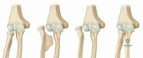
The tibiotalar joint is a highly congruent, inherently stable hinge joint that bears up to five times the body weight during the stance phase of the normal gait cycle. The surgical anatomy relevant to ankle arthrodesis involves meticulous management of the extra-articular soft tissue envelope and the intra-articular osseous structures. The blood supply to the talus is notoriously tenuous, relying on an extraosseous anastomotic ring formed by the artery of the tarsal canal, the artery of the sinus tarsi, and branches of the anterior tibial and peroneal arteries. Aggressive intra-articular debridement must preserve this extraosseous supply to prevent avascular necrosis (AVN) of the talar body, which catastrophically compromises the fusion bed. Furthermore, the anterior neurovascular bundle (deep peroneal nerve and anterior tibial artery) and the superficial peroneal nerve are at high risk during anterior approaches and percutaneous screw placement.
The biomechanical success of an ankle arthrodesis is entirely dependent on achieving the precise spatial orientation of the fused joint. Malpositioning leads to profoundly altered gait mechanics, exponentially increased energy expenditure, and accelerated, inevitable degeneration of the adjacent subtalar, transverse tarsal, and midfoot joints. The absolute optimal position for ankle arthrodesis dictates neutral dorsiflexion/plantarflexion (0 degrees). Plantarflexion must be avoided at all costs, as it forces a vaulting gait, induces genu recurvatum to achieve a plantigrade foot, and rapidly accelerates midfoot breakdown.
Furthermore, the ankle must be fused in 0 to 5 degrees of valgus. Slight valgus unlocks the transverse tarsal joint (talonavicular and calcaneocuboid joints), allowing for compensatory midfoot motion that absorbs the shock of heel strike. Varus positioning locks the midfoot, leading to a rigid, painful, and highly inefficient gait. External rotation should be set between 5 to 10 degrees, meticulously matched to the contralateral limb to ensure a symmetric foot progression angle during the stance phase. Finally, the talus should be translated slightly posterior relative to the mechanical axis of the tibia. This posterior translation decreases the anterior lever arm on the midfoot, reducing stress on the adjacent joints, and significantly improves the cosmetic appearance of the fused ankle.
Knee Arthrodesis Anatomy and Biomechanics
The knee is a complex bicondylar hinge joint, and its surgical anatomy in the context of arthrodesis is often profoundly distorted by previous arthroplasty, multiple revisions, or chronic infection. The critical anatomical considerations involve the management of massive metaphyseal bone defects in the distal femur and proximal tibia, the handling of a compromised or entirely absent extensor mechanism, and the protection of the posteriorly situated popliteal neurovascular bundle. Extensive scarring from prior surgeries necessitates meticulous dissection to avoid devascularizing the remaining cortical bone, which is essential for achieving union. Plastic surgery consultation for rotational or free tissue transfer (e.g., medial gastrocnemius flap) is often a prerequisite to ensure adequate soft tissue coverage over the fusion site.
Biomechanically, knee arthrodesis is a formidable, joint-sacrificing procedure that profoundly impacts the patient's kinematics. The loss of knee flexion completely alters sitting mechanics, stair climbing, and increases the energy cost of walking by up to 25% to 30%. However, compared to an above-knee amputation, which increases energy expenditure by up to 65%, a fused knee provides a highly functional salvage. Unlike the ankle, the knee must be fused in a position that delicate balances the demands of walking (which favors full extension for maximum stability and minimal quadriceps demand) and sitting/foot clearance (which favors slight flexion).
The optimal position for knee arthrodesis is 0 to 10 degrees of flexion. This slight flexion assists with foot clearance during the swing phase of gait and makes sitting in confined spaces marginally more comfortable. Fusing the knee in greater than 15 degrees of flexion is contraindicated, as it causes excessive functional limb shortening, induces severe quadriceps and hip extensor fatigue, and places excessive stress on the fusion construct. The mechanical axis must be restored to 5 to 7 degrees of valgus to match the normal anatomical alignment of the lower extremity, preventing excessive varus or valgus thrust during ambulation. Finally, rotation should be set from neutral to 5 degrees of external rotation, matching the normal tibial torsion and the contralateral limb.
Exhaustive Indications and Contraindications
The decision to proceed with a major lower extremity arthrodesis requires a comprehensive evaluation of the patient's physiologic age, functional demands, medical comorbidities, and the specific pathology of the joint. In the ankle, post-traumatic osteoarthritis remains the most common indication. Patients typically present with end-stage cartilage loss, subchondral cyst formation, and rigid deformity following complex pilon or rotational ankle fractures. Other indications include primary osteoarthritis, inflammatory arthropathies (such as rheumatoid arthritis or ankylosing spondylitis), and the salvage of a failed total ankle arthroplasty. Neuropathic (Charcot) arthropathy with severe instability or ulceration is a compelling indication, though it requires highly specialized fixation constructs, often combining intramedullary nailing with external fixation, due to the profound lack of protective sensation and poor bone quality.
In the knee, the indications are almost exclusively related to the salvage of a failed total knee arthroplasty. The most common modern indication is a recalcitrant periprosthetic joint infection (PJI) involving highly virulent or multi-drug resistant organisms, where a two-stage revision arthroplasty has failed or is deemed medically futile. Other critical indications include catastrophic failure of the extensor mechanism (e.g., irreparable patellar tendon rupture or severe patella baja) in the setting of a TKA, severe neuropathic arthropathy of the knee joint, post-traumatic destruction with unreconstructible massive bone loss, and oncologic resections requiring joint sacrifice where megaprostheses are contraindicated.
Contraindications must be strictly respected to prevent catastrophic complications, including the need for subsequent high-level amputation. Absolute contraindications include active, untreated systemic infection or local osteomyelitis (unless utilizing a staged approach or external fixation), severe peripheral vascular disease precluding wound healing, and a medically unfit patient unable to tolerate the physiologic stress of a major orthopedic reconstruction. Relative contraindications require nuanced clinical judgment. For instance, in the ankle, ipsilateral subtalar or transverse tarsal arthritis is a relative contraindication to isolated tibiotalar fusion; these patients are better served by a pantalar or tibiotalocalcaneal (TTC) fusion. In the knee, contralateral limb amputation or a contralateral stiff knee is a severe relative contraindication, as bilateral stiff knees make rising from a seated position nearly impossible.
| Parameter | Ankle Arthrodesis | Knee Arthrodesis |
|---|---|---|
| Primary Indications | Post-traumatic OA, Failed TAR, Charcot arthropathy, Paralytic deformities (drop foot), Inflammatory arthritis. | Infected TKA (recalcitrant), Extensor mechanism failure, Massive unreconstructible bone loss, Tumor resection. |
| Absolute Contraindications | Active untreated local infection (open hardware), severe peripheral vascular disease (ABI < 0.4), medically unfit. | Contralateral AKA/BKA (relative to functional status), active untreated infection (requires staged approach), severe vascular compromise. |
| Relative Contraindications | Ipsilateral subtalar/midfoot arthritis (consider extended fusion), severe osteopenia, active smoking (high nonunion risk). | Severe ipsilateral hip or ankle arthritis, inadequate soft tissue coverage (requires flap), contralateral stiff knee. |
| Optimal Positioning | Neutral DF/PF (0°), 0-5° Valgus, 5-10° External Rotation, slight posterior translation of talus. | 0-10° Flexion, 5-7° Valgus, Neutral to 5° External Rotation. |
Pre-Operative Planning, Templating, and Patient Positioning
Thorough pre-operative planning is the cornerstone of a successful lower extremity arthrodesis. The clinical evaluation must begin with a rigorous assessment of the vascular status; non-invasive arterial studies (Ankle-Brachial Index, toe pressures) and vascular surgery consultation are mandatory if peripheral arterial disease is suspected. The soft tissue envelope must be critically evaluated for prior surgical incisions, traumatic scarring, and the presence of sinus tracts. In the setting of knee arthrodesis for infected TKA, a plastic surgery consultation is frequently required to plan for local rotational flaps (e.g., medial gastrocnemius) or free tissue transfer to ensure robust coverage over the bulky arthrodesis construct.
Radiographic evaluation demands weight-bearing, full-length orthogonal views of the affected extremity to assess the mechanical axis, degree of deformity, and adjacent joint status. Advanced imaging is indispensable. Computed Tomography (CT) with metal artifact reduction sequences (MARS) is critical for evaluating the exact volume and geometry of bone stock. In the ankle, CT identifies hidden talar cysts or avascular necrosis that may compromise screw purchase. In the knee, CT dictates the necessity for structural allografts, custom 3D-printed titanium trabecular metal cones, or shortening osteotomies to achieve bleeding, viable bone apposition. Magnetic Resonance Imaging (MRI) or nuclear medicine scans (Indium-111 labeled WBC scan) may be utilized to delineate the extent of osteomyelitis in infected cases. Digital templating is mandatory to determine the appropriate length, diameter, and trajectory of intramedullary nails, the contouring of plates, and the precise level of bone resections required to correct deformity while minimizing limb length discrepancy.
Patient positioning is dictated by the chosen surgical approach and the specific joint. For ankle arthrodesis, the patient is typically positioned supine with a large bump under the ipsilateral hip to internally rotate the leg, bringing the lateral malleolus anteriorly for a transfibular approach. Alternatively, a lateral decubitus position can be utilized for isolated lateral approaches. A radiolucent table and unhindered access for the C-arm fluoroscope in both AP and lateral planes are absolute requirements. For knee arthrodesis, the patient is positioned supine on a radiolucent table. If a long antegrade intramedullary nail is planned, the patient may be placed on a fracture table or a flat table with a bump under the ipsilateral hip to allow access to the piriformis fossa or greater trochanter. A sterile tourniquet is highly recommended to minimize blood loss during the extensive debridement phase, though it must be let down prior to definitive fixation to assess bone viability and ensure meticulous hemostasis.
Step-by-Step Surgical Approach and Fixation Technique
Ankle Arthrodesis Techniques
1. Arthroscopic Ankle Arthrodesis:
Pioneered by Myerson and Glick, the arthroscopic approach is the preferred technique for patients with minimal to moderate deformity (less than 15 degrees of coronal or sagittal malalignment) and minimal bone loss. The advantages are profound: significantly lower morbidity, decreased blood loss, preservation of the critical extra-articular periosteal blood supply, and a substantially faster time to clinical and radiographic union (averaging 9 weeks compared to 14 weeks for open techniques). Standard anteromedial and anterolateral portals are established. Using a combination of curettes, aggressive shavers, and high-speed burrs, the articular cartilage of the tibial plafond and talar dome is meticulously resected until bleeding, punctate subchondral bone is exposed. The medial and lateral gutters must be aggressively cleared of osteophytes to allow for unhindered compression. Fixation is achieved percutaneously under fluoroscopic guidance using two or three large-diameter (6.5 mm or 7.3 mm) cannulated, partially threaded cancellous screws placed in a crossed configuration to provide rigid, interfragmentary compression.
2. Open Transfibular Approach:
The transfibular approach is the workhorse for open ankle arthrodesis, providing unparalleled exposure for the correction of severe deformities and meticulous joint preparation. A longitudinal incision is made over the distal fibula, curving slightly anteriorly toward the base of the fourth metatarsal. The fibula is osteotomized approximately 2 to 3 cm proximal to the joint line. The distal fibula is excised, meticulously denuded of soft tissue, morselized, and preserved for use as highly osteogenic autogenous bone graft. Alternatively, it can be split longitudinally and utilized as an onlay biological plate across the lateral fusion site.
The articular surfaces of the tibia and talus are then resected. Flat cuts made with an oscillating saw provide excellent bony apposition and inherent biomechanical stability but limit the surgeon's ability to fine-tune the alignment. Conversely, contour-matching (cup and cone) reaming preserves critical bone stock, maintains the anatomical center of rotation, and allows for multiplanar adjustability prior to fixation. Rigid internal compression is paramount. The standard, biomechanically superior construct involves three 6.5 mm or 7.3 mm cannulated screws. The "Home Run Screw" is placed from the posterior tibia, aimed anteriorly and inferiorly into the dense bone of the talar neck. Extreme care must be taken to avoid penetrating the subtalar joint. A second screw is placed from the medial malleolus into the talar body, and a third from the anterolateral tibia into the talar body. The morselized fibular autograft is then densely packed into the interstices and gutters.
Knee Arthrodesis Techniques
1. Intramedullary Nailing (The Gold Standard):
For the salvage of a failed TKA, intramedullary (IM) nailing provides the highest union rates (historically 85-95%) and the most rigid biomechanical load-sharing construct. If active periprosthetic infection is present, a rigorous staged approach is mandatory. Stage 1 involves the complete explantation of the prosthesis, thorough debridement of all cement and necrotic tissue, and the placement of an articulating or static antibiotic-impregnated cement spacer. Stage 2, performed only after clinical eradication of infection and normalization of inflammatory markers (ESR, CRP), involves definitive arthrodesis.
During the definitive procedure, the distal femur and proximal tibia are prepared. Cylindrical reaming or flat cuts can be utilized. Flat cuts provide excellent compression but result in significant limb shortening. In cases of massive metaphyseal bone loss, structural bulk allografts or custom titanium trabecular metal cones are utilized to bridge the defect and provide a stable foundation for the nail. A long, modular antegrade nail is passed from the greater trochanter, spanning the entire femoral diaphysis, across the prepared knee joint, and down to the distal tibial diaphysis. Alternatively, a retrograde nail can be inserted through the knee, locking into the femoral and tibial diaphyses. Dynamic compression is applied meticulously across the arthrodesis site using the nail's internal compression mechanism before final distal interlocking screws are placed.
2. Dual Plating and External Fixation:
Dual plating is advocated when the medullary canal is severely deformed, obliterated by prior hardware, or when an IM nail is absolutely contraindicated. Two heavy-duty, broad dynamic compression plates (or modern robust locking plates) are applied in orthogonal planes—typically one anteriorly and one medially or laterally. This requires extensive soft tissue stripping, which significantly increases the risk of wound breakdown and devascularization of the bone ends. As a load-bearing construct, it is highly susceptible to fatigue failure if union is delayed.
External fixation (using circular Ilizarov or Taylor Spatial Frames, or robust monolateral frames) is the treatment of choice in the presence of active, recalcitrant infection, massive soft tissue defects, or profound bone loss requiring simultaneous limb lengthening via distraction osteogenesis (bone transport). The frame allows for immense compression across the fusion site without placing permanent hardware in a potentially infected bed. However, it carries the burden of pin tract infections, requires meticulous patient compliance, and imposes a significant psychological toll due to the bulky apparatus.
Complications, Incidence Rates, and Salvage Management
The complication profile for major lower extremity arthrodesis is significant, reflecting the salvage nature of these procedures, the inherently compromised host beds, and the complex biomechanical forces at play. Nonunion is the most frequent major complication, occurring in approximately 5% to 10% of primary ankle arthrodeses and up to 15% to 20% of knee arthrodeses (particularly in the setting of prior infection or massive bone loss). Risk factors for nonunion include active smoking (which increases the nonunion rate by up to fourfold), uncontrolled diabetes mellitus, prior radiation therapy, and inadequate rigid internal compression. Management of an aseptic nonunion typically requires revision internal fixation, aggressive debridement of the fibrous nonunion site, and the application of autologous bone graft (e.g., iliac crest) or orthobiologics (e.g., BMP-2, demineralized bone matrix).
Infection is a devastating complication, particularly in knee arthrodesis performed for a prior periprosthetic joint infection. Recurrent or recalcitrant deep infection occurs in 10% to 15% of these cases. The presence of a biofilm on the massive intramedullary implants makes eradication exceedingly difficult. Salvage management often necessitates complete hardware removal, aggressive serial debridements, prolonged intravenous antibiotic therapy directed by an infectious disease specialist, and stabilization with a spanning external fixator. In cases where the infection cannot be eradicated, or the soft tissue envelope completely fails, a high-level above-knee amputation remains the ultimate, life-saving salvage procedure.
Malunion is a critical complication that profoundly affects patient function. In the ankle, fusion in plantarflexion leads to a vaulting gait, severe midfoot overload, and the rapid development of adjacent segment disease (subtalar and talonavicular osteoarthritis). Revision osteotomy and re-fusion are required to restore a plantigrade foot. In the knee, fusion in excessive flexion (>15 degrees) causes severe functional limb shortening and quadriceps fatigue, while fusion in varus or valgus leads to abnormal mechanical axis loading and stress fractures. Limb length discrepancy is expected in knee arthrodesis; a shortening of 1 to 2.5 cm is actually beneficial to allow for foot clearance during the swing phase of gait. However, shortening greater than 3 cm requires the permanent use of a shoe lift or, in severe cases, a staged limb lengthening procedure.
| Complication | Estimated Incidence | Primary Risk Factors | Salvage / Management Strategy |
|---|---|---|---|
| Nonunion (Aseptic) | Ankle: 5-10% Knee: 10-20% |
Smoking, AVN of talus, massive bone loss, inadequate compression. | Revision fixation, iliac crest autograft, orthobiologics (BMP-2), bone stimulators. |
| Deep Infection | Ankle: 2-5% Knee: 10-15% |
Prior PJI, severe soft tissue compromise, diabetes, prolonged OR time. | Hardware removal, radical debridement, IV antibiotics, external fixation, or Amputation. |
| Malunion | 5-10% | Poor intraoperative fluoroscopic assessment, inadequate bone cuts. | Corrective osteotomy, realignment, and revision arthrodesis. |
| Stress Fractures | 3-8% | Stress shielding at the tip of long IM nails or plates, altered modulus of elasticity. | Protected weight-bearing, extension of hardware (longer nail/plate), or bisphosphonates. |
| Adjacent Segment Disease | Ankle: >50% at 10 yrs | Altered biomechanics, compensatory hypermobility in midfoot/subtalar joint. | Orthotics, rocker-bottom shoes, eventual extension of fusion (e.g., TTC fusion). |
Phased Post-Operative Rehabilitation Protocols
Ankle Arthrodesis Rehabilitation
The postoperative rehabilitation following an ankle arthrodesis is a meticulously phased protocol designed to protect the highly vulnerable fusion mass while managing edema and preventing adjacent joint stiffness.
* Phase I (0-2 Weeks): The patient is placed in a well-padded, rigid short leg splint in the operating room. Strict non-weight-bearing (NWB) status is enforced. Absolute elevation of the limb above the level of the heart is critical to manage postoperative edema, which can threaten wound healing. Deep vein thrombosis (DVT) prophylaxis is initiated based on patient risk stratification.
* Phase II (2-6 Weeks): The patient is seen in the clinic for wound evaluation and suture removal. If the incisions are fully healed, the patient is transitioned to a rigid fiberglass short leg cast. Strict NWB status is maintained.
* Phase III (6-12 Weeks): At the 6-week mark, orthogonal radiographs are obtained. If there is evidence of early trabecular bridging and consolidation across the arthrodesis site, the cast is removed. The patient is transitioned to a controlled ankle motion (CAM) boot. Progressive partial weight-bearing is initiated, advancing to full weight-bearing as tolerated by 10 to 12 weeks. Physical therapy focuses on strengthening the proximal musculature and maintaining motion in the subtalar and midfoot joints.
* Phase IV (12+ Weeks): Once clinical and radiographic union is definitively confirmed, the patient is transitioned to regular footwear. The implementation of shoe modifications, specifically a rigid shank and a rocker-bottom sole (or a SACH - Solid Ankle Cushion Heel), is paramount. These modifications simulate the rollover action of the ankle during the stance phase, significantly improving gait kinematics and reducing stress on the adjacent joints.
Knee Arthrodesis Rehabilitation
Rehabilitation following knee arthrodesis is heavily dictated by the type of fixation construct utilized and the quality of the host bone.
* Intramedullary Nailing Protocol: The primary advantage of a long, load-sharing IM nail is the ability to allow early weight-bearing. Depending on bone quality and the rigidity of the fixation, patients are often permitted to begin immediate partial weight-bearing (touch-down to 25%) with the use of a walker or crutches. Progressive weight-bearing is advanced over the first 6 weeks. Physical therapy focuses heavily on gait training, hip and core strengthening, and energy conservation strategies, as the patient must adapt to a stiff-legged gait.
* Plate Fixation or External Fixation Protocol: If dual plating or external fixation is utilized, the construct is load-bearing rather than load-sharing. Consequently, strict non-weight-bearing or minimal touch-down weight-bearing is enforced for the first 6 to 8 weeks until robust radiographic callus is visible. Premature weight-bearing in these constructs inevitably leads to hardware fatigue failure or pin-site loosening. Once consolidation is evident, progressive weight-bearing is initiated. For patients with external fixators, rigorous daily pin site care is mandatory to prevent superficial infections from tracking into the medullary canal.
Summary of Landmark Literature and Clinical Guidelines
The operative principles of lower extremity arthrodesis are deeply rooted in landmark orthopedic literature and continuously refined by contemporary clinical guidelines. Sir John Charnley’s seminal work in the 1950s established the absolute necessity of rigid, dynamic compression to achieve primary bone healing without intermediate callus formation, a principle that remains the bedrock of modern fusion techniques.
In the realm of ankle arthrodesis, the transition toward minimally invasive techniques was spearheaded by Myerson and Glick, whose landmark studies demonstrated that arthroscopic fusion yields equivalent union rates to open procedures but with a statistically significant reduction in time to union, decreased blood loss, and lower complication rates in appropriately selected patients. Furthermore, the biomechanical studies by Zwipp and others have definitively established the optimal spatial positioning of the ankle (neutral dorsiflexion, slight valgus, and external rotation) to minimize the inevitable onset of adjacent segment disease in the subtalar and talonavicular joints.
For knee arthrodesis, the literature heavily focuses on the management of the infected total knee arthroplasty. Conway and colleagues provided foundational data demonstrating that intramedullary nailing provides superior union rates compared to external fixation, though they emphasized the necessity of a staged approach to eradicate infection prior to definitive nailing. Modern guidelines from the American Academy of Orthopaedic Surgeons (AAOS) and the Musculoskeletal Infection Society (MSIS) reinforce the use of articulating or static antibiotic spacers in the interim phase. Recent advancements prominently feature the use of highly porous titanium trabecular metal cones and sleeves
Detailed Chapters & Topics
Dive deeper into specialized chapters regarding references-1