Single-Incision Medial Approach for Triple Arthrodesis
Comprehensive Introduction and Patho-Epidemiology
The triple arthrodesis remains a cornerstone in the armamentarium of the orthopedic foot and ankle surgeon for the management of severe, rigid hindfoot and midfoot deformities. Traditionally, this procedure—encompassing the fusion of the subtalar, talonavicular, and calcaneocuboid joints—has been performed through a dual-incision approach utilizing both a medial and a lateral surgical window. However, in the setting of severe, longstanding rigid planovalgus foot deformity, the lateral skin and soft tissues become chronically contracted and severely compromised. Correcting a massive valgus deformity to a neutral, plantigrade position via a standard lateral sinus tarsi approach inherently places immense tension on these laterally contracted soft tissues. This tension frequently precipitates catastrophic wound healing complications, skin necrosis, and deep infection. To mitigate this significant morbidity, the single-incision medial approach for triple arthrodesis was developed. This technique affords comprehensive exposure of the subtalar, talonavicular, and calcaneocuboid joints without violating the high-risk lateral soft tissue envelope.
The pathogenesis of adult acquired flatfoot deformity (AAFD), recently reconceptualized as Progressive Collapsing Foot Deformity (PCFD), represents a complex failure of both dynamic and static stabilizers of the medial longitudinal arch. The dynamic component is primarily governed by the posterior tibial tendon, while the static components include the spring ligament (calcaneonavicular ligament), the plantar fascia, and the long and short plantar ligaments. As these structures become attenuated, the posterior tibial tendon undergoes tendinosis, elongation, and eventual dysfunction. It remains a topic of academic debate whether the static capsuloligamentous structures or the dynamic tendinous stabilizers fail first; however, the clinical manifestation is a progressive, cascading collapse of the midfoot and hindfoot.
As the deformity progresses to a severe flatfoot, the peroneal tendons and the laterally translated Achilles tendon gain a significant mechanical advantage. They overpower the dysfunctional posterior tibial tendon, driving the subtalar joint into rigid heel valgus. Concurrently, the transverse tarsal joints (the talonavicular and calcaneocuboid joints) are forcefully abducted by the relative overpull of the peroneus brevis. This unopposed abduction causes lateral subluxation of the talonavicular joint, clinically and radiographically presenting as the "uncovering" of the talar head.
If left untreated, the natural history of a severe hindfoot valgus deformity is progressive attenuation and eventual rupture of the deltoid ligament complex. Once the deltoid ligament becomes incompetent, the tibiotalar joint loses its congruent articulation and tilts into valgus, rapidly accelerating the onset of asymmetric ankle joint osteoarthritis. The association of severe hindfoot valgus with a valgus tilt of the ankle mortise presents an exceptionally challenging clinical scenario. Surgical management at this advanced stage generally necessitates either a pantalar arthrodesis or a total ankle arthroplasty combined with an underlying triple arthrodesis. Therefore, it is absolutely critical to intervene surgically in patients with severe hindfoot valgus before the deltoid ligament fails, thereby preserving the native ankle joint.
Detailed Surgical Anatomy and Biomechanics
A profound understanding of the complex three-dimensional anatomy of the hindfoot is a prerequisite for executing a single-incision medial triple arthrodesis. The approach relies heavily on leveraging medial anatomical windows to access lateral structures. The subtalar joint, a complex diarthrodial joint responsible for hindfoot inversion and eversion, consists of anterior, middle, and posterior facets. Through the medial approach, the posterior and middle facets of the subtalar joint lie directly deep to the sheath of the posterior tibial tendon. By excising the dysfunctional posterior tibial tendon—which is invariably sacrificed in this procedure due to its pathologic state—the surgeon gains direct, unhindered access to these critical articular surfaces.
Immediately posterior and plantar to the subtalar joint lie the flexor digitorum longus (FDL) tendon, the flexor hallucis longus (FHL) tendon, and the posterior tibial neurovascular bundle. The tibial nerve bifurcates into the medial and lateral plantar nerves in this vicinity, and the posterior tibial artery provides essential vascularity to the plantar aspect of the foot. These structures are at significant risk during medial joint preparation and must be meticulously protected with blunt retractors throughout the procedure.
The talonavicular joint, often referred to as the "coxa pedis" due to its ball-and-socket configuration, is the primary keystone of the transverse tarsal joint. It is easily accessible through the anterior extent of the extensile medial incision. The true anatomical challenge of the single-incision medial approach lies in accessing the calcaneocuboid joint. The calcaneocuboid joint is situated directly lateral to the talonavicular joint across the transverse plane of the foot. Biomechanically, the talonavicular and calcaneocuboid joints function in tandem; when the hindfoot is in eversion, their axes are parallel, unlocking the midfoot for shock absorption. In this surgical technique, the calcaneocuboid joint is accessed entirely through the medial incision by aggressively distracting the talonavicular joint with a laminar spreader, allowing the surgeon to look "across" the foot to prepare the lateral column.
Exhaustive Indications and Contraindications
While a single-incision medial triple arthrodesis is technically feasible in the vast majority of patients requiring a triple arthrodesis, its primary utility and highest value are realized in the most severe cases of rigid hindfoot valgus and in patients with a high risk of wound healing complications. Rheumatoid arthritis is a classic etiology of severe hindfoot valgus, with patients frequently presenting with greater than 30 degrees of valgus through the subtalar joint and gross radiographic subluxation of the posterior facet. Similarly, diabetic patients presenting with Charcot-like subtalar joint subluxation or dislocation are prime candidates. These patients possess a profoundly elevated risk of wound healing complications, making a lateral sinus tarsi approach highly unadvisable. Furthermore, any patient with a history of previous lateral soft tissue trauma, open lateral wounds, active lateral infection, or prior surgical incisions that compromise the lateral angiosomes will benefit immensely from this medial-only technique.
| Category | Indications for Single-Incision Medial Approach | Contraindications for Single-Incision Medial Approach |
|---|---|---|
| Clinical Deformity | Severe, rigid planovalgus (>30 degrees heel valgus); Rigid transverse tarsal abduction. | Flexible planovalgus (amenable to joint-sparing osteotomies/tendon transfers); Rigid varus deformity. |
| Soft Tissue Status | Contracted lateral skin; Prior lateral surgical scars; History of lateral trauma/flap coverage. | Severe medial soft tissue compromise; Active medial ulceration; Medial angiosome failure. |
| Patient Comorbidities | Rheumatoid Arthritis; Diabetes Mellitus with neuropathy; Charcot neuroarthropathy; Chronic corticosteroid use. | Active systemic infection; Uncontrolled peripheral arterial disease (PAD) without prior vascular optimization. |
| Radiographic Findings | Gross subluxation of the posterior subtalar facet; Uncovering of the talar head >50%; Subfibular impingement. | Isolated talonavicular or subtalar arthritis without deformity (standard approaches suffice). |
Pre-Operative Planning, Templating, and Patient Positioning
Comprehensive Clinical Examination
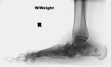
Pre-operative evaluation must be exhaustive. The examiner should visually inspect the posterior heel alignment with respect to the mechanical axis of the tibia while the patient is standing. Physiologic hindfoot valgus typically ranges from 5 to 7 degrees; valgus exceeding 15 degrees is pathologic, and deformities greater than 30 degrees virtually guarantee lateral skin tension upon reduction. Subtalar range of motion must be assessed by maximally inverting and everting the heel. Normal subtalar motion is approximately 5 degrees of eversion and 20 degrees of inversion. In the target demographic for this procedure, the pes planovalgus deformity will be rigidly fixed. If the hindfoot remains flexible, the surgeon should pivot toward joint-sparing procedures such as medial displacement calcaneal osteotomies or lateral column lengthening.
A critical component of the physical examination is the assessment of peroneal tendon contracture. With the heel manually forced into maximum inversion, the examiner must palpate the peroneal tendons to determine their contribution to the valgus contracture. If the peroneus longus and brevis are excessively tight, they act as a tether, preventing neutral realignment of the heel. These tendons will require a proximal release. Finally, the examiner must visually and tactilely inspect the skin overlying the lateral hindfoot. If the lateral skin is taut before any corrective maneuvers are applied, a sinus tarsi incision will be impossible to close without significant tension once the heel is reduced to neutral.
Imaging and Diagnostic Studies
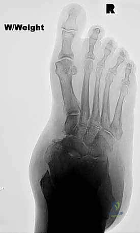
Standard weight-bearing radiographs of the foot and ankle are non-negotiable. Foot radiographs (AP, lateral, and oblique) quantify the degree of subluxation or dislocation of the subtalar and transverse tarsal joints. They allow the surgeon to assess Meary's angle, talonavicular coverage angle, and calcaneal pitch. Furthermore, radiographs dictate whether structural bone grafting (e.g., tricortical iliac crest or femoral head allograft) will be required to manage severe bone loss or to restore lateral column length. Weight-bearing ankle radiographs are equally critical to confirm that the severe valgus deformity is isolated to the hindfoot. If deltoid ligament incompetence has allowed the talus to tilt into valgus within the ankle mortise, a triple arthrodesis alone will fail to rebalance the tibiotalar joint, necessitating concurrent or staged ankle realignment or arthrodesis.
Patient Positioning and Setup
The patient is positioned supine on the operating table. A small, well-padded bump is placed beneath the ipsilateral hip (contrary to standard dual-incision approaches where a bump is placed under the ipsilateral hip to access the lateral side, here the goal is to externally rotate the leg). Placing a bump under the contralateral hip externally rotates the operative leg, placing the medial aspect of the foot directly facing the surgeon and nearly parallel to the ceiling. This positioning is absolutely critical because the entirety of the exposure, joint preparation, and hardware trajectory will be executed through the medial portal. A well-padded pneumatic tourniquet is applied to the proximal thigh to ensure a bloodless surgical field, which is vital for identifying the medial neurovascular structures.
Step-by-Step Surgical Approach and Fixation Technique
Release of the Peroneal Tendon Contracture
In cases of severe, rigid valgus, the peroneal tendons are invariably contracted and must be released to allow for varus reduction of the calcaneus. To avoid compromising the lateral skin around the hindfoot, this release is performed proximally. A 3-cm longitudinal incision is made posterolaterally, approximately 10 cm proximal to the tip of the lateral malleolus. This incision is sited directly over the peroneal musculotendinous junction, immediately posterior to the fibular border. The peroneal tendon sheath is incised longitudinally. Using a curved hemostat, the peroneus longus and brevis tendons are sequentially delivered from their sheath. Each tendon is completely transected (tenotomy) or Z-lengthened, depending on surgeon preference and the severity of the contracture. The tendons are then allowed to retract into the sheath, completely abolishing the lateral tethering force. The sheath and skin are closed in standard layered fashion.
Extensile Medial Exposure and Subtalar Joint Preparation
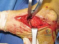
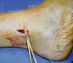
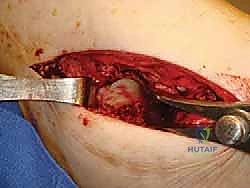
The primary surgical approach utilizes an extensile longitudinal incision over the medial aspect of the foot. The incision begins at the distal tip of the medial malleolus, curving gently along the course of the posterior tibial tendon, and terminates at the level of the first tarsometatarsal joint. Deep dissection proceeds directly to the posterior tibial tendon sheath, which is incised longitudinally. The posterior tibial tendon, which is typically hypertrophied, degenerated, and functionally useless in these advanced cases, is completely detached from its broad insertion onto the navicular tuberosity and medial cuneiform. A heavy Köcher clamp is applied to the distal stump of the tendon, and it is placed under maximum tension. The tendon is then sharply excised as far proximally as the incision allows to remove the pathologic tissue and expose the underlying structures.
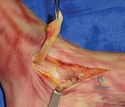
With the posterior tibial tendon removed, the surgeon must immediately identify and protect the flexor digitorum longus tendon and the posterior tibial neurovascular bundle. A blunt, curved retractor (such as a vascular or mini-Hohmann retractor) is carefully placed to retract these structures posteriorly and plantarly. Using a scalpel, the capsule overlying the medial aspect of the subtalar joint is incised. The posterior and middle facets of the subtalar joint are located directly deep to the excised posterior tibial tendon. An osteotome or Cobb elevator is utilized to release the robust interosseous talocalcaneal ligament and the medial joint capsule.
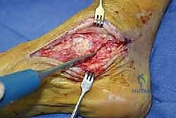
A lamina spreader is then inserted between the medial talar neck and the sustentaculum tali of the calcaneus. Opening the lamina spreader distracts the subtalar joint, providing excellent visualization of the posterior and middle facets. Utilizing a combination of straight and angled curettes, along with sharp osteotomes, all remaining articular cartilage is meticulously denuded down to bleeding subchondral bone. A curved osteotome is then employed to aggressively "feather" or shingle the articular surfaces. This feathering technique vastly increases the surface area available for osteogenesis, exposes highly vascular cancellous bone, and generates local autogenous bone graft.
Exposure and Preparation of the Talonavicular Joint
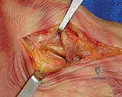
The talonavicular joint is inherently superficial and easily accessed through the anterior portion of the medial incision. A longitudinal capsulotomy is performed in line with the skin incision, directly over the medial prominence of the talonavicular joint. The dorsal and plantar capsular attachments are elevated off the joint margins using a periosteal elevator to expose the entire articular hemisphere of the talar head and the corresponding navicular socket.
A small lamina spreader is placed within the medial aspect of the talonavicular joint to distract it. Similar to the subtalar joint, aggressive cartilage removal is performed using curettes and osteotomes until the subchondral plate is reached. The articular surfaces of both the talus and navicular are aggressively feathered. To mobilize the joint for eventual reduction, a curved elevator is passed across the talonavicular joint from medial to lateral, blindly releasing the lateral capsule. This step is critical for allowing the navicular to be translated medially and plantarly over the talar head during the final correction.
Exposure and Preparation of the Calcaneocuboid Joint
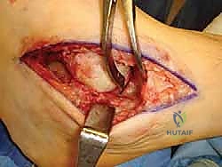
Accessing the calcaneocuboid joint represents the most technically demanding aspect of the single-incision medial approach. With the talonavicular joint fully mobilized, a large lamina spreader is inserted deep into the talonavicular interval. Opening this spreader vigorously distracts the medial column and provides a direct line of sight across the foot to the calcaneocuboid joint. The anatomical location of the calcaneocuboid joint is confirmed using a long scalpel or elevator, and its position is verified with intraoperative fluoroscopy to ensure accurate targeting.
Using a long elevator, the medial aspect of the calcaneocuboid joint is breached. A long-handled scalpel (e.g., a #15 blade on a #7 handle) is carefully passed across the joint to release the lateral capsule and the bifurcate ligaments (which connect the anterior process of the calcaneus to the cuboid and navicular). Crucially, the surgeon must exercise extreme caution to avoid violating the lateral skin from the inside out. The lateral skin will be placed under immense stretch once the valgus deformity is corrected; an iatrogenic puncture from the scalpel will create a stress riser that can lead to a devastating lateral soft tissue tear. Once the joint is opened, long-angled curettes and osteotomes are utilized to remove the articular cartilage down to subchondral bone, followed by aggressive feathering of the surfaces.
Reduction of the Deformity and Internal Fixation
The sequence of joint reduction and fixation is largely dictated by surgeon preference, though many advocate for securing the talonavicular joint first to establish the medial column, followed by the subtalar joint. Regardless of the sequence, the primary goal is to restore a plantigrade, biomechanically sound foot. The subtalar joint must be reduced out of its severe valgus position. The calcaneus is manually translated medially beneath the talus to restore the mechanical axis. The surgeon must meticulously verify that the hindfoot is positioned in 5 to 7 degrees of physiologic valgus. A hindfoot fused in varus is poorly tolerated, leading to rigid lateral column overload and intractable pain.
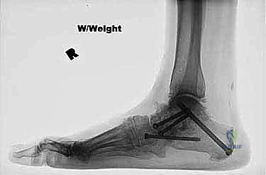
Once the subtalar joint is perfectly positioned, it is temporarily pinned with heavy Kirschner wires. Fixation is typically achieved using one or two large-diameter (6.5-mm or 7.0-mm) partially threaded cannulated screws directed from the non-weight-bearing portion of the posterior heel into the dense bone of the talar body.
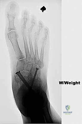
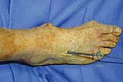
Attention is then turned to the transverse tarsal joints. The forefoot is supinated and plantarly flexed to reduce the abducted and dorsiflexed talonavicular joint, effectively covering the talar head. The talonavicular joint is rigidly fixed using multiple 4.0-mm or 4.5-mm cannulated screws, or alternatively, a medial column locking plate if bone quality is poor or structural grafting was required. Finally, the calcaneocuboid joint, which frequently compresses naturally once the medial column is reduced, is fixed percutaneously. A stab incision is made laterally, and a fully threaded or partially threaded screw is passed from the anterior process of the calcaneus into the cuboid under fluoroscopic guidance.
Complications, Incidence Rates, and Salvage Management
Despite the protective nature of the medial approach regarding the lateral soft tissue envelope, triple arthrodesis remains a major reconstructive procedure with a distinct complication profile. The most notorious complication is aseptic nonunion, with the talonavicular joint historically demonstrating the highest nonunion rates due to its complex spherical anatomy and watershed vascularity.
Malunion is another severe complication. Overcorrection into hindfoot varus locks the transverse tarsal joints, preventing the foot from accommodating uneven terrain and leading to lateral border ulcerations and fifth metatarsal stress fractures. Undercorrection leaves the patient with residual valgus, which may continue to place stress on the medial deltoid ligament, eventually leading to ankle joint deterioration.
| Complication | Estimated Incidence | Pathophysiology & Risk Factors | Salvage & Management Strategy |
|---|---|---|---|
| Talonavicular Nonunion | 5% - 15% | Poor vascularity of talar head; Inadequate cartilage debridement; Insufficient compression. | Revision arthrodesis with structural autograft (iliac crest) and rigid plate fixation; Bone marrow aspirate concentrate (BMAC). |
| Subtalar Nonunion | 2% - 8% | Smoking; Poor bone quality; Thermal necrosis during drilling. | Revision with large-diameter screws; Posterior approach for direct access and grafting. |
| Hindfoot Varus Malunion | 1% - 5% | Overzealous medial translation of calcaneus; Failure to recognize intraoperative alignment. | Valgus-producing calcaneal osteotomy; Revision arthrodesis if symptomatic and rigid. |
| Medial Wound Dehiscence | 3% - 7% | Excessive retraction on medial skin; Hematoma formation; Diabetic microangiopathy. | Aggressive local wound care; Negative pressure wound therapy (NPWT); Rarely requires flap coverage. |
| Adjacent Segment Disease | 20% - 40% (at 10+ years) | Loss of hindfoot shock absorption transfers stress to the ankle mortise and midfoot TMT joints. | Ankle arthrodesis or Total Ankle Arthroplasty (TAA); Midfoot corrective fusions. |
Phased Post-Operative Rehabilitation Protocols
The post-operative rehabilitation following a single-incision medial triple arthrodesis requires strict adherence to biological healing timelines. Because three major joints have been decorticated and stabilized, premature weight-bearing can lead to catastrophic hardware failure and nonunion.
Phase 1: Strict Non-Weight-Bearing (Weeks 0 to 6)
Immediately post-operatively, the patient is placed in a bulky, well-padded posterior splint to accommodate expected edema. The leg must remain strictly elevated above the level of the heart for the first 14 days to minimize swelling and protect the medial incision. At the two-week mark, sutures are removed, and clinical wound healing is assessed. The patient is then transitioned into a rigid, short-leg fiberglass cast. Strict non-weight-bearing (NWB) status is maintained using crutches, a walker, or a knee scooter.
Phase 2: Transition and Early Loading (Weeks 6 to 10)
At 6 weeks post-op, standard AP, lateral, and oblique radiographs are obtained to assess for early trabecular bridging across the arthrodesis sites. If radiographic progression is satisfactory, the cast is removed, and the patient is transitioned into a removable Controlled Ankle Motion (CAM) boot. The patient may begin progressive partial weight-bearing, starting at 25% of body weight and increasing by 25% each week. Active and passive range of motion of the ankle (tibiotalar joint) and toes is initiated to prevent capsular contracture, though hindfoot motion is obviously eliminated.
Phase 3: Full Weight-Bearing and Weaning (Weeks 10 to 14)
By 10 to 12 weeks, definitive radiographic consolidation is typically evident. The patient is permitted to bear full weight in the CAM boot. Physical therapy focuses on proprioceptive training, gait mechanics, and strengthening of the proximal kinetic chain (quad
