Ulnar Dimelia (Mirror Hand): Pathoanatomy, Biomechanics, and Surgical Reconstruction
Key Takeaway
Ulnar dimelia, or "mirror hand," is an exceedingly rare congenital anomaly characterized by a duplication of the ulnar half of the upper extremity. Patients typically present with an absent radius, duplicated ulna, and a symmetrical hand featuring six to eight digits with an absent thumb. Surgical reconstruction focuses on excising supernumerary preaxial digits, performing pollicization to restore pinch and grasp, and addressing associated elbow and wrist contractures.
Comprehensive Introduction and Patho-Epidemiology
Ulnar dimelia, classically referred to in the historical literature as "mirror hand," represents an exceedingly rare and profoundly complex congenital anomaly of the upper extremity. Unlike simple duplications or isolated hypoplasias, ulnar dimelia is characterized by the complete duplication of the ulnar half of the forearm, wrist, and hand, accompanied by a total absence of the radial components. Because there is a complete substitution of the radial structures with duplicated ulnar structures, this anomaly defies simple classification as a pure duplication and is instead considered a profound embryological patterning error. The condition presents with radial and ulnar clusters of fingers in the same hand that are near-mirror images of each other, typically resulting in a hand with seven or eight digits and an entirely absent thumb.
The exact etiology of ulnar dimelia remains largely unknown, and its occurrence is almost exclusively sporadic, with fewer than one hundred definitive cases reported in the global literature. However, advances in molecular embryology have elucidated the underlying mechanisms responsible for this striking phenotype. The deformity is believed to result from a severe aberration in the control process of the limb bud, specifically involving the Zone of Polarizing Activity (ZPA) and the Apical Ectodermal Ridge (AER). The ZPA, located at the posterior margin of the developing limb bud, secretes the Sonic Hedgehog (SHH) morphogen, which dictates the radioulnar (anterior-posterior) axis. An ectopic expression of SHH at the anterior margin of the limb bud creates a second ZPA, leading to a duplication of the posterior (ulnar) structures and a failure of anterior (radial) structure formation.
While typically sporadic, ulnar dimelia can occasionally present as part of a broader syndromic complex. When ulnar dimelia is associated with fibular dimelia (duplication of the fibula with an absent tibia) and other lower extremity anomalies, it may be explained as a single gene mutation transmitted as an autosomal dominant trait, most notably Laurin-Sandrow syndrome. Furthermore, ulnar dimelia is frequently associated with some degree of proximal upper extremity hypoplasia, including deficiencies of the proximal humerus, arm musculature, and scapular dyskinesia. Understanding these associations is critical for the orthopedic surgeon, as the evaluation of a patient with a mirror hand must prompt a comprehensive skeletal survey to rule out concomitant axial or appendicular anomalies.
The largest historical series reported in the literature is that of Harrison, Pearson, and Roaf, who meticulously described the deformity and its functional implications. Their early work, alongside the pioneering surgical principles established by Buck-Gramcko and Entin, laid the foundation for our modern reconstructive approach. Today, the management of ulnar dimelia requires a sophisticated understanding of both the unique pathoanatomy and the developmental needs of the pediatric patient, aiming to transform a non-functional, multi-digited appendage into a functional, socially acceptable hand.
Detailed Surgical Anatomy and Biomechanics
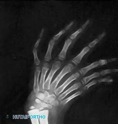
The clinical appearance of ulnar dimelia is visually striking and anatomically complex, dictating a highly customized approach to surgical reconstruction. The hand typically features six to eight well-formed fingers that dangle from a broadened, normal-appearing palm. The digits may all lie in nearly the same plane, or there may be slight opposition between the two halves of the hand. Crucially, the postaxial (true ulnar) digits generally appear slightly more normal in structure and function than the preaxial (duplicated ulnar/ectopic) digits. A true thumb is universally absent, which severely compromises pinch and grasp biomechanics, rendering the hand functionally equivalent to a primitive flipper. Complex or simple syndactyly is frequently present between the duplicated digits, and the digits are often held in a flexed posture due to deficient or absent extrinsic extensor tendons.
At the level of the wrist and forearm, the pathoanatomy becomes even more restrictive. The hand is usually severely radially deviated at the wrist, and active extension of the wrist may be impossible due to the absence of the radial wrist extensors (extensor carpi radialis longus and brevis). Imaging reveals a widened carpus, often with duplicated triquetrum, hamate, and pisiform bones, while the scaphoid and trapezium are congenitally absent. In the forearm, the radius is entirely missing, replaced by a preaxial (ectopic) ulna. Both the wrist and elbow appear abnormally thick and broad. Elbow motion is profoundly decreased due to the presence of two olecranons articulating with the distal humerus, often in a restricted, tethered, or completely fused manner. Forearm rotation (pronation and supination) is entirely absent because the two ulnas are rigidly tethered together by interosseous fibrous tissue or synostosis, and the normal radioulnar joints do not exist.
The vascular anatomy in ulnar dimelia is highly anomalous and represents the most critical surgical hazard. Surgeons must operate under the assumption that the vascular tree features a duplicated ulnar artery and a completely absent radial artery. The palmar arches are often incomplete or absent, relying on a delicate network of collateral vessels to perfuse the digits. Indiscriminate excision of preaxial digits without prior identification of the dominant vascular pedicle can inadvertently devascularize the remaining hand, leading to catastrophic ischemic necrosis. Similarly, the neural anatomy features duplicated ulnar nerves, with absent or highly rudimentary median and radial nerves, complicating sensory mapping and motor tendon transfers.
Biomechanically, the ulnar dimelia limb is severely disadvantaged. The absence of a thumb eliminates oppositional pinch, forcing the child to rely on inefficient scissoring maneuvers between adjacent digits for prehension. The wrist deformity, driven by the lack of radial skeletal support and unbalanced ulnar forces, creates a mechanically unstable carpus that further weakens grip strength. At the elbow, the mechanical block created by the duplicated olecranon prevents functional flexion, making it impossible for the child to bring the hand to the mouth for feeding or self-care. Therefore, surgical reconstruction must address the entire kinetic chain, from unlocking the elbow to stabilizing the wrist and creating a functional thumb ray.
Exhaustive Indications and Contraindications
The primary goal of surgical intervention in ulnar dimelia is to establish a functional upper extremity capable of independent activities of daily living. Because the natural history of the untreated mirror hand is one of profound functional impairment and significant psychosocial distress, operative reconstruction is almost universally indicated. Surgery is typically staged and initiated when the child is between 12 and 18 months of age. At this age, the anatomical structures are large enough to permit meticulous microsurgical dissection, yet the child's brain retains sufficient neuroplasticity to undergo cortical remapping, allowing them to integrate the newly pollicized digit as a functional thumb.
Indications for surgery are driven by both functional deficits and anatomical deformities. The absolute indication is the absence of oppositional pinch and grasp due to the missing thumb. Furthermore, the presence of supernumerary, non-functional digits creates mechanical interference and aesthetic concern, warranting excision to narrow the palm. At the elbow, severe restriction of flexion and extension due to the duplicated olecranon is a strong indication for early surgical release, as elbow mobility is a prerequisite for hand-to-mouth function. Wrist instability and severe radial deviation also necessitate surgical centralization to provide a stable platform for the reconstructed hand.
Contraindications to complex reconstruction are rare but must be carefully considered. Absolute contraindications include severe, life-threatening medical comorbidities or syndromic anomalies (e.g., severe congenital heart defects) that preclude prolonged general anesthesia. A relative contraindication is a completely intertwined or non-separable neurovascular network identified on preoperative angiography, where any attempt at digit excision would result in total hand ischemia; however, such cases are exceptionally rare. Delayed presentation in an older child or adult is also a relative contraindication for primary pollicization, as the lack of early cortical integration may result in a surgically successful but functionally neglected thumb, though aesthetic debulking may still be offered.
| Category | Specific Criteria | Clinical Rationale and Surgical Impact |
|---|---|---|
| Absolute Indications | Absent thumb / Lack of prehension | Requires pollicization of the most functional preaxial digit to restore pinch and grasp. |
| Absolute Indications | Severe elbow stiffness | Excision of the preaxial olecranon is required to unlock the joint and allow hand-to-mouth function. |
| Relative Indications | Supernumerary digits (Polydactyly) | Excision reduces mechanical interference, narrows the hand, and improves aesthetic appearance. |
| Relative Indications | Wrist radial deviation | Centralization over the dominant ulna provides a stable biomechanical platform for grip strength. |
| Absolute Contraindications | Severe cardiopulmonary instability | Prolonged, staged reconstructive surgeries carry unacceptable anesthetic risks in these patients. |
| Relative Contraindications | Inseparable neurovascular arch | High risk of catastrophic ischemic necrosis if supernumerary digits are excised without a clear pedicle. |
| Relative Contraindications | Late presentation (>5-7 years old) | Diminished neuroplasticity reduces the likelihood of functional cortical integration of a new thumb. |
Pre-Operative Planning, Templating, and Patient Positioning
Thorough preoperative planning is the cornerstone of successful ulnar dimelia reconstruction. The evaluation begins in the clinic with a meticulous physical examination to assess the active mobility, passive joint suppleness, and sensory responsiveness of each digit. The surgeon must identify the most functional preaxial digit to serve as the candidate for pollicization. This digit must possess adequate length, functional proximal and distal interphalangeal joints, and ideally, some degree of independent flexor and extensor tendon function. The digits slated for amputation are also evaluated, as their intrinsic muscles and skin flaps will be harvested to reconstruct the thenar eminence and the first web space.
Radiographic evaluation is mandatory and must include standard orthogonal radiographs of the entire upper extremity. Imaging will confirm the absence of the radius, the presence of two ulnas, and the specific carpal duplications. However, plain films are insufficient for surgical templating. A 3D computed tomography (CT) scan is highly recommended to delineate the complex bony anatomy of the elbow and carpus, allowing the surgeon to precisely plan the osteotomies required for olecranon excision and wrist centralization. The CT scan helps differentiate the dominant postaxial ulna from the ectopic preaxial ulna, ensuring that the critical triceps insertion is preserved during the elbow reconstruction.
Advanced vascular imaging is an absolute prerequisite. Never proceed to surgical reconstruction of a mirror hand without Magnetic Resonance Angiography (MRA) or conventional catheter angiography. The vascular tree is highly anomalous, and the surgeon must map the duplicated ulnar arteries, identify the presence or absence of the palmar arches, and trace the digital vessels to both the pollicization candidate and the digits planned for excision. This vascular roadmap dictates the surgical incisions and prevents inadvertent devascularization.
In the operating room, the patient is positioned supine with the affected upper extremity extended on a radiolucent hand table. A well-padded pneumatic tourniquet is applied high on the brachium to ensure a bloodless surgical field, which is critical for the meticulous neurovascular dissection required. The use of magnifying loupes (typically 3.5x to 4.5x) or an operating microscope is mandatory. A sterile marking pen is used to draw the planned racquet incisions for pollicization and the elliptical incisions for digit excision, referencing the preoperative vascular map. Intraoperative fluoroscopy must be readily available to confirm osteotomy levels and verify the alignment of the newly constructed carpometacarpal joint.
Step-by-Step Surgical Approach and Fixation Technique
Surgical reconstruction of ulnar dimelia is a formidable undertaking, typically divided into staged procedures to minimize tourniquet time and optimize functional recovery. Stage 1 focuses on hand reconstruction (pollicization and digit excision), while Stage 2 addresses the proximal mechanical blocks at the elbow and wrist.
Stage 1: Pollicization and Digit Excision
The most critical step in hand reconstruction is executing the Buck-Gramcko principles of pollicization on the selected preaxial digit. Usually, the second or third digit from the preaxial border is selected based on preoperative clinical assessment.
- Incision and Exposure: A racquet-shaped incision is designed around the base of the digit chosen for pollicization. Elliptical incisions are planned for the excision of the remaining supernumerary preaxial digits. The skin flaps are designed to maximize tissue preservation for the creation of a wide, deep first web space.
- Neurovascular Dissection: Meticulous dissection begins palmarly. The common digital nerves and arteries are identified and traced proximally into the palm. The anomalous vascular arch identified on preoperative MRA must be visually confirmed. The vessels to the digits slated for amputation are carefully ligated, strictly preserving the dominant neurovascular pedicle to the pollicized digit. The digital nerves must be separated longitudinally to prevent tethering during rotation.
- Digit Amputation and Intrinsic Harvest: The supernumerary digits are amputated at the carpometacarpal level. The metacarpals of these digits are excised to narrow the palm. Crucially, the intrinsic muscles (interossei and lumbricals) of the amputated digits are carefully dissected, detached distally, and preserved for later transfer to the new thumb.
- Skeletal Shortening and Rotation (Buck-Gramcko Principle): The metacarpal of the pollicized digit is exposed subperiosteally. A transverse osteotomy is performed to shorten the metacarpal. The metacarpal head is preserved and rotated to act as the new trapezium, while the original metacarpophalangeal (MCP) joint becomes the new carpometacarpal (CMC) joint of the thumb. The digit is rotated 160 degrees into pronation and palmar abducted to oppose the remaining postaxial fingers. It is temporarily stabilized with a smooth Kirschner wire.
- Tendon Transfers: The preserved intrinsic muscles from the amputated digits are transferred to the lateral bands and proximal phalanx of the new thumb to provide thenar function (abduction and opposition). The extensor digitorum communis of the pollicized digit is shortened, routed appropriately, and reattached to function as the extensor pollicis longus.
- Closure: The tourniquet is deflated to confirm robust perfusion to the pollicized digit. Hemostasis is achieved, and the skin flaps are transposed to create a wide first web space, avoiding linear scars that could lead to contractures.
Stage 2: Elbow and Wrist Reconstruction
Elbow stiffness is a major functional limiting factor. The presence of a duplicated, preaxial ulna creates a mechanical block to flexion and extension, which must be addressed to allow the hand to reach the mouth.
- Preaxial Olecranon Excision: Through a lateral approach to the elbow, the proximal portion of the preaxial (ectopic) ulna, including its olecranon, is exposed subperiosteally. An oscillating saw is used to excise the ectopic olecranon. Utmost care must be taken to identify and preserve the triceps insertion on the dominant, postaxial ulna.
- Capsular Release: Even after bony excision, the elbow may remain stiff due to capsular contracture. An anterior capsulotomy of the elbow is often required to achieve functional flexion. The radial nerve (or its anomalous equivalent) must be protected during this release.
- Wrist Centralization: If severe radial deviation persists despite the narrowing of the hand in Stage 1, a centralization procedure (similar to that used in radial clubhand) is performed. A dorsal approach is utilized to expose the carpus. The carpus is mobilized, and a centralizing notch is created in the distal articular surface of the dominant ulna. The carpus is seated squarely into this notch and stabilized with a stout Kirschner wire driven longitudinally down the ulnar shaft.
Management of Associated Polydactyly
While ulnar dimelia represents a profound duplication of the entire ulnar axis, surgeons frequently encounter isolated duplication anomalies (polydactyly) in pediatric orthopedics, which may present in the contralateral limb of syndromic patients.
Type 1 Duplications (Pedunculated Extra Digits):
Type 1 polydactyly consists of a soft-tissue appendage devoid of skeletal articulation, attached to the hand by a narrow skin pedicle. Suture ligation (tying off the digit with a silk tie to induce necrosis) is strictly not recommended due to documented reports of fatal hemorrhage, severe infection, and painful neuroma formation. Instead, the Evidence-Based Management (Katz and Linder Technique) is employed. In an outpatient setting under topical anesthesia (EMLA cream), digital traction is applied, and a single, decisive swipe of a #15 scalpel is used to excise the digit flush at its base. Direct pressure achieves hemostasis, and the wound is closed with a sterile adhesive strip.
Type 2 Duplications (Articulating Extra Digits):
Type 2 duplications contain skeletal elements and articulate with the hand skeleton. These require formal operating room excision. The extra digit is excised through an elliptical incision. The critical step is avoiding a painful bump caused by a retained segment of the duplicated metacarpal head. The surgeon must perform a longitudinal osteotomy to shave down the flaring of the remaining metacarpal head, ensuring a smooth contour. The collateral ligaments must be meticulously reconstructed to prevent angular deformity of the remaining digit.
Complications, Incidence Rates, and Salvage Management
The surgical reconstruction of ulnar dimelia is fraught with potential complications, stemming from the highly anomalous anatomy and the extensive nature of the soft tissue and bony rearrangements. The most devastating complication is vascular compromise of the pollicized digit or the remaining hand. Due to the unpredictable nature of the palmar arches, inadvertent ligation of a dominant feeding vessel during the excision of supernumerary digits can lead to acute ischemia. Surgeons must maintain a low threshold for utilizing intraoperative Doppler ultrasound and releasing tension on skin flaps if perfusion is sluggish.
Another frequent complication is stiffness of the newly constructed thumb. Despite meticulous tendon transfers, the transferred intrinsic muscles may fail to provide adequate thenar function, or the new CMC joint may become fibrotic. This results in a thumb that is aesthetically acceptable but functionally poor, acting merely as a static post rather than an active oppositional unit. Similarly, at the elbow, heterotopic ossification or recurrent capsular contracture can negate the gains achieved by the preaxial olecranon excision, leading to recurrent stiffness.
Recurrent radial deviation of the wrist is also a significant long-term concern. Because the fundamental lack of radial-sided skeletal support remains, the centralized carpus may gradually drift back into radial deviation as the child grows. This tethering effect is exacerbated by scar tissue formation and the unbalanced pull of the ulnar-sided musculature. Long-term surveillance is mandatory, and secondary corrective osteotomies or soft-tissue re-balancing procedures are frequently required during periods of rapid skeletal growth.
| Complication | Estimated Incidence | Prevention Strategy | Salvage Management |
|---|---|---|---|
| Vascular Compromise / Flap Necrosis | 5 - 10% | Mandatory preoperative MRA; meticulous dissection; avoid tight skin closures. | Immediate removal of sutures/dressings; application of nitroglycerin paste; revision vascular grafting if feasible; amputation if necrotic. |
| Stiff Pollicized Thumb | 20 - 30% | Precise tensioning of tendon transfers; early, aggressive occupational therapy. | Secondary tenolysis; revision tendon transfers (e.g., Huber transfer); CMC joint arthrodesis in functional position. |
| Recurrent Wrist Deviation | 30 - 40% | Robust intramedullary pin fixation during centralization; prolonged nighttime splinting. | Soft-tissue re-balancing; secondary closing wedge osteotomy of the ulna; eventual wrist arthrodesis near skeletal maturity. |
| Recurrent Elbow Stiffness | 15 - 25% | Adequate initial bony resection of the ectopic olecranon; meticulous hemostasis to prevent hematoma. | Aggressive physical therapy; secondary capsular release; excision of heterotopic ossification once mature. |
| Retained Metacarpal Bump (Type 2 Polydactyly) | 10 - 15% | Routine longitudinal osteotomy of the flaring metacarpal head during initial excision. | Secondary surgical exploration, osteophyte/bump excision, and collateral ligament reefing. |
Phased Post-Operative Rehabilitation Protocols
Following complex reconstruction for ulnar dimelia, meticulous postoperative care and specialized rehabilitation are paramount. The surgery merely provides the anatomical potential for function; it is the rigorous, phased rehabilitation protocol that ultimately determines the functional outcome and the child's ability to integrate the reconstructed hand into their activities of daily living.
Phase 1: Strict Immobilization (Weeks 0 to 4-6)
Immediately postoperatively, the upper extremity is immobilized in a bulky, long-arm spica cast or a highly reinforced, well-padded splint. The positioning is critical to protect the neurovascular pedicles and the tendon transfers. The elbow is held at 90 degrees of flexion, the wrist is maintained in a neutral position (or slight ulnar deviation to counteract the tendency for radial drift), and the newly pollicized thumb is rigorously protected in a position of palmar abduction and opposition. The hand is elevated to minimize edema. During this phase, parents are instructed on cast care and the signs of vascular compromise, though the critical window for acute ischemia is typically within the first 48 hours.
Phase 2: Early Mobilization and Splinting (Weeks 4 to 8)
At approximately 4 to 6 weeks postoperatively, the patient returns to the clinic for cast removal. If Kirschner wires were used to stabilize the CMC joint of the new thumb or the centralized wrist, they are removed at this time. Intensive, specialized pediatric hand therapy begins immediately. The therapist fabricates a custom thermoplastic thumb spica splint, which is worn continuously except during therapy sessions and bathing, to maintain the first web space and protect the healing tendon transfers. Therapy focuses on scar massage using silicone gels to prevent tethering, desensitization of the surgical sites, and gentle passive range of motion exercises for the digits, wrist, and elbow.
Phase 3: Cortical Integration and Strengthening (Weeks 8 and Beyond)
As healing progresses, the focus shifts to active motion and cortical integration. Because the child's brain previously recognized the pollicized digit as a finger, intensive play-based therapy is required to encourage the brain to remap and utilize the digit as an oppositional thumb. Therapists employ grasp-and-release games, picking up small objects (cheerios, blocks), and bilateral hand activities to promote active pinch and grasp. The daytime splint is gradually weaned, but a custom thermoplastic resting splint is often maintained for nighttime use to prevent recurrent wrist deviation and web space contracture.
Long-Term Surveillance (Skeletal Maturity)
Patients require continuous follow-up until skeletal maturity. Growth spurts are particularly perilous, as they can unmask tethering from deep scar tissue, leading to recurrent wrist deviation, progressive elbow stiffness, or angular deformities of the digits. Annual or biannual radiographic and clinical assessments are necessary to monitor the alignment of the centralized carpus and the growth plates of the forearm. Secondary soft-tissue releases, tendon lengthenings, or corrective osteotomies should be anticipated and discussed with the family as a normal part of the long-term management of this complex congenital anomaly.
Summary of Landmark Literature and Clinical Guidelines
The surgical management of ulnar dimelia is built upon a foundation of pioneering anatomical studies and refined reconstructive techniques. A thorough understanding of the landmark literature is essential for any orthopedic surgeon undertaking these complex cases.
The foundational description of the deformity and its functional implications was established by Harrison, Pearson, and Roaf, whose early case series provided the first detailed anatomical mapping of the mirror hand. Their work highlighted the critical absence of the radial structures and the mechanical blocks created by the duplicated ulnar elements. Entin (1959) further advanced the field by outlining the broad principles for the reconstruction of congenital abnormalities of the upper extremity, emphasizing the need to prioritize functional prehension over mere aesthetic improvement. His guidelines on determining which digits to sacrifice and which to retain remain relevant today.
The most significant advancement in the treatment of ulnar dimelia, however, came from the work of Dieter Buck-Gramcko (1964). Buck-Gramcko revolutionized the treatment of the absent thumb by codifying the principles of pollicization. His technique of skeletal shortening, 160-degree rotation, and specific intrinsic and extrinsic tendon transfers allowed surgeons to create a functional, opposable thumb from a non-functional preaxial digit. The "Buck-Gramcko principles" remain the gold standard for pollicization in ulnar dimelia, radial clubhand, and thumb hypoplasia.
More recently, literature has focused on the management of associated anomalies and the refinement of minor procedures. The work by Katz and Linder on the management of Type 1 polydactyly is a critical, evidence-based clinical guideline. Their prospective series conclusively demonstrated that sharp excision of pedunculated extra digits in the clinic setting is safe, effective, and vastly superior to the historically taught, yet dangerous, practice of suture ligation. Case reports, such as those by Bhaskaranand et al. (2003), continue to add to our understanding of the phenotypic variants of mirror hand, emphasizing the need for individualized, patient-specific preoperative planning utilizing modern advanced imaging modalities.
📚 Medical
