Operative Management of Thermal Hand Burns
Key Takeaway
Thermal burns of the hand require urgent evaluation to preserve viable tissue, prevent infection, and avoid debilitating contractures. Management dictates a systematic approach, prioritizing life-threatening injuries before assessing burn depth and distal perfusion. Treatment ranges from conservative topical therapies for superficial injuries to early tangential excision, skin grafting, and escharotomy for deep, circumferential burns. Rigorous postoperative rehabilitation is essential for optimal functional recovery.
Comprehensive Introduction and Patho-Epidemiology
The evaluation and operative management of patients sustaining severe thermal burns to the upper extremity must invariably be contextualized within the broader, highly protocolized framework of advanced trauma life support (ATLS). Life-threatening injuries, airway compromise, and extensive total body surface area (TBSA) burns take absolute and immediate precedence over isolated hand burns, regardless of the severity of the extremity trauma. A critical initial step in the primary survey is the rigorous assessment for inhalation injury, which drastically alters the patient's physiological baseline and overall prognosis. The literature demonstrates that mortality from extensive thermal burns associated with inhalation injury approaches 35%, whereas mortality from equivalent burns without inhalation injury remains approximately 2%. Once the patient is hemodynamically stabilized—encompassing appropriate intravenous fluid resuscitation utilizing the Parkland or modified Brooke formulas, securing of large-bore intravenous access, administration of tetanus prophylaxis, targeted empiric antibiotic therapy, and preparation for potential massive transfusion protocols—attention may be directed to the upper extremity.
Epidemiologically, the hand is involved in over 80% of all severe thermal burn injuries, reflecting its primary role in environmental interaction, occupational tasks, and reflex defensive posturing. The mechanisms of injury are broadly categorized into flame, scald, contact, and flash burns, each presenting with distinct pathophysiological profiles. Flame burns, frequently associated with structural fires or combustible accelerants, typically produce deep partial-thickness or full-thickness injuries due to prolonged high-temperature exposure. Scald injuries, commonly seen in pediatric and geriatric populations, vary in depth depending on the viscosity of the liquid, the temperature, and the duration of contact. Contact burns from industrial machinery or heated domestic surfaces often result in sharply demarcated full-thickness localized destruction. Flash burns, resulting from transient explosions, generally cause superficial partial-thickness injuries to exposed skin, though concomitant blast trauma must be ruled out.
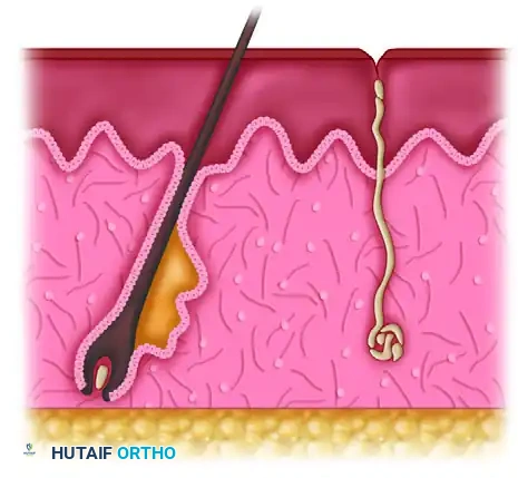
The pathophysiology of thermal injury is best conceptualized through Jackson’s burn wound model, which delineates three distinct concentric zones of tissue damage. The central zone of coagulation represents the area of maximum thermal energy transfer, characterized by irreversible protein denaturation, cellular necrosis, and complete microvascular thrombosis. Surrounding this is the zone of stasis, a critical area characterized by diminished tissue perfusion and intense local inflammation. The cells in this zone are viable but highly vulnerable; without meticulous resuscitation, infection control, and avoidance of secondary mechanical trauma or desiccation, this zone will inexorably progress to irreversible necrosis, effectively deepening the burn wound. The outermost zone of hyperemia exhibits intense vasodilation secondary to the release of inflammatory mediators (e.g., histamine, bradykinin, prostaglandins) but generally recovers spontaneously without permanent structural damage.
The overarching priorities in the operative management of thermal hand burns are multifaceted and demand a highly strategic approach. The surgeon must prioritize the preservation of viable tissue and the maintenance of distal perfusion, particularly in the acute setting of circumferential injuries. Subsequently, the focus shifts to the prevention of invasive wound infection through judicious debridement and early physiological closure. Furthermore, the control of profound fibrotic cascades and the avoidance of debilitating, irreversible contractures dictate both the timing of surgical intervention and the rigorous postoperative rehabilitation protocols. The initial examination of a burned hand is frequently confounded by severe pain, profound obligatory edema, and concomitant polytrauma; however, an accurate estimation of burn depth and a rigorous assessment of distal perfusion remain non-negotiable prerequisites for formulating a definitive surgical plan.
Detailed Surgical Anatomy and Biomechanics
The unique functional demands and specialized anatomical architecture of the hand necessitate a highly tailored, region-specific approach to thermal trauma. The fundamental dichotomy in hand burn management lies in the stark contrast between the dorsal and volar integumentary and fascial systems. The dorsal skin of the hand and digits is exceptionally thin, pliable, and highly mobile, designed to accommodate the extreme arcs of motion required for digital flexion. It lacks a robust subcutaneous adipose layer, leaving the underlying extensor mechanism, venous plexus, and interphalangeal joints highly vulnerable to thermal destruction. Even transient thermal exposure can result in deep partial-thickness or full-thickness injuries that rapidly compromise the central slip of the extensor tendon at the proximal interphalangeal (PIP) joint or the lateral bands.
Conversely, the palmar (volar) skin is heavily keratinized, glabrous, and mechanically resilient, designed to withstand immense frictional and compressive forces. It is securely tethered to the underlying rigid palmar aponeurosis by robust, vertically oriented fascial septa. The digital volar skin is similarly anchored by Cleland’s and Grayson’s ligaments, which prevent skin avulsion during power grip. The flexor tendons, superficial and deep palmar arterial arches, and digital neurovascular bundles are deeply situated beneath this thick fascial barrier, affording them significant protection from all but the most severe, prolonged thermal exposures. Consequently, volar burns often present as superficial or deep partial-thickness injuries that may tolerate a delayed surgical approach, allowing for potential spontaneous epithelialization from the deep epidermal appendages (sweat glands) before committing to excision.
The vascular anatomy of the upper extremity plays a pivotal role in the pathophysiology of acute burn complications. The hand is perfused by the superficial and deep palmar arches, which arise from the ulnar and radial arteries, respectively, and give rise to the common and proper digital arteries. In the setting of full-thickness thermal injuries, the denatured dermis loses all viscoelastic properties, transforming into a rigid, inelastic eschar. As the systemic inflammatory response syndrome (SIRS) drives massive capillary leak and obligatory interstitial burn edema accumulates, the volume of the hand and forearm expands rapidly within this unyielding envelope. This creates a severe tourniquet effect, leading to exponential increases in intracompartmental pressures. If left untreated, the venous outflow is initially occluded, exacerbating the edema, followed inevitably by the cessation of arterial inflow, resulting in acute tissue ischemia, myonecrosis of the intrinsic musculature, and irreversible ischemic neuropathy.
The biomechanics of post-burn contracture follow highly predictable patterns dictated by the relative strength of the intrinsic and extrinsic musculature, the resting posture of the hand, and the vectors of scar contraction. The classic "burn hand deformity" manifests as wrist flexion, metacarpophalangeal (MCP) joint hyperextension, interphalangeal (IP) joint flexion, and severe adduction contracture of the first web space. The MCP hyperextension occurs because the collateral ligaments are lax in extension; as dorsal edema accumulates and dorsal scar tissue contracts, the joint is pulled into extension, where the ligaments subsequently shorten and fibrose. Concurrently, the PIP joints are pulled into flexion, a deformity frequently exacerbated by the thermal destruction of the central slip, leading to a classic, rigid boutonnière deformity. The first web space, highly dependent on the supple dorsal skin for thumb abduction and opposition, rapidly develops a dense adduction contracture, devastating the hand's functional capacity for grasp and pinch.
Exhaustive Indications and Contraindications
Determining the precise depth of a thermal injury is the fundamental determinant of the surgical algorithm. While clinical judgment remains the cornerstone of assessment, it is inherently subjective and frequently inaccurate in the acute phase, particularly for intermediate-depth burns. Advanced modalities, such as noncontact laser Doppler imaging (LDI) and indocyanine green (ICG) angiography, have emerged as highly accurate, objective adjuncts for predicting burn depth and healing potential by quantifying dermal microvascular perfusion. Burns are anatomically classified based on the extent of dermal and epidermal destruction, which directly informs the indications for operative intervention.
Superficial burns (first-degree) involve only the epidermis, presenting as erythematous, exquisitely tender lesions without blister formation. Capillary refill remains brisk, and the underlying dermis is unharmed. These injuries typically heal spontaneously within 7 to 10 days without surgical intervention. Partial-thickness burns (second-degree) extend into the dermis and are subdivided. Superficial partial-thickness burns extend into the papillary dermis, characterized by blister (vesicle) formation, a pink, moist, hypersensitive wound bed, and intact capillary refill. Deep partial-thickness burns extend into the reticular dermis; the wound bed appears mottled, pale, or waxy, capillary refill is sluggish, and sensation is diminished due to the destruction of cutaneous nerve endings. Full-thickness burns (third and fourth-degree) involve the complete destruction of the epidermis and dermis, extending into the subcutaneous fat or deeper structures. The skin assumes a leathery, eschar-like appearance ranging from white to charred black, with a complete absence of capillary refill and sensation.
Clinical presentation of a superficial partial-thickness burn, demonstrating characteristic erythema and early blister formation with intact dermal perfusion.

Deep partial-thickness burn exhibiting a mottled, waxy appearance with sluggish capillary refill, indicating significant destruction of the reticular dermis.

Full-thickness thermal injury demonstrating dense, leathery eschar formation and complete absence of microvascular perfusion, mandating early operative excision.
The indications for operative management are dictated by the depth of the injury, the anatomical location, and the presence of vascular compromise. Immediate surgical intervention (escharotomy/fasciotomy) is absolutely indicated in the presence of circumferential full-thickness burns that compromise distal perfusion or elevate intracompartmental pressures above 30 mm Hg. Early tangential excision and autografting (within 3 to 5 days post-injury) is strongly indicated for all full-thickness burns and deep partial-thickness burns of the dorsal hand. This proactive approach ensures accurate early depth determination, rapid physiological closure, accelerated rehabilitation, and the mitigation of hypertrophic scarring associated with prolonged, secondary-intention healing. Delayed excision (up to 14-21 days) may be indicated for indeterminate depth volar burns to allow for potential spontaneous epithelialization.
Contraindications to early operative intervention in thermal hand burns are primarily systemic. Patients presenting with unresuscitated burn shock, profound hemodynamic instability, severe inhalation injury requiring maximal ventilatory support, or uncorrected severe coagulopathy are not candidates for prolonged excisional procedures. In such catastrophic scenarios, life-saving measures take precedence, and the hand burns must be managed non-operatively with topical antimicrobials (e.g., silver sulfadiazine, mafenide acetate) and biological dressings until the patient's physiological reserve can tolerate surgical stress. Local contraindications include active, uncontrolled invasive wound infection (which must be managed with aggressive debridement and targeted systemic antibiotics prior to grafting) and an inadequate or non-viable wound bed that cannot support a free skin graft, necessitating complex flap reconstruction.
| Category | Indications for Operative Management | Contraindications for Operative Management |
|---|---|---|
| Emergent Interventions | - Circumferential full-thickness eschar with distal ischemia - Intracompartmental pressure > 30 mm Hg - Loss of pulsatile Doppler signals in digital vessels - Impending compartment syndrome (intractable pain) |
- Profound hemodynamic instability (unresuscitated shock) - Active, uncontrolled coagulopathy - Non-survivable polytrauma or massive TBSA burns (comfort care) |
| Early Excision & Grafting (Days 3-5) | - Deep partial-thickness burns of the dorsal hand - All full-thickness thermal injuries - Burns crossing major joints (wrist, MCP, PIP) to prevent contracture - Chemical and high-voltage electrical injuries |
- Superficial partial-thickness burns capable of spontaneous healing - Severe concomitant inhalation injury precluding safe anesthesia - Lack of available donor sites in massive TBSA burns (requires temporization) |
| Delayed Reconstruction / Flaps | - Exposed bone stripped of periosteum - Exposed tendon devoid of paratenon - Exposed joint capsules or neurovascular bundles - Failure of primary split-thickness skin grafts |
- Active, invasive bacterial or fungal wound infection - Patient non-compliance with rigorous postoperative immobilization protocols - Severe peripheral vascular disease precluding microvascular free tissue transfer |
Pre-Operative Planning, Templating, and Patient Positioning
Pre-operative planning for the surgical management of thermal hand burns is a complex, multidisciplinary endeavor that begins immediately upon the patient's admission to the burn center. The timing of surgery is a critical determinant of functional outcome. The modern paradigm strongly favors early tangential excision and grafting (within 3 to 5 days post-injury) for deep burns of the hand. This approach, pioneered by Janzekovic, minimizes the duration of the systemic inflammatory response, reduces the bacterial bioburden of the eschar, and permits early mobilization, which is the sine qua non of excellent functional recovery. Prior to any surgical intervention, the patient must be adequately resuscitated, demonstrating normalized urine output (0.5-1.0 mL/kg/hr), stable hemodynamics, and corrected electrolyte and coagulation profiles.
Donor site selection is a vital component of the pre-operative templating process, particularly in patients with large TBSA burns where unburned skin is at a premium. For the dorsal hand, split-thickness skin grafts (STSG) harvested from the anterior or lateral thigh at a thickness of 0.012 to 0.016 inches are preferred. While meshed grafts (typically 1.5:1 expansion) are frequently utilized in major burns to maximize coverage and allow for the egress of hematoma and exudate, unmeshed (sheet) STSGs are vastly superior for the hand. Sheet grafts provide a significantly better cosmetic match, reduce the incidence of severe secondary contracture, and avoid the unsightly "crocodile skin" appearance associated with meshed interstices. For palmar defects, flexion creases, and the first web space, full-thickness skin grafts (FTSG) harvested from the groin or lower abdomen are indicated, as they undergo minimal secondary contracture and provide durable coverage capable of withstanding the mechanical shear forces of grasp.
The preparation of specialized surgical equipment is mandatory. The surgical tray must include precise excision instruments, such as Weck blades, Goulian knives, and Watson or Zimmer air-driven dermatomes. A sterile pneumatic tourniquet must be available to minimize catastrophic blood loss during the highly vascular tangential excision process, though the surgeon must be prepared to deflate it periodically to accurately assess tissue viability and achieve definitive hemostasis. Bipolar electrocautery is essential, as monopolar cautery causes excessive collateral thermal damage to the delicate underlying structures of the hand, potentially destroying the very paratenon required to support the subsequent skin graft.
Patient positioning must facilitate 360-degree access to the injured extremity and the selected donor sites. The patient is typically positioned supine on the operating table, with the affected extremity extended on a radiolucent hand table to allow for intraoperative fluoroscopy if skeletal stabilization (e.g., K-wire fixation for destroyed joints) is anticipated. The limb is prepped and draped in a standard sterile fashion, ensuring exposure up to the axilla to permit proximal tourniquet application and potential extension of incisions if fasciotomies are required. General anesthesia is usually required, given the intensely painful nature of burn excision and graft harvesting; however, regional anesthesia (e.g., supraclavicular or axillary brachial plexus blocks) may be employed as an adjunct for superior postoperative pain control and sympathectomy-induced vasodilation, provided there is no localized infection at the injection site.
Step-by-Step Surgical Approach and Fixation Technique
The operative sequence in the management of severe thermal hand burns follows a strict hierarchy: vascular decompression, meticulous excision of non-viable tissue, rigorous hemostasis, and definitive physiological closure. If the patient presents acutely with signs of vascular compromise secondary to a circumferential eschar, emergent escharotomies are performed. Digital escharotomies are executed longitudinally along the mid-axial lines of the digits (typically the ulnar border of the index, middle, and ring fingers, and the radial border of the thumb and small finger) to safely avoid the neurovascular bundles. Hand and forearm escharotomies are placed along the radial and ulnar borders. The incision must completely traverse the rigid eschar down to the yielding subcutaneous fat. If intracompartmental pressures remain elevated (>30 mm Hg) despite adequate escharotomy, or if the burn involves deep muscle compartments (e.g., high-voltage electrical injuries), formal fasciotomies of the forearm and the intrinsic compartments of the hand are mandatory to prevent catastrophic ischemic myonecrosis.
The core of the definitive surgical procedure is the tangential excision. Utilizing a Weck blade, Goulian knife, or a guarded Watson dermatome, the surgeon excises the burn eschar in sequential, paper-thin layers (typically 0.010 to 0.020 inches per pass). This precise shaving technique continues until a bed of indisputably viable tissue is reached. The clinical endpoints of adequate excision are characterized by the appearance of punctate, briskly bleeding dermal tissue (if the burn is partial-thickness) or healthy, glistening yellow subcutaneous fat (if the burn is full-thickness). Gray, thrombosed veins or dull, non-bleeding white dermis indicate retained necrotic tissue and mandate further excision. In instances of exceptionally deep burns, a full-thickness excision utilizing a scalpel down to the investing muscle fascia may be necessary to ensure a viable, vascularized graft bed. Extreme caution must be exercised over the dorsal PIP joints to avoid inadvertently excising the ultra-thin central slip of the extensor tendon.
Achieving absolute hemostasis is paramount; subgraft hematoma remains the leading cause of graft failure in burn surgery. Following the completion of the tangential excision, the pneumatic tourniquet is deflated. The immediate hyperemic response will result in profuse bleeding. Initial hemostasis is achieved through the application of warm saline compresses and topical epinephrine (1:100,000) soaks, combined with sustained manual pressure for 5 to 10 minutes. Subsequently, precise, pinpoint bipolar electrocautery is utilized to control any remaining arterial bleeders. The surgeon must avoid mass ligation or excessive cauterization, which creates necrotic foci that can harbor bacteria and impede graft neovascularization.
Once the recipient bed is perfectly hemostatic, the harvested skin graft is applied. For the dorsal hand, a thick split-thickness sheet graft (0.014-0.016 inches) is meticulously tailored to fit the defect. The graft must be applied with physiological tension—neither excessively stretched, which leads to sheer failure, nor excessively lax, which promotes seroma formation. The graft is secured at the margins utilizing surgical staples, fine absorbable sutures (e.g., 5-0 chromic gut), or modern fibrin sealants.
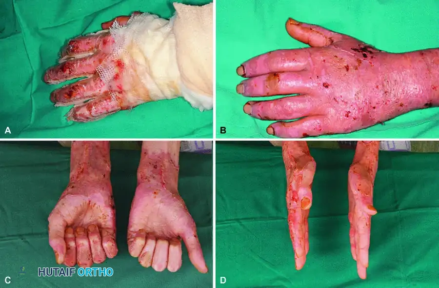
Clinical application of a synthetic dermal substitute (Biobrane) to a superficially burned hand, demonstrating excellent contouring and adherence prior to spontaneous healing or secondary grafting.
In complex injuries where thermal destruction extends beyond the subcutaneous tissue to involve bone, tendon without paratenon, or joint capsules, a free skin graft will inevitably fail due to the lack of a vascularized bed. In these scenarios, the surgical approach shifts to complex reconstruction. Destroyed joints may require temporary stabilization with axial Kirschner wires (K-wires) to maintain length and alignment. Soft tissue coverage must be achieved via vascularized tissue transfer. Depending on the defect size and patient physiology, options include local pedicled flaps (e.g., reverse radial forearm flap), regional flaps (e.g., pedicled groin flap, cross-finger flaps for digital defects), or microvascular free tissue transfer (e.g., anterolateral thigh flap, lateral arm flap). Finally, the advent of negative pressure wound therapy (VAC) has revolutionized graft take in the hand. A VAC dressing applied over the grafted site at -75 to -125 mm Hg continuous pressure effectively splints the graft, evacuates any micro-hematomas or seromas, minimizes shear forces, and profoundly stimulates rapid neoangiogenesis.
Complications, Incidence Rates, and Salvage Management
The operative management of thermal hand burns is fraught with potential complications, ranging from acute graft failure to chronic, debilitating contractures. Anticipation, early recognition, and aggressive salvage management are critical to preserving hand function. Acute graft loss is the most immediate complication, occurring in approximately 5% to 15% of cases. The primary etiologies are subgraft hematoma, invasive bacterial infection (frequently Pseudomonas aeruginosa or Staphylococcus aureus), and mechanical shear forces due to inadequate immobilization. If a hematoma is identified within the first 24 to 48 hours, salvage may be achieved by carefully incising the graft, evacuating the clot, and reapplying localized pressure or a VAC dressing. If the graft is lost to infection, the necrotic graft must be aggressively debrided, the wound bed treated with topical antimicrobials (e.g., mafenide acetate, which penetrates eschar effectively), and the patient returned to the operating room for re-grafting once quantitative tissue cultures confirm a bacterial load of less than $10^5$ organisms per gram of tissue.
Hypertrophic scarring and severe joint contractures represent the most profound long-term complications, affecting up to 40% of patients with deep dermal or full-thickness hand burns. The dorsal hand is particularly susceptible to extension contractures of the MCP joints and flexion contractures of the PIP joints (boutonnière deformity). The first web space is notoriously prone to severe adduction contractures, obliterating the functional span of the hand. Salvage management for established contractures requires complex secondary reconstructive procedures, typically delayed until the scar has fully matured (often 12 to 18 months post-injury). Web space syndactyly and adduction contractures are managed with local tissue rearrangement, such as Z-plasties, V-Y advancement flaps, or four-flap (jumping man) Z-plasties. Severe, diffuse dorsal contractures may require complete excision of the hypertrophic scar tissue, release of the contracted joint capsules (capsulotomy), and resurfacing with thick full-thickness skin grafts or fascial free flaps.
Vascular and neurological complications are also significant. Acute arterial thrombosis or delayed microvascular occlusion can result in digital necrosis, necessitating terminal amputation. Compartment syndrome, if unrecognized or inadequately decompressed via fasciotomy, leads to Volkmann's ischemic contracture—a devastating condition characterized by fibrotic replacement of the intrinsic and extrinsic musculature, rendering the hand a rigid, non-functional appendage. Salvage of established ischemic contractures is exceedingly difficult and often requires extensive muscle slides, tendon transfers, or free functioning muscle transfers (e.g., gracilis free flap) to restore rudimentary grasp.
| Complication | Estimated Incidence | Etiology / Risk Factors | Salvage Management & Prevention |
|---|---|---|---|
| Acute Graft Failure | 5% - 15% | Subgraft hematoma, shear forces, invasive infection (Pseudomonas, S. aureus), inadequate excision. | Evacuation of early hematoma; aggressive debridement of infected grafts; optimization of wound bed; re-grafting. Prevention via meticulous hemostasis and VAC application. |
| First Web Space Contracture | 20% - 30% | Inadequate splinting, delayed grafting, severe deep partial/full-thickness burns to the web space. | Z-plasty, V-Y advancement flaps, or full-thickness skin grafting after scar maturation. Prevention via rigorous "safe position" splinting. |
| Boutonnière Deformity | 10% - 20% | Direct thermal destruction of the central slip; prolonged PIP flexion posturing; secondary infection. | Splinting PIP in full extension; secondary tendon reconstruction (e.g., Fowler tenotomy) or PIP arthrodesis if joint is destroyed. |
| Hypertrophic Scarring | 30% - 40% | Delayed healing (>21 days), meshed skin grafts, genetic predisposition, lack of compression therapy. | Continuous compression garments (>30 mm Hg), silicone gel sheeting, intralesional corticosteroid injections, surgical excision and FTSG resurfacing. |
| Volkmann’s Ischemic Contracture | < 2% | Unrecognized compartment syndrome, delayed/inadequate escharotomy or fasciotomy. | Early: emergent fasciotomy. Late: intrinsic release, tendon transfers, free functioning muscle transfer. Prevention via rigorous serial compartment pressure monitoring. |
Phased Post-Operative Rehabilitation Protocols
The surgical intervention, regardless of its technical brilliance, is merely the preliminary phase of treatment; rigorous, protracted rehabilitation is the ultimate determinant of final hand function. A technically perfect tangential excision and sheet grafting procedure will inevitably fail, resulting in a rigid, non-functional claw hand, if the patient does not adhere to the demanding postoperative protocols. The rehabilitation team must be multidisciplinary, comprising specialized hand therapists, occupational therapists, burn surgeons, and psychiatric support personnel to address the profound psychological trauma and compliance issues associated with disfiguring burns.
The Early Phase (Days 0-5) focuses entirely on graft protection, edema control, and the prevention of initial contracture vectors. Immediately postoperatively in the operating theater, the hand must be immobilized in a bulky, rigid splint. The hand is strictly placed in the "Intrinsic Plus" (Safe) Position: the wrist is extended 20° to 30° to maintain the functional tenodesis effect; the metacarpophalangeal (MCP) joints are flexed 70° to 90° to maintain the collateral ligaments at their maximal length, thereby preventing devastating extension contractures; the interphalangeal (IP) joints are fully extended to prevent central slip attenuation and boutonnière deformities; and the thumb is widely abducted and opposed to maintain the critical dimensions of the first web space. The extremity must be continuously elevated above the level of the heart to facilitate venous and lymphatic drainage, minimizing obligatory edema that can compromise graft neovascularization.
The Intermediate Phase (Days 5-14) commences once definitive graft take is confirmed during the first major dressing down. The rigid immobilization is discontinued, and the focus shifts aggressively to restoring the gliding planes of the tendons and the articular arcs of motion. Active and active-assisted range of motion (ROM) exercises are initiated under the strict supervision of a hand therapist. If the patient is unable to achieve adequate active motion due to pain or profound weakness, continuous passive motion (CPM) machines may be utilized, particularly for the digits. The hand is still splinted in the intrinsic plus position during periods of rest and sleep to prevent nocturnal contracture development. Hydrotherapy may be employed to gently debride residual exudate and facilitate painless motion in a warm, buoyant environment.
The Late Phase (Weeks to Months) represents the longest and most arduous period of recovery, focusing primarily on scar management and the optimization of functional strength. As the grafted skin and donor sites undergo the remodeling phase of wound healing, they become highly susceptible to hypertrophic scarring and secondary contracture. This is mitigated through the mandatory, continuous use of custom-fitted compression garments, which provide >30 mm Hg of sustained pressure to induce localized ischemia within the scar, accelerating collagen degradation and realigning collagen bundles parallel to the skin surface. Silicone gel sheets are applied directly over the grafts to increase local hydration and further suppress hypertrophic cascades. Aggressive soft tissue mobilization and friction massage are performed daily. If specific contractures begin to develop despite these measures, dynamic splinting (utilizing outriggers and elastic tension bands) is employed to provide a low-load, prolonged stretch to the fibrotic tissues, gradually restoring anatomical alignment and functional capacity.
Summary of Landmark Literature and Clinical Guidelines
The evolution of operative management for thermal hand burns is deeply rooted in landmark surgical literature that has progressively refined the standard of care. Historically, deep burns were managed expectantly, allowing the eschar to separate spontaneously over weeks, a process fraught with overwhelming sepsis, massive catabolism, and devastating contractures. The paradigm shifted irrevocably with the seminal work of Janzekovic in the 1970s, who introduced the concept of early tangential excision and immediate grafting. Janzekovic demonstrated that sequential shaving of the burn wound to viable tissue within the first 5 days post-injury drastically reduced mortality, curtailed the systemic inflammatory response, and significantly improved long-term functional and cosmetic outcomes compared to delayed grafting.
Further refining the approach to the hand specifically, the prospective randomized trials by Engrav et al. in the 1980s provided definitive evidence supporting the early excision of indeterminate and deep partial-thickness burns of the dorsal hand. Their work established that early operative intervention (within 3 days) yielded superior range of motion, fewer days missed from work, and a lower incidence of hypertrophic scarring compared to conservative management that allowed the wounds to heal by secondary intention over 3 to 4 weeks. This literature forms the
