Orthopaedic Management of Epidermolysis Bullosa and Associated Hand Lesions
Key Takeaway
Epidermolysis bullosa (EB) presents profound challenges in hand surgery, primarily due to recurrent blistering, pseudosyndactyly, and flexion contractures. Surgical intervention focuses on repetitive degloving and contracture release to maintain functional prehension. Due to the high risk of recurrence and chronic infection, operative management must be meticulously timed, often delayed until subperiosteal bone maturation, and followed by rigorous, prolonged postoperative splinting to maximize functional longevity.
Comprehensive Introduction and Patho-Epidemiology
Epidermolysis bullosa (EB) encompasses a heterogeneous group of rare, inherited mechanobullous disorders characterized by profound cutaneous and mucosal fragility. For the orthopaedic hand surgeon, the severe dystrophic form of the disease—Recessive Dystrophic Epidermolysis Bullosa (RDEB)—represents one of the most formidable reconstructive challenges in the entirety of upper extremity surgery. Occurring in approximately one of every 300,000 live births, RDEB is driven by homozygous or compound heterozygous mutations in the COL7A1 gene. This genetic aberration leads to a quantitative or qualitative defect in type VII collagen, which is the indispensable primary structural component of the anchoring fibrils that secure the epidermis to the underlying dermal matrix at the dermoepidermal junction. Without these anchoring fibrils, the sublamina densa cleavage plane is inherently unstable, and even trivial shearing forces precipitate catastrophic tissue separation and blister formation.
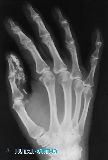
The clinical trajectory of RDEB in the upper extremity is devastatingly predictable and relentlessly progressive. At birth or shortly thereafter, minor mechanical trauma—such as the friction of clothing or the normal grasping reflex of an infant—induces the formation of widespread, tense bullae over the hands and forearms. As these bullae rupture, they leave raw, denuded dermis that heals via aggressive cicatrization. Over the first few years of life, this repetitive cycle of blistering, chronic ulceration, fibrinous exudation, and scarring leads to the continuing formation of a continuous, cocoon-like epidermal envelope over all the fingers of each hand. This process obliterates the interdigital web spaces and draws the digits into profound flexion contractures, resulting in the characteristic severe pseudosyndactyly colloquially termed the "mitten hand" deformity.
The systemic burden of RDEB cannot be overstated when considering surgical intervention. These patients exist in a state of chronic, severe hypermetabolism driven by continuous wound healing and ubiquitous colonization by virulent pathogens such as Staphylococcus aureus and Pseudomonas aeruginosa. Consequently, patients typically present with severe protein-calorie malnutrition, profound anemia of chronic disease, esophageal strictures requiring repeated dilations or gastrostomy tubes, and a severely compromised immune system. The reported mortality rate during childhood or adolescence is alarmingly high, approaching 25%, most frequently secondary to overwhelming sepsis, severe systemic debilitation, or the aggressive metastasis of early-onset cutaneous squamous cell carcinoma (SCC).
Given this grim pathophysiological landscape, the orthopaedic management of the EB hand is distinctly palliative rather than curative. The surgical philosophy must pivot away from the restoration of normal anatomical relationships and focus entirely on the temporary restoration of functional prehension. A highly cautious, multidisciplinary approach is mandatory, integrating the expertise of dermatologists, specialized anesthesiologists, plastic surgeons, and infectious disease specialists to mitigate the extraordinary perioperative risks associated with this fragile patient population.
Detailed Surgical Anatomy and Biomechanics
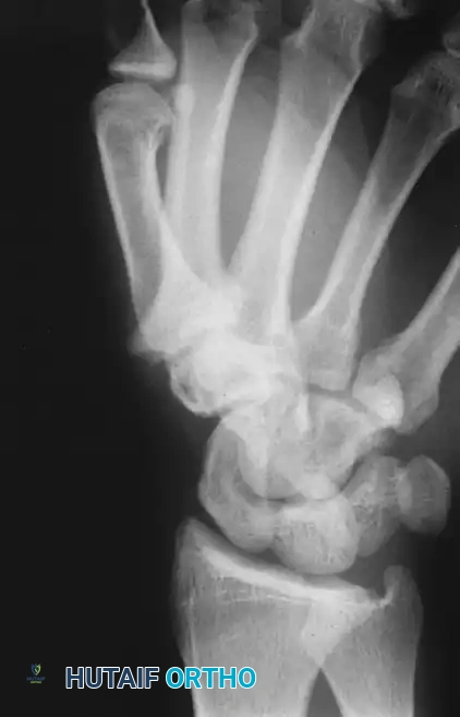
The orthopaedic manifestations of Epidermolysis Bullosa extend far beyond the superficial integument, profoundly altering the osseous, tendinous, and ligamentous structures of the upper extremity. Understanding the altered biomechanics of the EB hand is critical for safe surgical navigation. The pseudosyndactyly seen in RDEB is fundamentally different from congenital syndactyly. In EB, the native web spaces and the underlying fascial architecture of the digits are initially anatomically normal. The webbing is entirely a product of the enveloping epidermal cocoon. However, as the disease progresses, the continuous cicatricial contraction draws the native web spaces distally and pulls the metacarpophalangeal (MCP), proximal interphalangeal (PIP), and distal interphalangeal (DIP) joints into rigid, fixed flexion contractures.
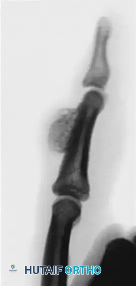
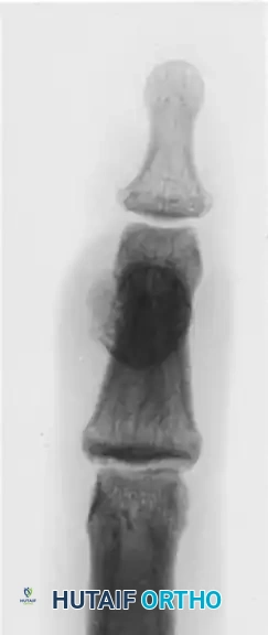
Subperiosteal Hemorrhage and Turret Exostosis
The extreme fragility of the tissues in RDEB extends to the periosteal layer of the phalanges. Minor blunt trauma to the dorsum of the digits frequently causes subperiosteal hemorrhage. In a normal physiological state, such a hematoma might resorb; however, in the highly inflammatory and dysregulated healing environment of the EB patient, this hemorrhage frequently undergoes heterotopic ossification. Over a period of months, this ossification matures into a firm, dome-shaped osseous prominence on the dorsal aspect of the phalanx, clinically referred to as a turret exostosis.
From a biomechanical perspective, the turret exostosis acts as a rigid, unyielding mechanical block to the extensor apparatus. The central slip and lateral bands of the extensor mechanism become densely adherent and tethered to the underlying newly formed bone. This tethering destroys the delicate balance of the digital sweep, completely arresting the proximal excursion of the extensor hood during active extension. Consequently, the interphalangeal joints distal to the lesion are forced into a fixed flexion posture, exacerbating the overall contracture of the digit and rendering conservative splinting entirely ineffective.
Calcinosis Circumscripta and Soft Tissue Contractures
In addition to subperiosteal ossification, the chronic inflammatory milieu and repetitive tissue necrosis in RDEB frequently lead to the development of calcinosis circumscripta. These are dense, localized deposits of insoluble calcium salts within the dermal and subcutaneous tissues. In the hand, these deposits exhibit a strong predilection for the volar pads of the fingertips and the thumb. Biomechanically, these calcific nodules act as rigid foreign bodies within the soft tissue envelope, exacerbating pain during attempted grasp, severely limiting the mobility of the terminal phalanges, and serving as a persistent nidus for recurrent ulceration and deep-space infections.
Beneath the cutaneous cocoon, the chronic flexed posture of the digits leads to profound secondary contractures of the articular structures. The volar plates of the PIP and DIP joints undergo severe shortening and fibrosis. The accessory collateral ligaments, which are normally lax in extension, become rigidly contracted, further locking the joints in flexion. Most critically for the operating surgeon, the digital neurovascular bundles are routinely displaced volarly and centrally by the bowstringing effect of the contracted flexor tendons and the shrinking soft tissue envelope. Any forceful attempt to passively extend the digits without meticulous, stepwise release of these contracted structures risks catastrophic traction injury to the digital arteries and nerves.
Exhaustive Indications and Contraindications
The decision to proceed with surgical intervention in the EB patient is fraught with complexity and must be carefully weighed against the formidable perioperative risks. Prophylactic surgery to prevent the progression of pseudosyndactyly is universally condemned, as the surgical trauma itself will inevitably trigger accelerated blistering and recurrent scarring. Therefore, the threshold for surgical intervention must be strictly defined by severe functional impairment.
The primary indication for soft tissue release and degloving is the absolute loss of functional prehension. When the epidermal cocoon progresses to the point where the patient can no longer grasp objects, perform basic activities of daily living, or feed themselves, surgical release is indicated to restore a functional, albeit temporary, pinch and grasp.
Timing is the most critical factor when addressing the osseous manifestations of RDEB, specifically the turret exostosis. Any indicated surgery to resect subperiosteal ossification must be delayed until the heterotopic bone has achieved full radiographic and clinical maturity. Operating prematurely on immature, actively forming heterotopic bone in an EB patient carries an unacceptably high risk of explosive, aggressive recurrence of the exostosis, which often returns larger and more restrictive than the original lesion. Maturation typically requires a minimum of 4 to 6 months following the inciting hemorrhagic event.
| Parameter | Indications | Absolute Contraindications | Relative Contraindications |
|---|---|---|---|
| Soft Tissue Degloving | Complete loss of functional prehension; inability to feed oneself; severe hygiene compromise due to deep web space maceration. | Active, uncontrolled systemic sepsis; profound uncorrected anemia/malnutrition; lack of patient/family commitment to lifelong postoperative splinting. | Mild-to-moderate webbing with preserved tripod pinch; active blistering flare-up in the proposed surgical field. |
| Excision of Turret Exostosis | Rigid mechanical block to extensor excursion; mature, well-corticated heterotopic bone on serial radiographs; severe pain over the prominence. | Immature, poorly corticated subperiosteal bone; highly active inflammatory phase of the exostosis. | Overlying active ulceration or squamous cell carcinoma (requires oncologic management first). |
| Joint Contracture Release | Severe PIP/DIP flexion contractures precluding functional grasp, addressed concurrently with degloving. | Fixed, bony ankylosis of the joint (unless corrective osteotomy is planned); neurovascular bundles at extreme risk of traction rupture. | Long-standing contractures where the flexor tendons have permanently shortened beyond the capacity for excursion. |
Pre-Operative Planning, Templating, and Patient Positioning
Preoperative optimization of the EB patient requires a highly orchestrated, multidisciplinary effort. The fragility of the patient's mucosal and cutaneous surfaces dictates every aspect of the perioperative protocol. Standard orthopaedic preoperative routines must be drastically modified to prevent iatrogenic catastrophe.
Anesthesia management is perhaps the most critical preoperative consideration. Endotracheal intubation is exceedingly hazardous; the friction of the endotracheal tube against the delicate oropharyngeal and tracheal mucosa can induce massive, life-threatening bullae formation, leading to acute airway obstruction upon extubation. Therefore, regional anesthesia—specifically an ultrasound-guided axillary or supraclavicular brachial plexus block—is the gold standard and strongly preferred. If general anesthesia is absolutely unavoidable, specialized techniques utilizing heavily lubricated, undersized airways and avoiding any adhesive securement must be employed by an anesthesiologist highly experienced with mechanobullous disorders.
Skin protection protocols in the operating room must be absolute. The cardinal rule of EB surgery is that no adhesive tape of any kind may come into contact with the patient's skin. Electrocardiogram leads must be modified (e.g., using needle electrodes or securing standard pads with soft silicone wraps). Blood pressure cuffs and the pneumatic tourniquet must be applied over multiple, thick layers of soft cotton cast padding (Webril) to completely eliminate shearing forces during inflation and deflation. Patient transfer to the operating table must be performed using a frictionless slide board, lifting the patient entirely rather than dragging them.
Radiographic evaluation is essential for preoperative templating, though early imaging can be deceptive. Radiographs taken during the acute phase of a blistering episode or immediately after blunt trauma typically reveal no osseous abnormalities. However, serial imaging over subsequent months will confirm the progressive ossification of subperiosteal hemorrhage. Advanced imaging is critical to assess the degree of disuse osteopenia, the presence of joint subluxations, and the exact location and maturity of calcific deposits or carpometacarpal bossing. The surgeon must carefully template the anticipated K-wire trajectories, recognizing that the severe osteopenia will compromise hardware purchase.
Infection control is paramount. Chronic colonization with multidrug-resistant Staphylococcus aureus and Pseudomonas aeruginosa is ubiquitous in the RDEB population. Preoperative swab cultures of any open wounds or chronic ulcerations should be obtained several weeks prior to surgery to guide targeted, culture-specific prophylactic intravenous antibiotic therapy. Standard empirical prophylaxis is often insufficient. Skin preparation in the operating room must be performed with extreme gentleness; aggressive scrubbing is strictly contraindicated. The surgical site should be prepared by gently pouring or lightly dabbing chlorhexidine or povidone-iodine solutions over the extremity.
Step-by-Step Surgical Approach and Fixation Technique
The surgical intervention for the "mitten hand" deformity is fundamentally a repetitious, palliative degloving procedure designed to liberate the digits and temporarily restore prehension. The goal is strictly functional, and the surgeon must resist the temptation to achieve perfect anatomical correction at the expense of tissue viability.
1. Incision and the Degloving Process
The patient is positioned supine with the operative arm extended on a radiolucent hand table. Following the inflation of the heavily padded tourniquet, a longitudinal incision is made along the fused distal margin of the pseudosyndactyly, effectively splitting the cocoon into dorsal and volar halves. Because this deformity is a pseudosyndactyly—representing a fusion of the epidermal layers rather than a true congenital failure of differentiation—the native, avascular scar planes between the digits can usually be identified.
Using blunt-tipped scissors or a fine mosquito hemostat, the surgeon carefully spreads the tissues to separate the digits. Sharp dissection is minimized to avoid inadvertent injury to the underlying native structures. The dissection must remain strictly superficial to the neurovascular bundles. Due to the severe, long-standing flexion contractures, the digital nerves and arteries are predictably displaced volarly and centrally, often lying immediately deep to the contracted volar skin. Meticulous care must be taken to identify and protect these structures as the epidermal cocoon is reflected proximally to recreate the web spaces.
2. Stepwise Contracture Release
Once the digits are individualized, the rigid PIP and DIP joint flexion contractures must be systematically addressed. Simple release of the volar skin envelope is universally insufficient. The surgeon must perform a stepwise release of the contracted deep structures. This begins with the division of the tight fascial bands and the check-rein ligaments of the volar plate. If the joint remains locked, the proximal attachments of the volar plate are incised, allowing it to slide distally. In severe cases, partial or complete excision of the contracted accessory collateral ligaments may be required to achieve functional extension.
Surgical Warning: The surgeon must exercise extreme caution during the correction of the joint contractures. Do not attempt to force the joints into full, anatomic extension if there is significant elastic resistance. Over-aggressive correction will stretch the chronically contracted neurovascular bundles, leading to acute vasospasm, digital ischemia, and catastrophic necrosis of the digit. A functional, mildly flexed posture is far superior to an ischemic, fully extended digit.
3. Skeletal Stabilization
Because the soft tissues in RDEB possess a relentless, memory-like tendency to contract, the digits must be rigidly stabilized in the maximum safe degree of extension achieved during the release. Longitudinal intramedullary Kirschner wires (K-wires) are the standard of care. Typically, 0.035-inch or 0.045-inch K-wires are driven retrograde through the tip of the distal phalanx, traversing the DIP and PIP joints, and advanced into the metacarpal shaft. Given the profound osteopenia characteristic of these patients, the surgeon must ensure the K-wires are centrally placed within the medullary canal to maximize purchase and prevent iatrogenic fracture or hardware cutout.
4. Soft Tissue Coverage Strategy
Historically, the massive soft tissue defects created by the degloving process were covered with split-thickness or full-thickness free skin grafts. However, extensive clinical experience has demonstrated that skin grafting offers limited long-term advantages and introduces significant donor site morbidity. The harvest sites for these grafts frequently become new epicenters for chronic blistering, intractable ulceration, and severe scarring.
Consequently, the modern paradigm has shifted toward allowing the surgically created web spaces and volar dermal defects to heal by secondary intention. This process is facilitated by the application of advanced biologic dressings, acellular dermal matrices, or cultured epidermal autografts. These modalities provide a protective scaffold that encourages epithelialization without inflicting further donor site trauma.
5. Excision of Turret Exostosis
When addressing a mature subperiosteal ossification that mechanically blocks extensor excursion, a targeted approach is required. A dorsal longitudinal or gently curved incision is made directly over the affected phalanx. The extensor mechanism is frequently found to be densely adherent to the underlying exostosis. A meticulous tenolysis is performed using fine surgical loops to elevate the delicate extensor hood and lateral bands off the bony mass without compromising their structural integrity.
Once the extensor apparatus is mobilized and retracted, the mature exostosis is excised flush with the native cortex of the phalanx. This is best accomplished using a sharp, fine osteotome or a high-speed burr under continuous, copious cold saline irrigation to prevent thermal necrosis of the fragile surrounding bone. To prevent the readherence of the extensor tendon to the raw, bleeding bony bed, a small flap of local adipose tissue or a synthetic anti-adhesion barrier membrane should be interposed between the bone and the tendon. The skin is then meticulously closed with fine, non-absorbable sutures (e.g., 5-0 or 6-0 nylon) using a tension-free technique, as any tension on the epidermal edges will induce immediate blistering.
Complications, Incidence Rates, and Salvage Management
The orthopaedic surgeon must approach the management of Epidermolysis Bullosa with a clear, sobering understanding of the disease's grim prognosis and exceptionally high complication rates. The surgical conversation with the patient and family must heavily emphasize that surgery is a temporary, palliative measure, not a permanent cure.
The most ubiquitous complication is the recurrence of the pseudosyndactyly and flexion contractures. Recurrence is not a possibility; it is an inevitable certainty. The underlying genetic defect ensuring dermal-epidermal instability remains untreated, and the continuous cycle of minor trauma and cicatrization will eventually re-cocoon the hand. Repetitious degloving procedures, often required every 2 to 5 years, will be necessary throughout the patient's life to maintain any semblance of functional prehension.
Infection is a constant threat. The chronic, open wounds and the extensive raw surfaces created during surgical degloving make these severely immunocompromised patients highly susceptible to deep-space hand infections, acute osteomyelitis, and overwhelming systemic sepsis. Aggressive, culture-directed antibiotic therapy and meticulous wound care are the only effective salvage strategies when deep infection occurs.
Perhaps the most devastating complication in the RDEB population is the malignant transformation of chronic wounds. The continuous, rapid cellular turnover and chronic inflammatory state in non-healing EB wounds lead to a remarkably high incidence of aggressive Squamous Cell Carcinoma (SCC), often referred to as Marjolin's ulcer. These tumors are biologically highly aggressive, exhibit early metastasis, and are a leading cause of mortality in young adult EB patients. Any suspicious, hypergranulating, indurated, or rapidly changing lesion within the hand must be biopsied immediately. If SCC is confirmed, wide local excision is rarely sufficient; digital ray amputation or even proximal extremity amputation is frequently required for definitive oncologic control.
| Complication | Estimated Incidence in RDEB | Pathomechanism | Salvage Management & Prevention |
|---|---|---|---|
| Recurrence of Pseudosyndactyly | > 95% within 2-5 years | Genetic lack of anchoring fibrils; continuous blistering and cicatricial healing. | Indefinite night splinting; repetitious palliative degloving procedures when prehension is lost. |
| Squamous Cell Carcinoma (Marjolin's Ulcer) | 70-90% lifetime risk (often presenting in 2nd/3rd decade) | Chronic inflammation, rapid cellular turnover, and secondary p53 mutations in chronic wounds. | High index of suspicion; immediate biopsy of atypical granulation; definitive oncologic amputation. |
| Digital Ischemia / Necrosis | 5-10% post-release | Over-aggressive extension of chronically contracted, volarly displaced neurovascular bundles. | Accept sub-maximal extension during surgery; immediate removal of K-wires and flexion of digit if ischemia is noted in OR. |
| Osteomyelitis / Sepsis | 15-25% | Ubiquitous colonization (MRSA, Pseudomonas) invading deep structures via chronic ulcerations. | Preoperative cultures; targeted IV antibiotics; aggressive surgical debridement of infected bone. |
| Recurrence of Turret Exostosis | High if resected prematurely | Excision of immature, actively forming heterotopic bone triggering an explosive inflammatory response. | Strict adherence to the 4-6 month maturation waiting period prior to any osseous resection. |
Phased Post-Operative Rehabilitation Protocols
The postoperative management of the EB hand is arguably more critical to the long-term success of the intervention than the surgical procedure itself. Without a rigorous, flawlessly executed rehabilitation and splinting protocol, the surgical gains will be lost within weeks.
Immediate Postoperative Care (Weeks 0-4)
In the immediate postoperative period, the primary goals are the protection of the fragile skin, the management of massive exudate from the secondary intention healing sites, and the maintenance of the surgically achieved extension. The wounds are meticulously covered with non-adherent, silicone-based contact layers (e.g., Mepitel) to prevent the dressing from adhering to the raw dermal beds. Petroleum-impregnated gauze can be used as an alternative, though silicone is preferred for its atraumatic removal profile. The hand is then wrapped in bulky, soft, highly absorbent dressings to manage exudate and protect against inadvertent shear forces.
The hand is rigidly immobilized in this bulky dressing, supported by a custom-molded volar resting splint. The longitudinal K-wires remain in place for a minimum of 3 to 4 weeks. This duration is critical; it allows the massive soft tissue defects to begin the process of epithelialization and early scar formation while the digits are held in an extended, abducted posture, resisting the immediate forces of contracture.
Long-Term Rehabilitation and Splinting (Week 4 and Indefinitely)
At the 3 to 4-week mark, the K-wires are carefully removed in the clinic. This marks the transition to the most challenging phase of EB management: the indefinite splinting regimen. The patient must be immediately transitioned to custom-fabricated, exceptionally well-padded thermoplastic splints.
Night splinting is absolutely mandatory and must be continued indefinitely. The splint must hold the digits in extension and maintain the web spaces. Failure of the patient or family to maintain a rigorous, nightly splinting regimen will result in the rapid, aggressive recurrence of the pseudosyndactyly and flexion contractures, often obliterating the surgical result within months.
During the day, specialized silicone web spacers or custom-made, seam-free compressive gloves are utilized to delay the proximal migration of the web spaces while allowing the patient to utilize their restored prehension for activities of daily living. Ongoing, meticulous wound care remains a daily requirement to manage the inevitable chronic ulcerations, manage exudate, and prevent secondary bacterial infections from taking hold in the newly reconstructed web spaces.
Summary of Landmark Literature and Clinical Guidelines
The orthopaedic approach to Epidermolysis Bullosa has evolved significantly over the past several decades, transitioning from aggressive attempts at anatomical reconstruction to a more measured, palliative, and tissue-sparing philosophy. Early literature, heavily influenced by the pioneering work of surgeons like E.C. Wieckesser in the mid-20th century, focused on the complex pathomechanics of the "mitten hand" and initially advocated for extensive full-thickness skin grafting to resurface the degloved digits.
However, long-term outcome studies published in the late 20th and early 21st centuries demonstrated the profound limitations of skin grafting in the RDEB population. Landmark retrospective reviews highlighted that donor site morbidity was unacceptably high, and the grafted skin ultimately succumbed to the same blistering and cicatrization as the native epidermis. This led to the current clinical consensus, strongly supported by multidisciplinary EB centers of excellence, advocating for secondary intention healing augmented by advanced biologic dressings and acellular matrices.
Furthermore, clinical guidelines regarding the management of osseous lesions in EB have been refined. The absolute necessity of waiting for the radiographic and clinical maturation of turret exostoses before attempting excision is now a foundational principle of EB orthopaedic management, significantly reducing the historically high rates of explosive heterotopic bone recurrence. Modern literature also increasingly emphasizes the critical need for vigilant oncologic surveillance, recognizing that the orthopaedic surgeon is often the first to evaluate chronic hand wounds that harbor early, aggressive squamous cell carcinoma. The integration of these historical lessons into current clinical pathways ensures that the modern surgical management of Epidermolysis Bullosa maximizes functional independence while meticulously minimizing iatrogenic harm.
