Comprehensive Management of Rheumatoid Thumb Deformities: Pathomechanics and Surgical Reconstruction
Key Takeaway
Rheumatoid thumb deformities present complex biomechanical challenges requiring precise surgical intervention. The Nalebuff classification categorizes these into four distinct types, with Type I (boutonnière) and Type III (swan-neck) being the most prevalent. Management depends on joint mobility and articular cartilage integrity, ranging from synovectomy and tendon reconstruction to arthrodesis. This guide details the pathomechanics, clinical evaluation, and step-by-step surgical techniques, including interphalangeal joint arthrodesis, to restore functional pinch and grip kinematics.
Comprehensive Introduction and Patho-Epidemiology
Rheumatoid arthritis (RA) is a chronic, systemic autoimmune and inflammatory disease that profoundly and progressively alters the biomechanics of the hand and wrist. Because the thumb is responsible for approximately 40% to 50% of overall hand function—serving as the critical opposing post for key pinch, tip-to-tip pinch, and power grasp—deformities in this single digit result in disproportionate and devastating functional impairment. The pathogenesis of rheumatoid thumb deformities is driven by an aggressive, cytokine-mediated synovial proliferation known as pannus. This hyperplastic synovial tissue invades the joint spaces, distends the delicate joint capsules, attenuates crucial collateral ligaments, and enzymatically destroys articular cartilage and subchondral bone. Ultimately, this structural degradation alters the moment arms of the crossing extrinsic and intrinsic tendons, initiating a cascade of biomechanical failures.
Epidemiologically, the hand and wrist are the most frequently involved anatomical regions in rheumatoid arthritis, with up to 90% of RA patients developing symptomatic hand and wrist manifestations within the first decade of disease onset. The thumb is specifically affected in over 60% to 70% of these individuals. Historically, before the advent of modern disease-modifying antirheumatic drugs (DMARDs) and targeted biologic therapies (such as TNF-alpha inhibitors), surgical intervention for rheumatoid thumb deformities was largely prophylactic, focusing on early synovectomies and tendon transfers to prevent joint destruction. In the contemporary biologic era, the incidence of early, aggressive joint destruction has decreased; however, orthopedic surgeons are now more frequently presented with late-stage, fixed deformities in older patients or those with refractory disease. This shift necessitates a profound understanding of salvage and reconstructive procedures.
To systematically approach these complex biomechanical failures, the classification of rheumatoid thumb deformities proposed by Nalebuff in 1968 remains universally utilized and clinically indispensable. Nalebuff originally described four primary types of rheumatoid thumb deformities based on the initiating joint of synovial disease and the subsequent predictable cascade of tendon imbalances. Later, Types V and VI were added to account for variations involving volar plate laxity and skeletal collapse. Understanding the primary epicenter of the disease is critical, as successful surgical correction must address the primary deforming force to prevent recurrence. Treating the compensatory deformity while ignoring the primary pathology is a fundamental error that inevitably leads to surgical failure.
The systemic nature of the disease also implies that the patient’s overall physiological state must be carefully managed. Rheumatoid patients frequently present with osteopenia or osteoporosis, driven both by the inflammatory process and chronic corticosteroid use, which significantly compromises bone stock for internal fixation. Furthermore, the cervical spine must be evaluated in all RA patients prior to any surgical intervention due to the high risk of atlantoaxial subluxation, which poses a catastrophic risk during intubation and patient positioning. Thus, the management of the rheumatoid thumb is not merely a localized mechanical exercise but a comprehensive orchestration of surgical biomechanics, rheumatologic medical management, and patient-specific functional goals.
Detailed Surgical Anatomy and Biomechanics
The thumb ray is a highly specialized, multi-articulated cantilever system comprising the trapeziometacarpal (CMC), metacarpophalangeal (MCP), and interphalangeal (IP) joints. The CMC joint is a biconcave saddle joint that affords the thumb its unique spatial mobility, allowing for flexion, extension, abduction, adduction, and the composite motion of opposition. Stability at the CMC joint relies heavily on the anterior oblique ligament (the "beak" ligament) and the dorsoradial ligament. In rheumatoid arthritis, synovial hypertrophy at the CMC joint selectively attenuates these primary stabilizers, leading to dorsal and radial subluxation of the first metacarpal base. As the metacarpal escapes its articular confines, the pull of the adductor pollicis muscle unopposedly draws the metacarpal shaft into a fixed adduction contracture, severely narrowing the first web space and obliterating the thumb's functional span.
Distal to the CMC joint, the MCP joint acts primarily as a hinge, though it possesses a crucial degree of lateral mobility and rotation to fine-tune pinch mechanics. The MCP joint is stabilized by the proper and accessory collateral ligaments, the volar plate, and the complex aponeurotic expansion of the extensor mechanism. In a Type I (Boutonnière) deformity, primary synovitis begins at this MCP joint. The proliferating pannus bulges dorsally, stretching the joint capsule and the extensor hood, which attenuates the insertion of the extensor pollicis brevis (EPB) at the base of the proximal phalanx. As dorsal support fails, the extensor pollicis longus (EPL) tendon subluxates ulnarly and volarly, dropping below the axis of rotation of the MCP joint. Consequently, the EPL paradoxically becomes an MCP joint flexor while maintaining its extension force on the IP joint. The intrinsic muscles (abductor pollicis brevis and flexor pollicis brevis) gain a mechanical advantage, further driving the proximal phalanx into fixed flexion, while the IP joint is driven into hyperextension.
The Type III (Swan-Neck) deformity represents a completely different biomechanical cascade, originating at the CMC joint. Following the aforementioned CMC subluxation and first metacarpal adduction, the patient struggles to grasp large objects. To compensate for the narrowed first web space, the patient forcefully hyperextends the MCP joint. Over time, this repetitive compensatory stress attenuates the volar plate of the MCP joint. The hyperextended posture of the MCP joint subsequently increases resting tension on the flexor pollicis longus (FPL) tendon. The FPL, now acting across a hyperextended MCP joint, exerts an exaggerated flexion force on the distal phalanx, pulling the IP joint into severe, eventually fixed, flexion. This highlights a critical biomechanical axiom in rheumatoid hand surgery: proximal joint pathology dictates distal joint posture.
The Type IV (Gamekeeper’s) deformity is less common and is characterized by primary inflammatory destruction of the ulnar collateral ligament (UCL) at the MCP joint. Unlike post-traumatic Skier's thumb, this is a gradual, attritional failure driven by targeted synovitis. The proximal phalanx drifts into severe radial deviation and abduction. To maintain pinch, the first metacarpal is forced into compensatory adduction. The adductor aponeurosis may interpose, but more importantly, the altered vector of the EPL and the intrinsic muscles exacerbates the valgus deformity, leading to rapid articular destruction of the MCP joint and profound weakness in key pinch. Understanding these precise vector alterations is the foundation of preoperative planning and surgical reconstruction.
Exhaustive Indications and Contraindications
Surgical intervention in the rheumatoid thumb is indicated when conservative measures—including rigid or dynamic splinting, targeted intra-articular corticosteroid injections, and optimization of DMARD and biologic therapies—fail to control debilitating pain, or when progressive deformity threatens overall hand function. The primary goals of surgery are pain relief, restoration of a stable post for pinch, correction of deformity, and prevention of further articular and tendinous destruction. Early surgical intervention (e.g., synovectomy, tendon realignment) is indicated in the presence of boggy synovitis that is unresponsive to 6 months of optimal medical management, provided the articular cartilage remains radiographically preserved and the deformity is passively correctable. Late surgical intervention (e.g., arthrodesis, arthroplasty) is indicated for fixed deformities, profound joint instability, severe pain associated with advanced articular destruction (Kellgren-Lawrence grade III or IV), or spontaneous tendon ruptures.
Contraindications must be meticulously evaluated, as the rheumatoid patient is inherently medically complex. Absolute contraindications include active local or systemic infection, critically compromised soft tissue envelopes (often exacerbated by chronic systemic corticosteroids and vasculitis), and a medically unstable patient who cannot tolerate regional or general anesthesia. Relative contraindications include profound non-compliance, active flare-ups of systemic rheumatoid disease (which should be medically optimized prior to elective surgery), and severe, unaddressed proximal joint instability. For instance, attempting to reconstruct a thumb while ignoring a severely subluxated, painful rheumatoid wrist will lead to predictable failure, as the patient will be unable to position the hand in space to utilize the reconstructed thumb.
Furthermore, the surgeon must carefully consider the timing of surgery in relation to biologic therapies. Current guidelines suggest withholding certain biologic agents (e.g., TNF inhibitors, IL-6 inhibitors) prior to major orthopedic surgery, timing the procedure at the end of the dosing cycle, and resuming therapy once wound healing is complete and there is no evidence of infection. Failure to manage these immunomodulatory medications appropriately can lead to catastrophic postoperative infections or severe disease flares that compromise rehabilitation.
| Category | Indications for Surgery | Contraindications for Surgery |
|---|---|---|
| Clinical Status | Refractory pain failing 6 months of medical management; progressive functional deficit | Active local or systemic infection; medically unstable patient |
| Deformity Type | Progressive correctable deformity (early); Fixed, debilitating deformity (late) | Severe, unaddressed proximal instability (e.g., wrist subluxation) |
| Soft Tissue | Impending or actual tendon rupture (e.g., EPL rupture at Lister's tubercle) | Poor soft tissue envelope; severe rheumatoid vasculitis |
| Radiographic | Erosive disease threatening bone stock; advanced cartilage destruction | Insufficient bone stock precluding any form of stable fixation |
| Patient Factors | Motivated patient with realistic functional expectations | Non-compliant patient; inability to participate in postoperative rehab |
Pre-Operative Planning, Templating, and Patient Positioning
Preoperative evaluation begins with a meticulous physical examination to differentiate between passively correctable and fixed deformities. This distinction dictates the surgical algorithm: correctable deformities may be amenable to soft-tissue rebalancing and joint-sparing procedures, whereas fixed deformities invariably require arthrodesis or arthroplasty. The examiner must assess the integrity of the EPL tendon, as spontaneous rupture at Lister's tubercle or the MCP joint is a frequent complication that requires concurrent tendon transfer (typically utilizing the extensor indicis proprius). The stability of adjacent joints, particularly the radiocarpal and midcarpal joints, must be documented. A severely deviated or subluxated wrist alters the extrinsic tendon tension on the thumb, and wrist arthrodesis or arthroplasty may need to precede or accompany thumb reconstruction.
Radiographic evaluation requires true posteroanterior (PA), lateral, and specialized views of the thumb ray. The Robert's view (true AP of the CMC joint) is essential for assessing the degree of trapeziometacarpal joint destruction, subluxation, and the presence of osteophytes or erosions. Stress radiographs may be utilized to quantify MCP joint collateral ligament laxity, particularly in Type IV deformities. The surgeon must scrutinize the radiographs for profound osteopenia, cystic erosions, and medullary canal narrowing, all of which influence the choice of internal fixation. Templating is performed to determine the appropriate sizes for headless compression screws, K-wires, or, in rare cases of MCP arthroplasty, silicone elastomer implants. Anticipating the need for bone grafting—often harvested from the distal radius or iliac crest—is a critical component of the preoperative plan, particularly when addressing severe erosive disease or revising a nonunion.
Patient positioning and anesthetic management require specialized attention in the rheumatoid population. The patient is typically positioned supine with the operative arm extended on a radiolucent hand table. A regional anesthetic block (axillary or supraclavicular) is highly preferred, as it provides excellent intraoperative anesthesia, mitigates postoperative pain, and avoids the risks of endotracheal intubation. If general anesthesia is required, preoperative flexion-extension radiographs of the cervical spine are mandatory to rule out atlantoaxial instability (C1-C2 subluxation) or subaxial cervical spine disease, which could lead to spinal cord compression during neck extension for intubation.
A pneumatic upper arm tourniquet is applied over abundant padding and typically elevated to 250 mm Hg after exsanguination of the limb with an Esmarch bandage. Prophylactic intravenous antibiotics (typically a first-generation cephalosporin, or clindamycin/vancomycin for penicillin-allergic patients) are administered prior to tourniquet inflation. The surgical site is meticulously prepped and draped, taking extreme care with the fragile rheumatoid skin, avoiding harsh scrubbing that could cause epidermal avulsion.
Step-by-Step Surgical Approach and Fixation Technique
Management of Type I (Boutonnière) Deformities
The surgical approach to the Type I deformity depends entirely on the stage of the disease. In the early stage, where the deformity is passively correctable and the MCP joint cartilage is preserved, a joint-sparing soft-tissue reconstruction is indicated. The classic Harrison-Ansell procedure involves a dorsal longitudinal or curvilinear incision over the MCP joint. A thorough synovectomy is performed. The attenuated EPB tendon is isolated, shortened, and securely advanced to the base of the proximal phalanx to restore active MCP extension. The subluxated EPL tendon is mobilized, rerouted dorsally over the MCP joint, and centralized. In some variations, the EPL is routed through the dorsal capsule or a slip of the extensor hood to maintain its central position. The intrinsic muscles may require fractional lengthening if a severe flexion contracture is present.
In the late stage, characterized by a fixed deformity and destroyed articular cartilage, MCP joint arthrodesis is the undisputed gold standard. The MCP joint is exposed via a dorsal longitudinal incision. The extensor mechanism is split longitudinally. The collateral ligaments are released, allowing the joint to be "shotgunned" to expose the articular surfaces. The remaining cartilage and subchondral bone are resected using a microsaw or a cup-and-cone reamer system. The cup-and-cone technique is advantageous as it preserves bone length and allows for multi-planar adjustments of the fusion angle. The ideal position for thumb MCP arthrodesis is 10 to 15 degrees of flexion, 5 degrees of abduction, and slight pronation to maximize pulp-to-pulp pinch. Fixation is typically achieved with crossed K-wires, a tension band construct, or modern headless compression screws, depending on bone quality. If the IP joint is fixed in severe hyperextension, a concurrent volar capsular release or IP joint arthrodesis must be performed to restore a functional pinch arc.
Management of Type III (Swan-Neck) Deformities
Surgical correction of the Type III deformity must address the primary pathology at the CMC joint. In the early stage, where the CMC joint is subluxated but the MCP joint hyperextension remains passively correctable, CMC arthroplasty combined with a first web space release is indicated. The CMC joint is approached via a Wagner or dorsal radiocarpal incision. The trapezium is excised (trapeziectomy). To reconstruct the volar beak ligament and provide a biological interposition, a ligament reconstruction and tendon interposition (LRTI) utilizing the flexor carpi radialis (FCR) tendon is commonly performed. Alternatively, hematoma distraction arthroplasty or suspensionplasty using the abductor pollicis longus (APL) may be utilized. Concurrently, the adductor aponeurosis and, if necessary, the adductor pollicis muscle origin must be released to open the first web space.
Crucially, if the MCP joint hyperextends more than 10 to 15 degrees passively, it must be stabilized to prevent recurrence of the swan-neck cascade. This is achieved via volar capsulodesis, EPB transfer to the metacarpal neck, or temporary transarticular K-wire pinning in slight flexion. In the late stage, where both the CMC and MCP joints are severely destroyed and fixed, the surgeon must perform a CMC arthroplasty combined with an MCP joint arthrodesis. A fundamental surgical warning must be strictly adhered to: never fuse both the CMC and MCP joints of the thumb. At least one of these joints must remain mobile to allow for the spatial positioning of the thumb during opposition. Fusing both joints results in a rigid, non-functional pillar.
Interphalangeal and Distal Interphalangeal Joint Arthrodesis
Arthrodesis of the interphalangeal (IP) joint of the thumb is frequently required for fixed, painful rheumatoid deformities, often in conjunction with proximal procedures. The surgical principles are identical to those used for finger DIP joints. Exposure must provide adequate visualization while preserving the delicate dorsal skin flaps, which are particularly fragile in rheumatoid patients on chronic corticosteroids.
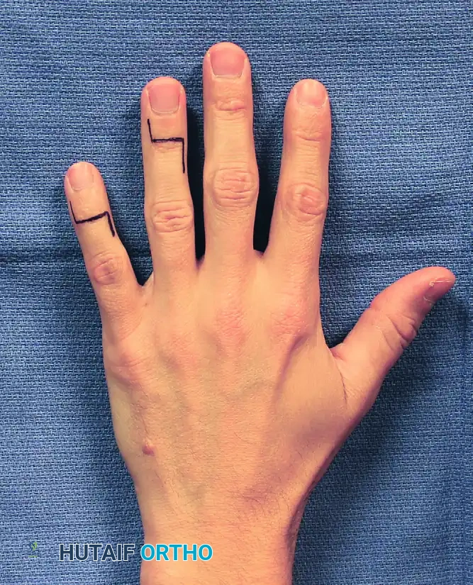
A step-cut (H-shaped), Y-shaped, or dorsal transverse incision is utilized to minimize the risk of longitudinal scar contracture. The dorsal skin flaps are elevated meticulously, preserving the subdermal venous plexus. The extensor tendon is incised transversely or split longitudinally to expose the joint capsule. A thorough synovectomy is performed, and the collateral ligaments are released completely to allow the joint to be hinged open volarly. Using a microsaw or rongeur, the articular cartilage is resected down to bleeding subchondral bone. For the thumb IP joint, the ideal position of fusion is 10 to 15 degrees of flexion.
Rigid internal fixation is paramount. Headless compression screws are currently the gold standard. A guidewire is driven antegrade through the distal phalanx, exiting the tip of the thumb. The joint is reduced in the desired position, and the wire is driven retrograde into the proximal phalanx. The screw is then advanced over the wire, burying the head beneath the distal phalangeal cortex to prevent nail bed irritation. If bone stock is extremely poor, crossed K-wires or a dorsal tension band wire may be utilized. The extensor tendon is repaired, and the skin is closed with interrupted nonabsorbable sutures to prevent wound breakdown.
Complications, Incidence Rates, and Salvage Management
Despite meticulous surgical technique and comprehensive preoperative planning, complications in the surgical management of rheumatoid thumb deformities are not uncommon. The inherent systemic nature of the disease, combined with the immunosuppressive effects of DMARDs and biologic therapies, significantly elevates the risk profile compared to osteoarthritis or post-traumatic cohorts. The surgeon must be prepared to identify and manage these complications aggressively to preserve hand function.
Nonunion is one of the most frequent complications following arthrodesis of the IP or MCP joints, with incidence rates reported between 5% and 12%. The etiology is multifactorial, stemming from profound osteopenia, compromised vascularity, inadequate rigid internal fixation, and the inhibitory effects of systemic medications on osteogenesis. Fortunately, many nonunions in the rheumatoid hand are stable, asymptomatic fibrous unions that do not require further surgical intervention. However, if a nonunion is painful, unstable, or associated with progressive deformity, salvage management is required. This typically involves revision surgery with meticulous debridement of the fibrous tissue, application of structural or cancellous bone graft (frequently harvested from the ipsilateral distal radius or iliac crest), and upgraded, rigid internal fixation, often transitioning from K-wires to a locked plate or a larger compression screw.
Infection is a devastating complication, occurring in 2% to 5% of cases. Superficial surgical site infections can usually be managed with targeted oral antibiotics and local wound care. Deep infections, however, threaten the integrity of the reconstruction and the viability of the digit. The risk is heightened by chronic corticosteroid use, which masks early signs of inflammation, and biologic therapies, which blunt the immune response. Management of a deep infection necessitates immediate return to the operating room for aggressive irrigation and debridement. Any loose or un-integrated hardware must be removed. If an arthrodesis site is infected, temporary stabilization with an external fixator or placement of an antibiotic-impregnated cement spacer is required until the infection is eradicated, followed by definitive revision arthrodesis.
Hardware prominence and soft tissue irritation are common, particularly when K-wires or tension band constructs are utilized beneath the atrophic rheumatoid skin. Screw back-out can lead to painful bursitis or nail bed deformities if the screw head violates the distal tuft. Symptomatic hardware should only be removed after solid, unequivocal radiographic union is confirmed, typically at 4 to 6 months postoperatively. Recurrent deformity is another significant concern, particularly if the primary deforming force was not adequately addressed (e.g., failure to release the adductor contracture during a Type III reconstruction). Salvage of a recurrent deformity often requires conversion of a joint-sparing procedure to a definitive arthrodesis.
| Complication | Estimated Incidence | Etiology in Rheumatoid Arthritis | Salvage Management Strategy |
|---|---|---|---|
| Nonunion (Symptomatic) | 5% - 12% | Osteopenia, inadequate fixation, systemic immunosuppressants | Revision arthrodesis, distal radius autograft, rigid compression fixation |
| Deep Space Infection | 2% - 5% | Biologic therapies, chronic corticosteroids, fragile soft tissue | Hardware removal, aggressive I&D, antibiotic spacers, external fixation |
| Hardware Prominence | 10% - 15% | Atrophic skin, screw back-out, K-wire migration | Hardware removal only after solid radiographic union is confirmed |
| Recurrent Deformity | 5% - 10% | Failure to address primary deforming force (e.g., unreleased web space) | Conversion of soft-tissue reconstruction to definitive arthrodesis |
| Tendon Rupture | 3% - 5% | Attritional wear over hardware or un-resected osteophytes | Tendon transfer (e.g., EIP to EPL) or tendon grafting |
Phased Post-Operative Rehabilitation Protocols
The postoperative rehabilitation protocol for the rheumatoid thumb is a delicate and highly individualized balancing act. The surgeon and hand therapist must work in close collaboration to achieve two inherently conflicting goals: providing rigid, uninterrupted immobilization to ensure successful osseous union at arthrodesis sites, while simultaneously initiating early, controlled mobilization of adjacent non-fused joints to prevent profound stiffness, which develops rapidly in the rheumatoid hand.
Phase I (0 to 2 weeks postoperatively) focuses on strict protection, edema control, and wound healing. Immediately following surgery, a bulky, non-adherent compressive dressing is applied, reinforced with a custom-molded volar or dorsal thermoplastic splint. The thumb is immobilized in a position that protects soft-tissue repairs and supports arthrodesis sites (typically slight palmar abduction for the CMC joint, and the specific fusion angles for the MCP and IP joints). The patient is instructed on strict elevation and active range of motion exercises for the shoulder, elbow, and uninvolved digits to maintain proximal lymphatic drainage and prevent complex regional pain syndrome. Suture removal is typically delayed in rheumatoid patients until 10 to 14 days postoperatively, as their skin heals more slowly and is prone to dehiscence.
Phase II (2 to 6 weeks postoperatively) marks the transition to controlled mobilization. The bulky dressing is removed, and a lighter, custom-fabricated thermoplastic splint is provided. If rigid internal fixation (e.g., headless compression screws) was achieved for an IP or MCP arthrodesis, the splint may be removed several times a day to allow for gentle, active range of motion of the un-fused joints. For example, following an isolated MCP arthrodesis, active IP joint flexion and extension, as well as CMC joint circumduction, are initiated. If K-wires were utilized, or if a CMC arthroplasty with soft-tissue reconstruction was performed, stricter immobilization is maintained for a full 4 to 6 weeks to allow for ligamentous healing. Pin tract care is meticulously performed daily using chlorhexidine or hydrogen peroxide solutions.
Phase III (6 to 12 weeks postoperatively) involves weaning from the orthosis and initiating progressive strengthening. Serial radiographs are obtained at 4, 8, and 12 weeks to assess the progression of osseous union. Once clinical stability and early radiographic bridging trabeculae are observed, the patient is gradually weaned from the splint, initially discontinuing daytime use and eventually nighttime use. Gentle passive stretching and isometric strengthening exercises are introduced. The therapist focuses on restoring functional pinch patterns, re-educating the patient to utilize the reconstructed thumb without placing excessive lateral stress on the joints. It is imperative to counsel the patient that maximal medical improvement may take 6 to 12 months, and that radiographic union frequently lags behind clinical stability by several months.
Summary of Landmark Literature and Clinical Guidelines
The surgical management of the rheumatoid hand is deeply rooted in decades of evolving biomechanical understanding and clinical outcomes research. The foundational cornerstone of this field remains the seminal work of Nalebuff (1968), who first categorized rheumatoid thumb deformities into predictable, sequential pathomechanical cascades. Nalebuff’s classification shifted the surgical paradigm from reactive, ad-hoc interventions to systematic, stage-specific reconstructive algorithms. His observation that proximal joint pathology inevitably dictates distal joint posture remains the guiding principle for all rheumatoid hand surgery today.
Subsequent landmark literature, particularly the extensive outcome studies published by Terrono and Feldon in the 1990s and early 2000s, refined these surgical indications. Their long-term follow-up studies definitively demonstrated that while early synovectomy and tendon rerouting (e.g., the Harrison-Ansell procedure) could delay progression in Type I deformities, MCP joint arthrodesis provided the most reliable, durable, and pain-free long-term outcome for the vast majority of patients with established deformity. Furthermore, their work reinforced the absolute contraindication of fusing both the CMC and MCP joints, highlighting the catastrophic loss of spatial positioning and opposition that results from creating a rigid, two-joint pillar.
In the modern era, the literature has heavily focused on the impact of biologic DMARDs on surgical indications and complication rates. Studies by the American College of Rheumatology (ACR) and the American Association of Hip and Knee Surgeons (AAHKS) have established critical perioperative guidelines for the management of immunomodulatory medications. The current consensus mandates a nuanced approach: while traditional DMARDs like methotrexate can often be continued perioperatively, targeted biologic agents (e.g., adalimumab, etanercept) should typically be withheld prior to surgery to mitigate the risk of deep space infection, balancing this against the risk of inducing a severe systemic disease flare.
Technologically, the shift from K-wire fixation to headless compression screws for small joint arthrodesis has been extensively validated in recent literature. Comparative biomechanical and clinical studies have consistently demonstrated that headless compression screws provide superior biomechanical rigidity, higher union rates, and significantly lower rates of hardware-related complications (such as pin tract infections and skin irritation) compared to traditional K-wire techniques. This technological advancement has allowed for earlier mobilization protocols and improved overall functional outcomes, cementing the contemporary, evidence-based approach to the complex, challenging, yet profoundly rewarding field of rheumatoid thumb reconstruction.
