Operative Management of Boutonnière Deformity: Soft Tissue Reconstruction to Salvage Arthroplasty
Key Takeaway
The surgical correction of boutonnière deformity requires meticulous restoration of the extensor mechanism. For moderate deformities, treatment focuses on central slip reconstruction and lateral band mobilization to restore proximal interphalangeal (PIP) joint extension. Severe, fixed deformities often necessitate PIP joint arthrodesis or resection arthroplasty. This guide details the biomechanical principles, step-by-step surgical techniques, and postoperative rehabilitation protocols essential for optimizing functional outcomes in complex digital deformities.
Comprehensive Introduction and Patho-Epidemiology
The boutonnière, or "buttonhole," deformity represents one of the most challenging and unforgiving biomechanical disruptions encountered in hand surgery. Characterized by a fixed or flexible flexion posture of the proximal interphalangeal (PIP) joint and a concomitant hyperextension of the distal interphalangeal (DIP) joint, the deformity arises from a primary disruption, attenuation, or elongation of the central slip of the extensor mechanism at its insertion on the dorsal base of the middle phalanx. The complexity of this deformity lies not merely in the primary tendinous injury, but in the subsequent cascade of secondary anatomical derangements that occur if the initial insult is left untreated or is inadequately managed. As the central slip fails, the delicate balance between the intrinsic and extrinsic extensor systems is obliterated, leading to a progressive deterioration of digital kinematics that severely compromises grip strength, fine pinch mechanics, and overall hand function.
Epidemiologically, the boutonnière deformity manifests through two primary etiologic pathways: traumatic and atraumatic (predominantly inflammatory). Traumatic boutonnière deformities frequently result from closed injuries, such as sudden forced flexion of an actively extended PIP joint—often seen in athletic endeavors like basketball or volleyball—which causes an avulsion of the central slip from the middle phalanx. Alternatively, dorsal lacerations over the PIP joint, industrial crush injuries, and deep full-thickness burns can directly transect or obliterate the extensor mechanism. In the acute traumatic setting, the deformity may not be immediately apparent; the lateral bands may initially remain in their anatomical position, held by an intact triangular ligament. However, over a period of 10 to 21 days, as the triangular ligament attenuates under the unabated pull of the flexor digitorum superficialis (FDS), the classic deformity slowly unmasks itself.
In the atraumatic or inflammatory cohort, rheumatoid arthritis (RA) is the overwhelming primary driver. The patho-epidemiology in the rheumatoid hand is insidious and driven by chronic synovial proliferation. Hypertrophic synovitis within the PIP joint gradually distends the dorsal capsule, directly stretching and attenuating the central slip and the triangular ligament. Unlike traumatic injuries, which are often isolated to a single digit, rheumatoid boutonnière deformities frequently present in multiple digits simultaneously and are often accompanied by metacarpophalangeal (MCP) joint volar subluxation and ulnar drift, further complicating the biomechanical landscape. The prevalence of boutonnière deformities in the rheumatoid population has historically been reported to be as high as 30% to 40% in patients with long-standing disease, though the advent of biologic disease-modifying antirheumatic drugs (DMARDs) has significantly altered the natural history and reduced the incidence of these severe manifestations.
Regardless of the etiology, the progressive nature of the deformity dictates the clinical presentation and surgical decision-making. The metaphorical "buttonhole" describes the herniation of the proximal phalanx head dorsally through the defect created by the ruptured central slip and the diverging lateral bands. Once this herniation occurs, the deformity transitions from a dynamic imbalance to a static contracture. The transverse retinacular ligaments contract, permanently tethering the lateral bands in their abnormal volar position. Concurrently, the collateral ligaments and volar plate of the PIP joint undergo adaptive shortening due to the persistent flexion posture, while the oblique retinacular ligaments (ORL) contract, exacerbating the DIP joint hyperextension. Recognizing this temporal progression from a flexible, passively correctable deformity to a rigid, fixed contracture with secondary joint arthrosis is the cornerstone of formulating an appropriate surgical strategy.
Detailed Surgical Anatomy and Biomechanics
A profound mastery of the extensor mechanism's surgical anatomy is non-negotiable for any surgeon attempting to reconstruct a boutonnière deformity. The digital extensor mechanism is an intricate, highly coordinated aponeurotic expansion that receives contributions from both the extrinsic system (extensor digitorum communis, extensor indicis proprius, extensor digiti minimi) and the intrinsic system (lumbricals and interossei). At the level of the MCP joint, the extrinsic tendon is stabilized by the sagittal bands. As it proceeds distally over the proximal phalanx, it trifurcates into a single central slip and two lateral slips. The central slip traverses the PIP joint and inserts firmly into the dorsal tubercle at the base of the middle phalanx, serving as the primary active extensor of the PIP joint.
The intrinsic tendons (lumbricals radially, interossei bilaterally) join the lateral slips of the extrinsic tendon to form the conjoined lateral bands. These lateral bands travel along the dorsolateral aspect of the PIP joint. Crucially, their position relative to the PIP joint's axis of rotation is maintained by a delicate retinacular balancing system. Dorsally, the triangular ligament spans the space between the two lateral bands over the middle phalanx, preventing them from subluxating volarly. Volarly, the transverse retinacular ligaments (TRL) originate from the flexor tendon sheath and volar plate, inserting into the lateral edges of the lateral bands, preventing them from subluxating dorsally. Distal to the PIP joint, the lateral bands converge to form the terminal tendon, which inserts on the dorsal base of the distal phalanx to extend the DIP joint.
Biomechanically, normal digital extension requires a precise, synchronous excursion of these interconnected structures. During active PIP extension, the central slip exerts a dorsal pull on the middle phalanx. Simultaneously, the lateral bands shift dorsally, moving above the PIP joint's axis of rotation, thereby contributing to PIP extension while transmitting force to the terminal tendon to extend the DIP joint. This coordinated movement is dependent on the precise resting length and tension of the triangular and transverse retinacular ligaments. The oblique retinacular ligament (Landsmeer's ligament), which originates from the volar proximal phalanx and flexor sheath, courses obliquely to insert on the terminal tendon. It acts as a passive tenodesis; as the PIP joint extends, the ORL tightens, passively extending the DIP joint, and as the PIP joint flexes, the ORL relaxes, allowing DIP flexion.
In the pathobiomechanics of the boutonnière deformity, this elegant system completely collapses. The primary failure of the central slip eliminates the direct extensor force on the middle phalanx. Consequently, the FDS acts unopposed, driving the PIP joint into flexion. As the proximal phalanx head herniates dorsally, the triangular ligament is stretched to the point of mechanical failure. Deprived of their dorsal tether, the lateral bands are pulled volarly by the intact TRLs. Once the lateral bands cross the critical equator—volar to the PIP joint's axis of rotation—their biomechanical function reverses. They cease to be PIP joint extensors and become paradoxical PIP joint flexors, exacerbating the deformity. Furthermore, because the lateral bands have displaced proximally and volarly, their entire excursion and tension are transmitted directly to the terminal tendon. This concentrated force, combined with the adaptive shortening of the ORL, creates the rigid, characteristic hyperextension of the DIP joint. Surgical correction must therefore systematically reverse this pathobiomechanics by re-establishing central slip continuity, releasing contracted volar structures, and relocating the lateral bands to their rightful dorsal apex.
Exhaustive Indications and Contraindications
Surgical intervention for the boutonnière deformity is dictated by a rigorous preoperative assessment of joint flexibility, articular cartilage integrity, soft tissue envelope viability, and patient compliance. The decision to proceed with operative management must be weighed against the reality that the extensor mechanism is notoriously prone to postoperative adhesions and stiffness. A fundamental tenet of hand surgery is that a stiff, straight finger is often more functionally debilitating than a flexible finger with a moderate boutonnière posture. Therefore, the indications for surgery are stratified based on the chronicity and rigidity of the deformity, typically categorized into mild (flexible), moderate (partially fixed), and severe (fixed with arthrosis) stages.
Conservative management remains the gold standard for acute, closed injuries and mild deformities where the PIP joint is fully, passively correctable to neutral. Continuous splinting of the PIP joint in absolute extension for 6 to 8 weeks, while allowing active DIP joint motion, is highly effective in these scenarios. Operative intervention for soft tissue reconstruction is indicated when conservative measures fail, or when the patient presents with a moderate deformity characterized by a partially fixed PIP flexion contracture (typically 30 to 60 degrees) but with well-preserved articular cartilage on radiographic evaluation. In these cases, the joint retains the potential for functional motion, provided the contracted transverse retinacular ligaments are released, the lateral bands are mobilized dorsally, and the central slip is reconstructed or plicated.
Salvage procedures, comprising PIP joint arthrodesis and resection arthroplasty, are strictly indicated for severe, chronic deformities. These are cases where the PIP joint exhibits a fixed flexion contracture exceeding 60 degrees, accompanied by severe secondary joint destruction, advanced osteoarthritis, or profound volar plate and collateral ligament contractures. Soft tissue reconstruction in this setting is universally doomed to fail, as the joint surfaces can no longer glide smoothly, and the chronically attenuated extensor mechanism lacks the excursion to overcome the fibrotic volar structures. Arthrodesis is the procedure of choice for the index and middle fingers, where rigid lateral stability is required for pinch and grasp. Arthroplasty utilizing a silicone implant is reserved for the ring and small fingers in lower-demand patients, particularly those with rheumatoid arthritis, provided the adjacent metacarpophalangeal joints are stable and functional.
Contraindications to surgical intervention must be respected to avoid catastrophic outcomes. Absolute contraindications include active local or systemic infection, profound vascular insufficiency of the digit, and a lack of patient compliance or cognitive capacity to adhere to the rigorous, months-long postoperative rehabilitation protocol. Relative contraindications include a severely compromised dorsal soft tissue envelope—such as massive scarring from a previous burn or crush injury—which would preclude adequate coverage of a delicate tendon repair or a silicone implant. Furthermore, in the rheumatoid patient, severe uncorrected proximal disease (e.g., destroyed, subluxated MCP joints or a collapsed wrist) is a strong relative contraindication to PIP joint reconstruction, as the distal procedures will fail without a stable proximal foundation.
| Clinical Scenario / Parameter | Indications for Soft Tissue Reconstruction | Indications for Salvage (Arthrodesis/Arthroplasty) | Contraindications (Absolute & Relative) |
|---|---|---|---|
| Deformity Flexibility | Partially fixed; passively correctable to <30° flexion. | Rigid, fixed contracture >60° flexion. | Completely stiff finger with poor functional demand (Relative). |
| Articular Cartilage | Preserved joint space; no significant osteophytes. | Advanced arthrosis; joint destruction; subluxation. | Active joint infection (Absolute). |
| Patient Profile | High demand; compliant with complex therapy. | Lower demand (Arthroplasty); High demand pinch (Arthrodesis). | Non-compliant; severe cognitive impairment (Absolute). |
| Soft Tissue Envelope | Intact, pliable dorsal skin capable of healing. | Scarred but adequate for arthrodesis approach. | Severe dorsal skin loss; radiation damage; burn scar (Relative). |
| Proximal Joint Status | Stable MCP and radiocarpal joints. | Stable MCP joints required for PIP arthroplasty. | Uncorrected, severe MCP volar subluxation/destruction (Relative). |
Pre-Operative Planning, Templating, and Patient Positioning
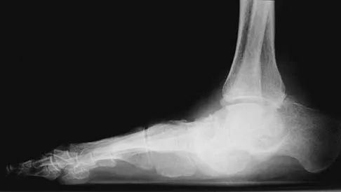
Meticulous preoperative planning is the vanguard against intraoperative surprises and suboptimal outcomes in boutonnière deformity correction. The clinical evaluation must be exhaustive. The surgeon must differentiate between a true boutonnière deformity and a pseudo-boutonnière deformity (which involves a PIP flexion contracture without true central slip disruption or DIP hyperextension, often due to volar plate injury). Elson's test is the most reliable clinical maneuver for assessing central slip integrity. The patient's PIP joint is flexed to 90 degrees over the edge of a table, and the patient is asked to extend the PIP joint against resistance. If the central slip is intact, the examiner will feel the extension force at the PIP joint, and the DIP joint will remain flail. If the central slip is ruptured, no extension force is felt at the PIP joint, and the lateral bands bypass the PIP joint to exert a rigid, paradoxical extension force on the DIP joint.
Radiographic evaluation is mandatory for all stages of the deformity. True anteroposterior, lateral, and oblique radiographs of the affected digit must be obtained. The surgeon must scrutinize these images for evidence of avulsion fractures at the dorsal base of the middle phalanx, joint space narrowing, osteophyte formation, and subtle volar subluxation of the middle phalanx. If salvage procedures are anticipated, radiographic templating is critical. For PIP joint arthroplasty, the medullary canals of the proximal and middle phalanges must be measured against standard templates to ensure they can accommodate the stems of a silicone implant (e.g., Swanson or NeuFlex) without cortical blowout. For arthrodesis, the desired angle of fusion must be planned according to the normal cascade of the hand: the index finger is typically fused at 25 to 30 degrees of flexion, the middle finger at 30 to 35 degrees, the ring finger at 35 to 40 degrees, and the small finger at 40 to 45 degrees.
The choice of anesthesia profoundly influences the surgical technique, particularly for soft tissue reconstructions. While regional blocks (axillary or supraclavicular) combined with a pneumatic arm tourniquet are traditional and provide excellent bloodless fields, the advent of Wide-Awake Local Anesthesia No Tourniquet (WALANT) has revolutionized extensor mechanism surgery. WALANT utilizes a mixture of lidocaine for anesthesia and epinephrine for hemostasis, injected locally. The paramount advantage of WALANT is the ability to assess active tendon excursion and joint kinematics intraoperatively. The surgeon can ask the awake, pain-free patient to actively flex and extend the digit after the central slip has been plicated. This real-time feedback allows for precise, millimeter-level adjustments of tendon tension, drastically reducing the risk of postoperative extension contractures or recurrent boutonnière deformities.
Patient positioning is standardized but requires careful attention to detail. The patient is placed supine with the operative extremity extended onto a radiolucent hand table. If WALANT is not utilized, a well-padded pneumatic tourniquet is applied to the upper arm and inflated to 250 mm Hg (or 100 mm Hg above systolic pressure) after exsanguination with an Esmarch bandage. The hand is secured in a lead hand or with custom taping to maintain the digits in a functional position while allowing the surgeon unhindered access to the dorsal aspect of the affected finger. A mini-C-arm fluoroscopy unit must be positioned perpendicular to the hand table, draped sterilely, and readily available for intraoperative assessment of joint reduction, K-wire placement, and implant seating.
Step-by-Step Surgical Approach and Fixation Technique
Surgical Approach and Soft Tissue Exposure
The surgical approach to the PIP joint must balance the need for wide exposure of the extensor mechanism with the imperative to preserve the delicate dorsal soft tissue envelope. A generous, curvilinear, or "lazy-S" dorsal longitudinal incision is centered over the PIP joint, extending from the mid-proximal phalanx to the level of the DIP joint. Straight longitudinal incisions directly over the PIP joint must be strictly avoided, as they are highly prone to hypertrophic scarring, wound breakdown, and secondary flexion contractures.
Full-thickness skin flaps are carefully elevated. The surgeon must stay in the subcutaneous plane, meticulously preserving the dorsal sensory nerve branches and the longitudinal venous drainage network. Compromising these veins leads to severe postoperative edema, which exacerbates stiffness and threatens wound healing. The flaps are retracted using fine skin hooks or stay sutures rather than crushing forceps. Once the flaps are elevated, the entire dorsal aponeurosis is visualized, revealing the attenuated central slip, the stretched triangular ligament, and the volarly displaced lateral bands.
Soft Tissue Reconstruction (Moderate Deformity)
The reconstruction begins with the systematic release of the secondary contractures. The transverse retinacular ligaments (TRL) are identified on the radial and ulnar aspects of the PIP joint. Longitudinal incisions are made through the TRLs, parallel to the lateral bands. Careful blunt and sharp dissection is performed underneath the displaced lateral slips to free them from the underlying joint capsule and collateral ligaments. This mobilization is critical; the lateral bands must be completely free to be transposed dorsally above the PIP joint's axis of rotation. Following mobilization, a thorough PIP joint synovectomy is performed, particularly in rheumatoid patients, to decompress the joint and remove the inflammatory tissue that stretches the extensor mechanism.
Next, the hyperextension contracture of the DIP joint is addressed via a terminal slip tenotomy, commonly known as the Fowler release. The terminal slips of the conjoined lateral bands are identified just proximal to the DIP joint. A complete tenotomy is performed. This release eliminates the deforming hyperextension force at the DIP joint and allows the lateral bands to shift proximally and dorsally, which inherently aids in PIP joint extension. The DIP joint is left free, and the patient will regain active DIP extension through the retained oblique retinacular ligaments.
Attention is then turned to the central slip. The attenuated, scarred segment is isolated. The central tendon is shortened by overlapping (plication) or by excising the redundant tissue and performing an end-to-end repair using non-absorbable 3-0 or 4-0 braided core sutures (e.g., Ethibond or FiberWire) and an epitendinous repair with 5-0 monofilament. Crucial Step: The tension must be perfect. If WALANT is used, the patient is asked to actively flex the PIP joint. If the joint cannot easily reach 80 degrees of flexion, the repair is too tight and must be lengthened immediately. If WALANT is not used, the surgeon must verify passive flexion to 80 degrees. Once tension is confirmed, the mobilized lateral bands are brought dorsally and sutured to the central slip and to each other, reconstructing the triangular ligament with 4-0 absorbable sutures to prevent recurrent volar subluxation. Finally, a 0.045-inch transarticular K-wire is driven obliquely across the PIP joint to hold it rigidly in 0 degrees of extension, protecting the repair.
Proximal Interphalangeal Joint Arthrodesis
For severe, fixed deformities, PIP arthrodesis provides a durable, stable salvage. Using the same dorsal approach, the scarred extensor mechanism is split longitudinally or excised to expose the joint. The collateral ligaments are sharply excised to allow full joint exposure. The articular cartilage and subchondral bone of the proximal phalanx head and the middle phalanx base are resected using a microsaw or rongeurs. The bone ends are contoured to create flat, highly congruent cancellous surfaces. Cup-and-cone reamers can also be used to maximize surface area and allow for fine-tuning of the fusion angle.
The joint is positioned in the pre-calculated angle of flexion (e.g., 30 degrees for the middle finger). Fixation is achieved using either crossed 0.045-inch K-wires, a dorsal tension band wiring construct, or a dedicated intramedullary compression device. Tension band wiring is biomechanically superior as it converts the dorsal tensile forces (from finger flexion) into dynamic compression across the arthrodesis site. Because the lateral bands are chronically contracted, fusing the PIP joint will exacerbate DIP hyperextension. Therefore, an oblique tenotomy of the lateral tendons (Fowler release) is mandatory to allow the DIP joint to drop into a neutral, functional position.
Interphalangeal Joint Arthroplasty
Resection arthroplasty utilizing a silicone implant is an excellent salvage for the ring and small fingers in appropriate candidates. Following joint exposure, a generous resection of the proximal phalanx head is performed at the metaphyseal flare. Minimal resection of the middle phalanx base is performed to preserve the vital insertions of the collateral ligaments. The medullary canals are sequentially broached using specialized rasps to accommodate the implant stems. The sizing must be exact; an oversized implant will buckle, while an undersized implant will fail to provide stability.
The Swanson or NeuFlex silicone implant is inserted using a strict "no-touch" technique to minimize the risk of devastating deep infection. The implant acts as a dynamic spacer, maintaining joint alignment while a fibrous pseudocapsule forms around it. Crucially, the extensor mechanism must be reconstructed over the implant. The central slip is reattached to the dorsal base of the middle phalanx, often utilizing small drill holes or suture anchors, to provide active extension. The lateral bands are relocated dorsally and sutured to the central slip. The joint is pinned in full extension with a temporary transarticular K-wire for 10 to 14 days to protect the soft tissue reconstruction over the implant.
Complications, Incidence Rates, and Salvage Management
The operative management of the boutonnière deformity is fraught with potential complications. The extensor mechanism is a delicate, unforgiving structure with minimal tolerance for error in tensioning or handling. Complications can arise from technical errors, poor patient compliance, or the inherent biological response to tendon surgery, namely profound scar formation. The surgeon must be prepared to identify and manage these complications aggressively, as early intervention is often the only way to salvage digital function.
The most common and dreaded complication following soft tissue reconstruction is an iatrogenic extension contracture of the PIP joint. This occurs primarily due to over-plication of the central slip, failure to achieve 80 degrees of intraoperative passive flexion, or excessive postoperative immobilization. The patient presents with a rigid, straight PIP joint that severely impedes grasp and grip strength. Management is initially non-operative, relying on aggressive hand therapy, dynamic flexion splinting, and serial casting. If conservative measures fail after 3 to 6 months, surgical tenolysis may be attempted, but it is technically demanding and carries a high risk of rupturing the central slip, leading to a recurrent boutonnière deformity.
Recurrent boutonnière deformity is another significant complication, resulting from failure of the central slip repair, inadequate release of the transverse retinacular ligaments, or premature removal of the transarticular K-wire. The deformity slowly re-establishes itself as the lateral bands migrate volarly. Revision soft tissue reconstruction is rarely successful due to the compromised, scarred soft tissue bed. In these cases, salvage via PIP joint arthrodesis is the most reliable and definitive management strategy.
Infection and skin necrosis are catastrophic complications, particularly in the presence of a silicone implant or K-wires. The dorsal skin over the PIP joint is thin and poorly vascularized. Excessive undermining, rough retraction, or tension on the wound closure can lead to skin edge necrosis, exposing the underlying tendon repair or implant. Superficial infections can be managed with oral antibiotics and local wound care. However, deep infections involving a silicone arthroplasty require immediate operative intervention, including hardware removal, aggressive pulsatile lavage, debridement of necrotic tissue, and placement of an antibiotic spacer, followed by a prolonged course of intravenous antibiotics.
| Complication | Estimated Incidence Rate | Pathogenesis / Primary Causes | Salvage Management Strategy |
|---|---|---|---|
| Extension Contracture | 15% - 25% | Over-plication of central slip; inadequate TRL release; prolonged immobilization. | Aggressive dynamic flexion splinting; late surgical tenolysis (high risk of failure). |
| Recurrent Deformity | 10% - 20% | Rupture of central slip repair; premature K-wire removal; unrecognized fixed contracture. | PIP joint arthrodesis; rarely, revision soft tissue reconstruction. |
| Skin Necrosis / Wound Breakdown | 2% - 5% | Poor incision design; excessive flap undermining; damage to dorsal venous network. | Local wound care; healing by secondary intention; reverse cross-finger flap if tendon exposed. |
| Deep Infection (Arthroplasty) | 1% - 3% | Contamination during implant insertion; hematogenous seeding. | Immediate implant removal; aggressive debridement; IV antibiotics; eventual arthrodesis. |
| Silicone Implant Fracture | 5% - 10% (Long-term) | Material fatigue; excessive shear forces; oversized implant buckling. | Revision arthroplasty (if bone stock allows) or conversion to PIP arthrodesis. |
Phased Post-Operative Rehabilitation Protocols
The ultimate success of a boutonnière deformity correction relies as heavily on meticulous, highly specialized postoperative hand therapy as it does on the precise execution of the surgical technique. The extensor mechanism is highly susceptible to adhesions, and the rehabilitation protocol must strike a delicate, perilous balance: the central slip repair must be protected from excessive tension to prevent rupture or attenuation, while simultaneous gliding of the adjacent structures must be encouraged to prevent catastrophic stiffness. A close, communicative partnership between the orthopedic surgeon and a certified hand therapist (CHT) is absolutely mandatory.
Phase I: Protection and Isolated Gliding (Weeks 0 to 4)
For patients who have undergone soft tissue reconstruction, the PIP joint is rigidly immobilized in full extension via the transarticular K-wire placed intraoperatively. During this initial phase, the primary goal is the protection of the central slip repair and the reconstructed triangular ligament. The digit is placed in a custom thermoplastic volar resting splint. However, immobilization of the PIP joint does not mean immobilization of the entire hand. The patient is instructed to perform active and passive range of motion exercises for the MCP and DIP joints immediately. Active DIP joint flexion is particularly critical; it pulls the lateral bands distally, maintaining their glide and preventing them from scarring to the dorsal capsule of the PIP joint. Pin site care is performed daily to prevent superficial tract infections.
Phase II: Controlled Mobilization (Weeks 4 to 8)
At the 3-to-4-week mark, the transarticular K-wire is removed in the clinic. The joint is immediately placed in a dynamic extension splint (e.g., a Capener splint or a reverse knuckle bender) or a static progressive extension splint, depending on the presence of any residual extensor lag. Active PIP joint motion is initiated under the strict supervision of the hand therapist. The patient performs short arc active flexion exercises, gradually increasing the arc of motion as tolerated. Crucial Rule: Passive PIP joint flexion is strictly forbidden during this phase. Forcing the joint into passive flexion will overwhelm the healing central slip, leading to elongation of the repair and immediate recurrence of the boutonnière deformity. Night splinting in full extension is mandatory to counteract the continuous resting tone of the flexor tendons.
Phase III: Strengthening and Weaning (Weeks 8 to 12+)
By the 8th week, the central slip repair has achieved sufficient tensile strength to withstand more rigorous forces. The patient is gradually weaned from the daytime extension splint, although it may be reapplied if an extensor lag begins to develop. Gentle strengthening exercises, such as using therapy putty or a hand exerciser, are introduced to rebuild the intrinsic and extrinsic muscle bulk that atrophied during immobilization. If the patient struggles to achieve functional flexion, gentle passive flexion and joint mobilization techniques may be cautiously introduced by the therapist. Maximum medical improvement is typically not reached until 6 to 9 months postoperatively, and patients must be counseled preoperatively regarding this protracted recovery timeline.
For patients who underwent salvage procedures, the protocols differ significantly. Following PIP arthrodesis, the digit is immobilized in a static protective splint for 6 to 8 weeks until definitive radiographic evidence of bony union (bridging trabeculae) is observed. Once fused, therapy focuses on maximizing motion at the adjacent MCP and DIP joints. For PIP arthroplasty, early controlled motion is paramount. A dynamic extension splint is applied within the first week, and active range of motion is initiated to promote the formation of a functional, pliable pseudocapsule around the silicone implant. Lateral stress (such as strong pinch) is strictly avoided for at least 6 weeks to prevent implant subluxation or fracture.
Summary of Landmark Literature and Clinical Guidelines
The evolution of operative management for the boutonnière deformity is deeply rooted in the historical contributions of pioneering hand surgeons whose anatomical studies laid the foundation for modern techniques. The seminal work of J. William Littler and J.H. Boyes in the mid-20th century elucidated the complex, interconnected
