Congenital Hand Reconstruction: Toe-Phalanx Transplantation and Ring Syndrome
Key Takeaway
Congenital hand anomalies, including symbrachydactyly and amniotic band sequence, require meticulous surgical intervention to restore function and aesthetics. This guide details the non-vascularized toe-phalanx transplantation technique for absent digits and the staged Z-plasty release for congenital constriction rings. Emphasizing biomechanics, soft-tissue handling, and postoperative protocols, these procedures are essential for optimizing pediatric hand development and preventing progressive neurovascular compromise.
Comprehensive Introduction and Patho-Epidemiology
The surgical management of congenital hand anomalies represents one of the most intellectually demanding and technically unforgiving disciplines within orthopedic surgery. It requires a profound, multidimensional understanding of pediatric biomechanics, microvascular anatomy, embryological development, and longitudinal growth potential. Among the most challenging and phenotypically diverse presentations encountered by the congenital hand surgeon are aphalangia—frequently presenting as a manifestation of symbrachydactyly—and Congenital Ring Syndrome, universally known as Amniotic Band Sequence (ABS) or Streeter Dysplasia. Surgical intervention in these complex conditions transcends mere aesthetic improvement; it is fundamentally reconstructive, aiming to provide a stable, sensate, and functional prehensile unit while mitigating progressive neurovascular compromise and harnessing the immense neuroplasticity of the infant brain.
Symbrachydactyly is characterized by a spectrum of developmental failures ranging from short, webbed fingers to complete absence of digits with only rudimentary soft-tissue nubbins remaining. The patho-epidemiology of symbrachydactyly is generally considered to be sporadic, with an incidence of approximately 1 in 32,000 live births. The most widely accepted etiologic theory is the subclavian artery supply disruption sequence, proposed by Bavinck and Weaver. This theory posits that a transient ischemic event during the critical period of limb embryogenesis (typically between the fourth and sixth weeks of gestation) leads to hypoplasia or necrosis of the developing mesoderm beneath the apical ectodermal ridge (AER). Because the insult is vascular and localized, systemic or syndromic associations are exceedingly rare, distinguishing symbrachydactyly from other longitudinal deficiencies such as radial clubhand.
Conversely, Congenital Ring Syndrome (Amniotic Band Sequence) presents an entirely different patho-epidemiological profile. Affecting between 1 in 1,200 to 1 in 15,000 live births, ABS is characterized by asymmetric, sporadic, and highly variable deformities caused by the entanglement of fetal parts in mesodermic fibrous bands. The extrinsic theory, originally championed by Torpin, remains the definitive pathophysiological explanation. It suggests that an early, spontaneous rupture of the amnion allows the fetus to enter the chorionic cavity, where it becomes entangled in the sticky, fibrous mesodermic bands emanating from the chorionic surface. The timing of the amniotic rupture dictates the severity of the phenotype: early ruptures lead to catastrophic intrauterine amputations, severe acrosyndactyly, and major craniofacial defects, whereas later ruptures typically result in isolated, shallow constriction rings.
The clinical burden of both symbrachydactyly and ABS is immense, necessitating early and decisive surgical strategy. In symbrachydactyly, the presence of flail, unsupported digital nubbins offers no functional utility for pinch or grasp. Here, the non-vascularized toe-phalanx transplantation serves as a critical reconstructive pillar, providing the necessary longitudinal osseous support to transform a useless soft-tissue bud into a functional post for opposition. In ABS, the progressive nature of deep constriction rings poses an immediate threat to the distal neurovascular integrity of the limb. The systematic, staged release of these rings via complex Z-plasties, combined with the meticulous separation of acrosyndactyly, is paramount to salvaging the extremity and optimizing long-term hand kinematics.
Detailed Surgical Anatomy and Biomechanics
Anatomy of the Toe-Phalanx Donor Site
The successful execution of a non-vascularized toe-phalanx transfer relies intrinsically on a masterful understanding of the osteology and microvascular anatomy of the pediatric foot. The proximal phalanges of the third and fourth toes are universally selected as the optimal donor grafts. These digits provide the most precise morphometric size match for the rudimentary hand digits while minimizing donor-site morbidity, particularly regarding the biomechanics of the developing arch and gait cycle. The vascular supply to these phalanges is derived from the plantar metatarsal arteries, which arborize into a rich periosteal plexus. For a non-vascularized transfer to survive and potentially exhibit growth, the phalanx must be harvested extraperiosteally. The periosteal sleeve harbors the vital osteoprogenitor cells and provides a robust vascular scaffold that facilitates rapid revascularization from the recipient bed via inosculation and neovascularization.
Furthermore, the capsuloligamentous anatomy of the donor phalanx must be meticulously preserved. The proximal interphalangeal (PIP) joint and metatarsophalangeal (MTP) joint attachments—specifically the volar plate, proper collateral ligaments, and accessory collateral ligaments—are harvested en bloc with the bone. The volar plate is an essential fibrocartilaginous structure that, when transferred to the hand, serves a dual purpose: it prevents catastrophic hyperextension deformities at the newly created pseudojoint and provides a durable, gliding surface for the rudimentary flexor tendon anlagen. The preservation of the cartilaginous endplates is equally critical, as chondrocytes, unlike osteocytes, can survive the initial period of transient ischemia through synovial fluid diffusion until the periosteal blood supply is re-established.
Biomechanics of the Reconstructed Digit
From a biomechanical standpoint, the primary objective of the toe-phalanx transfer is to establish a rigid, longitudinal post that enables functional pinch kinematics. In the normal hand, prehension requires a stable digital column against which the thumb can exert opposing force. The flail nubbins of symbrachydactyly collapse under compressive loads, rendering opposition impossible. By introducing a structural osseous scaffold, the surgeon alters the moment arms and force vectors within the hand, allowing the extrinsic flexors and extensors (even if rudimentary) to exert meaningful torque across the newly stabilized digit. The excursion of these tendons, often tethered in the dysplastic soft-tissue envelope, must be maximized during pouch creation to impart dynamic stability to the reconstructed unit.
The biomechanics of growth in a non-vascularized transfer present a unique clinical paradox. While the extraperiosteal harvest aims to preserve the physes, normal longitudinal growth is rarely achieved. The physes are highly sensitive to ischemia, and the transient avascular period prior to graft incorporation frequently results in premature physeal closure. However, the graft reliably undergoes appositional growth, increasing in girth and density in response to the mechanical stresses of pinch and grasp (Wolff's Law). The structural rigidity provided by the transferred phalanx prevents the progressive contracture of the soft-tissue nubbin, effectively acting as an internal tissue expander that maintains the spatial volume necessary for functional prehension.
Pathoanatomy of Constriction Rings and Acrosyndactyly
The pathoanatomy of Congenital Ring Syndrome is dictated by the constrictive properties of the amniotic bands. Histologically, these bands are composed of dense, avascular, mesodermic fibrous tissue that lacks the elasticity of normal dermis. As the fetus grows, the band remains static, effectively acting as a tourniquet. The level of vascular impairment is depth-dependent. Superficial rings obliterate the thin-walled superficial venous and lymphatic channels, leading to profound distal lymphedema and venous congestion. Deeper rings compress the robust arterial structures, precipitating ischemia, cyanosis, and eventual intrauterine or postnatal necrosis. The surgical release must therefore navigate this distorted vascular anatomy, prioritizing the decompression of the venous outflow tract without compromising the tenuous arterial inflow.
Acrosyndactyly represents the most severe manifestation of distal amniotic band entanglement. Unlike simple syndactyly, which is a failure of programmed cell death (apoptosis) in the interdigital webs, acrosyndactyly is a post-developmental fusion of previously separated digits. The pathoanatomy is characterized by a distal, disorganized fusion mass of bone, cartilage, and soft tissue, with proximal epithelialized fenestrations (sinus tracts) that represent the original web spaces. The neurovascular bundles in acrosyndactyly are highly aberrant; they are frequently shared between adjacent digits or tethered within the dense fibrotic scar of the fusion mass. Meticulous dissection is required to separate these digits while preserving these shared structures, a task complicated by the underlying symphalangism and joint contractures that invariably accompany this severe deformity.
Exhaustive Indications and Contraindications
The decision to proceed with surgical reconstruction in congenital hand anomalies requires a rigorous, individualized assessment of the patient's functional deficit, anatomical potential, and physiological readiness. The timing of intervention is paramount; the surgeon must balance the necessity of early intervention to harness neuroplasticity against the anesthetic risks inherent in infant surgery.
For non-vascularized toe-phalanx transfers, the ideal window for intervention is between 6 and 18 months of age. This timing capitalizes on the maximal osteogenic and chondrogenic potential of the transferred phalanx while integrating the reconstructed digit into the child's developing gross motor and fine pinch patterns. In contrast, the indications for Congenital Ring Syndrome are dictated entirely by the severity of the vascular compromise. Deep, constricting rings presenting with distal cyanosis, severe lymphedema, or impending necrosis demand emergent surgical release, regardless of the patient's age or weight.
| Clinical Scenario | Absolute Indications | Relative Indications | Contraindications (Absolute & Relative) |
|---|---|---|---|
| Toe-Phalanx Transfer (Symbrachydactyly) | - Presence of flail, unsupported digital nubbins. - Adequate soft-tissue envelope to accommodate the graft. - Presence of proximal skeletal elements (metacarpals) for fixation. |
- Desire to improve pinch kinematics and opposition. - Prevention of soft-tissue nubbin contracture. - Psychological/aesthetic considerations for the family. |
Absolute: Complete absence of soft-tissue envelope; severe systemic medical comorbidities precluding anesthesia. Relative: Severe hypoplasia of proximal motor units (extrinsic tendons); older children (>4-5 years) where neuroplastic integration is limited. |
| Constriction Ring Release (ABS) | - Emergent: Distal cyanosis, severe lymphedema, impending necrosis. - Circumferential deep rings threatening progressive neurovascular compromise. |
- Elective: Shallow rings causing cosmetic deformity. - Contour abnormalities persisting after the loss of infant "baby fat." |
Absolute: Simple excision with direct everting closure (high risk of circumferential scar contracture). Relative: Elective release in infants <6 months with stable, shallow rings and no vascular compromise. |
| Acrosyndactyly Separation (ABS) | - Border digits (thumb and small finger) tethered in a distal fusion mass (requires early release <6 months). - Progressive joint deformity due to tethering. |
- Central digit separation (staged at ~18 months). - Web space deepening to improve relative thumb length. |
Absolute: Separation of central digits sharing a single neurovascular bundle where division would result in digital ischemia. Relative: Severe symphalangism where separation offers no functional gain. |
Contraindications must be strictly respected to prevent catastrophic surgical failures. In symbrachydactyly, attempting a non-vascularized transfer into a deficient soft-tissue envelope will inevitably lead to wound breakdown, graft exposure, and catastrophic infection. In such cases, vascularized toe transfers or distraction osteogenesis may be more appropriate salvage strategies. For ring syndrome, the primary contraindication relates to surgical technique rather than patient selection: performing a simple circumferential excision and direct closure is universally condemned, as it invariably leads to a recurrent, unyielding cicatricial contracture that is often more severe than the original amniotic band.
Pre-Operative Planning, Templating, and Patient Positioning
Thorough pre-operative planning is the bedrock of successful congenital hand reconstruction. The clinical evaluation must be meticulous, assessing not only the local anatomical deficits but also the global functional capacity of the extremity. Standardized, high-quality radiographs of both the bilateral hands and bilateral feet are mandatory. In symbrachydactyly, these radiographs define the presence and morphology of the proximal skeletal elements (metacarpals or proximal phalanges) that will serve as the foundation for the transferred toe phalanx. In severe cases of Amniotic Band Sequence, advanced imaging modalities such as Magnetic Resonance Imaging (MRI) or high-resolution ultrasound may be indicated to delineate the aberrant neurovascular anatomy, assess the patency of deep arterial structures, and evaluate the integrity of rudimentary tendon anlagen.
Templating for the toe-phalanx transfer is a critical step that dictates the biomechanical success of the procedure. The surgeon must carefully measure the dimensions of the recipient soft-tissue pouch and select the corresponding donor phalanx (typically the 3rd or 4th proximal phalanx) that provides the optimal size match. Oversizing the graft will result in excessive tension on the delicate soft-tissue envelope, precipitating wound dehiscence and graft extrusion. Undersizing the graft fails to provide adequate structural support and compromises pinch kinematics. The pre-operative template must also define the boundaries of the extraperiosteal harvest, ensuring that the capsuloligamentous structures are included en bloc.
For the surgical management of constriction rings, pre-operative mapping is focused on the precise design of the Z-plasties. The surgeon must geometrically plan multiple 60-degree triangular flaps along the trajectory of the excised groove. This specific angle is chosen because it optimally recruits healthy subcutaneous tissue from the adjacent areas, breaks up the linear contracture, and theoretically lengthens the scar by 73%. The staging of the procedure must also be planned; releasing only 50% to 60% of the circumference during the index operation is a non-negotiable safety parameter to prevent catastrophic disruption of the remaining venous and lymphatic drainage networks.
Patient positioning and operating room setup require meticulous attention to detail. The pediatric patient is positioned supine, with dual operative fields prepared and draped simultaneously (the affected upper extremity and the donor lower extremity). A radiolucent hand table is utilized to facilitate intraoperative fluoroscopy. Pediatric pneumatic tourniquets are applied to both limbs, with pressures carefully calibrated to the patient's systolic blood pressure (typically 100 to 150 mmHg above systolic) to minimize the risk of tourniquet-induced neuropraxia. The use of high-powered loupe magnification (3.5x to 4.5x) or an operating microscope is absolute necessity, given the diminutive size and aberrant location of the neurovascular structures in these congenital anomalies.
Step-by-Step Surgical Approach and Fixation Technique
Toe-Phalanx Harvest and Transplantation
The non-vascularized toe-phalanx transplantation is a highly orchestrated procedure that demands technical precision at both the recipient and donor sites.
1. Recipient Site Preparation:
Under tourniquet control and loupe magnification, a volar zigzag (Bruner-type) incision is designed over the distal palm, extending meticulously into the apex of the soft-tissue bud of the absent digit. The incision must be planned to avoid linear contractures across the future flexion creases. Using fine tenotomy scissors and a small hemostat, the surgeon performs blunt dissection to create a precise, well-vascularized cavity (the pouch). It is imperative to avoid excessive electrocautery, as thermal necrosis will destroy the delicate capillary bed necessary for the inosculation and revascularization of the graft. The rudimentary neurovascular elements, often hypoplastic and displaced, are identified and protected. The flexor tendon anlagen are dissected, and a thorough adhesiolysis is performed proximal to their distal insertion to maximize tendon excursion. The distal attachments must remain intact to impart dynamic stability to the reconstructed digit.
2. Donor Site Harvest (Third or Fourth Toe):
Attention is then directed to the donor foot. A dorsal diagonal or lazy-S incision is made over the proximal phalanx of the selected toe. The dissection proceeds extraperiosteally; maintaining the integrity of the periosteal sleeve is the most critical step in ensuring graft survival, as it houses the osteoprogenitor cells. The soft-tissue attachments of the proximal interphalangeal (PIP) joint are incised flush with the bone. At the metatarsophalangeal (MTP) joint, the volar plate and collateral ligaments are incised at their metatarsal origins. The toe phalanx, encapsulated within its periosteum and retaining its volar plate and collateral ligaments, is removed as a single, contiguous en bloc unit. To prevent the significant donor-site morbidity of a "floppy toe," the dead space is meticulously closed, and the donor toe is syndactylized to the adjacent toe (e.g., 3rd to 4th toe) using a robust soft-tissue repair.
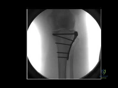
3. Graft Placement and Fixation:
On a separate sterile back table, the harvested phalanx is prepared. A small-diameter Kirschner wire (typically 0.028 or 0.035 inch) is passed retrogradely into the proximal aspect of the graft. The composite transfer is then gently inserted into the prepared soft-tissue pouch in the hand. The surgeon must ensure that the volar plate of the graft is oriented volarly; failure to do so will result in a severe, recalcitrant hyperextension deformity. The K-wire is advanced distally through the soft-tissue envelope, and then driven proximally into the recipient metacarpal to achieve rigid axial stability. The skin is closed with fine absorbable sutures (e.g., 5-0 or 6-0 chromic gut), avoiding any tension on the closure.
Staged Z-Plasty for Constriction Rings
The surgical release of amniotic constriction rings requires a disciplined, staged approach. The procedure begins with the precise excision of the fibrotic, dysplastic tissue of the ring. The incision is carried down through the dense, avascular mesodermic band until healthy, normal subcutaneous tissue and deep fascia are encountered. The surgeon must be hyper-vigilant to protect the underlying neurovascular structures, which may be displaced superficially due to the constrictive nature of the band.
Once the groove is excised, the pre-planned 60-degree Z-plasties are executed. The triangular flaps are sharply elevated, ensuring a robust subdermal plexus is maintained to prevent flap necrosis. The flaps are then transposed, effectively recruiting healthy subcutaneous fat into the deficient area and breaking the linear trajectory of the scar. The inset is performed meticulously with fine absorbable sutures. As mandated by evidence-based protocols, only 50% to 60% of the circumference is addressed during this index procedure; the remaining portion is left undisturbed to preserve the critical venous and lymphatic drainage, and will be released in a second stage 2 to 3 months later.
Acrosyndactyly Release and Web Space Deepening
The release of acrosyndactyly is a formidable challenge due to the disorganized distal fusion mass. The procedure prioritizes the preservation of the proximal epithelialized fenestrations (sinus tracts), which are ingeniously incorporated into the reconstruction of the new web spaces. The distal fusion mass is carefully separated using sharp dissection. The surgeon must navigate the highly distorted anatomy to identify and protect the shared neurovascular bundles; if a single dominant digital artery supplies two adjacent digits, the separation must be carefully designed to ensure both digits remain viable, sometimes requiring the sacrifice of separation in favor of perfusion.
Because the surface area of the separated digits vastly exceeds the available skin envelope, full-thickness skin grafts (FTSG) are almost universally required. These grafts are typically harvested from the groin or lower abdomen to ensure an adequate color and texture match, while concealing the donor site morbidity. The grafts are defatted and sutured into the raw defects on the facing aspects of the digits, and secured with tie-over bolster dressings to prevent hematoma formation and shear forces. Thumb reconstruction in these cases often requires advanced techniques, such as a four-flap Z-plasty for web space deepening, distraction osteogenesis for metacarpal lengthening, or Søiland’s on-top plasty for severe amputations.
Complications, Incidence Rates, and Salvage Management
The complication profile in congenital hand reconstruction is significant, reflecting the extreme technical demands of the surgery and the fragile, hypoplastic nature of the tissues involved. Meticulous surgical technique, rigorous pre-operative planning, and strict adherence to biomechanical principles are the only safeguards against catastrophic outcomes. The surgeon must be prepared to manage a wide array of intraoperative and postoperative complications, ranging from transient neuropraxia to complete graft necrosis.
In non-vascularized toe-phalanx transfers, the most ubiquitous complication is premature physeal closure. Despite extraperiosteal harvest, the transient ischemia inevitably damages the highly sensitive physeal plate. While this limits longitudinal growth, it is often considered an expected outcome rather than a failure, provided the graft achieves structural integration and appositional growth. Graft resorption is a more severe complication, typically resulting from thermal necrosis during pouch creation or overwhelming infection. Donor site morbidity, particularly the development of a "floppy toe" or angular deformity, can be profoundly disabling for the child's future gait and footwear fitting, underscoring the absolute necessity of primary syndactylization at the donor site.
In the management of Amniotic Band Sequence, the complications are frequently related to the vascular integrity of the limb. Flap necrosis following Z-plasty is a devastating complication that exposes the underlying neurovascular structures and leads to severe, recurrent cicatricial contracture. Iatrogenic injury to the digital nerves or arteries during the separation of acrosyndactyly is a constant threat, given the aberrant anatomy. Failure of the full-thickness skin grafts due to hematoma, infection, or shear forces requires prompt debridement and regrafting to prevent profound joint stiffness and syndactyly recurrence.
| Complication | Estimated Incidence | Etiology / Risk Factors | Salvage Strategy / Management |
|---|---|---|---|
| Premature Physeal Closure | 70% - 90% | Transient ischemia prior to revascularization; thermal injury during harvest. | Often requires no acute intervention if appositional growth and structural stability are maintained. Consider distraction osteogenesis at skeletal maturity if length is severely deficient. |
| Graft Resorption (Toe-Phalanx) | 5% - 15% | Infection; severe avascularity of the recipient pouch; excessive tension. | Debridement and systemic antibiotics. Salvage with a vascularized toe-to-hand transfer or delayed bone grafting once the soft-tissue envelope is optimized. |
| Donor Site "Floppy Toe" | 10% - 20% (if not syndactylized) | Failure to close dead space and reconstruct collateral ligamentous support. | Revision surgery with delayed syndactylization to the adjacent toe; orthotic management for footwear. |
| Flap Necrosis (Z-Plasty) | 2% - 8% | Excessive tension; aggressive defatting compromising the subdermal plexus; circumferential release. | Aggressive wound care; delayed healing by secondary intention; revision local flap coverage or full-thickness skin grafting once the bed is clean. |
| Recurrent Constriction/Contracture | 5% - 10% | Inadequate excision of the fibrotic band; failure to use Z-plasties (simple linear closure). | Revision surgery with wider excision of the scar tissue and execution of larger, multi-limb Z-plasties or W-plasties. |
| FTSG Failure (Acrosyndactyly) | 10% - 15% | Hematoma formation; inadequate bolster dressing; shear forces during early mobilization. | Early recognition; evacuation of hematoma; delayed regrafting or application of dermal regeneration templates (e.g., Integra) if the bed is compromised. |
Phased Post-Operative Rehabilitation Protocols
The surgical reconstruction is only the first phase of the therapeutic continuum; the ultimate functional outcome is heavily dependent on a rigorous, phased post-operative rehabilitation protocol. Pediatric hand therapy presents unique challenges, primarily related to the compliance and cognitive capacity of the infant or toddler. The therapist and surgeon must work in tandem to educate the parents, who serve as the primary facilitators of the rehabilitation process. The overarching goal is to protect the surgical reconstruction during the initial healing phase, and subsequently exploit the child's neuroplastic window to integrate the reconstructed digit into functional, bimanual play.
Phase I: Immobilization and Protection (Weeks 0 to 4-6)
Immediately post-operatively, the extremity is immobilized in a bulky, non-compressive soft dressing reinforced by a long-arm cast. The long-arm configuration (extending above the elbow with the elbow flexed at 90 degrees) is critical to prevent the child from sliding out of the cast or using the contralateral limb to remove it. During this phase, the primary objectives are strict protection of the K-wires, prevention of shear forces across the skin grafts or Z-plasties, and monitoring for any signs of cast complications (e.g., compartment syndrome, pressure sores, or infection). The donor foot is typically managed in a bulky soft dressing and a rigid post-operative shoe, with weight-bearing as tolerated.
Phase II: Mobilization and Integration (Weeks 6 to 12)
Following radiographic confirmation of graft incorporation and clinical assessment of wound healing, the long-arm cast and K-wires are removed in the clinic setting. The child is immediately transitioned into custom-fabricated thermoplastic splints. These splints are typically worn full-time initially, and then progressively weaned to nighttime use. Active range of motion is strongly encouraged through play-based therapy. The therapist utilizes age-appropriate toys to stimulate grasp, release, and pinch kinematics. Sensory re-education is initiated, employing various textures and stimuli to desensitize the surgical scars and promote cortical mapping of the reconstructed digits.
Phase III: Long-Term Functional Adaptation (Months 3 to Years)
The final phase of rehabilitation is an ongoing, longitudinal process that tracks the child through skeletal maturity. Bimanual play therapy remains the cornerstone of treatment, ensuring the child does not develop compensatory patterns of neglect for the operative hand. The orthopedic surgeon must monitor the longitudinal growth, joint stability, and the potential development of secondary contractures. As the child grows, the soft tissues may not keep pace with the skeletal elements, necessitating serial splinting or secondary soft-tissue releases to maintain optimal function and aesthetics.
Summary of Landmark Literature and Clinical Guidelines
The modern surgical approach to congenital hand reconstruction is built upon a foundation of landmark literature and evolving clinical guidelines. The evolution of the non-vascularized toe-phalanx transfer is inexorably linked to the pioneering work of Toby et al., Buck-Gramcko, and Goldberg. Their extensive clinical series and long-term follow-up studies elucidated the critical biomechanical principles of extraperiosteal harvest and the preservation of capsuloligamentous structures. These authors definitively demonstrated that while normal longitudinal growth is elusive, the structural stability and appositional
