العربية
Part of the Master Guide

AAOS & ABOS Basic Science MCQs (Set 3): Bone Biology, Biomechanics & Anatomy Review

Anatomy Board Review MCQs (Set 4): Peripheral Nerves, Major Joints, & Muscle Anatomy | USMLE & ABOS Prep

23 Apr 2026 38 min read 87 Views
Anatomy 2008 MCQs - Part 4

Key Takeaway

This high-yield question set (Set 4) for anatomy board review focuses on essential peripheral neuroanatomy, covering nerve plexuses and their distributions. It also includes challenging questions on major joint structures and the functional anatomy of the musculoskeletal system, critical for medical board preparation.

Anatomy Board Review MCQs (Set 4): Peripheral Nerves, Major Joints, & Muscle Anatomy | USMLE & ABOS Prep

Comprehensive 100-Question Exam


00:00

Start Quiz

Question 1

Bleeding is encountered while developing the internervous plane between the tensor fascia lata and the sartorius during the anterior approach to the hip. The most likely cause is injury to what artery?


Explanation

The ascending branch of the lateral femoral circumflex artery crosses the gap between the tensor fascia lata and the sartorious and must be identified and ligated or coagulated. The other vessels are out of the field of dissection. Barrack RL, Booth RE Jr, Lonner JH, et al (eds): Orthopaedic Knowledge Update: Hip and Knee Reconstruction 3. Rosemont, IL, American Academy of Orthopaedic Surgeons, 2006, p 312.

Question 2

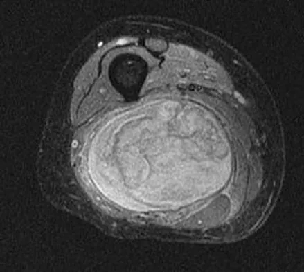
A 36-year-old woman with familial neurofibromatosis has an enlarging mass in the posterior thigh. The lesion has slowly increased in size and is now constantly painful. Pressure on the mass causes dysesthesias in the foot. Figures 44a through 44c show T1-weighted, STIR, and T1-weighted fat-saturated gadolinium scans, respectively. Figure 44d shows a PET scan. What does this lesion most likely represent?


Explanation

The images reveal a large mass in the posterior thigh arising from the sciatic nerve. The lesion is edematous, and the gadolinium image reveals rim enhancement, suggesting necrosis, given that the STIR image is not uniformly bright as would be seen in a cystic lesion. The PET scan has increased uptake, in this case a standard unit value (SUV) of greater than 2.0. These findings are all very suggestive of a malignant process. The history of neurofibromatosis makes a malignant peripheral nerve sheath tumor, or neurofibrosarcoma, the most likely diagnosis. The term "peripheral nerve sheath tumor" has replaced neurolemmoma and schwannoma. Menendez LR (ed): Orthopaedic Knowledge Update: Musculoskeletal Tumors. Rosemont, IL, American Academy of Orthopaedic Surgeons, 2002, pp 225-230.


Question 3

In Dupuytren's disease, the retrovascular cord typically displaces the radial proper digital nerve of the ring finger in what direction?


Explanation

Retrovascular cords are common in Dupuytren's disease and commonly require surgical treatment. Nerve injury in Dupuytren's surgery is an infrequent complication that occurs partly because the digital nerves can be displaced from their normal anatomic relationships by retrovascular cords. The nerves are displaced superficially, toward the center of the digit (palmarly and ulnarly). This displacement is typically seen at the level of the metacarpophalangeal joint.

Question 4

Ganglion cysts about the wrist most commonly arise from what structure?


Explanation

Ganglion cysts are the most common mass or mass-like lesions seen in the hand and wrist. They arise in a variety of locations, including synovial joints or tendon sheaths. The most common location is the dorsal/radial wrist arising from the dorsal scapholunate interosseous ligament.

Question 5

A 60-year-old woman has activity-related hip pain after undergoing arthroplasty 5 years ago. She has severe Parkinsonism and denies fevers or chills. Radiographs are shown in Figures 45a and 45b. What is the most likely cause of her pain?


Explanation

The radiographs reveal both cement debonding at the lateral shoulder of the prosthesis and a cement mantle fracture. Both of these indicate a loose femoral component. The radiographs show a stress fracture with reactive bone on the lateral femoral cortex in conjunction with the cement mantle fracture. The acetabular component shows no evidence of loosening. Heterotopic bone usually is not a source of pain when it is Brooker grade I, as in this case. Parkinsonism generally is not associated with hip pain. Harris WH, McCarthy JC, O'Neill DA: Femoral component loosening using contemporary techniques of femoral cement fixation. J Bone Joint Surg Am 1982;64:1063-1067. Callaghan JJ, Rosenberg AG, Rubash H (eds): The Adult Hip. Philadelphia, PA, Lippincott-Raven, 1998, pp 960, 1228-1229.


Question 6

A 15-year-old boy reports leg pain after being tackled during football practice. Radiographs and a CT scan are shown in Figures 46a through 46c. The patient has a pathologic fracture through what underlying lesion?


Explanation

The images show a lobulated, eccentric, well-marginated lesion that is typical of a nonossifying fibroma. The lesion is slightly expansile, and the CT scan findings show that the lesion is very well marginated and the cortex is disrupted, which is a common finding. None of the characteristics of this lesion is aggressive in nature. Menendez LR (ed): Orthopaedic Knowledge Update: Musculoskeletal Tumors. Rosemont, IL, American Academy of Orthopaedic Surgeons, 2002, pp 69-75.


Question 7

A 28-year-old man has left knee pain after a snow skiing accident. The MRI scan shown in Figure 47 reveals which of the following?


Explanation

Bone bruises are often noted on MRI after anterior cruciate and medial collateral ligament injuries. The significance of these injuries awaits long-term follow-up studies. The areas of increased signal on T2-weighted images represent areas of acute hemorrhage and are secondary to microfractures of the adjacent medullary trabeculae. Wright RW, Phaneuf MA, Limbird TJ, et al: Clinical outcome of isolated subcortical trabecular fractures (bone bruise) detected on magnetic resonance imaging in knees. Am J Sports Med 2000;28:663-667.


Question 8

Following application of a short leg cast, a patient reports a complete foot drop. A compression injury of the peroneal nerve at the fibular neck is confirmed by electrical studies. Which of the following muscles is expected to be the last to recover function during the ensuing months?


Explanation

The recovery process from peroneal nerve palsy may take many months as axonal regrowth occurs. Of the muscles listed, the extensor hallucis is innervated most distally by the peroneal nerve. The flexor digitorum longus is innervated by the tibial nerve.

Question 9

A 28-year-old man reports knee stiffness, swelling, and a constant ache that is worse with activity. Examination reveals an effusion, global tenderness, and warmth to the touch. Flexion is limited to 110 degrees. Figures 48a through 48d show sagittal T1-weighted, sagittal T2-weighted, axial T1-weighted fat-saturated gadolinium, and axial gradient echo MRI scans. Based on these findings, what is the most likely diagnosis?


Explanation

The MRI scans show multiple low-signal intensity lesions scattered throughout the knee, extending posteriorly inferior to the tibial plateau. The low-signal intensity on both the T1- and T2-weighted images, the modest vascularity noted on the gadolinium image, and the "blooming" noted on the gradient echo image (ferrous-laden tissue) are all strongly suggestive of diffuse PVNS. Whereas synovial chondromatosis can present as diffuse masses in the knee, they present as nodule masses that have low T1- and high T2-weighted signal characteristics. Resnick D (ed): Diagnosis of Bone and Joint Disorders. Philadelphia, PA, WB Saunders, 2002, pp 4241-4252.


Question 10

Figure 49 shows an acute axial MRI scan of a left knee. What is the most likely diagnosis?


Explanation

The MRI scan shows bone bruises in the medial aspect of the patella and the lateral aspect of the lateral femoral condyle. Both of these signs are typical for a lateral dislocation of the patella with spontaneous reduction. In addition, there may be associated tearing of the medial retinaculum or distal aspect of the vastus medialis. Elias DA, White LM, Fithian DC: Acute lateral patellar dislocation at MR imaging: Injury patterns of medial patellar soft-tissue restraints and osteochondral injuries of the inferomedial patella. Radiology 2002;225:736-743. Sanders TG, Miller MD: A systematic approach to magnetic resonance imaging interpretation of sports medicine injuries of the knee. Am J Sports Med 2005;33:131-148.


Question 11

Spontaneous entrapment of the posterior interosseous nerve most commonly occurs in which of the following locations?


Explanation

The extensor carpi radialis brevis, supinator muscle, arcade of Frohse, and leash of Henry are potential sites of compression for the posterior interosseous nerve. The most common location of spontaneous entrapment is the arcade of Frohse. The lateral intermuscular septum is a site of compression for the radial nerve.

Question 12

A 72-year-old man has had persistent pain after undergoing a hemiarthroplasty 18 months ago. Radiographs are shown in Figures 50a and 50b. What is the most likely cause of his problem?


Explanation

The radiographs demonstrate a rapid erosion of the bipolar component into the acetabulum. Although acetabular erosion is more common with unipolar hip arthroplasties, it can occur with bipolar components. Haidukewych and associates noted a very low erosion rate but none in the first 2 years. The second finding on the radiographs is the linear radiolucency progressing from the joint toward the end of the stem at the cement-bone interface suggesting chronic infection or diffuse loosening. The persistent pain since implantation also suggests chronic infection. High activity levels and osteoporosis do not lead to acetabular erosion in the first 2 years after hemiarthroplasty. While the cement technique is suboptimal, loosening and erosion should not be expected from this alone. An oversized bipolar head would extrude and not erode. Haidukewych GJ, Israel TA, Berry DJ: Long-term survivorship of cemented bipolar hemiarthroplasty for fracture of the femoral neck. Clin Orthop Relat Res 2002;403:118-126. Lestrange NR: Bipolar hemiarthroplasty for 496 hip fractures. Clin Orthop Relat Res 1990;251:7-19.


Question 13

What fibers of the anterior cruciate ligament tighten with extension of the knee?


Explanation

The anterior cruciate ligament consists of two functional bundles: anteromedial and posterolateral. During extension of the knee, the posterolateral bundle becomes taut. In flexion, the anteromedial bundle is tight and the posterolateral bundle relaxes. Traditionally, anterior cruciate ligament reconstruction primarily recreates the anteromedial bundle. Recently, techniques for double bundle reconstruction have been described to recreate the normal anatomic relationship of the two bundles. Girgis FG, Marshall JL, Monajem AS: The cruciate ligaments of the knee joint: Anatomical, functional and experimental analysis. Clin Orthop Relat Res 1975;106:216-231. Cha PS, Brucker PU, West RV, et al: Arthroscopic double-bundle anterior cruciate ligament reconstruction: An anatomic approach. Arthroscopy 2005;21:1275.

Question 14

In an effort to reduce costs, a limited MRI sequence is planned to detect a possible occult hip fracture. What is the anticipated fracture signal?


Explanation

At present, radiologists perform multiple MRI images to rule out all possible diagnoses. The ability to specify the anticipated changes on MRI should become more important as a means of reducing costs. MRI is sensitive to changes in free water (or hemorrhage) and thus this will appear dark on T1 and bright on T2. Miller MD: Review of Orthopaedics, ed 3. Philadelphia PA, WB Saunders, 2000, p 116.

Question 15

When using the direct lateral (or Hardinge) approach for hip arthroplasty, three muscles are detached from the femur. In addition to the vastus lateralis, they include the


Explanation

This approach is criticized for the episodic limp associated with the muscle detachment and reattachment. Classically, two thirds of the gluteus medius is detached as a sleeve with the vastus lateralis. This exposes the gluteus minimus and the ligament of Bigelow. These must also be detached to allow dislocation of the hip and osteotomy of the femoral neck. The rectus femoris lies medially and anteriorly and does not need to be addressed. The piriformis and obturator internus are exposed during the posterior approach. Neither the gluteus maximus nor tensor fascia lata attach to the anterior femur. The sartorius and iliopsoas are not exposed during this dissection. Hoppenfeld S, deBoer P (eds): Surgical Exposures in Orthopaedics: The Anatomic Approach. Philadelphia, PA, JB Lippincott, 1984, pp 333-335.

Question 16

Figure 51 shows an arthroscopic view of the patellofemoral joint from an inferolateral portal. The arrow points to which of the following structures?


Explanation

Synovial folds or plicae are the result of incomplete or partial resorption of the synovial membranes during fetal development of the knee. The arthroscopic view shows a medial patellar plica, which has been noted in 5% to 55% of all individuals but becomes symptomatic in only a small number of patients. Symptoms may include crepitus, pain, snapping, and swelling and often respond to nonsurgical management. Clarke HD, Scott WN, Insall JN: Anatomic aberrations, in Insall JN, Scott WN (eds): Surgery of the Knee, ed 4. Philadelphia, PA, Churchill Livingstone, 2006, vol 1, pp 67-85.


Question 17

In a juvenile Tillaux ankle fracture, what ligament causes the displacement of the fracture fragment?


Explanation

The juvenile Tillaux ankle fracture usually occurs because the lateral half of the distal tibial physis remains open. During an external rotational force, the anterior tibiofibular ligament holds the lateral tibial epiphysis, separating it through at the junction of the middle closed physis and lateral open physis.

Question 18

When harvesting an iliac crest bone graft from the posterior approach, what anatomic structure is at greatest risk for injury if a Cobb elevator is directed too caudal?


Explanation

If a Cobb elevator is directed caudally while stripping the periosteum over the iliac wing, it will encounter the sciatic notch. Although this puts the sciatic nerve at risk, the first structure encountered is the superior gluteal artery. Because it is tethered at the superior edge of the notch, it is very vulnerable to injury and can then retract inside the pelvis, making it difficult to obtain hemostasis. The inferior gluteal artery exits the sciatic notch below the piriformis and is more protected. The cluneal nerves are at risk only if the incision extends too anteriorly, and the sacroiliac joint can be entered while harvesting the graft. Banwart JC, Asher MA, Hassanein RS: Iliac crest bone graft harvest donor site morbidity: A statistical evaluation. Spine 1995;20:1055-1060.

Question 19

Figures 52a and 52b show the radiographs of a left proximal femoral lesion noted serendipitously following minor trauma to the left hip. The patient has no thigh pain and is fully active without limitation. What is the most likely diagnosis of this bony lesion?


Explanation

The radiographs reveal a geographic lesion of the proximal femur with the classic "ground glass" appearance noted in fibrous dysplasia. This intramedullary lesion is modestly expansile, demonstrates some minimal cortical thinning, and has no aggressive features. Chondroblastoma, giant cell tumor, and osteoblastoma are more lytic in appearance, and the location is not typical for giant cell tumor or chondroblastoma. While enchondroma may be considered, the uniform ground glass appearance, lack of punctuate mineralization, and distinct margination of the lesion make that diagnosis less likely.


Question 20

Figure 53 shows the arteriogram of a 45-year-old man who has severe vasculitis. What do the findings show?


Explanation

The arterial supply to the hand is abundant and normally duplicated. The deep palmar arch as shown in this arteriogram typically receives its primary contribution from the radial artery which travels deep to the first dorsal compartment tendons and then returns to the volar aspect of the palm through the first web space. The superficial palmar arch receives its supply from the ulnar artery and is not visualized in this patient.


Question 21

A patient with a left-sided C6-7 herniated nucleous pulposis would likely have which of the following constellation of findings?


Explanation

A C6-7 herniation affects the C7 root. The C7 root has the middle finger as its predominant sensory distribution. Its motor function is the triceps, wrist extension, and finger metacarpophalangeal extension. The reflex is the triceps. Magee D: Principles and concepts, in Orthopedic Physical Assessment, ed 3. Philadelphia, PA, WB Saunders, 1997, pp 1-18.

Question 22

Which of the following muscle tendons inserts just lateral to the long head of biceps tendon on the proximal humerus?


Explanation

The pectoralis major insertion is just lateral to the long head of the biceps tendon. Medial to the biceps is the insertion for the teres major and latissimus dorsi. The short head of the biceps originates on the coracoid process. The subscapularis inserts on the lesser tuberosity just medial to the biceps.

Question 23

A 4-month-old infant is unable to flex her elbow as a result of an obstetrical brachial plexus palsy. This most likely illustrates a predominate injury to what structure?


Explanation

Erb's palsy is the most common form of obstetrical plexus palsy resulting in C5, C6, or upper trunk deficits. This causes loss of shoulder abduction and elbow flexion. The biceps muscle and the brachialis muscles are predominately responsible for flexion of the elbow. Each of these muscles is innervated by individual branches of the musculocutaneous nerve which are supplied predominately by axons from the C6 nerve root and the upper trunk of the brachial plexus. Netter F: The Ciba Collection of Medical Illustrations: The Musculoskeletal System, Part 1: Anatomy, Physiology and Metabolic Disorders. West Caldwell, NJ, Ciba-Geigy Corporation, 1987, vol 8, pp 28-29. Wolock B, Millesi H: Brachial plexus-applied anatomy and operative exposure, in Gelberman RH (ed): Operative Nerve Repair and Reconstruction. Philadelphia, PA, JB Lippincott, 1991, pp 1255-1272.

Question 24

Following a fall from a height of 5 feet, a patient reports pain along the lateral border of the foot. The CT scan shown in Figure 54 indicates what pathology?


Explanation

The CT scan reveals an avulsion of the dorsal beak of the anterior process of the calcaneus. This common fracture is an avulsion of the origin of the bifurcate ligament, which runs from the anterior calcaneal process to both the cuboid and the lateral aspect of the navicular. An inversion mechanism is common, and the fracture is often missed in evaluation for a suspected ankle sprain. MRI may be useful in the diagnosis of these occult injuries, and suspicion should be present when tenderness exists over the superior portion of the anterior process of the calcaneus.


Question 25

The patient in Figure 55 is actively attempting to make a fist. This clinical scenario suggests which of the following anatomic lesions?


Explanation

The clinical presentation is characteristic of a high median nerve palsy. When trying to make a fist, the patient is unable to flex the thumb and index fingers due to paralysis of flexion of the distal interphalangeal joint of the thumb and the distal and proximal interphalangel joints of the index finger. This hand attitude differs from the anterior interosseous nerve lesion in which loss of distal interphalangeal joint flexion is seen in the thumb, index, and middle fingers. Posterior interosseous nerve syndrome presents with dropped fingers at the metacarpophalangeal joints with wrist extension in radial deviation. Wrist and finger drop is the typical posture of patients with radial nerve lesions.


Question 26

A patient presents with weakness in thumb and finger extension but normal wrist extension (with noticeable radial deviation) and no sensory deficits. Entrapment of the involved nerve most commonly occurs at which of the following structures?


Explanation

The scenario describes Posterior Interosseous Nerve (PIN) syndrome, which affects finger/thumb extensors and the extensor carpi ulnaris, leading to radial deviation on wrist extension. The most common site of PIN compression is the Arcade of Frohse, the proximal edge of the superficial supinator.

Question 27

During an open posterior approach to the shoulder, the internervous plane between the infraspinatus and teres minor is utilized. Which of the following nerve branches is most at risk if the dissection is carried too far medially?


Explanation

The suprascapular nerve winds around the spinoglenoid notch to innervate the infraspinatus. Dissection greater than 1-2 cm medial to the glenoid rim places this terminal motor branch at significant risk.

Question 28

An accessory head of the flexor pollicis longus (Ganzer's muscle) is implicated in the compression of a nerve that results in an inability to form a proper "OK" sign. Which of the following muscles is primarily innervated by the affected nerve?


Explanation

Ganzer's muscle can compress the Anterior Interosseous Nerve (AIN), causing weakness in the flexor pollicis longus, flexor digitorum profundus (index/middle), and pronator quadratus. The AIN is a purely motor branch of the median nerve.

Question 29

A 45-year-old overhead athlete presents with deep posterior shoulder pain and weakness in external rotation. Abduction strength is symmetric. MRI reveals a paralabral cyst. The cyst is most likely located in which of the following anatomic locations?


Explanation

Isolated infraspinatus weakness (diminished external rotation) with normal supraspinatus function (intact abduction) suggests distal suprascapular nerve compression at the spinoglenoid notch. A cyst at the suprascapular notch would typically affect both muscles.

Question 30

During a minimally invasive anterolateral approach to the distal tibia, the surgeon identifies a nerve crossing the surgical field from posteromedial to anterolateral. This nerve typically pierces the deep crural fascia to become subcutaneous at what location?


Explanation

The superficial peroneal nerve provides sensation to the dorsum of the foot. It typically pierces the deep fascia of the lateral compartment approximately 10-12 cm proximal to the lateral malleolus to become subcutaneous.

Question 31

A 24-year-old athlete sustains a posterolateral corner (PLC) knee injury. Surgical reconstruction involves repairing the structure that inserts onto the fibular head and is the primary restraint to varus opening at 30 degrees of knee flexion. What is this structure?


Explanation

The Lateral Collateral Ligament (LCL) inserts on the fibular head and is the primary restraint to varus stress at 30 degrees of knee flexion. The popliteofibular ligament and popliteus tendon primarily control external tibial rotation.

Question 32

In the setting of a perilunate dislocation, a patient develops profound weakness of the interossei and the lumbricals to the ring and small fingers, alongside decreased sensation over the volar small finger. The affected nerve is most likely compressed in which anatomical zone of Guyon's canal?


Explanation

Zone 1 of Guyon's canal contains the main ulnar nerve before it bifurcates; compression here causes mixed motor (intrinsic muscles) and sensory (volar ulnar digits) deficits. Zone 2 compression causes isolated motor deficits, and Zone 3 isolated sensory deficits.

Question 33

During a direct anterior approach to the hip (Smith-Petersen), the surgeon develops an internervous plane between muscles innervated by which two nerves?


Explanation

The direct anterior approach utilizes the true internervous plane between the sartorius (femoral nerve) and the tensor fasciae latae (superior gluteal nerve). The ascending branch of the lateral circumflex femoral artery crosses this plane.

Question 34

A 10-year-old boy sustains a supracondylar humerus fracture. After reduction, he is unable to flex the interphalangeal joint of his thumb and the distal interphalangeal joint of his index finger. If this specific nerve injury persists, which of the following muscles will also demonstrate denervation on electromyography?


Explanation

The patient has an anterior interosseous nerve (AIN) palsy, a branch of the median nerve. The AIN innervates the flexor pollicis longus, the flexor digitorum profundus to the index and long fingers, and the pronator quadratus.

Question 35

A 28-year-old professional baseball pitcher complains of posterior shoulder pain and numbness over the lateral deltoid. MRI demonstrates isolated atrophy of the teres minor muscle. Which artery travels through the specific anatomic space implicated in this nerve entrapment syndrome?


Explanation

Quadrilateral space syndrome involves compression of the axillary nerve and the posterior circumflex humeral artery. This space is bordered by the teres minor (superior), teres major (inferior), long head of the triceps (medial), and surgical neck of the humerus (lateral).

Question 36

During a submuscular ulnar nerve transposition, the surgeon must completely release the fascial roof of the cubital tunnel. After identifying the nerve, the surgeon notes the structures comprising the floor of the cubital tunnel. Which of the following forms the true floor of this anatomical space?


Explanation

The floor of the cubital tunnel is formed by the posterior bundle of the medial collateral ligament and the underlying joint capsule. The roof is formed by Osborne's fascia (the aponeurosis connecting the two heads of the flexor carpi ulnaris).

Question 37

A surgeon is performing a posterolateral corner reconstruction of the knee and must drill tunnels for the anatomic femoral attachments of the fibular collateral ligament (FCL) and the popliteus tendon. What is the typical anatomic relationship of the popliteus femoral footprint relative to the FCL footprint?


Explanation

The popliteus tendon inserts on the femur at the anterior aspect of the popliteal sulcus. This footprint is located just distal and anterior (typically 18.5 mm) to the femoral attachment of the fibular collateral ligament.

Question 38

A 65-year-old patient with a history of long-standing rheumatoid arthritis presents with a sudden inability to actively extend the interphalangeal joint of the thumb. The ruptured tendon implicated in this condition normally hooks around which bony prominence at the wrist?


Explanation

The third extensor compartment contains the extensor pollicis longus (EPL) tendon, which uses Lister's tubercle on the dorsal radius as a pulley. In rheumatoid arthritis, the EPL tendon is prone to attrition and rupture as it rubs against this prominent bony landmark.

Question 39

While performing the anterior (Henry) approach to the middle third of the radius for an open reduction and internal fixation, the surgeon develops an internervous plane. Which two nerves supply the muscles that define the proximal portion of this surgical interval?


Explanation

The Henry approach utilizes the internervous plane between the brachioradialis (innervated by the radial nerve) and the pronator teres (innervated by the median nerve) proximally. Distally, the plane is between the brachioradialis and the flexor carpi radialis (also median nerve).

Question 40

During a tarsal tunnel release for posterior tibial nerve entrapment, the surgeon meticulously dissects the structures passing posterior to the medial malleolus. What is the correct anatomic order of these structures from anterior to posterior?


Explanation

The structures traversing the tarsal tunnel from anterior/medial to posterior/lateral are the Tibialis posterior, Flexor digitorum longus, posterior tibial Artery, Tibial Nerve, and Flexor hallucis longus. This is commonly remembered by the mnemonic 'Tom, Dick, AND Very Nervous Harry'.

Question 41

A patient presents with a midshaft humerus fracture and an associated complete wrist drop. The injured nerve originally exits the axilla and enters the posterior compartment of the arm by traveling with the profunda brachii artery through which specific anatomic space?


Explanation

The radial nerve and profunda brachii artery pass through the triangular interval to enter the posterior compartment of the arm. The triangular interval is bounded by the teres major superiorly, the lateral head of the triceps laterally, and the long head of the triceps medially.

Question 42

During a posterior approach to the hip (Kocher-Langenbeck), the surgeon tags and releases the short external rotators. To preserve the primary blood supply to the adult femoral head, the surgeon must protect the ascending branch of the medial femoral circumflex artery. Where does this crucial vessel typically run?


Explanation

The ascending branch of the medial femoral circumflex artery (MFCA) typically courses anterior to the quadratus femoris and superior to its upper border. Staying at or above the superior border of the quadratus femoris during rotator release protects this critical blood supply.

Question 43

A patient suffers a severe laceration to the volar wrist, resulting in a complete, high ulnar nerve transection. Which of the following best describes the origin and insertion of the functional lumbrical muscles that are spared in this injury?


Explanation

An ulnar nerve injury spares the first and second lumbricals, which are innervated by the median nerve. All lumbricals originate from the flexor digitorum profundus tendons and insert into the radial lateral bands of the extensor expansions.

Question 44

A 25-year-old man presents with prominent medial scapular winging when asked to push against a wall, following a heavy traction injury to his shoulder. Electromyography confirms an isolated nerve palsy. From which specific nerve roots does the affected nerve originate?


Explanation

Medial scapular winging is characteristic of serratus anterior paralysis caused by a long thoracic nerve injury. The long thoracic nerve originates directly from the anterior rami of the C5, C6, and C7 nerve roots.

Question 45

A 28-year-old overhead athlete presents with isolated weakness in shoulder external rotation. Abduction is full and painless. Magnetic resonance imaging reveals a paralabral cyst. Compression of the nerve at which of the following locations is most likely responsible for this specific physical examination finding?


Explanation

A cyst at the spinoglenoid notch compresses the terminal branch of the suprascapular nerve, resulting in isolated infraspinatus weakness (external rotation). Compression at the suprascapular notch would affect both the supraspinatus and infraspinatus muscles.

Question 46

A 28-year-old overhead athlete presents with posterior shoulder pain and paresthesias over the lateral deltoid. MRI demonstrates isolated atrophy of the teres minor. Which of the following structures forms the inferior border of the anatomical space where the affected nerve is most likely compressed?


Explanation

The axillary nerve and posterior circumflex humeral artery pass through the quadrilateral space, where compression can lead to quadrilateral space syndrome. The boundaries are the teres minor (superior), teres major (inferior), long head of the triceps (medial), and humeral shaft (lateral).

Question 47

A 45-year-old man requires a sural nerve graft for a delayed brachial plexus reconstruction. During the harvest of the sural nerve in the distal posterior leg, which anatomical structure is located immediately adjacent to it and must be carefully protected?


Explanation

The sural nerve courses distally down the posterior aspect of the leg alongside the small (lesser) saphenous vein. In contrast, the great saphenous vein courses medially with the saphenous nerve.

Question 48

A 32-year-old cyclist presents with numbness in the small finger and the ulnar half of the ring finger, along with weakness of the intrinsic hand muscles. Compression of the ulnar nerve in Guyon's canal is suspected. What structure forms the floor of this canal?


Explanation

The floor of Guyon's canal is formed by the transverse carpal ligament and the pisohamate ligament. The roof consists of the volar carpal ligament and the palmaris brevis muscle.

Question 49

A thorough understanding of Hilton's Law is essential when performing diagnostic hip blocks or selective neurectomies for chronic hip pain. Which of the following nerves does NOT typically provide articular branches to the hip joint?


Explanation

Hilton's Law states that a joint is innervated by the same nerves that supply the muscles crossing that joint. The hip receives articular innervation from the femoral, obturator, superior gluteal, and quadratus femoris nerves, but not the ilioinguinal nerve.

Question 50

A 42-year-old mechanic presents with aching pain in the proximal lateral forearm and weakness in finger extension, but maintains normal strong wrist extension. The posterior interosseous nerve (PIN) is most commonly compressed by the proximal tendinous edge of which muscle?


Explanation

The Arcade of Frohse is the most common site of PIN compression. It is a fibrous band formed by the proximal tendinous edge of the superficial head of the supinator muscle.

Question 51

During an anatomic posterolateral corner reconstruction of the knee, the surgeon identifies the normal femoral attachment of the fibular collateral ligament (FCL). Where is this specific attachment located relative to the lateral epicondyle?


Explanation

The femoral footprint of the fibular collateral ligament is located proximal and posterior to the lateral epicondyle. In contrast, the popliteus tendon inserts proximal and anterior to the lateral epicondyle.

Question 52

A 60-year-old patient undergoes a lymph node biopsy in the posterior triangle of the neck and subsequently develops noticeable shoulder drooping and severe weakness with shoulder elevation. The nerve injured during this procedure exits the skull through which of the following foramina?


Explanation

The spinal accessory nerve (CN XI) is vulnerable to iatrogenic injury in the posterior triangle of the neck, leading to trapezius palsy. It exits the skull via the jugular foramen along with the glossopharyngeal (CN IX) and vagus (CN X) nerves.

Question 53

A 22-year-old man falls on an outstretched hand and sustains a scaphoid waist fracture, placing him at high risk for avascular necrosis of the proximal pole. The predominant intraosseous blood supply to the scaphoid enters at which specific location?


Explanation

Approximately 80% of the scaphoid's blood supply comes from branches of the radial artery that enter at the dorsal ridge. This blood supply travels in a retrograde fashion from distal to proximal, making proximal pole fractures highly susceptible to avascular necrosis.

Question 54

An anterolateral approach to the distal tibia and ankle joint is performed for a complex pilon fracture. During the superficial dissection, a specific nerve is identified crossing the surgical field and is protected. This nerve provides sensory innervation to which of the following areas?


Explanation

The anterolateral approach to the ankle passes between the peroneus tertius and the extensor digitorum longus, placing the superficial peroneal nerve at risk. This nerve supplies cutaneous sensation to the majority of the dorsum of the foot.

Question 55

Electromyography of a 35-year-old woman shows an anomalous neural connection in the forearm that carries motor fibers from the median nerve to the ulnar nerve. This normal variant is most likely to confound the clinical assessment of which of the following compressive neuropathies?


Explanation

The Martin-Gruber anastomosis is a median-to-ulnar nerve communication in the forearm. It can mask severe cubital tunnel syndrome because ulnar-innervated intrinsic hand muscles may receive functional innervation via the uncompressed median nerve.

Question 56

A 20-year-old collegiate baseball pitcher presents with medial elbow pain. Valgus stress testing reveals joint laxity at 30 degrees of elbow flexion. Which bundle of the ulnar collateral ligament (UCL) complex is the primary restraint to valgus stress at this angle, and what is its distal insertion site?


Explanation

The anterior bundle of the ulnar collateral ligament is the primary restraint to valgus stress at the elbow between 30 and 120 degrees of flexion. It originates on the medial epicondyle and inserts on the sublime tubercle of the anteromedial ulna.

Question 57

A 50-year-old woman undergoes arthroscopic shoulder stabilization for recurrent instability. The surgeon carefully evaluates and addresses pathology within the rotator interval. Which of the following structures is considered a standard normal content of the rotator interval?


Explanation

The rotator interval is bordered superiorly by the supraspinatus and inferiorly by the subscapularis. Its contents classically include the long head of the biceps tendon, the coracohumeral ligament, and the superior glenohumeral ligament.

Question 58

A patient sustains a laceration to the flexor digitorum profundus (FDP) tendon of the middle finger, which is repaired primarily. Postoperatively, the patient experiences limited active flexion of the uninjured ring and small fingers. What anatomical feature primarily accounts for this phenomenon?


Explanation

The "quadriga effect" occurs when an FDP tendon is advanced or repaired too tightly. Because the FDP tendons to the middle, ring, and small fingers share a common muscle belly, overtightening one restricts the normal proximal excursion of the others.

Question 59

During a standard deltopectoral approach for a total shoulder arthroplasty, the internervous plane is developed to expose the anterior shoulder. What are the respective nerves supplying the two muscles that define this internervous plane?


Explanation

The deltopectoral approach utilizes a true internervous plane between the deltoid (innervated by the axillary nerve) and the pectoralis major (innervated by the medial and lateral pectoral nerves).

Question 60

A surgeon is performing a dorsal approach for the excision of a Morton's neuroma in the third web space of the foot. To fully decompress or resect the lesion, the surgeon must understand its relationship to the deep transverse metatarsal ligament. In normal anatomy, where does the common plantar digital nerve course relative to this ligament?


Explanation

The common plantar digital nerves and vessels course plantar (superficial) to the deep transverse metatarsal ligament. Morton's neuroma is thought to result from mechanical tethering and compression of the nerve against the plantar edge of this ligament during weight-bearing.

Question 61

During a deltopectoral approach to the shoulder, the internervous plane is developed. This plane lies between muscles innervated by which of the following specific pairs of nerves?


Explanation

The deltopectoral approach utilizes a true internervous plane between the deltoid, innervated by the axillary nerve, and the pectoralis major, innervated by the medial and lateral pectoral nerves.

Question 62

A patient presents with progressive weakness in thumb adduction and finger abduction, but has intact sensation over the volar small finger and normal hypothenar muscle strength. A mass is suspected in Guyon's canal. Which anatomic zone is most likely affected?


Explanation

Zone 2 of Guyon's canal contains the deep motor branch of the ulnar nerve after it has given off branches to the hypothenar muscles. Compression here causes isolated weakness of the interossei and adductor pollicis with spared sensation and normal hypothenar strength.

Question 63

During a deltopectoral approach to the shoulder, a nerve is encountered piercing the clavipectoral fascia medial to the coracoid process. This nerve primarily innervates which of the following structures?


Explanation

The lateral pectoral nerve pierces the clavipectoral fascia to innervate the clavicular head of the pectoralis major. Conversely, the medial pectoral nerve pierces the pectoralis minor to supply the sternocostal portion of the pectoralis major.

None

Clinic OS
Medically Verified Content by
Prof. Clinic OS
Consultant Orthopedic & Spine Surgeon
Chapter Index