العربية
Part of the Master Guide

AAOS & ABOS Basic Science MCQs (Set 3): Bone Biology, Biomechanics & Anatomy Review

AAOS, ABOS & OITE Anatomy MCQs (Set 2): High-Yield Skeletal, Joint & Muscle Systems

27 Apr 2026 52 min read 88 Views
Anatomy 2005 MCQs - Part 2

Key Takeaway

This high-yield question set for AAOS/ABOS/OITE exams focuses on essential orthopedic anatomy. It covers detailed skeletal system components, joint structures and their biomechanics, and critical muscle origins, insertions, and functions. Additionally, it addresses key neurovascular pathways relevant to orthopedic practice.

AAOS, ABOS & OITE Anatomy MCQs (Set 2): High-Yield Skeletal, Joint & Muscle Systems

Comprehensive 100-Question Exam


00:00

Start Quiz

Question 1

To adequately expose the volar plate of the proximal interphalangeal joint of the finger, which of following pulleys is typically incised?

Anatomy Board Review 2005: High-Yield MCQs (Set 2) - Figure 1


Explanation

Full exposure of the volar plate of the proximal interphalangeal joint of the finger is best accomplished by incision of the distal C1, A3, and proximal C2 pulleys; followed by gentle retraction of the flexor digitorum superficialis and profundus tendons. Sacrifice of the A3 pulley, although associated with some biomechanic disadvantage, can be tolerated without causing functionally limiting bowstringing of the flexor tendon. Sacrifice of even a portion of the A2 or A4 pulleys can decrease the biomechanic leverage provided by the flexor tendon sheath, leading to bowstringing of the flexor tendons. Hoppenfeld S, deBoer P: Surgical Exposures in Orthopaedics, ed 2. Philadelphia, PA, Lippincott-Raven, 1994, pp 176-186. Strickland J: Flexor tendon-acute injuries, in Green DP, Hotchkiss RN, Pederson WC (eds): Green's Operative Hand Surgery, ed 4. New York, NY, Churchill Livingstone, 1999, vol 2, pp 1853-1855.

Question 2

A 42-year-old patient has had a fever and low back pain for several days. Laboratory studies show an elevated erythrocyte sedimentation rate and a WBC count of 9,500 mm3 with 75% neutrophils. A CT scan is shown in Figure 15. Examination will most likely reveal what other findings?

Anatomy Board Review 2005: High-Yield MCQs (Set 2) - Figure 2


Explanation

The CT scan reveals a left-sided psoas abscess. Irritation of the saphenous division of the femoral nerve can cause paresthesias along the medial aspect of the knee. Pain is usually improved with hip flexion. Cellier C, Gendre JP, Cosnes J, et al: Psoas abscess complication Crohn's disease. Gastroenterol Clin Biol 1992;16:235-238.

Question 3

Based on the diagram shown in Figure 16, what muscle derives its innervation from the nerve identified by the letter "A"?

Anatomy Board Review 2005: High-Yield MCQs (Set 2) - Figure 3


Explanation

The nerve labeled A is the axillary nerve, a branch from the posterior cord. The posterior cord innervates the subscapularis, latissimus dorsi, teres major and minor, deltoid, triceps, anconeus, brachioradialis, and extensors of the forearm. The axillary nerve innervates the teres minor and deltoid. The pectoralis minor is innervated by the medial cord. The supraspinatus and the subclavius are innervated by the superior trunk. The brachialis is innervated by the lateral cord. Moore K: Anatomy, ed 3. Philadelphia, PA, Williams and Wilkins, 1992.

Question 4

In performing an opening wedge high tibial osteotomy at the tibial tubercle, the osteotome extends 5 mm posteriorly and centrally out of the bone as shown in Figures 17a and 17b. What is the first structure it enters?


Explanation

The major risk of performing a high tibial osteotomy is neurovascular injury. The new version of the high tibial osteotomy makes a transverse osteotomy at the level of the tibial tubercle. The osteotome is protected by the oblique belly of the popliteus muscle. The popliteal artery and vein and tibial nerve all lie posterior to the muscle. The soleus muscle originates below this level. Clement CD: Anatomy: A Regional Atlas of Human Anatomy, ed 3. Baltimore, MD, Munich, Germany, Urban and Schwarzberg, 1987, Figure 422.

Question 5

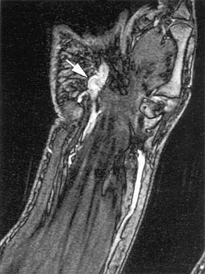
The arrow in the axial T1-weighted MRI scan shown in Figure 18 is pointing to which of the following structures?

Anatomy Board Review 2005: High-Yield MCQs (Set 2) - Figure 6


Explanation

The arrow is pointing to the ulnar nerve within Guyon's canal. Guyon's canal is approximately 4 cm long, beginning at the proximal extent of the transverse carpal ligament and ending at the aponeurotic arch of the hypothenar muscles. Many structures comprise the boundaries of Guyon's canal. The floor, for example, consists of the transverse carpal ligament, the pisohamate and pisometacarpal ligaments, and the opponens digiti minimi. Within Guyon's canal, the ulnar nerve bifurcates into the superficial and deep branches, with the deep branch of the ulnar nerve persisting distal to the canal. The ulnar artery is immediately adjacent and radial to the ulnar nerve. The median nerve is visualized within the carpal tunnel. The radial artery is on the radial side of the wrist. The hook of the hamate is clearly seen in the figure, orienting the observer to the ulnar side of the wrist. Goss MS, Gelberman RH: The anatomy of the distal ulnar tunnel. Clin Orthop 1985;196:238-247.

Question 6

Osteonecrosis of the femoral head after intramedullary nailing in children is thought to be the result of injury to the

Anatomy Board Review 2005: High-Yield MCQs (Set 2) - Figure 7


Explanation

All of these are possible explanations for the development of osteonecrosis following intramedullary nailing in children. However, the lateral ascending cervical artery, which supplies the epiphysis, is much more vulnerable to injury in children because it lies in the trochanteric fossa. Buckley SL: Current trends in the treatment of femoral shaft fractures in children and adolescents. Clin Orthop 1997;338:60-73.

Question 7

The illustration shown in Figure 19 shows a Chamberlain line. What is the most likely diagnosis?

Anatomy Board Review 2005: High-Yield MCQs (Set 2) - Figure 8


Explanation

Basilar invagination is best defined as vertical or compressive instability at the occiput-C1 joint. Such invaginations most commonly occur in patients with rheumatoid arthritis but also can occur secondary to trauma or tumor. A Chamberlain line is used as a method to determine basilar invagination. The odontoid tip should not be more than 5 mm above a Chamberlain line. Wiesel SW, Rothman RH: Occipito-atlantal hypermobility. Spine 1979;4:187-191.

Question 8

Figures 20a and 20b show the sagittal and coronal T1-weighted MRI scans of a patient's left knee. Abnormal findings include


Explanation

The MRI scans show meniscal tissue extending across the entire lateral compartment, revealing a discoid lateral meniscus. The increased signal within the lateral meniscal tissue indicates a tear. Discoid lateral menisci are congenital variants that often present with mechanical symptoms in adolescents. The other structures in the knee are normal. Ahn JH, Shim JS, Hwang CH, et al: Discoid lateral meniscus in children: Clinical manifestations and morphology. J Pediatr Orthop 2001;21:812-816.

Question 9

An ulnar nerve palsy at the level of the wrist is typically associated with deficits in the palmaris brevis, the hypothenar muscles, and what other groups of muscles?

Anatomy Board Review 2005: High-Yield MCQs (Set 2) - Figure 11


Explanation

The intrinsic muscles innervated by the ulnar nerve include the palmaris brevis, hypothenar muscles, all of the interossei, adductor pollicis, and the deep head of the flexor pollicis brevis. The superficial head of the flexor pollicis brevis is innervated by the median nerve. Goldfarb CA, Stern PJ: Low ulnar nerve palsy. JASSH 2003;3:14-26.

Question 10

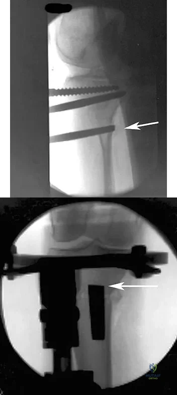
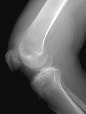
Figures 21a and 21b show the radiographs of a 22-year-old man who has had progressive pain and swelling about the knee for the past 6 weeks. Examination reveals limited range of motion and fullness about the knee. What is the most likely diagnosis?


Explanation

The radiographs reveal a destructive lesion in the metaphysis of the distal femur with periosteal changes and an associated soft-tissue mass with subtle mineralization. This suggests an aggressive malignant process. In this age group, the most likely diagnosis is osteosarcoma. Giant cell tumor, which usually is in a more subchondral location, is not typically so aggressive. Aneurysmal bone cyst is usually more geographic, with a well-marginated reactive rim. Menendez LR (ed): Orthopaedic Knowledge Update: Musculoskeletal Tumors. Rosemont, IL, American Academy of Orthopaedic Surgeons, 2002, pp 175-186.

Question 11

The anterolateral (Watson-Jones) approach to the hip exploits the intermuscular interval between the

Anatomy Board Review 2005: High-Yield MCQs (Set 2) - Figure 14


Explanation

The Watson-Jones approach to the hip uses the intermuscular interval between the gluteus medius and the tensor fascia lata. This is not a true internervous plane, as both muscles are supplied by the superior gluteal nerve. Hoppenfeld S, deBoer P: Surgical Exposures in Orthopaedics: The Anatomic Approach. Philadelphia, PA, JB Lippincott, 1984, pp 316-332.

Question 12

An 8-month-old infant has an infection of the fingertip as shown in Figure 22. If neglected, the anticipated path of ascending infection is the fingertip, the flexor sheath, and the

Anatomy Board Review 2005: High-Yield MCQs (Set 2) - Figure 15


Explanation

The flexor sheaths are in continuity with the deep spaces of the hand. The flexor sheaths of the thumb and little finger communicate with the radial and ulnar bursae, respectively, and these two bursae commonly communicate. The central digits do not communicate as readily with deep spaces of the hand but if flexor tendon sheath infection of the index, long, and right fingers is neglected, the potential exists for rupture into the deep midpalmar spaces. Peimer CA (ed): Surgery of the Hand and Upper Extremity: Acute and Chronic Sepsis. New York, NY, Mcgraw Hill, 1996, pp 1735-1741.

Question 13

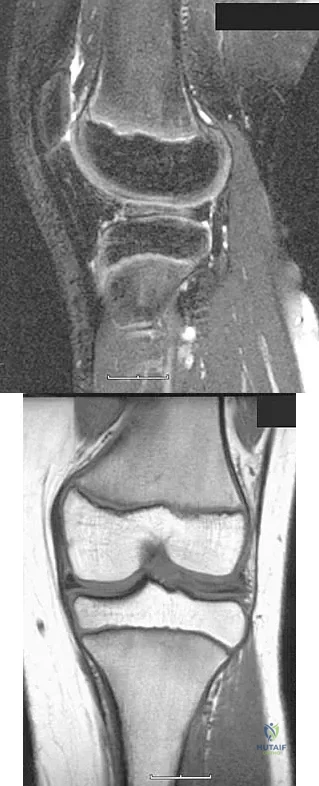
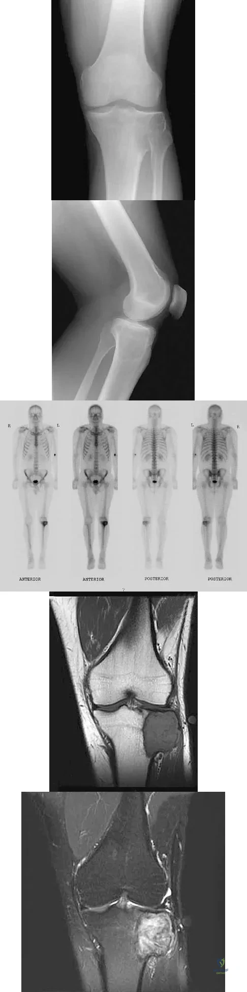
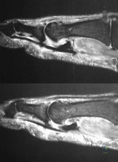
A 24-year-old man has had pain in the left knee for the past several months. He reports that initially the pain was associated with weight-bearing activities, but it has now become more constant. He denies any swelling but reports a lateral fullness at the tibial plateau. Figures 23a through 23e show radiographs, a bone scan, and T1- and T2-weighted MRI scans. What is the most likely diagnosis?


Explanation

The radiographs reveal a lytic subchondral lesion that has a poorly defined margin and lacks mineralization. The bone scan confirms an active lesion that has central photopenia, producing the characteristic doughnut configuration. The MRI scans confirm the presence of a subchondral lesion that is modestly expansile at the lateral plateau and has low signal intensity on the T1-weighted image and a mixed high signal on the T2-weighted image. These features strongly suggest giant cell tumor of bone, more than 50% of which appear around the knee. Simple cyst is excluded by the MRI characteristics. Fibrous dysplasia is unlikely to be in a subchondral location and typically does not show this intensity of uptake on bone scan. Parsons TW: Benign bone tumors, in Fitzgerald RH, Kaufer H, Malkani AL (eds): Orthopaedics. St Louis, MO, Mosby, 2002, pp 1027-1035.

Question 14

Figure 24 shows an axial MRI scan of the ankle. The arrowhead is pointing to what structure?

Anatomy Board Review 2005: High-Yield MCQs (Set 2) - Figure 21


Explanation

The peroneus brevis is easily identified by its location behind the fibula and its distal muscle belly. Axial MRI images provide a reliable guide even when one of the peroneals is completely ruptured, subluxated out of the peroneal groove, or absent. Sarrafian SK: Anatomy of the Foot and Ankle: Descriptive, Topographic, Functional, ed 2. New York, NY, Lippincott, 1993, pp 234-235.

Question 15

During total hip arthroplasty, profuse bleeding is noted following predrilling for placement of an acetabular component screw. The drill most likely penetrated too deep in the

Anatomy Board Review 2005: High-Yield MCQs (Set 2) - Figure 22


Explanation

The acetabular quadrants are defined by two lines: one drawn from the anterosuperior iliac spine to the posterior fovea, forming acetabular halves, and a second drawn perpendicular to the first at the midpoint of the acetabulum, forming four quadrants. The anterior quadrants should be avoided because improper screw placement may injure the external iliac artery and vein, as well as the obturator nerve, artery, and vein. These structures lie close to the pelvic bone, with little protective interposition of soft tissue. Wasielewski RC, Cooperstein LA, Kruger MP, et al: Acetabular anatomy and the transacetabular fixation of screws in total hip arthroplasty. J Bone Joint Surg Am 1990;72:501-508.

Question 16

A posterolateral approach to the tibial plafond proceeds between what two muscles?

Anatomy Board Review 2005: High-Yield MCQs (Set 2) - Figure 23


Explanation

A posterolateral approach to the posterior malleolus proceeds between the lateral and deep posterior compartments. Distally, the peroneus brevis muscle lies most medially within the lateral compartment, and the flexor hallucis longus lies most laterally in the deep posterior compartment. Henry AK: Extensile Exposure, ed 2. Edinburgh, UK, Churchill Livingstone, 1973, pp 269-270.

Question 17

The brachialis muscle is innervated by what two nerves?

Anatomy Board Review 2005: High-Yield MCQs (Set 2) - Figure 24


Explanation

The brachialis is innervated by two nerves: medially, the musculocutaneous nerve; laterally, the radial nerve. The muscle is split longitudinally to approach the humerus anteriorly. Henry AK: The distal part of the humerus and front of the forearm, in Henry AK (ed): Extensile Exposure, ed 2. Edinburgh, UK, Churchill Livingstone, 1973, pp 90-115.

Question 18

Figure 25 shows the CT scan of an adult patient who has neck pain following a motor vehicle accident. What is the most likely diagnosis?

Anatomy Board Review 2005: High-Yield MCQs (Set 2) - Figure 25


Explanation

If the atlanto-dens interval is greater than 3 mm in an adult, a transverse ligament rupture usually is suspected. The atlanto-dens interval can be seen with CT or in lateral radiographs of the upper cervical spine. Transverse ligament rupture can occur as an isolated entity or in association with an odontoid or a Jefferson's fracture. Patients with this type of injury usually require fusion. Dickman CA, Greene KA, Sonntag VK: Injuries involving the transverse atlantal ligament: Classification and treatment guidelines based upon experience with 39 injuries. Neurosurgery 1996;38:44-50.

Question 19

Which of the following best describes the course of the ulnar nerve in the midforearm?

Anatomy Board Review 2005: High-Yield MCQs (Set 2) - Figure 26


Explanation

In the midforearm, the ulnar nerve travels deep to the flexor carpi ulnaris muscle and ulnar to the ulnar artery as it lies on the flexor digitorum profundus muscle. In this region, the ulnar nerve and artery lie side-by-side, whereas more proximal in the forearm, the ulnar artery originates from the brachial artery in the antecubital fossa, and the ulnar nerve lies within the cubital tunnel. Hoppenfeld S, deBoer P: Surgical Exposures in Orthopaedics, ed 2. Philadelphia, PA, Lippincott-Raven, 1994, pp 118-131.

Question 20

A 70-year-old former baseball catcher reports long-standing pain in the ring and little fingers. A gradient-echo MRI scan is shown in Figure 26. What is the most likely diagnosis?

Anatomy Board Review 2005: High-Yield MCQs (Set 2) - Figure 27


Explanation

The gradient-echo MRI scan highlights the ulnar and radial arteries, as indicated by the arrow. This technique suppresses the signal of the surrounding fat and causes the stationary surrounding tissues to become intermediate in signal intensity. The flowing blood is then easily identified with a bright signal because it does not absorb the radiofrequency pulse. Based on the findings, the diagnosis is an ulnar artery aneurysm, most likely caused by years of repetitive trauma as the result of catching baseballs. Neurolemmoma and giant cell tumor of the tendon sheath would be intermediately enhanced on this image sequence, and the continuity with the ulnar artery, demonstrated here, would not be expected. Lipomas are not enhanced using the gradient-echo technique. The chronic nature of the patient's symptoms is not indicative of a hematoma, and the hematoma would be dark on this imaging sequence since it is stationary tissue. Koman LA, Ruch DS, Patterson Smith B, et al: Vascular disorders, in Green DP, Hotchkiss RN, Pederson WC (eds): Green's Operative Hand Surgery, ed 4. New York, NY, Churchill Livingstone, 1999, vol 2, pp 2254-2302.

Question 21

In a postganglionic brachial plexus lesion at Erb's point (point of formation of the upper trunk by the C5 and C6 nerve roots), which of the following nerves will still function normally?

Anatomy Board Review 2005: High-Yield MCQs (Set 2) - Figure 28


Explanation

In a postganglionic injury to the brachial plexus, the rhomboid muscle, innervated by the dorsal scapular nerve, would still be expected to function. This is a useful clinical sign that the brachial plexus lesion is postganglionic as opposed to preganlionic. The musculocutaneous, axillary, and suprascapular nerves are all located distal to Erb's point (the most common location of an upper nerve root brachial plexus injury), and all contain fibers from the C5 and C6 nerve roots. Therefore, these nerves are not expected to function normally following a postganglionic C5 and C6 nerve root injury. Netter F: The Ciba Collection of Medical Illustrations: The Musculoskeletal System: Part 1, Anatomy, Physiology and Metabolic Disorders. West Caldwell, NJ, Ciba-Geigy, 1991, vol 8, pp 28-29.

Question 22

The posterior circumflex humeral artery and the axillary nerve usually lie in a space bordered superiorly by the

Anatomy Board Review 2005: High-Yield MCQs (Set 2) - Figure 29


Explanation

The quadrangular space is bordered superiorly by the teres minor, medially by the long head of the triceps, laterally by the humerus, and inferiorly by the teres major. The posterior circumflex humeral artery and the axillary nerve lie in this space. Rockwood CA Jr, Matsen FA III: The Shoulder, ed 2. Philadelphia, PA, WB Saunders, 1998, vol 1, pp 70-71.

Question 23

A patient notes pain under the first metatarsophalangeal joint following a soccer injury. The MRI scans shown in Figures 27a and 27b reveal what pathologic finding?


Explanation

The MRI scans show a complete disruption of the sesamoid complex with proximal retraction of the medial sesamoid and high signal originating from the site normally occupied by the plantar plate (metatarsophalangeal ligament). This injury is the result of a hyperextension injury and is a severe variant of a turf toe. Watson TS, Anderson RB, Davis WH: Periarticular injuries to the hallux metatarsophalangeal joint in athletes. Foot Ankle Clin 2000;5:687-713.

Question 24

When performing the exposure for an anterior approach to the cervical spine, excessive retraction of the trachea and esophagus should be avoided to prevent injury of the

Anatomy Board Review 2005: High-Yield MCQs (Set 2) - Figure 32


Explanation

The recurrent laryngeal nerve lies between the trachea and the esophagus and is subject to stretch injury if excessive retraction is applied. The vagus nerve lies in the carotid sheath. The sympathetic trunk lies anterior to the longus colli muscles. The hypoglossal nerve and superior laryngeal nerve are both at risk during the exposure but are not located between the trachea and esophagus. An HS: Principles and Techniques of Spine Surgery. Baltimore, MD, Williams and Wilkins, 1998, chapter 2.

Question 25

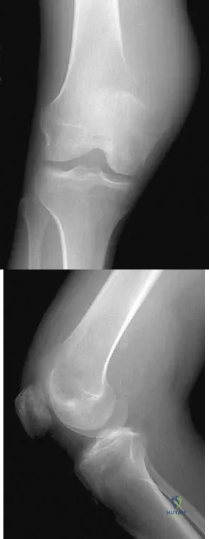
What is the first ossification center to appear radiographically in the pediatric elbow?

Anatomy Board Review 2005: High-Yield MCQs (Set 2) - Figure 33


Explanation

The first ossification center to appear in the pediatric elbow is the capitellum. This ossification center generally appears between the first month and the 11th month in girls and between the first month and the 26th month in boys. The other ossification centers in the elbow appear in the following progression: radial head (3.8 to 4.5 years), medial epicondyle (5 to 6 years), olecranon (6 to 7 years), trochlea (9 to 10 years), and the lateral epicondyle (10 years). Wilkins KE, Beaty JH, Chambers HG, et al: Fractures and dislocation of the elbow region, in Rockwood CA, Green DP, Bucholz RW, et al (eds): Rockwood and Green's Fractures in Adults, ed 4. Philadelphia, PA, Lippincott-Raven, 1996, pp 657-662.

Question 26

During the anterior (Henry) approach to the forearm, the surgeon must exploit a specific internervous plane to safely expose the radius. Which of the following correctly describes this proximal internervous plane?


Explanation

The proximal internervous plane of the Henry approach is between the brachioradialis (radial nerve) and the pronator teres (median nerve). This allows safe anterior exposure of the proximal radius.

Question 27

The anterior (Smith-Petersen) approach to the hip provides excellent exposure for pelvic osteotomies and total hip arthroplasty. The superficial internervous plane for this approach utilizes which two muscles?


Explanation

The superficial plane of the Smith-Petersen approach lies between the sartorius (femoral nerve) and the tensor fasciae latae (superior gluteal nerve). The deep plane lies between the rectus femoris and gluteus medius.

Question 28

A posterior approach to the shoulder may place the axillary nerve at risk as it exits the quadrangular space. Which of the following structures forms the superior border of the quadrangular space?


Explanation

The quadrangular space is bordered superiorly by the teres minor, inferiorly by the teres major, medially by the long head of the triceps, and laterally by the humeral surgical neck. It transmits the axillary nerve and posterior humeral circumflex artery.

Question 29

During a posterolateral approach to the distal humerus, the radial nerve must be identified and protected. At approximately what distance proximal to the lateral epicondyle does the radial nerve pierce the lateral intermuscular septum?


Explanation

The radial nerve travels from the posterior to the anterior compartment by piercing the lateral intermuscular septum approximately 10 cm proximal to the lateral epicondyle. This is a critical anatomical landmark during distal humerus surgery.

Question 30

Avascular necrosis of the proximal pole of the scaphoid is a known complication of scaphoid waist fractures due to its retrograde blood supply. The primary blood supply to the proximal pole enters via which of the following vessels?


Explanation

The dorsal carpal branch of the radial artery enters the distal pole of the scaphoid and provides retrograde flow to the proximal 80% of the bone. This unique vascular anatomy explains the high rate of proximal pole AVN following waist fractures.

Question 31

In severe rotational ankle fractures, the deltoid ligament may be disrupted. Which component of the deltoid ligament complex is the primary static restraint to lateral displacement and external rotation of the talus?


Explanation

The deep posterior tibiotalar ligament is the strongest component of the deltoid ligament complex. It acts as the primary restraint against lateral shift and external rotation of the talus within the mortise.

Question 32

During clinical assessment of the distal radioulnar joint (DRUJ) for instability, understanding the tensioning of the radioulnar ligaments is essential. Which ligamentous structure is under maximum tension when the forearm is placed in full supination?


Explanation

The volar (palmar) radioulnar ligament of the TFCC is tightest in full supination, preventing dorsal translation of the ulna. Conversely, the dorsal radioulnar ligament becomes tightest in full pronation.

Question 33

Following a displaced femoral neck fracture in an adult, the femoral head is at high risk for osteonecrosis. The most significant contributor to the vascular supply of the adult femoral head is the:


Explanation

The deep branch of the medial femoral circumflex artery (MFCA) provides the predominant blood supply to the adult femoral head. It courses posterior to the femoral neck, making it vulnerable in displaced fractures.

Question 34

Fractures of the talar neck frequently lead to avascular necrosis of the talar body. The body of the talus receives its major blood supply from the artery of the tarsal canal, which is a direct branch of the:


Explanation

The artery of the tarsal canal arises from the posterior tibial artery and provides the dominant blood supply to the talar body. It forms an anastomotic sling with the artery of the sinus tarsi.

Question 35

When evaluating a patient with recurrent anterior shoulder instability, the primary static restraint to anterior translation must be assessed. At 90 degrees of shoulder abduction and external rotation, which structure is the primary restraint?


Explanation

The anterior band of the inferior glenohumeral ligament (IGHL) complex is the primary static restraint to anterior and inferior humeral head translation when the arm is abducted to 90 degrees and externally rotated.

Question 36

The lumbrical muscles of the hand are unique in that they both originate from and insert onto tendons. Which of the following correctly describes the innervation of the lumbrical muscle associated with the ring finger?


Explanation

The third and fourth lumbricals (ring and small fingers) are innervated by the deep branch of the ulnar nerve. The first and second lumbricals (index and middle fingers) are innervated by the median nerve.

Question 37

The anterior cruciate ligament (ACL) is composed of two functional bundles named for their tibial insertion sites. Which of the following statements regarding the anteromedial (AM) bundle is correct?


Explanation

The anteromedial (AM) bundle of the ACL is tightest in flexion and is the primary restraint to anterior tibial translation. The posterolateral (PL) bundle is tightest in extension and primarily controls rotatory stability.

Question 38

The posterolateral (Kocher) approach to the elbow is frequently used for radial head fractures. This approach exploits an internervous plane between which two muscles?


Explanation

The Kocher approach utilizes the internervous plane between the extensor carpi ulnaris (posterior interosseous nerve) and the anconeus (radial nerve). This protects the neural structures during joint access.

Question 39

When placing pedicle screws in the lumbar spine, understanding the changing regional anatomy is critical for safe trajectory. Compared to the T12 pedicle, the typical L5 pedicle is:


Explanation

As you move caudally from the thoracolumbar junction to the lower lumbar spine, the pedicles generally become wider in diameter and have an increased medial (convergent) transverse angle.

Question 40

During a surgical release for a recalcitrant trigger finger, a specific annular pulley is incised. What is the location of this pulley relative to the digit's joints?


Explanation

Trigger finger release involves sectioning the A1 pulley, which overlies the metacarpophalangeal (MCP) joint. The A2 and A4 pulleys must be preserved to prevent flexor tendon bowstringing.

Question 41

In the proximal forearm, the median nerve passes between the two heads of the pronator teres. Which of the following structures anatomically separates the median nerve from the ulnar artery at this level?


Explanation

The deep (ulnar) head of the pronator teres separates the median nerve from the underlying ulnar artery. The median nerve passes deep to the humeral head but superficial to the ulnar head.

Question 42

A surgeon is performing a posterolateral approach to the ankle for fixation of a posterior malleolus fracture. During the superficial dissection, the sural nerve must be protected. The sural nerve runs in close proximity to which structure?


Explanation

The sural nerve provides sensation to the posterolateral aspect of the distal third of the leg and lateral foot. It travels alongside the small saphenous vein in the posterolateral ankle.

Question 43

During an anterolateral approach to the distal tibia, the superficial peroneal nerve must be identified and protected. At what approximate level does this nerve typically pierce the deep fascia to become subcutaneous?


Explanation

The superficial peroneal nerve provides motor innervation to the lateral compartment before piercing the deep crural fascia approximately 10 to 15 cm proximal to the lateral malleolus to provide dorsal foot sensation.

Question 44

The pes anserinus is frequently utilized as a harvest site for autograft in anterior cruciate ligament reconstruction. From anterior to posterior, what is the anatomical arrangement of these tendinous insertions on the proximal medial tibia?


Explanation

The correct anterior-to-posterior order of the pes anserinus tendons is Sartorius, Gracilis, and Semitendinosus (mnemonic: "Say Grace before Tea").

Question 45

During a radical axillary dissection, a nerve passing posterior to the axillary artery and innervating the latissimus dorsi is inadvertently injured. Which of the following describes the origin of this nerve?


Explanation

The thoracodorsal nerve innervates the latissimus dorsi. It branches from the posterior cord of the brachial plexus and courses posterior to the axillary artery.

Question 46

The primary bony stabilizer of the Lisfranc joint complex is the base of the second metatarsal.

Which of the following accurately describes its articulation with the cuneiforms?


Explanation

The base of the second metatarsal forms a keystone that is recessed between the medial and lateral cuneiforms, articulating proximally with the middle cuneiform and medially/laterally with the medial and lateral cuneiforms, respectively.

Question 47

A patient sustains an injury to the posterolateral corner of the knee. The popliteofibular ligament is identified as a critical stabilizer. From which structure does it anatomically originate?


Explanation

The popliteofibular ligament is a static stabilizer of the posterolateral corner. It originates from the musculotendinous junction of the popliteus and inserts onto the posteromedial aspect of the fibular styloid.

Question 48

In the hand, the lumbrical muscles flex the metacarpophalangeal joints and extend the interphalangeal joints. What is the origin of the third lumbrical?


Explanation

The first and second lumbricals are unipennate, originating from the radial side of their respective FDP tendons. The third and fourth are bipennate, originating from the adjacent sides of the FDP tendons.

Question 49

The rotator interval is a critical anatomical space in the anterior shoulder. Which of the following structures is NOT considered a border or content of the rotator interval?


Explanation

The rotator interval is bordered by the supraspinatus superiorly and the subscapularis inferiorly. It contains the long head of the biceps tendon, the coracohumeral ligament, and the superior glenohumeral ligament.

Question 50

An intracapsular femoral neck fracture frequently compromises the blood supply to the femoral head. Which artery provides the primary blood supply to the mature adult femoral head?


Explanation

The medial femoral circumflex artery (MFCA) provides the dominant blood supply to the mature femoral head via its lateral epiphyseal branches. The artery of the ligamentum teres provides a negligible supply in adults.

Question 51

When performing a standard posterior approach to the lumbar spine, the surgeon exposes the pars interarticularis. Which neural structure lies immediately anterior to the pars interarticularis?


Explanation

The exiting nerve root lies immediately anterior to the pars interarticularis within the neural foramen. It is highly vulnerable during overly aggressive bone removal at the pars.

Question 52

The central band of the forearm interosseous membrane is essential for longitudinal load transfer. What is the anatomical orientation of its fibers?


Explanation

The central band of the interosseous membrane runs obliquely from the proximal radius to the distal ulna. This orientation facilitates the transfer of axial loads from the radius to the ulna.

Question 53

The anterior inferior tibiofibular ligament (AITFL) is commonly torn in syndesmotic ankle sprains. Where does this ligament primarily insert on the tibia?


Explanation

The AITFL inserts anteriorly on the tibia at Chaput's tubercle and on the fibula at Wagstaffe's tubercle. Volkmann's tubercle is the posterior tibial insertion for the PITFL.

Question 54

During an ilioinguinal approach to the acetabulum, the surgeon must ligate the corona mortis to prevent massive hemorrhage. This structure is an anastomosis between which two vascular systems?


Explanation

The corona mortis is a vascular anastomosis between the external iliac (or inferior epigastric) vessels and the internal iliac (obturator) vessels. It traverses the superior pubic ramus.

Question 55

The triangular fibrocartilage complex (TFCC) stabilizes the distal radioulnar joint (DRUJ). Which component is the primary restraint to dorsal translation of the distal ulna when the forearm is in pronation?


Explanation

During forearm pronation, the dorsal radioulnar ligament becomes taut and acts as the primary restraint against dorsal translation of the distal ulna. Conversely, the volar radioulnar ligament tightens in supination.

Question 56

The anterior bundle of the ulnar collateral ligament (UCL) of the elbow is the primary restraint to valgus stress. Where is its primary anatomical insertion on the ulna?


Explanation

The anterior bundle of the UCL originates on the anteroinferior surface of the medial epicondyle. It inserts on the sublime tubercle of the proximal ulna.

Question 57

Entrapment of the suprascapular nerve at the spinoglenoid notch, often due to a paralabral cyst, typically results in isolated weakness of which muscle?


Explanation

The suprascapular nerve innervates the supraspinatus before passing through the spinoglenoid notch. Entrapment at the spinoglenoid notch therefore causes isolated denervation and weakness of the infraspinatus.

Question 58

Following a proximal fibular fracture, a patient develops a deep peroneal nerve palsy. On physical examination, where would sensation most likely be decreased?


Explanation

The deep peroneal nerve provides motor innervation to the anterior compartment of the leg. Its sensory distribution is exclusively limited to the first dorsal web space of the foot.

Question 59

During anterior cervical spine surgery, aggressive dissection lateral to the uncovertebral joints puts the vertebral artery at risk. At which cervical level does the vertebral artery typically first enter the foramen transversarium?


Explanation

The vertebral artery typically enters the transverse foramen at the C6 level. It is vulnerable during dissection lateral to the uncovertebral joints at C6 and superiorly.

Question 60

When performing a lateral approach to the fibula, the superficial peroneal nerve is at risk as it exits the deep fascia to become subcutaneous. At approximately what distance proximal to the lateral malleolus does this typically occur?


Explanation

The superficial peroneal nerve pierces the crural fascia to become subcutaneous in the lateral leg approximately 10 to 12 cm proximal to the tip of the lateral malleolus.

Question 61

The recurrent motor branch of the median nerve (the "million dollar nerve") provides critical motor function to the hand. Which of the following muscle combinations does it innervate?


Explanation

The recurrent motor branch of the median nerve innervates the thenar eminence musculature. This includes the abductor pollicis brevis, opponens pollicis, and the superficial head of the flexor pollicis brevis.

Question 62

The spring ligament is a critical static stabilizer of the longitudinal arch of the foot, often implicated in adult acquired flatfoot deformity. What are its precise anatomical attachments?


Explanation

The spring ligament, or plantar calcaneonavicular ligament, is a major support for the talar head. It originates from the sustentaculum tali of the calcaneus and inserts onto the plantar-medial navicular.

Question 63

The quadrilateral space of the shoulder transmits the axillary nerve and the posterior circumflex humeral artery.

Which muscle defines the superior boundary of this space?


Explanation

The quadrilateral space is bordered superiorly by the teres minor, inferiorly by the teres major, medially by the long head of the triceps, and laterally by the surgical neck of the humerus.

Question 64

During a posterior approach to the hip (Kocher-Langenbeck), which of the following short external rotators should be preserved to protect the deep branch of the medial circumflex femoral artery?


Explanation

The medial circumflex femoral artery courses anterior to the obturator externus and quadratus femoris. Preserving the obturator externus minimizes the risk of injury to the vessel and subsequent avascular necrosis of the femoral head.

Question 65

When performing a deltoid-splitting surgical approach to the shoulder, the axillary nerve is typically found at what approximate distance distal to the lateral edge of the acromion?


Explanation

The axillary nerve courses circumferentially from posterior to anterior approximately 5 to 7 cm distal to the lateral tip of the acromion. Extending a deltoid split beyond this safe zone places the nerve at significant risk.

Question 66

During a volar (Henry) approach to the proximal radius, how should the forearm be positioned to best protect the posterior interosseous nerve (PIN)?


Explanation

Supination of the forearm moves the insertion of the supinator muscle and the enclosed posterior interosseous nerve (PIN) laterally. This draws the nerve away from the surgical field during anterior exposure of the proximal radius.

Question 67

The popliteus tendon is a critical component of the posterolateral corner of the knee. In relation to the lateral collateral ligament (LCL) footprint, where does the popliteus tendon insert on the lateral femoral condyle?


Explanation

The femoral insertion of the popliteus tendon is located anterior and inferior to the lateral collateral ligament (LCL) origin on the lateral femoral condyle. Understanding this anatomy is essential for anatomic posterolateral corner reconstructions.

Question 68

During a percutaneous repair of an Achilles tendon rupture, the sural nerve is at highest risk of iatrogenic injury. At approximately what distance proximal to the calcaneal tuberosity does the sural nerve typically cross the lateral border of the Achilles tendon?


Explanation

The sural nerve crosses the lateral border of the Achilles tendon approximately 9.8 to 10 cm proximal to the calcaneal insertion. Sutures placed at this level or more proximally on the lateral side must be placed with caution.

Question 69

The "corona mortis" is a significant anatomic structure encountered during the ilioinguinal approach to the acetabulum. It represents a vascular anastomosis between which two systems?


Explanation

The corona mortis is a critical anastomotic connection between the obturator vessels (internal iliac system) and the inferior epigastric vessels (external iliac system). It crosses over the superior pubic ramus and can cause life-threatening hemorrhage if avulsed.

Question 70

During anterior cervical spine surgery, recognizing the course of the vertebral artery is vital. In the majority of individuals, the vertebral artery enters the transverse foramen at which cervical level?


Explanation

In over 90% of the population, the vertebral artery enters the transverse foramen of the cervical spine at the C6 level. Anatomical variants exist where it may enter at C7 or higher levels like C5.

Question 71

Which of the following describes the typical motor innervation of the lumbrical muscles of the hand?


Explanation

The first and second lumbricals are unipennate and innervated by the median nerve. The third and fourth lumbricals are bipennate and innervated by the deep branch of the ulnar nerve.

Question 72

A 25-year-old athlete presents with medial winging of the scapula after a traction injury to the shoulder. Which nerve is most likely injured, and what are its contributing nerve roots?


Explanation

Medial winging of the scapula is caused by paralysis of the serratus anterior muscle. This muscle is innervated by the long thoracic nerve, which arises from the C5, C6, and C7 nerve roots.

Question 73

The short head of the biceps femoris muscle plays a unique anatomical role in the posterior compartment of the thigh. It receives its motor innervation from which of the following nerves?


Explanation

Unlike the other hamstring muscles which are innervated by the tibial division of the sciatic nerve, the short head of the biceps femoris is innervated by the common peroneal division. It originates from the linea aspera and aids in knee flexion.

Question 74

The subscapularis muscle is a crucial dynamic anterior stabilizer of the glenohumeral joint. What is its primary bony footprint insertion site?


Explanation

The subscapularis tendon inserts primarily onto the lesser tuberosity of the proximal humerus. It acts as the primary internal rotator of the shoulder and provides critical anterior joint stability.

Question 75

Surgical decompression of the ulnar nerve at the elbow requires an understanding of the cubital tunnel boundaries. Which structure forms the true floor of the cubital tunnel?


Explanation

The floor of the cubital tunnel is formed by the elbow joint capsule and the posterior bundle of the medial collateral ligament (MCL). Osborne's ligament and the FCU aponeurosis form the roof.

Question 76

During a carpal tunnel release, caution is required to avoid injuring the recurrent motor branch of the median nerve. In the majority of individuals, what is the anatomical relationship of this branch to the transverse carpal ligament?


Explanation

The recurrent motor branch of the median nerve is extraligamentous in approximately 50-80% of individuals, taking a recurrent course distal to the ligament. Subligamentous and transligamentous variants occur less frequently but are at higher risk during surgery.

Question 77

The anterior cruciate ligament (ACL) consists of two main functional bundles. During knee flexion, which bundle is tightest and what is its primary stabilizing function?


Explanation

The anteromedial (AM) bundle of the ACL is tightest in knee flexion and provides the primary restraint to anterior tibial translation. The posterolateral (PL) bundle is tightest in extension and resists rotatory loads.

Question 78

The anterior inferior tibiofibular ligament (AITFL) is a critical stabilizer of the ankle syndesmosis. It originates from the Chaput tubercle on the tibia and inserts onto which bony landmark on the fibula?


Explanation

The AITFL courses obliquely from the anterolateral tibial prominence (Chaput's tubercle) to the anterior fibular tubercle (Wagstaffe's tubercle). Avulsion fractures can occur at either of these insertions during syndesmotic injuries.

Question 79

In the normal lumbar spine anatomy, the exiting nerve root travels through the intervertebral foramen in what relation to the pedicle of the corresponding numbered vertebral body?


Explanation

In the lumbar spine, the exiting nerve root travels inferior to the pedicle of the corresponding vertebra (e.g., the L4 nerve root exits below the L4 pedicle). This relationship is critical during foraminal decompression and pedicle screw placement.

Question 80

Vascular supply to the hand is provided by an extensive anastomotic network. The superficial palmar arch is primarily formed by the direct continuation of which vessel?


Explanation

The superficial palmar arch is primarily formed by the continuation of the ulnar artery, which anastomoses with the superficial palmar branch of the radial artery. Conversely, the deep palmar arch is primarily formed by the radial artery.

Question 81

Talar neck fractures are notorious for causing avascular necrosis of the talar body. The dominant blood supply to the body of the talus is provided by the artery of the tarsal canal, which is a branch of which major artery?


Explanation

The artery of the tarsal canal is typically a branch of the posterior tibial artery and provides the dominant blood supply to the talar body. It forms an anastomotic sling with the artery of the sinus tarsi under the talar neck.

Question 82

During a deltopectoral approach for shoulder arthroplasty, the conjoined tendon is retracted medially. The nerve that pierces the coracobrachialis muscle typically enters it at what distance distal to the coracoid process?


Explanation

The musculocutaneous nerve pierces the coracobrachialis approximately 3 to 8 cm distal to the coracoid tip. Vigorous medial retraction of the conjoined tendon during a deltopectoral approach can cause neuropraxia of this nerve.

Question 83

During a direct lateral (Hardinge) approach to the hip, proximal extension of the gluteus medius split is typically limited to 3-5 cm superior to the greater trochanter to prevent injury to which nerve?


Explanation

The superior gluteal nerve innervates the gluteus medius, gluteus minimus, and tensor fasciae latae. It courses roughly 3 to 5 cm proximal to the tip of the greater trochanter; splitting the muscle beyond this point risks denervation of the anterior hip abductors.

Question 84

During a surgical exploration for radial tunnel syndrome, the surgeon identifies the most common site of compression of the posterior interosseous nerve. This structure is a fibrous band at the proximal edge of which of the following muscles?


Explanation

The Arcade of Frohse is a fibrous arch at the proximal edge of the superficial head of the supinator muscle. It is the most common anatomic site of compression for the posterior interosseous nerve (PIN).

Question 85

An orthopedic surgeon is performing an anterior ilioinguinal approach for an acetabular fracture. Severe hemorrhage is encountered near the superior pubic ramus. This bleeding is most likely from an anastomotic vessel connecting which two arterial systems?


Explanation

The corona mortis is a vascular anastomosis between the obturator (internal iliac) and external iliac (or inferior epigastric) systems located over the superior pubic ramus. Iatrogenic injury during anterior acetabular approaches can cause life-threatening hemorrhage.

Question 86

Following a radical mastectomy, a patient presents with a noticeable "winging" of the scapula with arm elevation. The injured nerve originates from which of the following brachial plexus segments?


Explanation

A winged scapula in this context is typically caused by injury to the long thoracic nerve, which innervates the serratus anterior. This nerve originates directly from the ventral rami of the C5, C6, and C7 nerve roots.

Question 87

The anterolateral (Watson-Jones) approach to the hip utilizes a superficial interval between the tensor fasciae latae and the gluteus medius. What is the innervation of these two muscles respectively?


Explanation

The superficial interval of the anterolateral approach to the hip passes between the tensor fasciae latae and gluteus medius. This is not a true internervous plane, as both muscles are innervated by the superior gluteal nerve.

Question 88

A 28-year-old sustains a displaced talar neck fracture. The primary blood supply to the body of the talus, which is at highest risk of disruption in this injury, is provided by the artery of the tarsal canal. This artery is a direct branch of which of the following?


Explanation

The artery of the tarsal canal provides the dominant blood supply to the body of the talus. It arises as a branch from the posterior tibial artery, usually about 1 cm proximal to its bifurcation.

Question 89

During a deltopectoral approach to the shoulder, the coracoid process may be osteotomized to improve exposure. The surgeon must be careful to avoid placing retractors too distally on the conjoined tendon to prevent injury to which nerve?


Explanation

The musculocutaneous nerve enters the coracobrachialis (part of the conjoined tendon) approximately 5 to 8 cm distal to the tip of the coracoid process. Retractors placed distal to this point can cause neurapraxia or permanent nerve injury.

Question 90

In reconstructing the posterolateral corner (PLC) of the knee, identifying anatomic landmarks is critical. On the lateral femoral condyle, where is the popliteus tendon attachment located relative to the fibular collateral ligament (FCL) origin?


Explanation

On the lateral femoral condyle, the popliteus tendon inserts into a sulcus that is located distal and anterior to the origin of the fibular collateral ligament. This precise anatomical relationship is critical for isometric PLC reconstructions.

Question 91

A surgeon is performing an open reduction and internal fixation of a calcaneus fracture via an extensile lateral approach. Which nerve is at greatest risk of iatrogenic injury during the full-thickness subperiosteal dissection of the posterior vertical limb?


Explanation

The sural nerve runs posterior to the lateral malleolus alongside the small saphenous vein. It is at significant risk of injury during the posterior vertical limb of the extensile lateral approach to the calcaneus.

Question 92

When performing the volar (Henry) approach to the proximal radius, the surgeon develops the interval between the pronator teres and the brachioradialis. Which vascular structure must be ligated and divided to fully mobilize the mobile wad laterally?


Explanation

The radial recurrent artery (along with its accompanying veins, known as the "leash of Henry") crosses the surgical field transversely in the proximal volar approach to the forearm. It must be ligated to allow lateral retraction of the brachioradialis and radial nerve.

Question 93

A patient develops weakness of the deltoid and teres minor following a posterior shoulder dislocation. The injured nerve passes through the quadrangular space. What muscle forms the superior border of this anatomic space?


Explanation

The quadrangular space transmits the axillary nerve and posterior circumflex humeral artery. Its superior border is the teres minor, inferior border is the teres major, medial border is the long head of the triceps, and lateral border is the humeral surgical neck.

Question 94

A hamstring autograft is being harvested for an ACL reconstruction. The surgeon isolates the gracilis and semitendinosus tendons. What is the respective nerve supply to the individual muscles comprising the pes anserinus (Sartorius, Gracilis, Semitendinosus)?


Explanation

The pes anserinus is composed of the conjoined tendons of the sartorius (innervated by the femoral nerve), gracilis (obturator nerve), and semitendinosus (tibial nerve).

Question 95

Avascular necrosis of the proximal pole of the scaphoid is a known complication following a waist fracture. This occurs because the primary arterial supply to the scaphoid enters at which location?


Explanation

The primary blood supply to the scaphoid comes from the dorsal carpal branch of the radial artery, which enters at the dorsal ridge near the distal pole. Blood flows retrograde to the proximal pole, making proximal fractures highly susceptible to osteonecrosis.

Question 96

During a lateral transpsoas approach to the lumbar spine (LLIF), the surgeon must navigate the lumbar plexus carefully to avoid neurologic deficit. Which nerve is classically found emerging directly from the anterior surface of the psoas major muscle?


Explanation

The genitofemoral nerve pierces and emerges from the anterior surface of the psoas major muscle. In contrast, the femoral nerve emerges from its lateral border, and the obturator nerve emerges from its medial border.

Question 97

A deep laceration to the hypothenar eminence severs the deep branch of the ulnar nerve. Assuming isolated injury to this branch, which of the following intrinsic hand muscles would most likely retain normal function?


Explanation

The first and second lumbricals are unipennate muscles innervated by the median nerve. The third and fourth lumbricals, along with the adductor pollicis and all interossei, are innervated by the deep branch of the ulnar nerve.

Question 98

The anterior (Smith-Petersen) approach to the hip exploits a true internervous plane. Which two nerves supply the respective muscles that form the superficial interval of this approach?


Explanation

The superficial interval of the Smith-Petersen approach is between the sartorius (innervated by the femoral nerve) and the tensor fasciae latae (innervated by the superior gluteal nerve), establishing a true internervous plane.

None

Clinic OS
Medically Verified Content by
Prof. Clinic OS
Consultant Orthopedic & Spine Surgeon
Chapter Index