العربية
Part of the Master Guide

AAOS & ABOS Basic Science MCQs (Set 3): Bone Biology, Biomechanics & Anatomy Review

Orthopedic Anatomy MCQs (Set 3): Bone Structure, Joint Kinematics & Ligaments | ABOS & AAOS Review

27 Apr 2026 53 min read 87 Views
Anatomy 2000 MCQs - Part 3

Key Takeaway

This high-yield question set, Set 3, for the ABOS, AAOS, and OITE exams, thoroughly tests core orthopedic anatomical knowledge. It includes MCQs on detailed bone structure and histology, joint kinematics, ligament and tendon anatomy, and essential muscle origins and insertions.

Orthopedic Anatomy MCQs (Set 3): Bone Structure, Joint Kinematics & Ligaments | ABOS & AAOS Review

Comprehensive 100-Question Exam


00:00

Start Quiz

Question 1

A 41-year-old man who plays golf regularly has had ulnar-sided wrist pain for the past several days after striking a tree root with a golf club. Examination reveals significant pain with resisted flexion of the ring and small fingers and tenderness over the hook of the hamate. Which of the following radiographic views would be most helpful in identifying the pathology of this injury?

Anatomy 2000 Practice Questions: Set 3 (Solved) - Figure 1


Explanation

The history and examination findings suggest an acute fracture of the hook of the hamate. The radiographic study considered most helpful in identifying this type of fracture is the carpal tunnel view. PA and lateral views of the wrist will not adequately visualize the hook of the hamate. Bruerton's view is intended for the assessment of the metacarpophalangeal joints. Pathology would not be suspected in the scaphoid, metacarpals, or the phalanges, so the scaphoid view and the PA, lateral, and oblique views of the hand would not be helpful. Green DP, Hotchkiss RN, Pederson WC (eds): Operative Hand Surgery, ed 4. New York, NY, Churchill Livingstone, 1999, p 855.

Question 2

What is the primary limiting membrane and mechanical support for the periphery of the physis?

Anatomy 2000 Practice Questions: Set 3 (Solved) - Figure 2


Explanation

The perichondrial fibrous ring of La Croix acts as a limiting membrane that provides mechanical support for the bone-cartilage junction of the growth plate. It is continuous with the ossification groove of Ranvier, which contributes chondrocytes for the increase in width of the growth plate. The zone of provisional calcification lies at the bottom of the hypertrophic zone and is the site of initial calcification of the matrix. It is quite weak and usually is the cleavage plane for fractures; therefore, it does not qualify as mechanical support. The last intact transverse septum separates the zone of provisional calcification from the primary spongiosa and provides no real support to the physis. The primary spongiosa is the part of the metaphysis nearest the physis. Netter FH: Growth plate, in Woodburne RT, Crelin ES, Kaplan FS, Dingle RV (eds): The Ciba Collection of Medical Illustrations. Summit, NJ, Ciba-Geigy Corporation, 1987, vol 8, pp 166-167.

Question 3

What normal tissue has a low signal intensity (appears black) on both T1- and T2-weighted images?

Anatomy 2000 Practice Questions: Set 3 (Solved) - Figure 3


Explanation

Tendons, cortical bone, ligaments, menisci, and fibrous tissue will show low signal intensity (SI) on both T1- and T2-weighted images. Fat-containing tissues, such as subcutaneous fat and bone marrow, will show high SI on T1-weighted images and low SI on T2-weighted images. Tissues with high water content, such as joint fluid, intervertebral disk, and edema, will show low SI on T1-weighted images and high SI on T2-weighted images. Kasser JR (ed): Orthopaedic Knowledge Update 5. Rosemont, IL, American Academy of Orthopaedic Surgeons, 1996, pp 65-70.

Question 4

A positive Froment sign indicates weakness of which of the following muscles?

Anatomy 2000 Practice Questions: Set 3 (Solved) - Figure 4


Explanation

Thumb adduction is powered by the adductor pollicis (ulnar nerve). Testing involves having the patient forcibly hold a piece of paper between the thumb and radial side of the index proximal phalanx. When this muscle is weak or nonfunctioning, the thumb interphalangeal joint flexes with this maneuver, resulting in a positive Froment sign. The paper is held by action of the thumb flexion (flexor pollicis longus and flexor pollicis brevis; median innervated).

Question 5

Figure 28 shows an AP radiograph of a 54-year-old woman who underwent lumbar laminectomy and fusion at the L4 and L5 levels with placement of a bone stimulator 8 years ago. She also underwent a left total hip arthroplasty 2 years ago; aspiration of that joint now reveals that it is infected with a gram-positive cocci organism. History is also significant for IV drug use and human immunodeficiency virus (HIV). The patient reports fever, chills, and left flank and abdominal pain. Examination reveals significant pain with resisted left hip flexion and passive hip extension. She also has lumbar hyperlordosis. Which of the following studies would best identify the underlying cause of her infection?

Anatomy 2000 Practice Questions: Set 3 (Solved) - Figure 5


Explanation

The patient's clinical signs (fever and flank, hip, and abdominal pain) suggest a primary iliopsoas abscess. With an increased patient population who abuse drugs and/or who are HIV-positive, iliopsoas abscess may be more prevalent because of systemic bacterial seeding and may be potentially unrecognized. Diagnostic imaging studies provide a better understanding of the anatomic magnitude of the infection, give concrete confirmation of the diagnosis, and may suggest an underlying cause. Neither standard abdominal radiographs nor ultrasound studies are sensitive enough to be diagnostic of this disease process. CT has been established as the standard study for identifying the underlying cause of this abscess. The hip infection has most likely developed as a result of hematogenous spread from an infected skin lesion from the patient's IV drug use. Santaella RO, Fishman EK, Lipsett PA: Primary vs secondary iliopsoas abscess: Presentation, microbiology, and treatment. Arch Surg 1995;130:1309-1313.

Question 6

A 55-year-old woman who underwent a left total hip arthroplasty 8 months ago using a modified Hardinger approach reports a persistent painless limp. Examination reveals that when she is not using a cane, she lurches to the left during weight bearing on the left lower extremity. An AP radiograph is shown in Figure 29. Which of the following hip muscle groups should be strengthened to improve the gait abnormality?

Anatomy 2000 Practice Questions: Set 3 (Solved) - Figure 6


Explanation

The modified Hardinger approach includes a partial anterior trochanteric osteotomy creating a trochanteric wafer (as seen on the radiograph) that is displaced anterior and medial in continuity with the gluteus medius and vastus lateralis. Failure of abductor reattachment, migration of the trochanter, nonunion of the osteotomy site, and excessive splitting of the gluteus medius muscle causing injury to the inferior branch of the superior gluteal nerve can result in weakness of the abductor mechanism. Abductor strength should be evaluated with the patient lying on the opposite side and elevating the affected limb. Although slight weakness may manifest itself as a limp only after prolonged muscular activity, significant weakness results in a constant limp without associated discomfort. Morrey BF (ed): Joint Replacement Arthroplasty. New York, NY, Churchill Livingstone, 1991, pp 512-526.

Question 7

Figure 30 shows an axial cross section of extensor tendon anatomy in zone 7 of the wrist. What letter best depicts the location of the posterior interosseous nerve?

Anatomy 2000 Practice Questions: Set 3 (Solved) - Figure 7


Explanation

The posterior interosseous nerve in contained in the floor of the fourth dorsal compartment of the wrist, which is labelled C in this diagram. Tubiana R, McCullough CJ, Masquelet AC: An Atlas of Surgical Exposures of the Upper Extremity. Philadelphia, PA, JB Lippincott, 1990, p 224.

Question 8

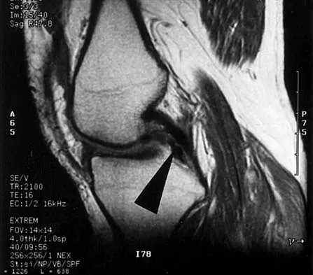
Figures 31a and 31b show the T1- and T2-weighted MRI scans of a patient's knee joint. What is the most likely diagnosis?


Explanation

The scans show a lipohemarthrosis. There is the characteristic layering of a superior zone containing fat (high signal intensity), a central zone containing serum (low signal intensity), and an inferior zone that contains red blood cells (low signal intensity). The most common cause of a lipohemarthrosis is an intra-articular fracture with leakage of marrow fat into the joint. Resnick D, Kang HS: Synovial joints, in Resnick D, Kang HS (eds): Internal Derangements of Joints: Emphasis on MR Imaging. Philadelphia, PA, WB Saunders, 1997, pp 49-53.

Question 9

A 15-year-old boy reports feeling a pop and notes sudden giving way of the left knee while playing basketball. He has immediate pain and swelling in the knee. An AP radiograph is shown in Figure 32. A small avulsion fragment from the lateral tibial margin is the only finding. What is the most likely diagnosis?

Anatomy 2000 Practice Questions: Set 3 (Solved) - Figure 10


Explanation

An avulsion fracture from the lateral tibial margin carries the eponym Segond fracture and is pathognomonic for an anterior cruciate ligament (ACL) tear. The fragment is located posterior to Gerdy's tubercle and is superior and anterior to the fibular head. It represents an avulsion of the lateral capsular ligament of the knee and is caused by the same mechanism that causes the ACL tear. The pes anserinus is the insertion point of the medial hamstrings and would not be affected in a lateral avulsion injury. The posterior cruciate ligament may be seen on a lateral view if associated with an avulsion fragment, but a tear of the PCL generally cannot be diagnosed on an AP view. The insertion of the iliotibial band is broad and is unlikely to produce an avulsion injury such as that seen in the radiograph. This view is not consistent with the appearance of a lateral collateral ligament injury. Beaty JH (ed): Orthopaedic Knowledge Update 6. Rosemont, IL, American Academy of Orthopaedic Surgeons, 1999, pp 533-557.

Question 10

What neurovascular structure is most at risk when performing an inside-out repair of the posterior horn of the medial meniscus?


Explanation

The saphenous nerve is located on the posterior medial aspect of the knee and must be protected when performing an inside-out repair of the medial meniscus. The peroneal nerve is most at risk with lateral meniscal repairs. The other structures usually are not at risk with meniscal repair. Cannon WD Jr, Morgan CD: Meniscal repair: Arthroscopic repair techniques. Instr Course Lect 1994;43:77-96.

Question 11

Figure 33 shows the AP and lateral radiographs of an obese 58-year-old man who underwent a cementless total hip arthroplasty 6 years ago. He reports no pain, and examination reveals a normal gait and painless hip range of motion. What is the most likely diagnosis?

Anatomy 2000 Practice Questions: Set 3 (Solved) - Figure 11


Explanation

Osteolysis of an otherwise well-functioning total hip arthroplasty is a recognized complication, and its radiographic appearance is typical, as shown here. Distal osteolysis, such as that shown here, is more prevalent when there is noncircumferential sealing of the proximal femoral canal. Pellicci PM, Tria AJ Jr, Garvin KL (eds): Orthopaedic Knowledge Update: Hip and Knee Reconstruction 2. Rosemont, IL, American Academy of Orthopaedic Surgeons, 2000, pp 175-180.

Question 12

Which of the following muscles attaches to the coracoid process of the scapula?


Explanation

The insertion of the pectoralis minor is on the base of the coracoid process. The coracoid helps define the interval between the subscapularis and supraspinatus muscles but neither attaches to it. The coracobrachialis and short head of biceps attach to the tip of the coracoid but are not listed as options. The long head of the biceps attaches to the supraglenoid tubercle. The serratus arises from the vertebral border of the scapula. Jobe CM: Anatomy and surgical approaches, in Jobe FW (ed): Operative Techniques in Upper Extremity Sports Injuries. St Louis, MO, Mosby, 1996, pp 140-142.

Question 13

Turf toe typically involves injury to which of the following structures of the great toe?


Explanation

The term turf toe includes a range of injuries of the capsuloligamentous complex of the first metatarsophalangeal joint with or without osteochondral fracture of the first metatarsal head or one of the sesamoids. The mechanism of injury is hyperextension. Clanton TO, Butler JE, Eggert A: Injuries to the metatarsophalangeal joints in athletes. Foot Ankle 1986;7:162-176.

Question 14

Figures 34a through 34c show an axial proton density (spin echo long TR, short TE) image, a sagittal inversion recovery (STIR) image, and a sagittal T1-weighted (short TR, short TE) image of the left thigh. What is the most likely diagnosis?
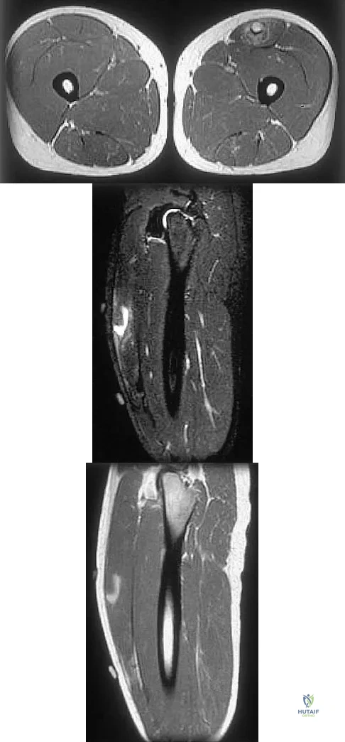


Explanation

The images reveal a region of increased signal within the rectus femoris muscle with mild, ill-defined surrounding edema. The presence of high intensity signal on the T1-weighted image favors acute blood, in this case associated with a rectus femoris muscle tear or fatty tissue. However, because of fat suppression, a fatty lesion or lipoma would be dark on STIR, rather than bright as in this image. Most foreign bodies are low intensity signal and if small, are difficult to evaluate with MRI. The lack of adjacent subcutaneous soft-tissue edema or surrounding fluid makes pyomyositis an unlikely diagnosis.

Question 15

The artery located within the substance of the coracoacromial ligament is a branch of what artery?


Explanation

The acromial branch of the thoracoacromial artery courses along the medial aspect of the coracoacromial ligament and may be encountered when performing an open or arthroscopic subacromial decompression. Bleeding can be controlled by ligation of its branch from the thoracoacromial artery. The other arteries may be injured in other surgical exposures of the shoulder. Esch JC, Baker CL: The shoulder and elbow, in Whipple TL (ed): Arthroscopic Surgery. Philadelphia, PA, JB Lippincott, 1993, pp 65-66.

Question 16

Figures 35a and 35b show the axial T2-weighted and coronal T1-weighted MRI scans of a patient who has enlargement of the right thigh. What is the most likely diagnosis?
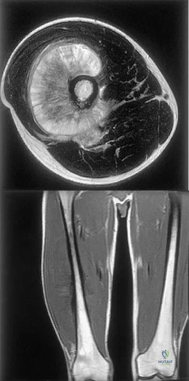


Explanation

The images show a large, almost circumferential, mass surrounding the diaphysis of the femur. The intramedullary signal is normal with minimal cortical destruction, both findings that should be abnormal in conventional osteosarcoma and Ewing's sarcoma. There are very low-signal striations representing osteoid formation that would have a sunburst radiographic pattern. This indicates an osteogenic lesion. Myositis ossificans is not indicated because studies would reveal zonal ossification starting in the periphery rather than the more central pattern seen in this patient. This appearance is typical for periosteal osteosarcoma.

Question 17

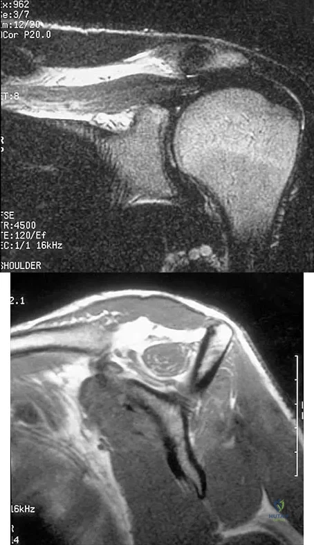
Figures 36a and 36b show the MRI scans of a patient who has shoulder weakness. What is the most likely diagnosis?


Explanation

The sagittal image reveals increased signal and decreased size of the supraspinatus and infraspinatus muscles, indicating muscle atrophy. The rotator cuff tendon signal is normal. The subscapularis and teres minor muscles are unaffected. Muscular dystrophy and thoracic outlet syndrome would be expected to have a more global effect. Although muscular atrophy can occur in the setting of a rotator cuff tear, the coronal image shows an intact supraspinatus. The suprascapular nerve supplies the supraspinatus and infraspinatus muscles. Therefore, suprascapular nerve entrapment would result in atrophy of these muscles with sparing of the surrounding musculature. Any lesion within the suprascapular notch, including neoplastic disease, a venous varix, or neuroma, can place pressure on the suprascapular nerve. Suprascapular nerve entrapment most commonly results from extension of a paralabral cyst or ganglion, often with associated labral pathology. Spinal accessory nerve disruption would show trapezius muscle atrophy. Resnick D, Kang HS (eds): Internal Derangement of Joints: Emphasis on MR Imaging. Philadelphia, PA, WB Saunders, 1997, pp 308-317.

Question 18

Figure 37 shows the T2-weighted MRI scan of the hip joint. What structure is labeled A?

Anatomy 2000 Practice Questions: Set 3 (Solved) Figure 1


Explanation

The obturator internus originates on the obturator membrane and adjacent bone, including the quadrilateral plate, and exits the lesser sciatic notch to insert on the posterior medial greater trochanter. The structure labeled C is the pectineus, B is the sartorius, and D is the gluteus medius. Last RJ: Anatomy: Regional and Applied, ed 6. London, England, Churchill Livingstone, 1978, pp 145-150, 324.

Question 19

The great medullary artery, also known as the Adamkiewicz artery, originates from which of the following arteries?


Explanation

The great medullary artery originates as a direct or indirect branch of the left posterior intercostal artery, usually between T8 and T12. It becomes intradural and crosses over one to three disk spaces before turning to the midline where it anastomoses with the anterior spinal artery. Injury to this artery can result in devastating ischemia of the lower spinal cord. Lu J, Ebraheim NA, Biyani A, Brown JA, Yeasting RA: Vulnerability of great medullary artery. Spine 1996;21:1852-1855.

Question 20

A patient who underwent total knee arthroplasty now reports a loss of sensation in the area circled in Figure 38. This area is innervated by which of the following nerves?

Anatomy 2000 Practice Questions: Set 3 (Solved) - Figure 19


Explanation

The saphenous nerve follows the saphenous vein, giving off the infrapatellar branch that crosses the knee anteriorly to supply the peripatellar skin. A longitudinal incision can interrupt the nerve, leaving the terminal distribution without sensation.

Question 21

During an anterior approach to the shoulder, excessive traction on the conjoined tendon is most likely to result in loss of


Explanation

The musculocutaneous nerve travels through the conjoined tendon approximately 8 cm distal to the tip of the acromion. The musculocutaneous nerve innervates the biceps muscle and the bracialis muscle, both of which are responsible for elbow flexion. Shoulder flexion is facilitated by the anterior fibers of the deltoid muscle (axillary nerve) and the supraspinatus muscle (suprascapular nerve). The subscapular muscle facilitates internal rotation of the shoulder (upper and lower subscapularis nerve). Shoulder abduction is performed by the deltoid muscle (axillary nerve), and forearm pronation is facilitated by the pronator teres (median nerve). Hollinshead WH: Anatomy for Surgeons: The Back and Limbs, ed 3. Philadelphia, PA, Harper and Row, 1982, pp 391-393.

Question 22

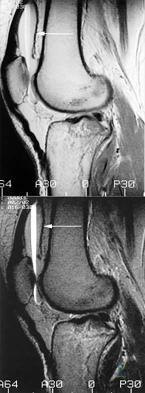
Figure 39 shows the sagittal T1-weighted MRI scan of a 27-year-old man who twisted his knee 2 weeks ago. The arrow is pointing to

Anatomy 2000 Practice Questions: Set 3 (Solved) - Figure 20


Explanation

The arrow identifies a transverse dark line that represents primary trabeculae of the physeal scar. A similar finding is seen in the proximal tibia. These lines may persist indefinitely. They do not represent ongoing growth, an abnormally open physeal plate, a stress fracture, or Looser's line (fatigue fracture in osteomalacia).

Question 23

Figure 40 shows the AP radiograph of a 55-year-old man who reports left knee pain. Which of the following conditions is least likely to produce this radiographic presentation?

Anatomy 2000 Practice Questions: Set 3 (Solved) - Figure 21


Explanation

The radiograph reveals densities within the articular cartilage of the knee commonly referred to as chondrocalcinosis. The term chondrocalcinosis refers to the presence of calcium-containing crystals detected as radiodensities in cartilage. Calcium-containing crystals other than calcium pyrophosphate dihydrate may also deposit in articular cartilage and menisci, producing both radiographically detectable densities in cartilage and joint inflammation or degeneration. Hemochromatosis, alkaptonuria (ochronosis), and Wilson's disease are characterized by cellular deposition of iron, calcium, and copper ions, respectively, into various tissues including articular cartilage and can give this appearance. Septic arthritis does not usually cause chondrocalcinosis. Klippel JH (ed): Primer on the Rheumatic Diseases, ed 11. Atlanta, GA, Arthritis Foundation, 1997, pp 226-229 and 328-331.

Question 24

Figure 41 shows the MRI scan of a 39-year-old man who has severe left groin and anterior thigh pain. What is the most likely diagnosis?

Anatomy 2000 Practice Questions: Set 3 (Solved) - Figure 22


Explanation

The MRI scan shows near complete involvement of the femoral head with bone marrow changes and some collapse of the necrotic segment. This is most suggestive of osteonecrosis.

Question 25

Iliosacral screws placed for stabilization of posterior pelvic ring injuries (eg, sacroiliac dislocation) that exit the sacrum anteriorly are most likely to injure which of the following structures?


Explanation

Iliosacral screws have gained popularity for posterior stabilization of pelvic ring disruptions, but complications attributed to incorrect placement are a clinical problem. The L5 nerve root is at greatest risk and is in closest proximity to a malpositioned screw (exiting the sacrum). The L4 root is more anterior at this level. The S1 root is still intraosseous at this level and is at risk but not from the screw exiting anteriorly at this level. The arteries are at risk but are more anterior and are at less risk than the L5 nerve root.

Question 26

In an anterior cruciate ligament (ACL)-deficient knee, which of the following structures acts as the primary secondary restraint to anterior tibial translation?


Explanation

The posterior horn of the medial meniscus acts as the primary secondary wedge against anterior tibial translation in an ACL-deficient knee. This accounts for the high rate of medial meniscus tears seen in chronic ACL deficiency.

Question 27

The coracoclavicular ligament complex is crucial for acromioclavicular joint stability. Which of the following describes the specific biomechanical function of the conoid ligament?


Explanation

The conoid ligament is the primary restraint to superior clavicular displacement. The trapezoid ligament is positioned anterolateral to the conoid and provides resistance against posterior translation and axial compression.

Question 28

During the late cocking phase of throwing, the shoulder is maximally abducted and externally rotated. Which structural component is the primary restraint to anterior glenohumeral translation in this specific position?


Explanation

The anterior band of the inferior glenohumeral ligament (IGHL) is the primary restraint to anterior translation when the arm is positioned at 90 degrees of abduction and maximal external rotation.

Question 29

A 24-year-old athlete sustains an inversion ankle sprain. Biomechanical studies indicate that the primary ligamentous restraint to inversion of the talus when the ankle is placed in maximal dorsiflexion is the:


Explanation

The calcaneofibular ligament (CFL) becomes taut in dorsiflexion and acts as the primary restraint to inversion in this position. The ATFL is the primary restraint to inversion when the ankle is in plantarflexion.

Question 30

Reconstruction of the medial ulnar collateral ligament (MUCL) of the elbow typically targets the anterior bundle. The anterior bundle is the primary restraint to valgus stress at which of the following elbow flexion angles?


Explanation

The anterior bundle of the MUCL is the primary restraint to valgus stress from approximately 30 degrees to 120 degrees of elbow flexion. In full extension, the radiocapitellar bony articulation provides the primary valgus stability.

Question 31

In mature lamellar bone, osteons are bounded by a structurally distinct boundary that separates them from the surrounding interstitial lamellae. This histological structure is known as the:


Explanation

The cement line marks the outer boundary of an osteon in cortical bone. It indicates the physiological reversal point where osteoclastic bone resorption stopped and osteoblastic new bone formation began.

Question 32

Which of the following accurately describes the anatomy and function of Cleland's ligaments in the hand?


Explanation

Cleland's ligaments are strong fascial bands that pass dorsal to the digital neurovascular bundles. They stabilize the digital skin during flexion and are uniquely spared in Dupuytren's disease, unlike Grayson's ligaments.

Question 33

The dart-throwing motion of the wrist is clinically significant for rehabilitation after specific carpal injuries. Biomechanically, this functional motion occurs primarily at which of the following articulations?


Explanation

The dart-throwing motion, which couples radial deviation/extension with ulnar deviation/flexion, occurs predominantly at the midcarpal joint. This unique kinematic pattern minimizes movement and shear forces across the scaphoid and lunate.

Question 34

During a lateral patellar dislocation, the medial patellofemoral ligament (MPFL) is typically ruptured. Biomechanically, the MPFL provides its greatest contribution to restraining lateral patellar translation at which knee flexion angle?


Explanation

The MPFL is the primary soft-tissue restraint to lateral patellar translation in early knee flexion (0 to 30 degrees). Beyond 30 degrees, the patella fully engages in the trochlear groove, and bony geometry dictates stability.

Question 35

A patient with suspected anterior hip instability requires an assessment of the hip capsular ligaments. Which ligament is the thickest in the body and serves as the primary restraint to hip extension and external rotation?


Explanation

The iliofemoral ligament (Y ligament of Bigelow) spans the anterior hip capsule and is the strongest ligament in the human body. It severely limits hip extension and anterior translation of the femoral head.

Question 36

The sternoclavicular (SC) joint relies heavily on ligamentous stabilizers due to poor bony congruity. Which ligament acts as the primary restraint to superior displacement of the medial clavicle?


Explanation

The costoclavicular ligament (also known as the rhomboid ligament) strongly anchors the inferior aspect of the medial clavicle to the first rib. It is the primary restraint to superior translation of the medial clavicle.

Question 37

Half of all cervical spine rotation occurs at the atlantoaxial (C1-C2) articulation. Which of the following ligaments is the primary restraint to anterior translation of the atlas on the axis?


Explanation

The transverse ligament of the atlas courses posterior to the dens, holding it tightly against the anterior arch of C1. It is the crucial primary stabilizer preventing anterior translation of C1 relative to C2.

Question 38

The lateral meniscus is structurally and kinematically distinct from the medial meniscus. Which of the following is a defining anatomical feature of the lateral meniscus?


Explanation

The lateral meniscus is highly mobile, O-shaped, and is physically separated from the LCL by the popliteus tendon. It receives capsular attachments from the anterior (Humphrey) and posterior (Wrisberg) meniscofemoral ligaments.

Question 39

A pediatric patient sustains a distal radius fracture with apex volar angulation. Over a year, the bone straightens as formation occurs on the concave side and resorption on the convex side. This biomechanical response is described by:


Explanation

Wolff's Law states that bone in a healthy person or animal will adapt to the loads under which it is placed. Remodeling restores the axis by depositing bone on the compressive (concave) side and resorbing it on the tensile (convex) side.

Question 40

A 55-year-old female presents with progressive flattening of her longitudinal arch. Pathology of the posterior tibial tendon is suspected. Which associated ligamentous structure is most commonly attenuated and primarily responsible for supporting the head of the talus?


Explanation

The plantar calcaneonavicular ligament (spring ligament) forms a critical sling under the talar head. Its attenuation, often concurrent with posterior tibial tendon dysfunction, directly contributes to adult acquired flatfoot deformity.

Question 41

In an anteroposterior compression (APC) pelvic ring injury, progressive disruption of ligaments leads to widening of the symphysis. Which sacroiliac (SI) ligament is the strongest and typically tears last, differentiating an APC-II from an APC-III injury?


Explanation

The posterior sacroiliac ligament complex is the strongest ligamentous restraint of the pelvic ring. It remains intact in rotationally unstable but vertically stable APC-II injuries, but is torn in globally unstable APC-III injuries.

Question 42

The alar ligaments play a critical role in craniocervical stability. What is their primary biomechanical function?


Explanation

The alar ligaments connect the superior aspect of the dens to the medial aspects of the occipital condyles. They act as the primary structural restraints to excessive axial rotation and lateral bending in the upper cervical spine.

Question 43

A 28-year-old athlete undergoes knee arthroscopy. The surgeon identifies a ligamentous structure originating from the posterior horn of the lateral meniscus and inserting on the lateral aspect of the medial femoral condyle, passing anterior to the posterior cruciate ligament (PCL). What is this structure?


Explanation

The meniscofemoral ligament of Humphrey passes anterior to the PCL. The ligament of Wrisberg passes posterior to the PCL.

Question 44

Regarding the microscopic structure of cortical bone, which of the following statements most accurately describes Volkmann's canals?


Explanation

Volkmann's canals run transversely through cortical bone to connect the longitudinal Haversian canals with the periosteal and endosteal blood supplies. This network ensures adequate vascularity throughout the dense osteonal system.

Question 45

The screw-home mechanism of the knee is essential for locking the joint in terminal extension. Which of the following kinematic coupled motions occurs during the final 20 degrees of open-chain knee extension?


Explanation

During the final 20 degrees of open-chain extension, the tibia must externally rotate relative to the femur to properly articulate the unequal articular surfaces of the femoral condyles. This locks the knee into its most stable position.

Question 46

A 22-year-old baseball pitcher presents with anterior shoulder pain and apprehension. Biomechanical evaluation of the glenohumeral joint during the late cocking phase of throwing (90 degrees of abduction and maximal external rotation) demonstrates that anterior translation is primarily restrained by which structure?


Explanation

The anterior band of the inferior glenohumeral ligament (IGHL) complex acts like a hammock. It becomes the primary restraint to anterior and inferior humeral head translation when the arm is abducted to 90 degrees and externally rotated.

Question 47

A 34-year-old carpenter presents with wrist pain. Evaluation of his wrist kinematics reveals normal synchronous carpal motion. During active radial deviation of the wrist from a neutral position, what is the normal kinematic behavior of the scaphoid?


Explanation

During radial deviation, the scaphoid normally flexes (palmarflexes) to allow the distal carpal row to move radially without impinging on the radial styloid. Conversely, the scaphoid extends during ulnar deviation.

Question 48

A 26-year-old football player sustains a direct blow to the proximal tibia with the knee flexed, resulting in a posterior cruciate ligament (PCL) tear. The PCL consists of two functional bundles. Which of the following best describes their tension patterns during knee range of motion?


Explanation

The PCL's larger anterolateral (AL) bundle is primarily taut in flexion, preventing posterior translation of the tibia. The smaller posteromedial (PM) bundle becomes taut in extension.

Question 49

In the process of bone remodeling, a distinct basophilic line is formed that marks the outer boundary of a newly created osteon. It is rich in osteopontin and deficient in collagen. What is this histological structure called?


Explanation

The cement line (reversal line) marks the outer limit of bone resorption by osteoclasts and the starting point of new bone deposition by osteoblasts. It is characteristically lacking in collagen fibers.

Question 50

A 19-year-old soccer player suffers an inversion ankle sprain. During the physical examination, the anterior drawer test is positive when the ankle is plantarflexed. Which ligament is primarily assessed, and what is its normal kinematic behavior?


Explanation

The anterior talofibular ligament (ATFL) is the most commonly injured ankle ligament and is maximally taut in plantarflexion. The calcaneofibular ligament (CFL) becomes taut in dorsiflexion.

Question 51

In an acromioclavicular (AC) joint dislocation, the coracoclavicular (CC) ligaments are disrupted. Which of the following describes the correct anatomy and primary biomechanical function of the conoid ligament?


Explanation

The conoid ligament is the medial and posterior component of the CC ligament complex. It acts as the primary restraint to superior translation of the distal clavicle.

Question 52

A 40-year-old man falls on an outstretched hand and develops radial-sided wrist pain. MRI reveals a complete rupture of the scapholunate interosseous ligament (SLIL). The SLIL has three distinct anatomic regions. Which region is the thickest, strongest, and primary restraint to abnormal scapholunate translation?


Explanation

The dorsal region of the scapholunate interosseous ligament is composed of thick, transversely oriented collagen fibers. It is biomechanically the strongest component and the most critical restraint to scapholunate dissociation.

Question 53

A 25-year-old professional javelin thrower presents with medial elbow pain. Valgus stress testing is positive at 30 degrees of elbow flexion. Which structure is the primary restraint to valgus instability in this position?


Explanation

The anterior bundle of the medial collateral ligament (MCL) is the primary restraint to valgus stress at the elbow from approximately 30 to 120 degrees of flexion. The posterior bundle contributes more in deep flexion.

Question 54

During the stance phase of normal human gait, the hip undergoes significant extension. Which capsuloligamentous structure is the primary restraint to hip hyperextension and external rotation, often referred to as the strongest ligament in the body?


Explanation

The iliofemoral ligament (Y ligament of Bigelow) spans anteriorly across the hip joint. It is uniquely structured to prevent hyperextension of the hip while maintaining upright posture without constant muscular effort.

Question 55

A 60-year-old woman complains of anterior knee pain when descending stairs. Patellofemoral joint contact forces change dynamically with knee flexion. At what degree of knee flexion does the patella typically exhibit the maximum contact area with the femoral trochlea, distributing the highest joint reaction forces?


Explanation

The patellofemoral contact area progressively increases as the knee flexes, reaching a maximum at roughly 60 to 90 degrees. This large contact area helps distribute the massively increased joint reaction forces generated during deep flexion activities.

Question 56

Fracture healing progresses through several overlapping phases. During the reparative phase, what type of bone is initially deposited by osteoblasts within the hard callus, characterized by a random, disorganized orientation of collagen fibers?


Explanation

Woven bone is immature bone that is laid down rapidly during fetal development and early fracture healing (hard callus). It is eventually remodeled into highly organized, mechanically stronger lamellar bone.

Question 57

A clinician applies a constant deformation by placing a limb in a corrective cast. Over time, the internal tension within the constrained ligamentous structures gradually decreases. This viscoelastic property of ligaments is best defined as:


Explanation

Stress relaxation is the decrease in stress (tension) over time when a viscoelastic material is held at a constant length or strain. Creep, in contrast, is the increase in length over time under a constant load.

Question 58

During the final 15 degrees of knee extension, the tibia must externally rotate relative to the femur to reach the fully locked position. Which anatomic feature is the primary driver of this 'screw-home' kinematic mechanism?


Explanation

The screw-home mechanism is primarily dictated by the asymmetry of the femoral condyles, where the medial condyle is longer anteriorly than the lateral condyle. Tension in the ACL also assists in driving this terminal external rotation of the tibia.

Question 59

A 12-year-old is evaluated for osteopetrosis. A genetic defect affecting osteoclast function at the ruffled border is suspected. Which cell surface integrin is essential for the sealing zone of the osteoclast to adhere to the bone surface?


Explanation

Osteoclasts utilize the AlphaV Beta3 integrin to bind to bone matrix proteins like osteopontin at the sealing zone. This creates the isolated microenvironment required for acidic bone resorption.

Question 60

A 24-year-old overhead athlete presents with recurrent anterior shoulder instability. Biomechanical testing demonstrates maximum instability when the arm is positioned in 90 degrees of abduction and maximum external rotation. Which specific structure is the primary restraint to anterior translation in this position?


Explanation

The inferior glenohumeral ligament (IGHL) complex is the primary restraint to anterior and inferior subluxation at 90 degrees of abduction. Specifically, the anterior band tightens during external rotation in this abducted position.

Question 61

During rehabilitation of a scapholunate ligament repair, the therapist utilizes the 'dart thrower's motion' to minimize strain on the healing ligament. This motion primarily restricts movement at the midcarpal joint by linking which of the following coupled wrist positions?


Explanation

The 'dart thrower's motion' couples radial extension with ulnar flexion. It is beneficial during early scapholunate rehabilitation because it occurs almost entirely at the radiocarpal joint, thereby minimizing midcarpal (scapholunate) motion.

Question 62

A 13-year-old obese boy presents with groin pain and an obligatory external rotation of the hip with flexion. Radiographs confirm a slipped capital femoral epiphysis (SCFE). Through which specific histologic zone of the physis does this biomechanical failure primarily occur?


Explanation

A slipped capital femoral epiphysis occurs primarily through the hypertrophic zone of the physis. This zone lacks the structural strength of collagen and is the weakest area under shear stress.

Question 63

During an arthroscopic anterior cruciate ligament (ACL) reconstruction, careful attention is paid to the vascular supply of the notch. Which artery provides the primary blood supply to the native ACL?


Explanation

The middle genicular artery pierces the posterior capsule to provide the primary blood supply to the cruciate ligaments. It arborizes within the synovial fold enveloping the ACL and PCL.

Question 64

In mature diaphyseal cortical bone, vascular channels that run transversely and connect the longitudinally oriented central canals of adjacent osteons are known as:


Explanation

Volkmann canals are transverse channels that transmit blood vessels between the longitudinal Haversian canals and connect the periosteal and endosteal blood supplies. Canaliculi are the microchannels connecting osteocyte lacunae.

Question 65

A 35-year-old runner complains of anterior knee pain during deep squats. Biomechanically, as the knee transitions from full extension to 90 degrees of flexion, how does the primary contact area on the articular surface of the patella migrate?


Explanation

As the knee flexes, the patella glides distally into the trochlea, causing the point of contact on the patellar articular surface to migrate from inferior (distal) in early flexion to superior (proximal) in deep flexion. Deep squats place the highest joint reactive forces on the proximal patella.

Question 66

Following a grade II medial collateral ligament (MCL) sprain, the injured tissue undergoes a multiphase healing process. During the early remodeling phase, which type of collagen is most predominantly synthesized to bridge the defect before maturing into stronger tissue?


Explanation

During the early phases of ligament and tendon healing, fibroblasts predominantly synthesize Type III collagen, which forms a disorganized preliminary matrix. As the tissue remodels and matures, this is gradually replaced by the stronger, more organized Type I collagen.

Question 67

Normal kinematics of the subaxial cervical spine exhibit obligate coupled motions due to the spatial orientation of the facet joints. When a patient performs active lateral bending of the neck to the right, which obligate coupled motion naturally occurs?


Explanation

In the subaxial cervical spine (C2-C7), lateral bending is biomechanically coupled with axial rotation to the ipsilateral side. Therefore, right lateral bending couples with right axial rotation.

Question 68

A newborn is evaluated for delayed closure of the cranial sutures and absent clavicles. Genetic testing reveals a mutation in a master transcription factor essential for osteoblast differentiation. Which gene is most likely affected?


Explanation

The patient has cleidocranial dysplasia, which is caused by a mutation in RUNX2 (Cbfa1). RUNX2 is the master transcription factor required for the commitment of mesenchymal stem cells to the osteoblast lineage.

Question 69

A patient is evaluated for chronic ankle instability. Clinical examination includes the anterior drawer test and talar tilt test. Regarding the kinematics of the lateral ankle ligaments, in which position is the calcaneofibular ligament (CFL) under the greatest tension?


Explanation

The calcaneofibular ligament (CFL) becomes tightest in ankle dorsiflexion and inversion. Conversely, the anterior talofibular ligament (ATFL) is tightest in plantarflexion and inversion.

Question 70

When designing a hinged external fixator for a complex elbow fracture-dislocation, precise alignment of the fixator axis is critical. The instant center of rotation of the normal ulnohumeral joint is best described as:


Explanation

The elbow approximates a true hinge joint. The instant center of rotation is tightly constrained to a very small area (essentially a single fixed point) passing through the center of the trochlea and capitellum.

Question 71

Intramedullary nailing of a diaphyseal femur fracture profoundly affects local hemodynamics. In an intact long bone, the normal direction of arterial blood flow within the diaphyseal cortex is primarily:


Explanation

In the mature, uninjured diaphyseal cortex, the high-pressure nutrient artery system drives blood flow centrifugally from the endosteum to the periosteum. Following injury or intramedullary reaming, this flow temporarily reverses to a centripetal direction as the periosteal supply takes over.

Question 72

During a pivot shift test for an ACL deficient knee, the tibia subluxates anteriorly in extension and reduces in flexion. Which specific anatomic bundle of the ACL is primarily responsible for resisting rotatory loads and anterior translation near full knee extension?


Explanation

The anterior cruciate ligament has two distinct bundles. The posterolateral (PL) bundle is tight in extension and primarily controls rotatory stability, whereas the anteromedial (AM) bundle is tight in flexion and controls anterior translation.

Question 73

A military recruit develops a tibial stress fracture during week 4 of basic training. At a cellular level, fatigue failure in cortical bone under cyclical loading initially propagates through the formation of microcracks. What is the primary biological mechanism that attempts to repair this microdamage before macroscopic failure occurs?


Explanation

Microdamage in cortical bone triggers targeted remodeling by basic multicellular units (BMUs). Osteoclasts resorb the damaged matrix, followed by osteoblasts depositing new bone; a stress fracture occurs when repetitive loading outpaces this targeted repair.

Question 74


A 22-year-old female presents with activity-related groin pain. Radiographic evaluation of her hip joint includes measurement of the lateral center-edge angle of Wiberg. This specific angle is utilized to evaluate the joint's biomechanical environment by quantifying:


Explanation

The lateral center-edge angle of Wiberg is measured on an AP pelvis radiograph to assess the superolateral coverage of the femoral head. An angle less than 20 degrees is indicative of acetabular dysplasia.

Question 75

When biomechanically testing a biological ligament, a cyclic loading and unloading curve demonstrates that the unloading path does not trace the same path as the loading curve. The area between these two curves represents energy lost as heat. This property is defined as:


Explanation

Hysteresis describes the phenomenon where energy is lost (typically as heat) during the cyclic loading and unloading of a viscoelastic material. It explains why a ligament does not snap back exactly along the same load-deformation curve during unloading.

Question 76

Histological analysis of articular cartilage from a patient with early osteoarthritis reveals duplication of a specific boundary layer. What is the normal functional significance of the tidemark in articular cartilage?


Explanation

The tidemark is a distinct histological line that demarcates the transition between the deep zone of uncalcified articular cartilage and the underlying calcified cartilage. Duplication or advancement of the tidemark is a hallmark of osteoarthritis.

Question 77

A 25-year-old athlete undergoes ACL reconstruction. During graft tensioning, the surgeon considers the kinematics of the native ACL bundles. Which of the following statements correctly describes the tension patterns of the native ACL bundles during knee range of motion?


Explanation

The anteromedial (AM) bundle of the ACL is tight in knee flexion and primarily restrains anterior translation. The posterolateral (PL) bundle is tight in extension and is the primary restraint to rotatory loads.

Question 78

A 30-year-old mechanic falls onto an outstretched hand. Examination reveals tenderness over the dorsal wrist and a positive Watson scaphoid shift test. A complete tear of the scapholunate interosseous ligament (SLIL) is suspected. Which portion of this ligament is the most critical for maintaining carpal stability?


Explanation

The dorsal portion of the scapholunate interosseous ligament (SLIL) is the thickest and biomechanically strongest region. It serves as the primary stabilizer of the scapholunate articulation, and its rupture leads to significant dorsal intercalated segment instability (DISI).

Question 79

A 22-year-old rugby player sustains an acromioclavicular (AC) joint separation. During surgical reconstruction, the surgeon reconstructs the coracoclavicular (CC) ligaments. Which of the following accurately describes the anatomy of the native CC ligaments?


Explanation

The conoid ligament inserts posteromedially on the clavicle and is the primary restraint to superior clavicular translation. The trapezoid ligament inserts anterolaterally and primarily resists axial compression of the AC joint.

Question 80

A 12-year-old boy presents with right hip pain and a limp. Radiographs demonstrate a slipped capital femoral epiphysis (SCFE). Through which specific zone of the growth plate does the failure primarily occur in this condition?


Explanation

Slipped capital femoral epiphysis (SCFE) and most Salter-Harris fractures typically occur through the zone of hypertrophy. This zone is mechanically the weakest due to the increased ratio of cell volume to extracellular matrix.

Question 81

During clinical examination of knee kinematics, you observe the "screw-home" mechanism as the patient's knee reaches terminal extension. Which of the following best describes the osteokinematics and primary anatomical driver of this phenomenon?


Explanation

The screw-home mechanism involves obligatory external rotation of the tibia relative to the femur during terminal knee extension. This is primarily driven by the asymmetry of the femoral condyles, as the medial condyle has a larger articular surface than the lateral condyle.

Question 82

A 24-year-old overhead throwing athlete presents with anterior shoulder instability. The physical examination reveals apprehension when the shoulder is placed in 90 degrees of abduction and maximum external rotation. Which ligamentous structure is the primary restraint to anterior translation in this specific position?


Explanation

In 90 degrees of abduction and external rotation, the anterior band of the inferior glenohumeral ligament (IGHL) is the primary restraint to anterior glenohumeral translation. The superior and middle glenohumeral ligaments provide restraint at lower degrees of abduction.

Question 83

A bone biopsy from a patient with a suspected metabolic bone disorder is examined under microscopy. The pathologist identifies multiple vascular channels running longitudinally through the center of osteons, surrounded by concentric lamellae. What are these specific anatomical structures called?


Explanation

Haversian canals are the longitudinally oriented central vascular channels within osteons (Haversian systems) in cortical bone. Volkmann canals are transversely oriented and connect adjacent Haversian canals to each other and the periosteum.

Question 84

A 28-year-old skier sustains an isolated medial collateral ligament (MCL) injury. To properly evaluate the integrity of the superficial MCL, the examiner applies a valgus stress test. At which degree of knee flexion does the superficial MCL provide the maximum percentage of the total restraining force against valgus stress?


Explanation

The superficial MCL is the primary restraint to valgus stress at the knee. Its relative contribution to valgus restraint is maximized at approximately 30 degrees of knee flexion, where the posterior capsule and secondary stabilizers are relaxed.

Question 85

A 32-year-old male dashboard injury victim undergoes a posterior cruciate ligament (PCL) reconstruction. The surgeon opts for a double-bundle technique to anatomically restore native kinematics. Which of the following accurately describes the behavior of the native PCL bundles?


Explanation

The native PCL consists of the larger anterolateral (AL) bundle and the smaller posteromedial (PM) bundle. The AL bundle is tight in knee flexion (maximal at 90 degrees) and restrains posterior translation, whereas the PM bundle is tight in extension.

Question 86

A 26-year-old soccer player sustains a high ankle sprain with widening of the tibiofibular clear space on radiographs. Which of the following ligaments of the distal tibiofibular syndesmosis provides the greatest biomechanical strength and resistance to diastasis?


Explanation

The posterior inferior tibiofibular ligament (PITFL) is the strongest and thickest of the syndesmotic ligaments, providing the greatest stabilizing force against diastasis. The AITFL is the most commonly injured but is biomechanically weaker.

Question 87

A patient is undergoing rehabilitation following a partial scapholunate ligament injury. The therapist emphasizes the "dart-thrower's motion" to allow functional wrist movement while minimizing stress on the healing scapholunate interval. Which of the following best describes the coupled motion and primary articulation of this kinematic pathway?


Explanation

The dart-thrower's motion couples radial deviation with wrist extension, and ulnar deviation with wrist flexion. This unique oblique planar motion occurs primarily at the midcarpal joint, significantly reducing rotation and stress at the radiocarpal joint and scapholunate interval.

Question 88

During a total hip arthroplasty, the surgeon uses a high-offset femoral stem to restore the patient's native anatomy. What is the primary biomechanical effect of increasing femoral offset on hip kinematics and joint reaction forces?


Explanation

Increasing femoral offset lateralizes the greater trochanter, which increases the abductor moment arm. This mechanical advantage requires less force from the abductor muscles to maintain pelvic stability, thereby decreasing the overall joint reaction force across the hip.

Question 89

A 65-year-old man presents with neurogenic claudication. Lumbar MRI reveals severe central canal stenosis at L4-L5, largely secondary to hypertrophy of the posterior ligamentous structures. Which of the following extracellular matrix components is disproportionately high in the normal anatomy of the specific ligament contributing most to this compression?


Explanation

The ligamentum flavum contributes significantly to central canal stenosis when hypertrophied or buckled. Unlike most ligaments that are primarily Type I collagen, the ligamentum flavum is composed of approximately 80% elastin, giving it high elasticity and a yellow appearance.

Question 90

A 45-year-old woman is being evaluated for a custom orthosis to correct a flexible pes planovalgus deformity. Understanding normal hindfoot kinematics is essential. The axis of rotation of the subtalar joint is best described by which of the following orientations?


Explanation

The subtalar joint functions as a mitered hinge with a complex axis of rotation. This axis deviates approximately 42 degrees superiorly from the horizontal plane and 16 degrees medially from the sagittal plane, allowing coupled triplanar motion.

Question 91

A pathologist evaluates a bone biopsy from a healing fracture callus in a 19-year-old patient. The tissue is identified as primary woven bone. Compared to mature lamellar bone, which of the following histological or material properties is characteristic of woven bone?


Explanation

Woven bone is immature or reactive bone formed rapidly during early fracture healing. It is characterized by randomly oriented collagen fibrils, a higher osteocyte density, and lower mineral content compared to highly organized mature lamellar bone.

None

Clinic OS
Medically Verified Content by
Prof. Clinic OS
Consultant Orthopedic & Spine Surgeon
Chapter Index