Unlock Wrist Diagnosis: Cases Guided by Figure Sunil Thirkannad
Key Takeaway
Discover the latest medical recommendations for Unlock Wrist Diagnosis: Cases Guided by Figure Sunil Thirkannad. Radiographic findings, such as those in a figure sunil thirkannad, can diagnose pisotriquetral arthritis, a cause of ulnar-sided wrist pain. The article also covers the pisiform bone's anatomy, Watson's test for SLAC wrist (Scapholunate Advanced Collapse), and SNAC wrist (Scaphoid Nonunion Advanced Collapse) management. Understanding these conditions is crucial for accurate wrist assessments.
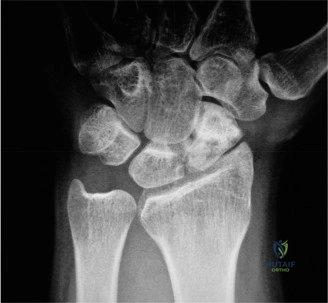
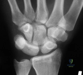
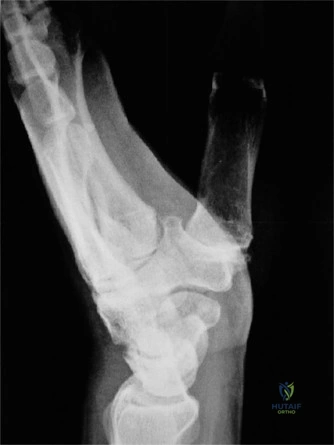
A 55-year-old tennis player presents with chronic ulnar-sided wrist pain, worse with follow-through. You suspect pisotriquetral arthritis. Describe the radiographic imaging required to confirm your diagnosis and explain why standard views may be misleading.
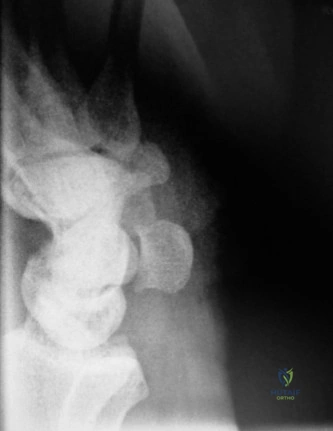
Candidate: I would request a 30-degree supinated AP view of the wrist, also known as the pisotriquetral view. Standard PA views are often unreliable because the pisiform is superimposed over the triquetrum and capitate, obscuring the joint space. This specific view projects the pisiform off the carpal row, allowing clear assessment of the pisotriquetral joint space for narrowing, sclerosis, or osteophytes.
Failing to mention the specific projection and simply stating "wrist X-rays." A borderline candidate might also incorrectly suggest an MRI as the first-line investigation, showing a lack of understanding regarding the radiographic "low-hanging fruit" for this diagnosis.
Clearly state the "30-degree supinated AP view." Explain the biomechanical reason for the failure of standard views: "The pisiform is a sesamoid bone that lies volar to the triquetrum; standard PA projection results in bony overlap. The supinated view effectively 'unmasks' the pisotriquetral articulation." Mentioning the "Pisotriquetral Grind Test" as the clinical correlate adds further depth.
You are examining a 68-year-old patient who sustained a wrist injury years ago. You perform a Watson's Scaphoid Shift Test and get a positive result. Explain the mechanism behind this test and what the "clunk" signifies.
Candidate: The Watson test assesses scapholunate ligament competence. In a normal wrist, as the wrist moves from ulnar to radial deviation, the scaphoid flexes to accommodate the radial styloid. By placing my thumb on the scaphoid tuberosity, I block this normal flexion. In an SL-deficient wrist, the proximal pole of the scaphoid subluxates dorsally over the dorsal rim of the radius because the scapholunate ligament cannot stabilize it. When I release the pressure, the scaphoid snaps back into the elliptical fossa, producing the 'clunk'.
Confusing the direction of subluxation or failing to emphasize that the test requires an *incompetent* ligament. Some candidates forget that the *clunk* is the hallmark of the reduction, not just the presence of pain.
Structure the answer by describing the normal kinematics vs. the pathological kinematics. Define the "clunk" specifically as the reduction of the dorsally subluxated scaphoid into the lunate fossa. Explicitly note that the test is positive only when there is both pain *and* a palpable mechanical shift.
Our 68-year-old farmer has a Stage III SLAC wrist. Explain why a Proximal Row Carpectomy (PRC) is contraindicated in this specific patient and identify the definitive surgical salvage procedure.
Candidate: A PRC is contraindicated in Stage III SLAC because the capitate head is arthritic. The PRC requires a healthy, congruent articular surface on the head of the capitate to articulate with the lunate fossa of the distal radius. If the capitate is damaged, the procedure will result in immediate, painful radiocapitate arthritis. The procedure of choice is a Scaphoid Excision and Four-Corner Arthrodesis (4CF).
Suggesting a "total wrist fusion" as the first line of surgical treatment. While a salvage procedure, it is an endpoint. The candidate must show they understand the selection criteria (i.e., state of the capitate head) that differentiate between a PRC and a 4CF.
Use the "Watson and Ballet" staging context. State clearly: "PRC success depends entirely on the chondral health of the capitate head." Contrast this with the 4CF, which bypasses the need for the capitate-radius articulation, making it the superior choice for Stage III disease.
