Mastering the Open Repair of Palmer Class 1B Triangular Fibrocartilage Complex Injuries
Key Takeaway
Palmer Class 1B injuries represent traumatic avulsions of the triangular fibrocartilage complex (TFCC) from its ulnar foveal insertion, often resulting in distal radioulnar joint (DRUJ) instability. This comprehensive surgical guide details the open repair technique, emphasizing precise anatomical exposure between the fifth and sixth extensor compartments, meticulous foveal debridement, and secure transosseous suture fixation to restore DRUJ kinematics and grip strength in the active patient.
Comprehensive Introduction and Patho-Epidemiology
The triangular fibrocartilage complex (TFCC) functions as the primary static stabilizer of the distal radioulnar joint (DRUJ) and the ulnocarpal articulation, serving as a critical load-bearing structure that facilitates smooth, multidirectional kinematics of the human wrist. According to the universally adopted Palmer classification system, a Class 1B injury denotes a traumatic, acute avulsion of the TFCC from its ulnar insertion site. This avulsion typically occurs at the anatomical fovea located at the base of the ulnar styloid, and it presents either as an isolated ligamentous disruption or in conjunction with an associated ulnar styloid basilar fracture. The patho-epidemiology of these injuries is predominantly linked to high-energy trauma, most frequently occurring from a fall onto an outstretched hand (FOOSH) with the wrist in extension and the forearm in hyper-pronation, which places maximal tensile stress on the ulnar-sided capsuloligamentous structures.
Because the deep, robust fibers of the radioulnar ligaments—often referred to as the ligamentum subcruentum—attach directly to the foveal footprint, their traumatic disruption leads to profound DRUJ instability. Patients typically present with debilitating ulnar-sided wrist pain, a subjective sensation of "clunking" or giving way during rotational movements, and a significant, measurable decrease in grip strength. The natural history of an untreated Class 1B foveal avulsion is progressive DRUJ subluxation, accelerated articular wear, and eventual post-traumatic osteoarthritis of the ulnar head and sigmoid notch.
While the last two decades have seen a surge in arthroscopic techniques for managing partial tears, central perforations (Class 1A), and peripheral capsular detachments, the open repair of Palmer Class 1B injuries remains the unequivocal gold standard for massive foveal avulsions, chronic retractions, and complex cases requiring simultaneous osteosynthesis of ulnar styloid nonunions. The open approach provides unparalleled, direct visualization of the foveal footprint, allowing the operating surgeon to perform meticulous decortication and achieve robust, anatomic transosseous fixation. This direct structural reattachment is paramount for restoring the true isometric axis of the DRUJ, thereby preventing the high rates of recurrent instability often associated with non-anatomic or superficial arthroscopic repairs.
Detailed Surgical Anatomy and Biomechanics
A profound, three-dimensional understanding of DRUJ biomechanics and ulnar-sided wrist anatomy is mandatory for the operating surgeon. The DRUJ is an inherently unstable diarthrodial joint. The osseous architecture—comprising the convex ulnar head articulating with the shallow, concave sigmoid notch of the radius—confers minimal intrinsic stability. The radius of curvature of the sigmoid notch is substantially larger than that of the ulnar head, meaning stability is almost entirely dependent on the extrinsic soft-tissue envelope, primarily the TFCC.
The TFCC itself is a heterogeneous, complex structure composed of the central articular disc, the dorsal and volar radioulnar ligaments (DRUL and VRUL), the meniscus homologue, the ulnocarpal ligaments (ulnolunate and ulnotriquetral), and the extensor carpi ulnaris (ECU) subsheath. The articular disc acts as a shock absorber, transmitting approximately 20% of the axial load from the carpus to the ulna. However, it is the peripheral ligamentous margins—specifically the DRUL and VRUL—that dictate DRUJ kinematics.
The Foveal Attachment and Isometric Axis
The critical stabilizing components of the TFCC are the deep, stout fibers of the DRUL and VRUL, which converge to insert directly into the fovea of the ulna. The fovea, a small depression located at the base of the ulnar styloid, represents the true isometric axis of rotation for the forearm. During the complex motions of pronation and supination, the radius and the carpus rotate around the fixed ulnar head. Because the deep fibers attach at this isometric center, they maintain consistent tension throughout the entire arc of rotation.
If the foveal attachment is disrupted (as in a Palmer Class 1B injury), the radius and the attached articular disc are no longer tethered to the ulnar axis. This allows the radius to translate dorsally during pronation or volarly during supination relative to the ulnar head, resulting in gross clinical instability. A critical biomechanical principle that must be respected during surgery is that reattaching the TFCC to the tip of the ulnar styloid (where only the superficial fibers insert) will not restore DRUJ stability. The repair must be anchored deeply to the fovea to recreate the isometric axis. Failure to recognize this biomechanical mandate is the leading cause of recurrent instability and catastrophic failure following TFCC repair.
The Extensor Carpi Ulnaris Subsheath and Secondary Stabilizers
Beyond the radioulnar ligaments, the surgeon must intimately understand the role of the secondary stabilizers, most notably the ECU tendon and its subsheath. The ECU subsheath is a distinct fascial layer that lies deep to the extensor retinaculum, firmly anchoring the ECU tendon in the ulnar groove. The deep layer of this subsheath is intimately blended with the dorsal capsule of the DRUJ and the dorsal margin of the TFCC.
During supination, the ECU tendon dynamically tightens its subsheath, which in turn applies a stabilizing volar-directed force to the ulnar head, assisting the TFCC in preventing dorsal subluxation of the ulna. Consequently, during the surgical exposure for an open TFCC repair, absolute care must be taken to preserve the integrity of the ECU subsheath. Iatrogenic violation of the sixth extensor compartment not only compromises this vital secondary dynamic stabilizer but also predisposes the patient to painful ECU tendon subluxation and chronic tendinopathy postoperatively.
Exhaustive Indications and Contraindications
The decision to proceed with an open transosseous repair of a Palmer Class 1B injury requires a careful synthesis of the patient's clinical presentation, chronicity of the injury, functional demands, and radiographic findings. Patient selection is critical; while acute and subacute repairs yield excellent functional outcomes, chronic injuries with significant soft-tissue contracture or underlying osseous deformity may require adjunctive or alternative procedures.
The primary indication for an open repair is an acute or subacute Palmer Class 1B tear accompanied by gross, demonstrable DRUJ instability that is not amenable to closed reduction and long-arm casting. In the chronic setting, patients presenting with persistent ulnar-sided wrist pain, profound grip weakness, and positive provocative testing (such as the DRUJ ballottement test or the foveal sign) are prime candidates, provided the TFCC tissue remains viable and excursion is sufficient for repair. Furthermore, open repair is strongly indicated when a TFCC avulsion is associated with a displaced, symptomatic, or ununited basilar ulnar styloid fracture. In these scenarios, the open approach allows for simultaneous excision of the fracture fragment and direct repair of the ligamentous complex into the bleeding cancellous bed of the fovea. Open repair is also the procedure of choice for revising failed arthroscopic repairs, where structural augmentation or precise anatomical realignment is required.
Conversely, absolute contraindications include advanced osteoarthritis of the DRUJ or ulnocarpal joint. In the presence of significant degenerative changes, restoring soft-tissue stability will not alleviate pain; instead, salvage procedures such as the Darrach procedure, Sauvé-Kapandji procedure, or total DRUJ arthroplasty are indicated. Fixed ulnocarpal impaction syndrome with severe ulnar positive variance is a relative contraindication to isolated soft-tissue repair; these cases typically require a concurrent ulnar shortening osteotomy to decompress the ulnocarpal articulation and reduce tension on the TFCC repair.
| Category | Specific Clinical Scenarios | Rationale / Clinical Impact |
|---|---|---|
| Primary Indications | Acute/Subacute Class 1B tears with gross DRUJ instability. | Restores isometric axis; prevents progression to chronic instability and secondary osteoarthritis. |
| Primary Indications | Chronic tears with persistent pain, weakness, and positive foveal sign. | Eliminates "clunking" and restores functional grip strength in symptomatic patients. |
| Primary Indications | TFCC avulsion with displaced/ununited basilar ulnar styloid fracture. | Allows simultaneous excision of the non-union fragment and direct transosseous foveal repair. |
| Primary Indications | Failed prior arthroscopic repair. | Open approach provides superior visualization for revision, debridement, and robust structural fixation. |
| Absolute Contraindications | Advanced DRUJ or ulnocarpal osteoarthritis. | Soft-tissue repair will not relieve osteoarthritic pain; necessitates salvage arthroplasty or resection. |
| Absolute Contraindications | Active deep space infection or severe soft-tissue compromise. | High risk of catastrophic postoperative infection, hardware failure, and wound dehiscence. |
| Relative Contraindications | Severe ulnar positive variance (>3mm) with ulnocarpal impaction. | High tension on the repair leads to failure; requires concurrent ulnar shortening osteotomy. |
| Relative Contraindications | Fixed, chronic DRUJ dislocation with severe soft-tissue contracture. | Primary repair may be impossible due to tissue retraction; may require Adams-Berger reconstruction. |
Pre-Operative Planning, Templating, and Patient Positioning
Meticulous preoperative planning is the foundation of a successful open TFCC repair. The surgeon must evaluate not only the soft-tissue integrity but also the underlying osseous morphology, ulnar variance, and the presence of concomitant carpal pathology.
Imaging Modalities and Diagnostic Criteria
Standard posteroanterior (PA) and lateral radiographs of the wrist are the initial diagnostic step. The PA view must be taken with the shoulder abducted 90 degrees, the elbow flexed 90 degrees, and the forearm in neutral rotation ("zero PA view") to accurately assess ulnar variance. Positive ulnar variance increases the axial load transmitted through the ulnocarpal joint and places the TFCC under elevated baseline tension, which may necessitate a concurrent ulnar shortening osteotomy to protect the repair. The lateral radiograph is scrutinized for dorsal or volar subluxation of the ulnar head relative to the radius, indicating gross DRUJ incompetence.
High-resolution, 3-Tesla Magnetic Resonance Imaging (MRI), preferably with intra-articular contrast (MR arthrography), is the gold standard advanced imaging modality. MR arthrography provides exceptional sensitivity and specificity for delineating the exact location and extent of the foveal avulsion. The surgeon must meticulously review the axial and coronal sequences to assess the integrity of the articular disc, identify any central perforations (which may require concurrent debridement), and evaluate the degree of retraction of the radioulnar ligaments. In cases of diagnostic ambiguity, diagnostic wrist arthroscopy may be utilized as a prelude to the open repair to directly visualize the tear pattern and assess the quality of the remaining TFCC tissue.
Anesthesia, Positioning, and Tourniquet Setup
The procedure is typically performed under regional anesthesia (supraclavicular or axillary brachial plexus block), which provides excellent intraoperative muscle relaxation and prolonged postoperative analgesia. General anesthesia is a viable alternative based on patient preference or specific medical comorbidities.
The patient is placed in the supine position on the operating table, with the operative extremity extended onto a radiolucent hand table. A non-sterile pneumatic tourniquet is placed high on the brachium over generous cast padding. The entire upper extremity is prepped and draped in a standard sterile fashion. The limb is exsanguinated using an Esmarch bandage, and the tourniquet is inflated (typically to 250 mm Hg or 100 mm Hg above the patient's systolic blood pressure). A completely bloodless field is absolutely critical for this procedure. The surgical dissection, identification of delicate sensory nerves, and precise decortication of the fovea occur in a highly confined, anatomically dense space where even minor bleeding can obscure the operative field and compromise the quality of the repair.
Step-by-Step Surgical Approach and Fixation Technique
Mastery of the open TFCC repair demands surgical precision, a deep respect for the dorsal sensory neural anatomy, and strict adherence to the biomechanical principles of transosseous fixation. The following masterclass details the operative sequence.
Incision and Superficial Dissection
The surgical approach begins with a longitudinal skin incision over the dorsal ulnar aspect of the wrist, meticulously centered between the extensor digiti quinti (EDQ, fifth extensor compartment) and the extensor carpi ulnaris (ECU, sixth extensor compartment). The incision should be centered directly over the ulnar head and extend for approximately 5 to 6 cm. This length ensures adequate, tension-free exposure of the DRUJ without requiring excessive retraction that could damage the skin edges.
Once the dermis is breached, the surgeon must proceed with extreme caution. The dorsal sensory branch of the ulnar nerve (DSBUN) is a critical structure that crosses from the volar to the dorsal aspect of the forearm approximately 3 to 5 cm proximal to the ulnar styloid. Blunt dissection techniques using a hemostat or tenotomy scissors are mandatory in the subcutaneous tissues to identify, mobilize, and protect these delicate arborizing nerve branches. Retraction of the DSBUN should be performed with soft vessel loops rather than rigid retractors. Iatrogenic injury, transection, or entrapment of the DSBUN within scar tissue is a devastating complication that leads to debilitating postoperative neuromas and complex regional pain syndrome (CRPS).
Extensor Compartment Management and Capsulotomy
Following the identification and protection of the neural structures, the extensor retinaculum is exposed. The surgeon carefully incises the retinaculum to open the fifth extensor compartment. The EDQ tendon is mobilized and retracted radially. It is imperative at this stage to maintain the absolute integrity of the sixth extensor compartment (the ECU subsheath). As previously detailed, the ECU and its subsheath are vital secondary dynamic stabilizers of the ulnar wrist; violating this compartment will create iatrogenic instability and postoperative tendinopathy.
Through the floor of the fifth compartment, the dorsal capsule of the DRUJ is exposed. The joint is opened utilizing an angular, L-shaped capsular incision. The longitudinal limb of the incision begins proximal to the ulnar head and extends distally to the level of the attachment of the dorsal radioulnar ligament (DRUL). A crucial technical mandate during this step is to strictly preserve the radial attachment of the DRUL. Detaching the ligament from the radius will destabilize the entire complex. At the proximal margin of the DRUL, the incision is turned transversely and directed medially (toward the ulnar side), stopping precisely at the lateral (radial) border of the sixth extensor compartment.
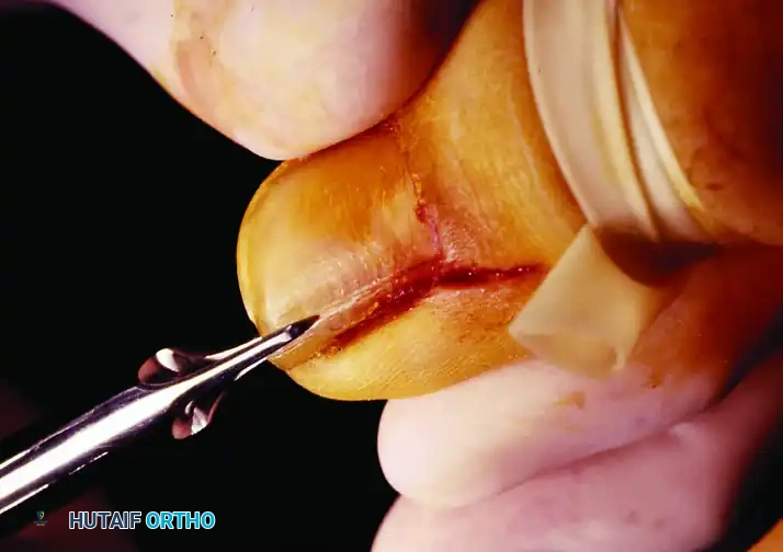
Joint Exposure, Styloid Management, and Foveal Debridement
Elevation of this right-angled capsular flap provides a panoramic view of the proximal surface of the triangular fibrocartilage, the ulnar neck, the ulnar head, and the sigmoid notch. The surgeon first inspects the ulnar styloid. If there are remnants of an ununited, symptomatic ulnar styloid fracture, they must be meticulously excised using a rongeur or sharp osteotome. Retaining a mobile, ununited styloid fragment can cause persistent impingement pain and physically interfere with the anatomic seating of the TFCC repair.
Attention is then directed to the fovea at the base of the ulnar styloid. The foveal footprint is often covered with fibrous scar tissue or degenerative debris. Using a small angled curette, a motorized arthroscopic shaver, or a high-speed spherical burr, the surgeon must thoroughly débride the fovea down to bleeding, healthy cancellous bone. This decortication step is non-negotiable. The biological success of the procedure relies on a robust healing response, which can only occur if the avulsed fibrocartilage is compressed against a highly vascularized bony bed. Following decortication, the torn edge of the TFCC is grasped with a fine hemostat or tissue forceps. The tissue is assessed for contracture, friability, and excursion. It must be mobilized sufficiently to reach the foveal trough without excessive tension.
Transosseous Tunnel Preparation and Suture Passage
Once the TFCC is mobilized and the fovea is prepared, the surgeon proceeds with the bony preparation for transosseous fixation. To facilitate suture passage, the DRUJ is opened slightly further with a second, transverse incision along the distal border of the DRUL, again strictly preserving its radial attachment.
Using a 0.045-inch Kirschner wire (K-wire) or a 1.25 mm drill bit, the surgeon creates the transosseous tunnels. The standard technique involves drilling two or three side-by-side tunnels, or a converging V-shaped tunnel configuration. The trajectory of these tunnels is of paramount importance. The drill should start on the ulnar neck (extra-articular, on the ulnar cortex), just volar to the ECU subsheath, and aim obliquely distal and radial to exit precisely at the center of the decorticated fovea. The surgeon must ensure that the cortical bone bridge between the entry holes on the ulnar neck is sufficiently wide (at least 5 to 7 mm). A narrow bone bridge risks cortical blowout and catastrophic loss of fixation when the sutures are tensioned and tied.
A heavy, non-absorbable braided suture—preferably a 2-0 or 0 ultra-high-molecular-weight polyethylene (UHMWPE) suture due to its superior tensile strength and low profile—is passed through the robust deep fibers of the TFCC. A horizontal mattress or a locking Krackow-type configuration is utilized to ensure a secure grasp of the ligamentous tissue without tearing through the substance of the disc. A suture-passing wire, a Hewson suture passer, or a specialized nitinol loop is then used to shuttle the free ends of the suture through the pre-drilled foveal tunnels, exiting at the ulnar neck.
Tensioning, Fixation, and Closure
With the sutures passed, the critical step of tensioning and fixation is performed. The assistant reduces the DRUJ by holding the forearm in neutral rotation. The surgeon applies firm, steady traction to the sutures, drawing the TFCC down tightly into the bleeding foveal trough. While maintaining this tension, the DRUJ stability is assessed in full supination, full pronation, and neutral rotation. The joint should feel rock-solid, with no dorsal or volar subluxation of the ulnar head relative to the radius.
Once stability is confirmed, the sutures are tied securely over the cortical bone bridge on the ulnar neck. The knot should be buried deep to the subcutaneous tissues to prevent hardware prominence. The joint is then irrigated thoroughly with sterile saline to remove any bone debris generated during drilling. The angular capsular flap is repaired using absorbable sutures (e.g., 3-0 Vicryl), ensuring a watertight closure without over-constraining the joint. The EDQ tendon is allowed to fall back into its anatomical position. The extensor retinaculum is repaired over the fifth compartment; however, leaving a small gap or performing a partial step-cut lengthening may be necessary to prevent tendon friction or bowstringing. The subcutaneous tissue and skin are closed in a standard layered fashion, and a sterile compressive dressing is applied.
Complications, Incidence Rates, and Salvage Management
While the open repair of Palmer Class 1B injuries is a highly successful and reproducible procedure in the hands of an experienced wrist surgeon, complications can and do occur. A thorough understanding of these potential pitfalls is essential for preoperative patient counseling and prompt postoperative management.
The most devastating complication is recurrent instability. This typically results from a failure to anchor the repair to the true fovea (the isometric point), inadequate bony decortication leading to non-healing, or a premature return to high-torque activities by a non-compliant patient. When primary repair fails and the tissue is deemed non-viable for revision, salvage reconstruction is required. The Adams-Berger procedure, which utilizes a free tendon graft (such as the palmaris longus) routed through precisely drilled tunnels in the radius and ulna to anatomically reconstruct the DRUL and VRUL, is the preferred salvage technique for restoring stability.
Postoperative stiffness, specifically a loss of terminal pronation or supination, is the most frequently encountered complication. It is often secondary to prolonged immobilization, capsular contracture, or iatrogenic over-tensioning of the capsular repair. The vast majority of these cases resolve with aggressive, dedicated hand therapy and dynamic splinting. Rarely, refractory stiffness may necessitate an arthroscopic or open capsular release.
Neurological complications, specifically injury to the DSBUN, represent a significant source of postoperative morbidity. Neuromas of the DSBUN cause severe, burning, dysesthetic pain that can be more debilitating than the original TFCC injury. Prevention through meticulous blunt dissection is paramount. Established neuromas that fail conservative management (gabapentinoids, targeted nerve blocks) require surgical excision and burying of the proximal nerve stump deep into the pronator quadratus muscle belly or into a drilled bone hole to prevent superficial mechanical irritation.
| Complication | Estimated Incidence | Etiology / Risk Factors | Prevention and Salvage Management |
|---|---|---|---|
| Recurrent DRUJ Instability | 5% - 10% | Non-isometric placement (styloid tip instead of fovea); premature return to heavy lifting; poor tissue quality. | Prevention: Precise foveal decortication and tunnel placement. Salvage: Adams-Berger tendon reconstruction. |
| Postoperative Stiffness (Loss of ROM) | 15% - 25% | Prolonged immobilization (>6 weeks); over-tensioning of the dorsal capsule during closure. | Prevention: Early, supervised ROM protocols. Management: Aggressive hand therapy; rare open/arthroscopic capsular release. |
| DSBUN Neuroma / Injury | 2% - 5% | Iatrogenic sharp transection or aggressive retraction during the superficial surgical approach. | Prevention: Blunt subcutaneous dissection; use of soft vessel loops. Management: Neuroma excision and deep intramuscular burying. |
| ECU Tendinopathy / Subluxation | 3% - 8% | Violation of the 6th extensor compartment (ECU subsheath) during exposure; prominent suture knots irritating the tendon. | Prevention: Strict preservation of the ECU subsheath; burying suture knots. Management: NSAIDs, steroid injections, surgical subsheath reconstruction. |
| Infection / Wound Dehiscence | < 2% | Poor soft-tissue envelope; hematoma formation; medical comorbidities (diabetes, smoking). | Prevention: Meticulous hemostasis (tourniquet deflation prior to closure); layered closure. Management: Oral/IV antibiotics; operative debridement if deep. |
Phased Post-Operative Rehabilitation Protocols
The ultimate clinical success of an open TFCC repair relies as much on a disciplined, scientifically structured postoperative rehabilitation protocol as it does on flawless surgical execution. The biological healing of avascular fibrocartilage to a cortical bone bed is an inherently slow process that requires strict protection from shear and tensile forces during the early phases of remodeling. The surgeon must work in close concert with a certified hand therapist (CHT) to guide the patient through a rigorous four-phase protocol.
Phase 1: Immobilization (Weeks 0-6)
Immediately following surgery, the patient is placed in a bulky, well-padded Muenster cast or a rigid sugar-tong splint. The critical element of this phase is immobilizing the forearm in absolute neutral rotation. Neutral rotation minimizes the tension on the newly repaired radioulnar ligaments at the foveal footprint. Finger, thumb, and elbow range of motion (ROM) exercises are initiated on postoperative day one. Active digit motion is essential to prevent intrinsic hand stiffness, reduce dependent edema, and promote tendon gliding of the extrinsic flexors and extensors. The cast is maintained continuously for a full 6 weeks to allow for the initial osseous integration and collagen cross-linking of the TFCC footprint.
Phase 2: Early Mobilization (Weeks 6-8)
At the 6-week mark, the rigid cast is removed, and the clinical stability of the DRUJ is gently assessed. The patient is immediately transitioned to a custom-molded, removable thermoplastic wrist splint. During this phase, active and active-assisted ROM exercises for the wrist (flexion and extension) and the forearm (pronation and supination) are initiated. These exercises must be performed strictly under the guidance of the CHT. The emphasis is on restoring the "dart-throwing motion" arc, which minimizes stress on the DRUJ. Passive stretching, forceful overpressure, and weight-bearing are strictly prohibited during this vulnerable phase, as the maturing scar tissue is still susceptible to elongation or rupture.
Phase 3: Strengthening and Proprioception (Weeks 8-12)
Once full, pain-free active ROM is achieved—typically around the 8-to-10-week mark—progressive isometric and isotonic strengthening begins. A specific focus is placed on strengthening the ECU and the pronator quadratus muscles, as these structures act as vital dynamic stabilizers of the DRUJ. Proprioceptive neuromuscular facilitation (PNF) exercises are introduced to restore the dynamic reflex stabilization of the wrist. The removable splint is gradually weaned for light, activities of daily living (ADLs) but must still be worn during sleep, commuting, or in unpredictable, high-risk environments.
Phase 4: Return to Unrestricted Activity (Months 3-6)
The final phase focuses on work-hardening and return to sport. Heavy lifting, forceful gripping, and torque-producing activities (e.g., using a screwdriver, turning heavy wrenches, swinging a golf club or baseball bat) are strictly restricted until at least 3 to 4 months postoperatively. A full return to contact sports, heavy manual labor, or competitive athletics is typically permitted between 4 and 6 months. This return is entirely contingent upon the patient meeting specific objective criteria: the restoration of grip strength to at least 80% to 85% of the uninjured contralateral side, full and symmetric forearm rotation, and the complete absence of pain or apprehension during provocative DRUJ testing.
Summary of Landmark Literature and Clinical Guidelines
The evolution of TFCC repair is deeply rooted in landmark anatomical and biomechanical studies. Palmer's original 1989 classification system remains the foundational framework for diagnosing and treating these injuries, establishing the critical distinction between central disc perforations (Class 1A) and peripheral foveal avulsions (Class 1B). The biomechanical imperative of the foveal attachment was elegantly elucidated by Garcia-Elias and later by Adams, who definitively demonstrated that the deep fibers of the radioulnar ligaments dictate the isometric stability of the DRUJ, and that superficial repairs to the styloid tip are biomechanically doomed to fail.
Furthermore, the work of Atzei and colleagues has significantly advanced our understanding of the "ice cream cone" geometry of the ulnar head and the precise trajectory required for transosseous tunnel placement. While arthroscopic techniques have advanced rapidly, comparative literature consistently reinforces that for massive, retracted Class 1B injuries, or those complicated by ulnar styloid nonunions, the open transosseous repair provides superior visualization, more reliable anatomic reduction, and equivalent or superior long-term functional outcomes regarding grip strength and joint stability. It is incumbent upon the modern orthopedic surgeon to master this open technique, ensuring that the foundational principles of biomechanics and tissue healing are meticulously applied to every patient.
