Carpal Ligament Injuries and Instability Patterns: A Comprehensive Surgical Guide
Key Takeaway
Carpal instability represents a complex spectrum of wrist pathology resulting from ligamentous disruption. This comprehensive guide details the biomechanical foundations, advanced radiographic evaluation, and evidence-based surgical management of carpal ligament injuries. From scapholunate dissociation to progressive perilunar instability, mastering these concepts is essential for orthopedic surgeons to restore wrist kinematics, prevent degenerative collapse, and optimize patient outcomes through precise operative intervention and structured postoperative rehabilitation.
Comprehensive Introduction and Patho-Epidemiology
The human carpus is an exquisitely complex, intercalated biomechanical linkage system that bridges the forearm to the hand, permitting a remarkable combination of global mobility and structural stability. Posttraumatic loss of alignment of the carpal bones leads to profound kinematic derangements, progressive articular wear, and debilitating functional loss. Describing the posttraumatic loss of alignment of the carpal bones, Linscheid et al. established the foundational framework for understanding these pathologies, grouping carpal instabilities into primary types based on the directional collapse of the intercalated proximal row. When this delicate mechanical equilibrium is disrupted by high-energy trauma or repetitive microtrauma, the resultant instability initiates a predictable, catastrophic cascade of degenerative arthrosis if left untreated.
Epidemiologically, carpal ligament injuries predominantly affect young, active, and working-age populations, most commonly males between the ages of 20 and 40. The classic mechanism of injury is a fall onto an outstretched hand (FOOSH), subjecting the wrist to forceful hyperextension, ulnar deviation, and intercarpal supination. Despite their severity, carpal instabilities—particularly isolated scapholunate dissociations—are notoriously misdiagnosed or missed entirely during initial emergency department evaluations. Up to 25% of scapholunate ligament tears are overlooked on initial presentation, often dismissed as simple wrist sprains until the patient presents months later with chronic mechanical pain, weakness, and established carpal collapse.
Instability in the carpus is categorized temporally and kinematically. It is considered static if the radiographic intercarpal relationships are persistently abnormal and do not change with motion, indicating complete failure of both primary and secondary ligamentous stabilizers. Conversely, it is considered dynamic if the resting intercarpal relationships appear normal on standard radiographs but become abnormal under physiologic loads, manipulation, or active motion. Dynamic instability represents an earlier, potentially more salvageable stage of ligamentous failure. Provocative maneuvers and stress radiography (e.g., clenched-fist views, ulnar deviation views) are absolutely mandatory when a patient presents with mechanical wrist pain, clicking, or giving way, but exhibits normal static radiographs.
The classification of carpal instability is primarily dictated by the anatomical location of the ligamentous disruption. Linscheid et al. advocated the vital concept of dissociative and nondissociative instabilities. Dissociative Carpal Instability (CID) is characterized by the disruption of the intrinsic interosseous ligaments between the bones of the proximal carpal row (e.g., scapholunate or lunotriquetral ligaments), meaning the proximal row is "dissociated" from itself. Nondissociative Carpal Instability (CIND) involves the disruption of the extrinsic radiocarpal or midcarpal ligaments, while the intrinsic ligaments remain intact; the entire proximal row moves as a coupled unit but is unstable relative to the radius or the distal carpal row. Complex or Combined Instabilities (CIC) involve both elements, such as perilunate dislocations, and Adaptive Carpus describes malposition secondary to extrinsic skeletal deformity like a distal radius malunion.
Detailed Surgical Anatomy and Biomechanics
Osteology and the Intercalated Segment
The carpus consists of eight bones arranged in two distinct rows. The distal carpal row (trapezium, trapezoid, capitate, and hamate) functions as a tightly bound, rigid unit articulated with the metacarpals, effectively acting as the base of the hand. In stark contrast, the proximal carpal row (scaphoid, lunate, and triquetrum) is an intercalated segment. It possesses absolutely no direct tendinous insertions; its movement is entirely passive, dictated by the mechanical forces exerted by the surrounding articular contours and ligamentous constraints. The scaphoid uniquely bridges both rows, serving as a critical mechanical link that stabilizes the midcarpal joint and dictates the kinematics of the proximal row.
Intrinsic and Extrinsic Ligamentous Anatomy
The stability of the carpus relies on a highly specialized network of intrinsic and extrinsic ligaments. The intrinsic ligaments originate and insert entirely within the carpus. The most clinically significant are the scapholunate interosseous ligament (SLIL) and the lunotriquetral interosseous ligament (LTIL). The SLIL is a C-shaped structure divided into three regions: the dorsal, membranous, and volar zones. The dorsal SLIL is the thickest, strongest, and most critical stabilizer against distraction and translation. Conversely, the LTIL is thickest and strongest in its volar aspect. The extrinsic ligaments connect the radius or ulna to the carpus. Volarly, the robust radioscaphocapitate (RSC), long radiolunate (LRL), and short radiolunate (SRL) ligaments provide critical check-reins to extension and ulnar translation. The space between the RSC and LRL, known as the Space of Poirier, is an area of relative capsular weakness through which the lunate can dislocate in severe trauma.
Carpal Kinematics and Collapse Patterns
Normal carpal kinematics rely on a delicate balance of opposing forces. The scaphoid naturally tends to flex and pronate under axial load, while the triquetrum naturally tends to extend and supinate. The lunate, caught in the middle, is balanced between these opposing forces by the intact SLIL and LTIL. When the SLIL fails, the scaphoid flexes uninhibited, and the lunate extends with the triquetrum, resulting in a Dorsal Intercalated Segment Instability (DISI) pattern. When the LTIL fails, the triquetrum extends, and the lunate flexes with the scaphoid, resulting in a Volar Intercalated Segment Instability (VISI) pattern. Furthermore, the functional axis of the wrist during most activities of daily living is not pure flexion/extension, but rather the "dart-thrower's motion" (coupled radial extension and ulnar flexion), which occurs primarily at the midcarpal joint, minimizing stress on the proximal row intrinsic ligaments.
Progressive Perilunar Instability
Mayfield, Johnson, and Kilcoyne described the pathomechanics of carpal failure through four sequential stages of progressive disruption resulting from forced wrist hyperextension, ulnar deviation, and intercarpal supination. Stage I (Scapholunate Failure) involves disruption of the SLIL and volar radioscaphocapitate ligament, manifesting as scapholunate dissociation. Stage II (Capitolunate Failure) occurs as the force propagates ulnarly through the Space of Poirier, disrupting the capitolunate articulation. Stage III (Triquetrolunate Failure) involves disruption of the LTIL, separating the entire carpus from the lunate and resulting in a perilunate dislocation. Finally, Stage IV (Lunate Dislocation) involves the failure of the dorsal radiocarpal ligament, forcing the capitate proximally and extruding the lunate volarly into the carpal tunnel, creating an acute orthopedic emergency due to severe median nerve compression.
Exhaustive Indications and Contraindications
The overarching philosophy of surgical intervention in carpal ligament injuries is the anatomical restoration of carpal kinematics to prevent the inevitable progression to Scapholunate Advanced Collapse (SLAC) or other post-traumatic arthropathies. The timing of the injury profoundly influences the surgical decision-making process. Acute injuries (less than 4 weeks) are generally amenable to primary ligament repair and capsulodesis. Subacute injuries (4 to 12 weeks) occupy a gray zone where direct repair may be attempted but frequently requires augmentation. Chronic injuries (greater than 12 weeks) typically present with irreparable, retracted, or attenuated ligaments, necessitating complex ligamentous reconstructions (e.g., tenodesis) or, if arthrosis is present, salvage procedures.
Specific indications for acute open reduction and primary ligament repair include any acute scapholunate dissociation with a gap greater than 3mm on standard or stress radiographs, any acute perilunate or lunate dislocation, and highly symptomatic dynamic instabilities that have failed a rigorous trial of conservative management (immobilization and targeted proprioceptive hand therapy). Reconstructive procedures, such as the modified Brunelli tenodesis, are indicated for chronic, reducible scapholunate dissociations where the articular cartilage of the radiocarpal and midcarpal joints remains pristine. Salvage procedures (Proximal Row Carpectomy or Four-Corner Fusion) are strictly indicated when irreversible articular cartilage damage has already occurred.
Contraindications to primary repair or soft-tissue reconstruction must be rigorously respected to avoid catastrophic surgical failures. Absolute contraindications include the presence of established arthrosis (e.g., SLAC wrist stages II-III), fixed/irreducible carpal deformity due to severe chronicity, active intra-articular infection, and severe osteopenia that precludes reliable osseous fixation of anchors or screws. Relative contraindications include heavy tobacco use (which significantly impairs ligamentous healing), poorly controlled diabetes mellitus, non-compliant patients unable to adhere to strict postoperative immobilization protocols, and systemic inflammatory arthropathies (e.g., rheumatoid arthritis) where intrinsic ligament repair is destined to fail due to ongoing synovial disease.
| Pathology / Injury Stage | Primary Surgical Indications | Absolute Contraindications | Relative Contraindications |
|---|---|---|---|
| Acute SL Dissociation (<4 wks) | SL gap >3mm, DISI deformity, failed closed reduction | Pre-existing SLAC arthrosis, active local infection | Heavy smoking, patient non-compliance |
| Chronic SL Dissociation (>12 wks) | Reducible carpus, preserved cartilage, chronic pain | Fixed deformity, radioscaphoid arthrosis | Advanced age, low functional demand |
| Acute Perilunate Dislocation | All acute presentations (orthopedic emergency) | None (emergent decompression required) | Severe soft tissue compromise (delay definitive fixation) |
| SLAC Wrist (Stage II/III) | Intractable pain, progressive weakness, failed conservative care | Active infection, medically unstable for anesthesia | Preserved scaphoid facet (consider lesser procedure) |
Pre-Operative Planning, Templating, and Patient Positioning
Clinical Evaluation and Provocative Testing
Meticulous preoperative planning begins with a comprehensive clinical evaluation. The surgeon must ascertain the precise mechanism of injury, chronicity, and the patient's functional demands. Provocative maneuvers are essential. The Watson scaphoid shift test evaluates the integrity of the SLIL; a palpable "clunk" and reproduction of pain as the scaphoid subluxates over the dorsal rim of the radius is highly specific for SL instability. The lunotriquetral ballottement (shuck) test assesses the LTIL. A thorough neurovascular examination is paramount, particularly in the setting of perilunate or lunate dislocations, where acute median nerve neuropathy is frequently present and dictates the urgency of intervention.
Advanced Imaging Modalities
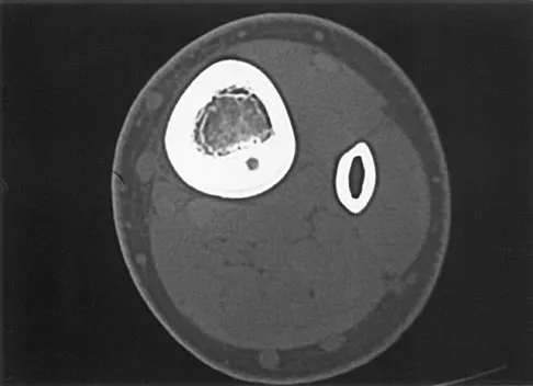
While standard zero-rotation posteroanterior (PA) and true lateral radiographs are the cornerstone of diagnosis, advanced imaging is frequently required for surgical templating. On the PA view, the Terry Thomas sign (scapholunate gap >3mm) and disruption of Gilula's carpal arcs are classic findings. The lateral view is critical for measuring the scapholunate angle (normal 30-60 degrees; >70 degrees indicates DISI) and the capitolunate angle. High-resolution Magnetic Resonance Imaging (MRI), particularly MR arthrography, is the gold standard for evaluating the intrinsic and extrinsic ligaments, assessing the vascularity of the proximal pole of the scaphoid, and identifying occult chondral shear injuries that may alter the surgical plan. Computed Tomography (CT) is invaluable for evaluating complex fracture-dislocations (e.g., trans-scaphoid perilunate dislocations) to assess comminution and plan screw trajectories.
Templating and Implant Selection
Surgical templating involves anticipating the need for various fixation modalities. The surgeon must ensure the availability of 0.045-inch and 0.062-inch Kirschner wires for provisional and definitive stabilization of the intercalated segment. For ligament repairs, micro-suture anchors (typically 1.5mm to 2.5mm, loaded with high-tensile non-absorbable suture) must be selected based on the patient's bone density. If a tenodesis is planned, interference screws or specialized tenodesis anchors must be templated to match the diameter of the harvested tendon graft (e.g., ECRB or FCR strip). In cases of fracture-dislocations, headless compression screws of various lengths must be available for scaphoid or capitate fixation.
Patient Positioning and Anesthesia
The procedure is typically performed under regional anesthesia (supraclavicular or axillary brachial plexus block) combined with intravenous sedation or general anesthesia, depending on patient preference and the anticipated duration of the surgery. The patient is positioned supine with the operative extremity extended on a radiolucent hand table. A pneumatic tourniquet is applied to the proximal arm, padded with cast padding, and inflated to 250 mmHg following exsanguination with an Esmarch bandage to ensure a bloodless surgical field. The fluoroscopy C-arm is positioned parallel to the hand table, entering from the distal or contralateral side, allowing for unimpeded orthogonal views of the carpus throughout the procedure without compromising the sterile field.
Step-by-Step Surgical Approach and Fixation Technique
The Dorsal Approach and Capsulotomy
The dorsal approach provides unparalleled access to the proximal carpal row and the intrinsic ligaments. A dorsal longitudinal incision is centered over the Lister tubercle, extending distally to the base of the third metacarpal. The extensor retinaculum is exposed and incised over the third dorsal compartment. The extensor pollicis longus (EPL) tendon is mobilized and transposed radially. The second and fourth dorsal compartments are elevated subperiosteally. A ligament-sparing dorsal capsulotomy, such as the Berger flap, is then created. This involves a V-shaped or inverted T-shaped incision in the dorsal capsule, meticulously preserving the dorsal intercarpal (DIC) and dorsal radiocarpal (DRC) ligaments. The capsular flap is elevated distally, exposing the radiocarpal and midcarpal joints.
Acute Scapholunate Ligament Repair
Once the joint is exposed, the scapholunate interval is inspected and debrided of hematoma and interposed fibrous tissue. The articular surfaces are examined for iatrogenic or traumatic chondral damage. To achieve anatomical reduction, 0.045-inch K-wires are inserted as joysticks into the dorsal non-articular surfaces of the scaphoid and lunate. The scaphoid is extended and supinated, while the lunate is flexed, correcting the DISI deformity and closing the scapholunate gap. This reduction is provisionally held by driving multiple 0.045-inch K-wires from the scaphoid into the lunate and capitate. The avulsed dorsal SLIL is then directly repaired to its footprint (usually the scaphoid) using one or two micro-suture anchors. The repair is frequently augmented with a dorsal capsulodesis (e.g., Blatt capsulodesis), utilizing a slip of the dorsal capsule tethered to the distal pole of the scaphoid to prevent recurrent volar flexion.
Chronic Reconstruction: Modified Brunelli Tenodesis
For chronic, reducible scapholunate dissociations where the primary ligament is irreparable, the modified Brunelli tenodesis (using a strip of the flexor carpi radialis or extensor carpi radialis brevis) is the gold standard. When utilizing the FCR, a volar incision is made to harvest a distally based strip of the tendon. A bone tunnel is drilled through the distal pole of the scaphoid from volar to dorsal. The tendon graft is passed through this tunnel, effectively recreating the volar stabilizing vectors. The graft is tensioned to correct the scaphoid flexion, passed across the dorsal scapholunate interval, and secured to the dorsum of the lunate using a suture anchor or interference screw. The remaining graft is then looped back and sutured to the dorsal radiotriquetral ligament. The entire construct is protected with rigid K-wire fixation across the scaphoid, lunate, and capitate.
Management of Perilunate and Lunate Dislocations
Surgical management of perilunate (Stage III) and lunate (Stage IV) dislocations requires a combined volar and dorsal approach. The volar phase begins with an extended carpal tunnel incision. The transverse carpal ligament is released to decompress the median nerve, which is frequently contused. The rent in the volar capsule (Space of Poirier) is identified, and the extruded lunate is gently manipulated back into the lunate fossa of the radius. The dorsal phase then proceeds as described above. The carpus is anatomically reduced around the lunate using joysticks. The scapholunate and lunotriquetral intervals are pinned. The dorsal SLIL and LTIL are repaired with suture anchors. The volar capsular rent is repaired if accessible, though the dorsal ligamentous repair remains the biomechanical priority for long-term stability.
Complications, Incidence Rates, and Salvage Management
Intraoperative and Early Postoperative Complications
The surgical management of carpal instability is fraught with potential complications. Intraoperatively, iatrogenic injury to the delicate articular cartilage during K-wire insertion or drilling for suture anchors is a significant risk. Inadequate reduction, characterized by residual scapholunate gapping or persistent DISI deformity, is a technical failure that guarantees poor outcomes. Early postoperative complications include pin tract infections, which occur in up to 10-15% of cases involving percutaneous K-wires, necessitating early pin removal and potentially compromising the repair. Median nerve neuropathy may persist following perilunate dislocations, requiring careful monitoring and potential re-exploration if symptoms worsen. Complex Regional Pain Syndrome (CRPS) is a devastating complication seen in 2-5% of patients, characterized by disproportionate pain, vasomotor instability, and severe stiffness.
Late Complications and Hardware Failure
Late complications are primarily mechanical and degenerative. Wrist stiffness is a ubiquitous finding; patients universally lose a degree of terminal flexion and extension, which must be clearly communicated preoperatively. Hardware failure, such as K-wire breakage or suture anchor pullout, can lead to recurrent instability. The most significant late complication is the progression to Scapholunate Advanced Collapse (SLAC). Despite anatomical repair, the altered micro-kinematics and initial traumatic chondral insult frequently lead to progressive degenerative arthritic changes, beginning at the radioscaphoid styloid (Stage I), progressing to the entire radioscaphoid joint (Stage II), and eventually involving the capitolunate joint (Stage III).
Salvage Management Strategies
When primary repair or reconstruction fails, or when patients present with established SLAC arthrosis, salvage procedures are indicated to provide a painless, stable wrist at the expense of motion. Proximal Row Carpectomy (PRC) involves the excision of the scaphoid, lunate, and triquetrum, allowing the capitate to articulate directly with the lunate fossa of the radius. PRC is indicated only if the capitate head and lunate fossa cartilage are pristine; it offers a relatively rapid recovery and preserves roughly 50% of normal wrist motion. If the capitate head is arthritic (SLAC Stage III), a Four-Corner Fusion (4CF) is required. This involves excision of the scaphoid and arthrodesis of the capitate, lunate, triquetrum, and hamate using a circular plate or headless screws. In cases of pan-carpal arthrosis or failed partial fusions, a Total Wrist Arthrodesis is the ultimate salvage procedure, providing reliable pain relief but completely eliminating wrist motion.
| Complication | Estimated Incidence | Prevention Strategy | Salvage / Management |
|---|---|---|---|
| Pin Tract Infection | 10 - 15% | Meticulous pin care, burying pins under skin | Oral antibiotics, early pin removal |
| Recurrent Instability | 15 - 25% | Rigid intra-op fixation, strict rehab adherence | Revision reconstruction, PRC, or 4-Corner Fusion |
| CRPS Type I | 2 - 5% | Early digital ROM, adequate pain control | Aggressive hand therapy, sympathetic nerve blocks |
| Post-Traumatic Arthrosis (SLAC) | 30 - 50% (Long-term) | Anatomical reduction, early intervention | Proximal Row Carpectomy, Total Wrist Fusion |
Phased Post-Operative Rehabilitation Protocols
Phase I: Immediate Postoperative Period (0-2 Weeks)
The success of carpal ligament surgery relies as much on rigid adherence to postoperative rehabilitation as it does on flawless surgical execution. The immediate postoperative goal is the protection of the surgical repair, mitigation of edema, and prevention of complex regional pain syndrome. The wrist is immobilized in a bulky, well-padded volar and dorsal splint, typically incorporating the thumb if a scaphoid procedure was performed. The patient is instructed to keep the hand strictly elevated above the level of the heart. Active motion of the fingers, elbow, and shoulder is begun immediately in the recovery room. The patient must achieve full composite digital flexion and extension to prevent intrinsic contractures and promote tendon gliding, which also acts as a muscle pump to reduce carpal edema.
Phase II: Intermediate Immobilization (2-6 Weeks)
At the two-week postoperative mark, the initial surgical dressing and skin sutures are removed. The wound is inspected for signs of dehiscence or infection. A custom-molded thermoplastic splint is fabricated by a certified hand therapist. This orthosis must be worn at all times, removed only for supervised hygiene and specific, isolated digit exercising. In noncompliant patients, or in cases of severe high-energy trauma (e.g., perilunate dislocations), a rigid short-arm cast is a more reliable form of immobilization for this critical healing phase. Formal hand therapy is initiated, focusing heavily on edema control (using compressive garments and retrograde massage), aggressive scar management to prevent tethering of the extensor tendons, and differential tendon gliding exercises.
Phase III: Pin Removal and Mobilization (6-12 Weeks)
Protective immobilization and percutaneous K-wires are typically discontinued between 6 to 8 weeks postoperatively, contingent upon radiographic evidence of maintenance of reduction and adequate clinical healing. Once the pins are removed, active and active-assisted range of motion (ROM) of the wrist is initiated. The therapy program initially focuses on the dart-thrower's motion, which minimizes stress on the newly repaired proximal row ligaments. Pure flexion and extension are introduced gradually. It is absolutely critical that passive stretching and aggressive strengthening are strictly avoided during this phase to prevent stretching out the delicate, immature ligamentous repair or tenodesis construct.
Phase IV: Strengthening and Return to Function (12+ Weeks)
At 10 to 12 weeks postoperatively, assuming stable clinical and radiographic parameters, progressive strengthening exercises are incorporated. Isometric exercises transition to isotonic strengthening using graded resistance. Patients are gradually weaned from their protective orthoses during activities of daily living, though a rigid sports brace may be recommended for heavy labor or athletic participation for up to 6 months. Patients must be rigorously counseled preoperatively and throughout the rehabilitation process that maximal medical improvement may take up to 12 to 18 months. Furthermore, they must understand that a mild to moderate permanent loss of terminal flexion and extension is not a complication, but rather an expected outcome of a successful, stable carpal stabilization procedure.
Summary of Landmark Literature and Clinical Guidelines
The evolution of carpal instability management is deeply rooted in several landmark anatomical and clinical studies. Linscheid et al. (1972) published the seminal work that first defined the kinematic patterns of carpal collapse, introducing the terminology of DISI and VISI that remains the universal language of hand surgeons today. This foundational paper shifted the orthopedic paradigm from viewing wrist sprains as benign soft-tissue injuries to recognizing them as potentially devastating biomechanical derangements. Building upon this, Dobyns and Cooney refined the classification into CID, CIND, and CIC, providing a comprehensive framework that dictates modern surgical algorithms.
The pathomechanics of high-energy carpal trauma were masterfully elucidated by Mayfield, Johnson, and Kilcoyne (1980). Through elegant cadaveric biomechanical studies, they defined the four stages of progressive perilunar instability. Their work proved that scapholunate dissociation and lunate dislocations are not isolated, random events, but rather points along a predictable continuum of ligamentous failure driven by wrist hyperextension, ulnar deviation, and intercarpal supination. This understanding is critical for the modern surgeon, as it dictates the necessity of combined dorsal and volar approaches and the mandatory repair of specific ligamentous sequences during perilunate reconstruction.
In the realm of chronic instability, the work of Brunelli and subsequent modifications by Garcia-Elias have been revolutionary. The original Brunelli tenodesis utilized the entire FCR tendon, which often resulted in severe stiffness and donor site morbidity. Garcia-Elias modified the technique, utilizing only a strip of the FCR or ECRB and altering the routing to more accurately recreate the isometric vectors of the native SLIL. Current clinical guidelines from the American Academy of Orthopaedic Surgeons (AAOS) and the American Society for Surgery of the Hand (ASSH) heavily emphasize the early recognition of dynamic instability through stress radiography, advocating for prompt surgical intervention before the onset of fixed carpal collapse and irreversible SLAC arthropathy.
This academic synthesis is based on established protocols from Hutaifortho's Operative Orthopaedics and has been medically reviewed by Prof. Dr. Mohammed Hutaif, Consultant Orthopedic & Spine Surgeon. It is designed to assist orthopedic residents, fellows, and practicing surgeons in surgical preparation and board reviews (AAOS, FRCS, Arab Board).

Chapter Index
- Comprehensive Introduction and Patho-Epidemiology
- Detailed Surgical Anatomy and Biomechanics
- Exhaustive Indications and Contraindications
- Pre-Operative Planning, Templating, and Patient Positioning
- Step-by-Step Surgical Approach and Fixation Technique
- Complications, Incidence Rates, and Salvage Management
- Phased Post-Operative Rehabilitation Protocols
- Summary of Landmark Literature and Clinical Guidelines
Back to Master Guide