Management of Inflammatory Hand Pathologies: Synovitis, Nodules, and Surgical Staging
Key Takeaway
The management of the rheumatoid and inflammatory hand requires a meticulous balance of nonoperative modalities and precisely staged surgical interventions. Persistent synovitis often responds to targeted corticosteroid injections, though refractory cases necessitate synovectomy. When surgical reconstruction is indicated, prioritizing pain relief and functional restoration is paramount. This involves strategic staging of procedures, addressing the wrist as the foundational keystone, and employing specialized fixation techniques tailored to osteopenic bone.
Comprehensive Introduction and Patho-Epidemiology
The management of inflammatory arthropathies and tenosynovitis in the hand requires a highly structured, evidence-based approach that integrates an understanding of systemic rheumatologic pathology with intricate biomechanical principles. Rheumatoid arthritis (RA), the most prevalent of these conditions, is a chronic, systemic autoimmune disorder characterized by symmetric, erosive synovitis. The primary target tissue in RA is the synovial lining of joints and tendon sheaths. In the healthy state, the synovium is a delicate, one-to-two cell layer thick membrane that provides lubrication and nutrition to avascular articular cartilage and gliding tendons. In the rheumatoid state, this membrane undergoes massive hypertrophy, transforming into a locally invasive tissue known as pannus. This hyperplastic tissue is densely infiltrated with macrophages, T-cells, and B-cells, which orchestrate a relentless cascade of pro-inflammatory cytokines, most notably tumor necrosis factor-alpha (TNF-α), interleukin-1 (IL-1), and interleukin-6 (IL-6).
The pathophysiological consequences of this inflammatory cascade are devastating to the delicate structures of the hand and wrist. The hyperplastic pannus upregulates the production of matrix metalloproteinases (MMPs) and osteoclasts, leading to the enzymatic degradation of collagenous tendon substance, articular cartilage, and subchondral bone. Persistent tenosynovitis or joint synovitis characterized by obvious, boggy swelling that remains refractory to several weeks of systemic medical management (including nonsteroidal anti-inflammatory drugs [NSAIDs], conventional disease-modifying antirheumatic drugs [DMARDs], and targeted biologic therapies) warrants critical evaluation for localized intervention. The natural history of untreated, florid tenosynovitis is inexorable progression toward attritional tendon rupture, joint subluxation, and profound biomechanical collapse.
Rheumatoid subcutaneous nodules represent another hallmark of seropositive rheumatoid arthritis, occurring in approximately 20% to 30% of patients. Histologically, these lesions are characterized by a central zone of fibrinoid necrosis surrounded by palisading histiocytes and a peripheral layer of chronic inflammatory cells. These nodules typically manifest in areas subjected to repeated mechanical pressure or friction, such as the dorsal aspect of the hand, the metacarpophalangeal (MCP) joints, the palmar surface of the fingers, and the subcutaneous border of the proximal ulna. While often initially asymptomatic, these masses can grow to a size where they severely interfere with digital motion, cause significant discomfort during activities of daily living (ADLs), and pose a high risk for skin breakdown, ulceration, and subsequent secondary infection.
The window for nonoperative management of these inflammatory pathologies is finite and must be carefully monitored. If florid synovitis and tenosynovitis persist after 4 to 6 months of adequate medical and localized injection therapy, surgical intervention must be strongly considered. Prolonged tenosynovitis inevitably leads to invasive pannus formation, which enzymatically destroys the tendon substance. Prophylactic synovectomy or tenosynovectomy is indicated to prevent catastrophic complications such as Vaughan-Jackson syndrome (attritional rupture of the ulnar-sided extensor tendons) or flexor tendon ruptures within the rigid confines of the carpal tunnel or digital sheaths.
Detailed Surgical Anatomy and Biomechanics
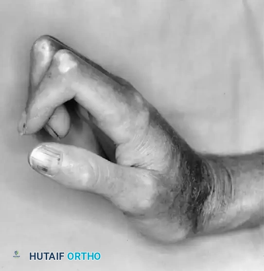
A profound understanding of the complex anatomical and biomechanical interplay in the hand and wrist is a prerequisite for successful surgical intervention in the rheumatoid patient. The wrist serves as the foundational keystone of hand function; its position and stability dictate the biomechanical balance of the extrinsic digital flexor and extensor tendons. In rheumatoid arthritis, the typical collapse pattern involves a predictable sequence of events driven by ligamentous attenuation and altered vector forces. The radiocarpal and midcarpal ligaments become distended by chronic synovitis, leading to carpal supination, ulnar translation of the carpus along the inclined radius articular surface, and subsequent radial deviation of the metacarpals.
This radial deviation of the metacarpals creates a secondary, zigzag biomechanical force that exacerbates the ulnar drift of the digits at the MCP joints. As the metacarpals deviate radially, the extrinsic flexor and extensor tendons are subjected to altered moment arms. The extensor tendons slip ulnarly off the apex of the MCP joints into the intermetacarpal valleys, transitioning from primary extensors to deforming flexors and ulnar deviators. Therefore, wrist reconstruction (whether via partial arthrodesis, total wrist arthrodesis, or total wrist arthroplasty) must precede or be performed concurrently with digital realignment. Correcting MCP ulnar drift without first stabilizing the foundational wrist will inevitably lead to rapid, frustrating recurrence of the digital deformity.
The anatomy of the extensor retinaculum and the dorsal compartments is of particular surgical importance during dorsal tenosynovectomy. The extensor retinaculum is a fibrous band that prevents bowstringing of the extensor tendons but also serves as a rigid roof over the expanding synovial tissue. The caput ulnae syndrome, a frequent precursor to Vaughan-Jackson syndrome, involves destructive synovitis of the distal radioulnar joint (DRUJ). The distended DRUJ capsule and the dorsally subluxated, eroded ulnar head mechanically abrade the overlying extensor tendons (typically starting with the extensor digiti minimi in the fifth compartment, followed by the extensor digitorum communis to the ring and long fingers). Understanding this anatomical relationship is critical for timely prophylactic intervention, such as a Darrach or Sauvé-Kapandji procedure combined with tenosynovectomy.
Systemic conditions like scleroderma present unique anatomical and biomechanical staging challenges due to severe soft-tissue contractures, dermal fibrosis, and profound microvascular compromise. The classic presentation involves rigid flexion contractures of the interphalangeal joints, extension contractures of the MCP joints, and a severe adduction contracture of the thumb, driven by progressive fibrosis of the skin, subcutaneous tissues, and joint capsules.
In such complex connective tissue deformities, reconstructive surgery must be staged meticulously. The surgeon must first release the first web space to restore thumb opposition before addressing the digital contractures, always prioritizing the preservation of the precarious vascular supply. The digital arteries in scleroderma patients are often affected by profound intimal hyperplasia and adventitial fibrosis, making any surgical dissection fraught with the risk of digital ischemia.
Exhaustive Indications and Contraindications
The decision-making process for surgical intervention in the inflammatory hand requires a delicate balance between maximizing functional preservation and minimizing the risks associated with operating on immunocompromised patients with fragile soft tissues. The primary, overarching indication for surgery in the rheumatoid hand is pain that limits function. Esthetics, while understandably important to the patient, are strictly secondary. Patients who function well despite significant, visually striking deformities may be less inclined—and are generally poorer candidates—for surgical intervention than patients whose activities of daily living are severely restricted by pain or progressive tendon dysfunction.
Local injections of a corticosteroid preparation combined with a local anesthetic serve as the first-line localized treatment for persistent inflammatory swelling. This nonoperative modality is particularly efficacious for stenosing tenosynovitis (reducing the volume of the A1 pulley and inflamed flexor tendon sheath), carpal tunnel syndrome (decreasing flexor tenosynovitis within the rigid canal), and providing symptomatic relief in early-stage osteoarthritis or rheumatoid arthritis of the small joints. However, while initial corticosteroid injections often yield dramatic results, the efficacy of repeated injections diminishes over time. Furthermore, repeated intratendinous or peritendinous injections carry a well-documented risk of tendon attenuation and spontaneous rupture, particularly in the rheumatoid patient where enzymatic degradation is already occurring.
If conservative measures fail, surgical indications follow a highly structured hierarchy. To optimize outcomes, Souter recommended a strategic staging protocol, beginning with procedures that carry the highest probability of success and the lowest morbidity. He grouped hand procedures from the most effective (Group I) to the least effective (Group V). Group I includes highly predictable procedures such as carpal tunnel release, trigger finger release, dorsal tenosynovectomy, and DRUJ reconstruction. Group II encompasses arthrodesis of the thumb MCP or interphalangeal joints. Group III includes MCP joint arthroplasty (silicone elastomer implants) and wrist arthrodesis. Groups IV and V are considered less predictable salvage procedures, including complex tendon transfers for multiple ruptures and complex soft-tissue realignments.
| Pathology / Condition | Primary Indications for Surgery | Absolute & Relative Contraindications |
|---|---|---|
| Persistent Tenosynovitis | Failure of medical management > 4-6 months; impending tendon rupture; caput ulnae syndrome. | Active systemic infection; medically unstable patient; lack of functional deficit. |
| Rheumatoid Nodules | Severe mechanical interference with ADLs; impending skin ulceration; recurrent secondary infections. | Asymptomatic nodules; extremely poor overlying skin envelope precluding closure (relative). |
| MCP Joint Ulnar Drift | Painful, rigid dislocation; severe functional deficit; failure of orthotic management. | Uncorrected proximal wrist deformity (absolute); absent intrinsic muscle function without planned transfer. |
| Advanced Wrist Collapse | Intractable pain; severe carpal subluxation threatening extensor tendons; loss of foundational stability. | Painless, stable, functional spontaneous ankylosis; active local soft tissue infection. |
| Thumb Boutonniere/Swan Neck | Painful instability pinching; fixed contracture preventing grasp; Nalebuff Type I-III deformities. | Lack of patient compliance for post-operative rehabilitation; severe generalized ischemia (e.g., advanced scleroderma). |
Pre-Operative Planning, Templating, and Patient Positioning
Pre-operative planning for the patient with inflammatory hand pathology extends far beyond the localized surgical site; it demands a comprehensive, holistic evaluation of the patient's entire musculoskeletal involvement and medical status. The rheumatoid hand is rarely an isolated problem; it is part of a systemic biomechanical collapse. Frequently, the rheumatoid patient requires surgical intervention not only on the opposite hand but also on weight-bearing joints such as the hips, knees, and feet. A crucial rule of staging is that surgery is almost universally performed on only one hand at a given time. The patient must retain the use of one hand for daily independent living, personal hygiene, and feeding.
Logistical considerations for bilateral disease dictate that when multiple small joint procedures (e.g., all four MCP arthroplasties or multiple PIP joint fusions) are indicated, they should be performed concurrently on the same hand to minimize the total number of surgical events and anesthetic exposures. Furthermore, if the patient's lower extremity involvement requires the use of external walking aids (canes, crutches, or walkers), standard grip devices will be impossible to use during the hand's postoperative recovery. In these instances, a platform walker or forearm crutch must be provided to allow weight-bearing through the forearms rather than the healing hand, and this must be coordinated with physical therapy well before the day of surgery.
Medical optimization is paramount. The surgeon must collaborate closely with the patient's rheumatologist regarding the perioperative management of DMARDs and biologic agents. Current American College of Rheumatology (ACR) and American Association of Hip and Knee Surgeons (AAHKS) guidelines generally recommend continuing conventional synthetic DMARDs (like methotrexate) through the perioperative period to prevent disease flare, while biologic agents (like TNF inhibitors) are typically withheld for one dosing cycle prior to surgery and resumed once wound healing is secure (typically 14 days post-operatively). Corticosteroid stress dosing may be required for patients on chronic systemic steroids to prevent adrenal crisis under surgical stress.
In the operating room, meticulous attention to patient positioning is required. The patient is typically positioned supine with the operative extremity extended on a radiolucent hand table. Given the high prevalence of cervical spine instability in rheumatoid patients (atlantoaxial subluxation or subaxial subluxation), extreme care must be taken during intubation and positioning to avoid hyperextension of the neck. A well-padded pneumatic tourniquet is applied to the proximal arm, but the surgeon must be mindful of the fragile rheumatoid skin; cast padding should be applied generously beneath the tourniquet. Regional anesthesia (brachial plexus block) is highly preferred, as it avoids airway manipulation, provides excellent intraoperative conditions, and offers prolonged postoperative analgesia, which is critical for early rehabilitation.
Step-by-Step Surgical Approach and Fixation Technique
Surgical intervention in the inflammatory hand demands meticulous, tissue-respecting techniques. When performing a dorsal tenosynovectomy, a longitudinal midline or slightly curvilinear incision is made over the dorsal wrist. Full-thickness skin flaps, including the subcutaneous tissue and superficial veins, are elevated to preserve the precarious dermal blood supply. The extensor retinaculum is identified and carefully incised. To prevent postoperative bowstringing while allowing for complete synovectomy, a step-cut or Z-lengthening of the retinaculum is often performed. The hypertrophic, boggy synovium is then meticulously dissected from the extensor tendons using tenotomy scissors and blunt dissection. Care must be taken to excise the synovium from between the tendon slips and from the floor of the compartments without violating the tendon substance. If caput ulnae syndrome is present, a Darrach resection of the distal ulna or a Sauvé-Kapandji procedure is performed concurrently to remove the offending bony prominence. The extensor retinaculum is then repaired, often placing half of it deep to the tendons to provide a smooth gliding surface over the carpus.
The surgical excision of rheumatoid nodules requires equally meticulous planning. A carefully designed incision—often a lazy-S or longitudinal incision avoiding the very apex of the nodule—is utilized. The overlying skin in rheumatoid patients is notoriously thin, fragile, and ischemic. Careful blunt and sharp dissection is mandatory. On the palmar aspect of the digits, the neurovascular bundles are frequently displaced, splayed, or densely adherent to the pseudocapsule of larger nodular masses. Magnification loupes must be used to meticulously separate the delicate digital nerves and arteries from the nodule. Because the expanding nodule often causes pressure necrosis of the overlying dermis, primary closure may result in excessive tension. The surgeon must be prepared to utilize full-thickness or split-thickness skin grafts to achieve tension-free closure and prevent postoperative wound dehiscence. Incomplete excision of the nodule's base or failure to address the underlying bony prominence will result in a high rate of local recurrence.
Kirschner wires (K-wires) remain a fundamental tool for osseous fixation in rheumatoid hand surgery, particularly for arthrodesis of the PIP, DIP, and thumb MCP joints. However, the rheumatoid patient presents unique biomechanical and biological challenges, primarily severe periarticular osteopenia. In the rheumatoid hand, the osteopenic nature of the bone means that most K-wires will eventually loosen due to micro-motion at the pin-bone interface. Fortunately, because rheumatoid bone is often highly vascularized due to chronic inflammation, osseous fusion occurs remarkably rapidly after meticulous joint preparation (decortication down to bleeding cancellous bone). The primary role of the K-wire is to provide temporary alignment rather than rigid, long-term biomechanical stability.
The trajectory and final resting position of the K-wires are critical to preventing severe postoperative morbidity. K-wires are usually cut beneath the skin at a level that makes them easily recoverable once fusion is achieved. Under no circumstances should K-wires be left embedded in the tactile pulp of the fingers or near the palmar aspect of the MCP joint of the thumb. The volar skin is highly innervated, and a prominent wire in these areas will cause excruciating pain during pinch and grasp, completely negating the functional benefits of the surgery. To avoid volar pain, wires in these regions must always be inserted or driven so that the end nearest the skin rests on the dorsal surface of the digit or hand, where the soft tissue is more forgiving and less involved in tactile interaction.
Complications, Incidence Rates, and Salvage Management
Despite meticulous surgical technique, complication rates in the surgical management of inflammatory hand pathologies remain higher than in the general population, largely due to systemic immunosuppression, poor bone stock, and compromised soft-tissue envelopes. Wound healing complications are the most frequent adverse events. The fragile, steroid-thinned skin of the rheumatoid patient is highly susceptible to marginal necrosis, dehiscence, and delayed healing. Meticulous hemostasis, tension-free closure, and the judicious use of drains and bulky, non-constrictive dressings are essential preventive measures.
Infection is a devastating complication, particularly in patients on concurrent biologic therapies and high-dose corticosteroids. Superficial pin-tract infections associated with percutaneous K-wires occur in up to 10-15% of cases. These must be managed aggressively with oral antibiotics and prompt removal of the offending hardware if early consolidation has been achieved. Deep space infections or septic arthritis following arthroplasty require emergent surgical debridement, implant removal, and placement of antibiotic-impregnated spacers, followed by a prolonged course of intravenous antibiotics. Salvage in these scenarios typically culminates in a delayed arthrodesis once the infection is definitively eradicated.
Hardware failure and loss of fixation are common due to severe osteopenia. K-wires may migrate, bend, or back out before osseous union is complete. If a nonunion occurs following an attempted arthrodesis (e.g., at the PIP joint or the wrist), the patient may remain surprisingly asymptomatic due to fibrous ankylosis. However, if the nonunion is painful or unstable, revision surgery utilizing more robust fixation methods—such as tension band constructs, locking plates, or intramedullary compression devices—supplemented with autologous bone grafting is indicated. Recurrence of deformity, particularly ulnar drift of the MCP joints, is another significant complication, almost always resulting from a failure to recognize and correct proximal radiocarpal or midcarpal instability at the time of the index procedure.
| Complication | Estimated Incidence | Prevention Strategy | Salvage / Management |
|---|---|---|---|
| Wound Dehiscence / Necrosis | 5% - 12% | Tension-free closure; avoid incisions over nodule apices; handle tissues with skin hooks. | Local wound care; secondary intention healing; split-thickness skin grafting if extensive. |
| Pin Tract Infection | 10% - 15% | Buried pins when possible; meticulous pin site care; early removal at 4-6 weeks. | Oral antibiotics; immediate pin extraction; local debridement if purulence is present. |
| Tendon Rupture (Post-Op) | 2% - 5% | Complete synovectomy; address bony prominences (e.g., Darrach); avoid aggressive early passive ROM. | Tendon transfer (e.g., EIP to EDC); intercalary tendon grafting; side-to-side tenodesis. |
| Recurrent Ulnar Drift | 15% - 30% (Long-term) | Correct wrist radial deviation first; adequate intrinsic release; robust radial collateral ligament reconstruction. | Revision arthroplasty; crossed intrinsic transfer; eventual MCP joint arthrodesis. |
| Symptomatic Nonunion | 5% - 10% | Meticulous joint decortication; rigid temporary fixation; avoid premature hardware removal. | Revision arthrodesis with locking plates or intramedullary devices; autologous bone graft. |
Phased Post-Operative Rehabilitation Protocols
The post-operative rehabilitation of the rheumatoid hand is arguably as critical to the final outcome as the surgical execution itself. The rehabilitation protocol must be carefully phased to balance the competing demands of protecting delicate soft-tissue repairs, achieving osseous union, and preventing profound joint stiffness. The immediate post-operative phase (Days 1 to 14) is focused entirely on wound healing, edema control, and pain management. The hand is immobilized in a bulky, well-padded compressive dressing reinforced with a plaster splint. Elevation of the extremity strictly above the level of the heart is mandatory to minimize dependent edema, which can compromise the microcirculation of the skin flaps. Finger motion is typically restricted, except for gentle, active range of motion of joints not involved in the surgical field (e.g., shoulder and elbow) to prevent proximal stiffness.
The intermediate phase (Weeks 2 to 6) begins with the first post-operative clinic visit. Sutures are removed at 10 to 14 days, provided the wound edges are well-apposed and viable. At this stage, a custom-fabricated thermoplastic orthosis is provided by a specialized hand therapist. The design of the orthosis depends on the procedure performed; for example, following MCP arthroplasty, a dynamic extension splint with outriggers to prevent recurrent ulnar deviation is utilized. Because of the rapid fusion rates and the tendency for wires to loosen in osteopenic bone, prolonged retention of K-wires is rarely necessary or advised. Once clinical and radiographic evidence of early consolidation is present (typically 4 to 6 weeks), the wires should be extracted. Most percutaneous or superficially buried K-wires can be easily removed in the outpatient office setting using a local anesthetic. Prompt removal minimizes the risk of deep pin-tract infections.
The late phase of rehabilitation (Weeks 6 to 12 and beyond) focuses on maximizing functional range of motion, restoring grip and pinch strength, and educating the patient on joint protection strategies. Following K-wire removal, the limb is typically protected in a custom orthosis during heavy activities until final, robust osseous union is achieved. Strengthening exercises are introduced gradually, utilizing putty and graded resistance devices. However, the therapist must remain vigilant; overzealous strengthening in the rheumatoid patient can precipitate a flare of localized synovitis or stress newly reconstructed soft tissues. Patients are educated on ergonomic adaptations, such as using built-up handles for utensils and tools, to minimize the mechanical stress across the small joints of the hand during daily activities.
Summary of Landmark Literature and Clinical Guidelines
The surgical management of the inflammatory hand is built upon a foundation of landmark literature and established clinical guidelines that have evolved over decades. William Souter’s seminal work on the staging of rheumatoid hand surgery remains a cornerstone of orthopedic education. Souter’s hierarchy accurately categorized procedures based on their predictability and efficacy, establishing the paradigm that prophylactic and decompressive procedures (Group I) yield the highest patient satisfaction and should be prioritized. His assertion that the wrist is the keystone of hand function fundamentally changed how surgeons sequence multi-level deformities, ensuring that foundational stability is achieved before distal digital realignment is attempted.
Arthur Nalebuff’s contributions to the classification and management of rheumatoid thumb deformities are equally critical. Nalebuff described the classic patterns of thumb collapse—most notably the Type I (boutonniere) and Type III (swan neck) deformities—and provided a clear, algorithm-based approach to surgical reconstruction based on the flexibility of the joints and the integrity of the articular cartilage. His principles emphasize that isolated soft-tissue reconstructions are doomed to failure if underlying joint destruction or fixed contractures are present, advocating for arthrodesis as the most reliable, durable procedure for the rheumatoid thumb interphalangeal and MCP joints.
Contemporary clinical guidelines, particularly those jointly published by the American College of Rheumatology (ACR) and the American Association of Hip and Knee Surgeons (AAHKS), provide critical directives on the perioperative management of antirheumatic medications. These guidelines, updated to reflect the proliferation of biologic and targeted synthetic DMARDs, emphasize a stratified approach based on the medication's half-life and the patient's individual risk of infection versus disease flare. The consensus dictates that conventional DMARDs should generally be continued, while biologics require careful, timed cessation prior to surgery. Mastery of these historical principles and modern guidelines is essential for the orthopedic surgeon striving to deliver optimal, comprehensive care to the complex patient with inflammatory hand pathology.
