Christine M Kleinert Discussion: Solving Difficult Wrist Fractures
Key Takeaway
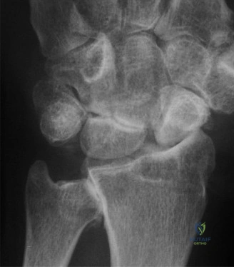
For anyone wondering about Christine M Kleinert Discussion: Solving Difficult Wrist Fractures, Scaphoid fractures require suspicion with anatomical snuff box tenderness, even if initial X-rays are negative; treat with splinting and re-imaging in 2-4 weeks. The christine m kleinert discussion highlights CT scans as more reliable for assessing healing than X-rays. For persistent non-healing, Open Reduction Internal Fixation (ORIF) with cancellous bone graft and headless compression screws is the recommended treatment.
A 14-year-old male presents following a FOOSH. You suspect an "occult" scaphoid fracture based on clinical examination. You obtain initial radiographs, which are unremarkable. Describe your immediate management and the rationale behind your decision-making.
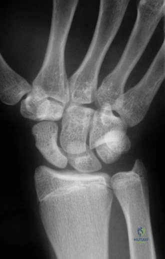
Candidate: I would clinically diagnose this as a suspected scaphoid fracture. Since the initial films are negative, I would immobilize the wrist in a thumb spica splint and arrange for a follow-up with repeat radiographs in 10-14 days. If the pain persists, I might consider an MRI if available, or continue casting.
Candidates often fail to mention the "axial loading" test or specific landmarks (tubercle vs. snuff box) during examination. They also often fail to mention the role of MRI as the definitive "gold standard" for ruling out occult fractures early, relying too heavily on outdated "wait and see" protocols without considering the socioeconomic benefits of early diagnosis.
A perfect answer emphasizes the high clinical suspicion: "I would examine for tenderness at the anatomical snuff box, scaphoid tubercle, and pain with axial thumb loading. Given the negative X-rays, the patient has an 'occult' fracture until proven otherwise. Standard care is immobilization in a thumb spica splint with follow-up at 10-14 days. However, given the current evidence base, I would favor early MRI to confirm or exclude the diagnosis within 48 hours. This prevents unnecessary prolonged immobilization, allows for earlier return to activity if negative, and avoids the diagnostic delay associated with repeat radiographs."
The patient returns at 8 weeks post-injury. The fracture line is now visible, and the patient reports persistent pain. How do you assess for healing, and what criteria determine your next steps?
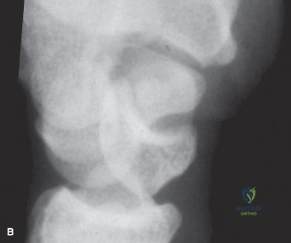
Candidate: I would examine the patient for tenderness and then get a CT scan. I'm looking for trabecular bridging. If there is less than 50% bridging, I would consider surgery.
Failing to specify the CT scan orientation. It is critical to mention the CT must be oriented along the long axis of the scaphoid to accurately assess bridging. Also, failing to distinguish between the adolescent's healing potential and an adult's requirement for more aggressive management.
The candidate must state: "Clinical assessment of tenderness is unreliable for union. I would order a CT scan reformatted along the longitudinal axis of the scaphoid. A fracture is defined as united when there is >50% trabecular bridging. In an adolescent with an asymptomatic fracture, I might accept a longer duration of cast treatment; however, symptomatic non-union at 8 weeks with <50% bridging necessitates surgical intervention (ORIF) to restore anatomy and prevent long-term sequelae like SNAC wrist."
Describe the "humpback" deformity and its significance in scaphoid non-union. How does this dictate your surgical approach?
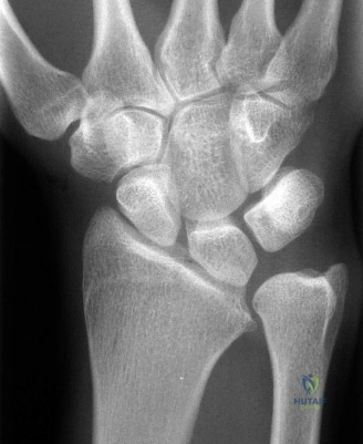
Candidate: The humpback deformity is when the scaphoid collapses into a flexed position, leading to DISI instability. This makes the wrist arthritic. I would do a volar approach to fix it and maybe use a bone graft.
Vague terminology. Candidates should use specific angles (Intrascaphoid angle >35°, Radiolunate angle >15°) to define the deformity. They must also explain the *why*—that this creates a midcarpal instability pattern that progresses to SNAC wrist.
The perfect answer: "The humpback deformity is a volar angular collapse of the scaphoid fragments. It is characterized by an intrascaphoid angle >35° and is associated with DISI (dorsal intercalated segment instability), evidenced by a radiolunate angle >15°. Because this alters carpal kinematics and inevitably leads to SNAC (Scaphoid Non-union Advanced Collapse), it mandates a volar approach. The volar approach allows for formal open reduction, restoration of the length of the scaphoid using an interpositional structural bone graft (usually from the distal radius or iliac crest), and rigid fixation with a headless compression screw."
