Wrist Recovery Guide: Mastering Degrees with Forearm Movement
Key Takeaway
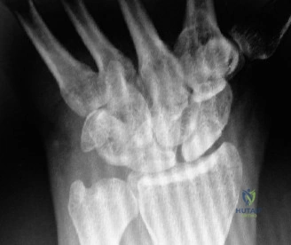
Here are the crucial details you must know about Wrist Recovery Guide: Mastering Degrees with Forearm Movement. Initial treatment for a transverse pisiform fracture includes short arm splinting. Should this fail, surgical excision of the pisiform is the preferred procedure for chronic pain, generally without major functional deficit. Post-operative rehabilitation aims to restore full wrist motion, progressing through various degrees with forearm support and stability to ensure optimal recovery and function.
A 28-year-old rugby player presents with persistent ulnar-sided wrist pain 4 months after a direct blow to the hypothenar eminence. He has failed conservative management. Please describe the relevant anatomy of the pisiform and the potential neurological implications of your management plan.
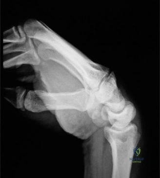
Candidate: The pisiform is a sesamoid bone within the FCU tendon. It articulates with the triquetrum. Because it forms the medial wall of Guyon’s canal, it is closely related to the ulnar nerve. If I perform a pisiform excision, I need to be very careful not to injure the deep motor branch or the superficial sensory branch of the ulnar nerve.
Candidates often fail to describe the dynamic nature of the ulnar nerve relative to the pisiform or ignore the critical role of the pisohamate and pisometacarpal ligaments in maintaining the FCU's moment arm. Merely stating "it's near the nerve" is insufficient at the FRCS level.
The candidate should state: The pisiform is a sesamoid in the FCU, acting as an anterior fulcrum to increase the FCU moment arm. It forms the ulnar wall of Guyon's canal. The ulnar neurovascular bundle bifurcates just distal to the pisiform; the deep motor branch dives between the abductor and flexor digiti minimi, making it highly vulnerable during resection. Key anatomical landmarks include the pisohamate and pisometacarpal ligaments, which must be released carefully. Pre-operative documentation of distal ulnar nerve function (motor/sensory) is mandatory.
Look at this radiograph of a 65-year-old patient with chronic thumb base pain. What is your staging and management strategy?
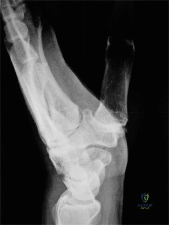
Candidate: This shows advanced osteoarthritis at the first CMC joint with subchondral sclerosis and osteophytes. This is likely Eaton-Littler Stage III or IV. I would try splinting and injections first. If that fails, I would suggest a trapeziectomy with LRTI.
Failure to mention the status of the scaphotrapezial-trapezoid (STT) joint or failing to differentiate between a manual laborer (where arthrodesis might be preferred) and an elderly patient. Also, failing to recognize that "radiographic subsidence" after LRTI does not necessarily correlate with poor clinical outcomes.
Identify the findings as Eaton-Littler Stage III/IV. Structure the response by addressing: 1) Clinical symptoms (rest pain/pinch deficit). 2) Failure of non-operative management (min 6 months). 3) Surgical options: Trapeziectomy ± LRTI is standard, but mention that recent literature shows simple trapeziectomy is equally effective. 4) For high-demand younger patients, discuss CMC arthrodesis. 5) Highlight the risk of radial sensory nerve injury during the dorsal-radial approach.
Following a successful excision of the pisiform, what is your specific rehabilitation protocol for this athlete to ensure they return to rugby safely?
Candidate: I would put them in a splint for a few weeks, then start moving the wrist. Eventually, I would introduce light weights and then sports-specific training once the FCU repair is strong.
Being too vague. The examiner is looking for the "Dart-Thrower's Motion" and the timing of FCU stress. Missing the importance of early digit mobilization to prevent CRPS is a significant error.
Phased protocol: 1) 0-2 weeks: Immobilization in 20° flexion to protect the FCU repair; prioritize edema control and digit AROM. 2) 2-6 weeks: Transition to custom splint; initiate gravity-eliminated AROM, avoiding passive stretch. 3) 6-10 weeks: Initiate "Dart-Thrower’s Motion" (coupled radial extension/ulnar flexion) to utilize midcarpal motion while protecting radiocarpal/pisotriquetral stress. 4) 10-16+ weeks: Eccentric FCU strengthening and plyometrics for return-to-sport. Emphasize that FCU strength typically recovers well if the repair is robust.
