Ulnar Shortening Osteotomy: Comprehensive Surgical Technique and Biomechanical Principles
Key Takeaway
Ulnar shortening osteotomy is a highly effective surgical intervention for ulnar impaction syndrome and symptomatic ulnar-positive variance. By precisely resecting a measured diaphyseal segment and applying rigid dynamic compression plating, surgeons can decompress the ulnocarpal joint while preserving the distal radioulnar joint (DRUJ) kinematics. This guide details the Chun and Palmer technique, emphasizing preoperative templating, meticulous soft-tissue handling, oblique osteotomy execution, and robust internal fixation to ensure optimal biomechanical restoration and union.
Comprehensive Introduction and Patho-Epidemiology
The Ulnar Shortening Osteotomy (USO) stands as a foundational, joint-leveling operative intervention within the specialized domain of hand and upper extremity surgery. Originally conceptualized and subsequently popularized by the landmark biomechanical and clinical investigations of Chun and Palmer, this procedure is meticulously designed to address ulnar impaction syndrome (ulnocarpal abutment) and its associated constellation of degenerative pathologies. Ulnar impaction syndrome is a progressive, debilitating condition characterized by excessive, pathological load transmission across the ulnar aspect of the radiocarpal and ulnocarpal articulations, leading to the sequential degradation of the Triangular Fibrocartilage Complex (TFCC), the chondral surfaces of the ulnar head, the lunate, and the triquetrum.
The patho-epidemiology of ulnar impaction syndrome is intimately linked to both congenital variance and acquired anatomical distortions. Congenitally, patients with a naturally occurring ulnar-positive variance are predisposed to this condition, as the distal ulna projects further distally than the articular surface of the radius, creating a mechanical conflict during functional arcs of motion. Acquired ulnar impaction frequently manifests as a sequela of distal radius fractures that have healed with a loss of radial height, dorsal angulation, or radial inclination, thereby creating a state of relative ulnar overgrowth. Furthermore, premature closure of the distal radial physis secondary to pediatric trauma (e.g., Salter-Harris fractures) or repetitive microtrauma (as seen in gymnast's wrist) can precipitate severe ulnar-positive variance in young, active demographics.
Clinically, patients present with insidious, progressive ulnar-sided wrist pain that is characteristically exacerbated by forceful grip, forearm pronation, and ulnar deviation. This triad of movements dynamically increases the ulnocarpal load, grinding the ulnar head against the carpus. Over time, the chronic repetitive abutment leads to focal chondromalacia of the proximal ulnar pole of the lunate, central perforation of the TFCC articular disc (Palmer Class 2 degenerative tears), and eventual lunotriquetral (LT) ligament attenuation. The progressive nature of this cascade underscores the necessity of early recognition and biomechanical correction through a precisely executed ulnar shortening osteotomy to halt joint degradation and restore physiological load distribution.
Beyond the simple mechanical decompression of the ulnar head against the proximal carpal row, the ulnar shortening osteotomy exerts a profound stabilizing effect through the principle of "ligamentotaxis." By shortening the ulnar diaphysis, the procedure effectively increases the resting tension within the ulnocarpal ligaments (ulnolunate and ulnotriquetral ligaments) and the volar radioulnar ligaments of the TFCC. This tensioning stabilizes the ulnar carpus, provides secondary support to the lunotriquetral interval, and enhances the dynamic stability of the distal radioulnar joint (DRUJ). Consequently, USO has evolved from a purely decompressive operation into a versatile, multifaceted tool for managing complex ulnar-sided wrist instability.
Detailed Surgical Anatomy and Biomechanics
Osteology and Articular Geometry
A profound understanding of the complex osteology and articular geometry of the distal forearm is paramount for the successful execution of an ulnar shortening osteotomy. The distal radioulnar joint (DRUJ) is a highly specialized trochoid (pivot) joint that facilitates forearm pronation and supination. The articular congruity of the DRUJ is dictated by the relationship between the convex articular surface of the ulnar head (the seat) and the concave sigmoid notch of the distal radius. The morphology of the sigmoid notch is highly variable and significantly influences the biomechanical outcome of ulnar shortening. Tolat et al. classified the DRUJ into three distinct morphological types based on the inclination of the sigmoid notch relative to the longitudinal axis of the ulna: Type 1 (parallel), Type 2 (oblique, facing proximally), and Type 3 (reverse oblique, facing distally).
In Type 1 configurations, ulnar shortening typically maintains joint congruity and is well-tolerated. However, in Type 2 and particularly Type 3 configurations, proximal translation of the ulnar head can lead to severe articular incongruity, resulting in dramatically increased joint reactive forces, focal chondral overload, and the rapid onset of iatrogenic DRUJ osteoarthritis. Therefore, meticulous preoperative radiographic evaluation of the sigmoid notch morphology is a critical determinant of patient suitability for this procedure. The surgeon must recognize that altering the length of the ulna fundamentally changes the contact mechanics of the DRUJ, and failure to account for an unfavorable sigmoid notch inclination is a primary cause of postoperative failure.
Ligamentous Anatomy and the TFCC
The Triangular Fibrocartilage Complex (TFCC) is the primary stabilizing structure of the ulnar aspect of the wrist, serving both as a load-bearing cushion and a complex ligamentous tether. The TFCC comprises the central articular disc, the dorsal and volar radioulnar ligaments, the meniscus homologue, the ulnocarpal ligaments (ulnolunate and ulnotriquetral), and the extensor carpi ulnaris (ECU) subsheath. The radioulnar ligaments are the primary stabilizers of the DRUJ, with the deep (foveal) fibers providing the majority of the isometric restraint during forearm rotation. The ulnocarpal ligaments originate from the base of the ulnar styloid and insert onto the volar aspects of the lunate and triquetrum, acting as critical stabilizers of the ulnar carpus.
During an ulnar shortening osteotomy, the proximal translation of the distal ulnar segment exerts a direct, tensioning force on these ligamentous structures. This ligamentotaxis effect is particularly beneficial in the presence of mild to moderate lunotriquetral (LT) instability or peripheral TFCC laxity. By tightening the ulnocarpal ligaments, the lunate and triquetrum are drawn proximally and ulnarly, reducing abnormal carpal kinematics and alleviating dynamic instability. Furthermore, the tensioning of the ECU subsheath and the volar radioulnar ligaments enhances the containment of the ulnar head within the sigmoid notch, thereby improving DRUJ stability. This intricate interplay between bony architecture and soft-tissue tension underscores the elegance of the USO as a comprehensive reconstructive procedure.
Biomechanics of Load Transfer and Ligamentotaxis
The biomechanical rationale for ulnar shortening is rooted in the precise quantification of axial load transmission across the radiocarpal and ulnocarpal joints. In a physiologically normal wrist exhibiting neutral ulnar variance, the distal radius absorbs approximately 80% of the axial compressive load during activities of daily living, while the distal ulna and the interposed TFCC absorb the remaining 20%. This physiological distribution ensures optimal stress distribution across the chondral surfaces and prevents focal overloading. However, minor alterations in ulnar length precipitate dramatic shifts in this biomechanical equilibrium.
Extensive cadaveric and biomechanical studies, pioneered by Palmer and Werner, have unequivocally demonstrated that an increase in ulnar variance of merely 2.5 mm (ulnar-positive variance) shifts the load distribution catastrophically. In this state, the ulnar axial load transmission escalates from 20% to nearly 42%, subjecting the TFCC and the ulnar carpus to forces that far exceed their physiological tolerance. This chronic overload initiates the degenerative cascade characteristic of ulnar impaction syndrome. Conversely, the biomechanical efficacy of the USO is equally profound: shortening the ulna by 2.5 mm from a neutral position decreases the load at the ulnocarpal joint to approximately 4.3%. By strategically resecting a meticulously measured diaphyseal wafer, the surgeon effectively unloads the ulnar compartment, allowing for the potential healing of early chondral lesions and providing immediate, definitive relief from mechanical abutment.
Exhaustive Indications and Contraindications
Primary Indications
The decision to proceed with an ulnar shortening osteotomy must be predicated on a rigorous clinical and radiographic evaluation. The hallmark indication is symptomatic ulnar impaction syndrome, characterized by recalcitrant ulnar-sided wrist pain, swelling, and a demonstrable loss of grip strength. This is frequently accompanied by positive provocative maneuvers, such as the ulnocarpal stress test (pain elicited with the wrist in ulnar deviation, axial compression, and passive pronosupination). USO is the definitive treatment for patients who have failed conservative management modalities, including non-steroidal anti-inflammatory drugs (NSAIDs), splinting, and intra-articular corticosteroid injections.
Beyond classic impaction, USO is highly indicated for the management of specific TFCC pathologies. Palmer Class 2 (degenerative) tears, particularly Class 2C (perforation of the articular disc with chondromalacia of the lunate/triquetrum) and Class 2D (addition of LT ligament perforation), are prime indications. In these scenarios, repairing the degenerative tissue is futile without addressing the underlying biomechanical overload. Furthermore, USO is indicated for Palmer Class 1 (traumatic) peripheral tears that are associated with a pre-existing ulnar-positive variance, where primary ligamentous repair alone would be subjected to excessive, deleterious tension. Additionally, early, dynamic lunotriquetral instability without fixed carpal collapse benefits immensely from the ligamentotaxis generated by ulnar shortening.
Relative and Absolute Contraindications
Despite its high success rate, USO is not a panacea and carries stringent contraindications that must be respected to avert disastrous postoperative outcomes. The absolute contraindication to a diaphyseal ulnar shortening is the presence of advanced, symptomatic osteoarthritis of the distal radioulnar joint. Shortening the ulna in the setting of pre-existing DRUJ arthrosis will inevitably exacerbate the joint reactive forces, leading to severe, intractable pain and a profound loss of forearm rotation. In such instances, salvage procedures that eliminate the DRUJ articulation, such as the Darrach procedure (distal ulnar resection) or the Sauvé-Kapandji procedure (DRUJ arthrodesis with proximal pseudoarthrosis), are the mandated alternatives.
Relative contraindications include a fundamentally incompatible sigmoid notch morphology (Tolat Type 3, reverse oblique), which predisposes the patient to postoperative DRUJ incongruity and subluxation. Fixed, irreducible DRUJ subluxation or frank dislocation also precludes a simple shortening osteotomy, as the procedure cannot overcome severe anatomical distortion. Furthermore, active local or systemic infection, severe osteopenia or osteoporosis (which compromises rigid internal fixation), and a history of noncompliance with postoperative rehabilitation protocols are significant deterrents. The surgeon must also cautiously evaluate patients who smoke tobacco, as the diaphyseal ulna possesses a tenuous blood supply, and nicotine-induced vasoconstriction exponentially increases the risk of delayed union and nonunion.
| Clinical Scenario | Indication Status | Biomechanical Rationale / Alternative Strategy |
|---|---|---|
| Ulnar Impaction Syndrome (Positive Variance) | Primary Indication | Unloads the ulnocarpal joint, reducing axial load from ~42% to <5%. |
| Palmer Class 2C/2D TFCC Tears | Primary Indication | Decompresses degenerative perforations; ligament repair is futile without unloading. |
| Early Dynamic LT Instability | Primary Indication | Utilizes ligamentotaxis to tension ulnocarpal ligaments, stabilizing the LT interval. |
| Advanced DRUJ Osteoarthritis | Absolute Contraindication | Shortening exacerbates joint reactive forces. Alternative: Sauvé-Kapandji or Darrach. |
| Tolat Type 3 Sigmoid Notch (Reverse Oblique) | Relative Contraindication | High risk of creating iatrogenic DRUJ incongruity and subluxation upon shortening. |
| Active Infection (Local/Systemic) | Absolute Contraindication | High risk of hardware seeding and osteomyelitis. Requires eradication prior to surgery. |
Pre-Operative Planning, Templating, and Patient Positioning
Radiographic Evaluation and Variance Measurement
The foundation of a successful ulnar shortening osteotomy lies in obsessive, meticulous preoperative planning and precise radiographic evaluation. Standardized, high-quality radiographs are non-negotiable. The critical view is the strict posteroanterior (PA) radiograph of the wrist, obtained with the patient's shoulder abducted to 90 degrees, the elbow flexed to 90 degrees, and the forearm in absolute neutral rotation. This "zero position" is imperative because forearm rotation dynamically alters apparent ulnar variance: pronation artificially increases ulnar-positive variance due to the proximal migration of the radius around the fixed ulna, whereas supination decreases it. Measurements taken from non-standardized films will lead to erroneous calculations and catastrophic over- or under-shortening.
Ulnar variance is quantified by measuring the perpendicular distance between two parallel lines drawn perpendicular to the longitudinal axis of the radius. The first line is drawn tangent to the sclerotic subchondral bone of the lunate fossa of the distal radius. The second line is drawn tangent to the most distal articular surface of the ulnar head (excluding the ulnar styloid process). In a symptomatic patient, this measurement dictates the magnitude of the required resection. The surgical objective is to achieve a final postoperative ulnar variance of 0 to −1 mm (neutral to slightly ulnar-negative).
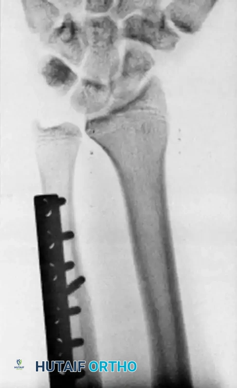
Figure 1: Preoperative PA radiograph of a patient with symptomatic ulnar-positive variance, demonstrating the need for ulnar shortening.
Preoperative Templating and Implant Selection
Once the required magnitude of shortening is determined (e.g., a 3.5 mm positive variance requires a 4.0 to 4.5 mm resection to achieve a −1 mm final variance), the surgeon must engage in rigorous preoperative templating. Templating involves overlaying digital or physical templates of the selected osteotomy plate onto the preoperative radiographs to determine the optimal plate size, screw length, and the precise anatomical location for the osteotomy. The osteotomy is typically planned at the junction of the middle and distal thirds of the ulnar diaphysis, ensuring adequate bone stock distally for the secure purchase of at least three bicortical screws.
Implant selection is a critical variable. While a standard 6-hole or 7-hole 3.5-mm dynamic compression plate (DCP) or limited contact dynamic compression plate (LC-DCP) can be utilized, contemporary practice frequently favors dedicated, specialized ulnar shortening osteotomy systems (e.g., Rayhack, Trimed, or Synthes USO systems). These dedicated systems incorporate precision cutting guides that attach directly to the plate or bone, facilitating perfectly parallel oblique cuts and controlled, millimeter-specific compression. The choice of implant dictates the specific surgical steps, but the underlying biomechanical goal of achieving absolute stability with interfragmentary compression remains universal.
Patient Setup and Anesthesia
Optimal patient positioning and anesthetic management are essential for facilitating a smooth, unhindered surgical workflow. The procedure is typically performed under regional anesthesia (supraclavicular or axillary brachial plexus block), which provides excellent intraoperative analgesia, profound muscle relaxation, and beneficial postoperative pain control. General anesthesia is reserved for patients with contraindications to regional blocks or based on patient preference. Following the induction of anesthesia, the patient is positioned supine on the operating table, with the operative extremity extended onto a radiolucent hand table.
A well-padded pneumatic tourniquet is applied to the proximal arm to ensure a bloodless surgical field, which is critical for identifying delicate neurovascular structures. The limb is prepped and draped in a standard sterile fashion, encompassing the entire forearm and hand to allow for intraoperative assessment of forearm rotation and wrist kinematics. Prior to incision, the limb is exsanguinated using an Esmarch bandage, and the tourniquet is inflated to a standard pressure (typically 250 mm Hg or 100 mm Hg above the patient's systolic blood pressure). The intraoperative C-arm fluoroscopy unit must be positioned to allow for seamless, unobstructed orthogonal imaging (PA and lateral views) of the forearm and wrist throughout the procedure, ensuring real-time verification of hardware placement and osteotomy reduction.
Step-by-Step Surgical Approach and Fixation Technique
Incision and Soft Tissue Dissection
The surgical approach to the distal ulnar diaphysis utilizes the highly reliable internervous and intermuscular plane between the Flexor Carpi Ulnaris (FCU), innervated by the ulnar nerve, and the Extensor Carpi Ulnaris (ECU), innervated by the posterior interosseous nerve (PIN). Beginning at the level of the ulnar neck, a longitudinal incision of approximately 8 to 10 centimeters is made along the subcutaneous, ulnar (medial) border of the distal forearm. The length of the incision must be generous enough to accommodate the selected plate (typically a 6-hole or 7-hole construct) without subjecting the soft tissues to excessive, ischemic retraction.
Meticulous subcutaneous dissection is paramount. The surgeon must possess a heightened awareness of the Dorsal Sensory Branch of the Ulnar Nerve (DSBUN). This critical sensory nerve emerges from beneath the FCU approximately 5 to 8 centimeters proximal to the ulnar styloid and courses dorsally over the distal ulna to provide sensation to the ulnar dorsum of the hand. The DSBUN is exquisitely sensitive to traction and compression; iatrogenic injury or aggressive retraction can result in debilitating postoperative neuromas and complex regional pain syndrome (CRPS). Once identified, the nerve is gently mobilized and protected with vessel loops. The deep antebrachial fascia is then incised in line with the skin incision, and the interval between the FCU and ECU is developed. The periosteum over the dorsal and ulnar aspects of the ulna is incised longitudinally and elevated subperiosteally to expose the diaphysis. Crucially, periosteal stripping must be strictly limited to the footprint of the plate to preserve the tenuous periosteal blood supply, thereby mitigating the risk of nonunion.
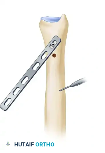
Plate Application and Rotational Marking
Following adequate bony exposure, the selected plate is introduced into the surgical field. For a standard technique utilizing a 3.5-mm DCP, the plate is positioned on the dorsal or volar-ulnar surface of the distal ulna. Dorsal placement is biomechanically advantageous as it acts as a tension band against the physiological volar bowing forces, but it carries a higher risk of symptomatic hardware prominence. The distal end of the plate should rest just proximal to the proximal border of the sigmoid notch to avoid interfering with DRUJ kinematics. The plate may require subtle contouring using plate benders to perfectly match the native anatomy of the ulnar diaphysis.
Once optimally positioned, the plate is temporarily secured to the bone using bone reduction forceps. The two most distal screw holes are drilled, measured, tapped, and filled with 3.5-mm cortical screws, firmly anchoring the plate to the distal ulnar segment.
Figure 2: A six-hole 3.5-mm plate is placed on the dorsal surface of the ulna, and the two most distal screws are inserted. A longitudinal mark is made with electrocautery for rotational orientation.
Before proceeding with the osteotomy, it is absolutely critical to establish a reliable reference for rotational alignment. The ulna is not a simple cylinder; any inadvertent rotation of the distal fragment relative to the proximal fragment during the osteotomy closure will profoundly alter the DRUJ mechanics, leading to a mechanical block to pronation or supination. Using a fine-tipped electrocautery or a sterile surgical marker, a distinct, longitudinal line is scored across the planned osteotomy site on the exposed ulnar shaft. This mark serves as the definitive guide for restoring perfect anatomical rotation during the final reduction. Once marked, the most distal screw is slightly loosened, and the second distal screw is completely removed. Hinging on the loosened distal screw, the plate is swung out of the surgical field to provide unobstructed access to the diaphysis.
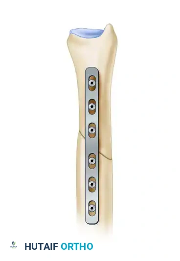
Figure 3: The plate is swung out of the way, hinging on the distal-most screw, to allow unobstructed access for the osteotomy.
Execution of the Oblique Osteotomy
The osteotomy is the most technically demanding phase of the procedure. The site is centered precisely within the footprint of the swung-away plate. An oblique osteotomy is universally preferred over a transverse cut for several biomechanical reasons: it provides a significantly larger surface area for osteogenesis and bony union, it offers inherent rotational stability upon reduction, and crucially, it allows for the placement of an independent interfragmentary lag screw perpendicular to the osteotomy plane, achieving absolute stability.
Using a fine-toothed oscillating saw under continuous, copious cold saline irrigation to prevent thermal necrosis of the osteocytes, the initial cut is initiated. The trajectory of the cut should be from proximal-medial to distal-lateral, angled at approximately 45 degrees to the longitudinal axis of the bone. The saw blade is advanced through approximately 70% of the ulnar diameter, leaving the far cortex intact temporarily to maintain structural stability for the second cut.
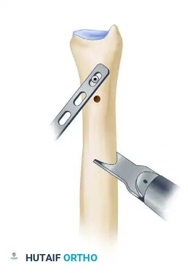
Figure 4: An oscillating saw is used to make the initial oblique osteotomy through 70% of the ulna.
Precision in the thickness of the resected bone wafer is paramount and must correspond exactly to the preoperative templating (e.g., 2.5 mm). To ensure the second cut is perfectly parallel to the first—preventing angular deformity upon closure—a free, unattached saw blade of known thickness is inserted into the first osteotomy kerf to act as a physical, parallel guide. The oscillating saw is then positioned adjacent to this guide, separated by the exact measured distance of the planned resection, and the second cut is executed.
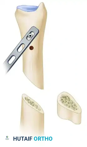
Figure 5: A second cut is made parallel to the first, utilizing a free saw blade as a guide to ensure precision.
Once both cuts are established, they are carefully completed through the remaining far cortex. The precisely measured, oblique diaphyseal bone wafer is then gently extracted using a Freer elevator or small rongeur. The osteotomy surfaces are inspected to ensure they are clean, parallel, and free of thermal damage.

Figure 6: The precisely measured oblique bone segment is removed.
Reduction, Compression, and Rigid Internal Fixation
With the bone wafer removed, the osteotomy is reduced by bringing the proximal and distal ulnar fragments into intimate contact. The surgeon must meticulously verify that the previously scored electrocautery marks align perfectly, ensuring zero rotational malalignment. The plate is then rotated back into its original footprint over the dorsum of the ulna. The first distal screw is retightened, and the second distal screw is replaced and secured.
To achieve dynamic compression across the osteotomy site, an articulated bone reduction clamp (e.g., a Verbrugge or specialized compression clamp) is applied. One tine of the clamp engages the plate, while the other engages the proximal ulnar shaft. As the clamp is tightened, the osteotomy gap is forcibly closed, compressing the oblique bony surfaces together. At this critical juncture, intraoperative C-arm fluoroscopy is deployed to assess the reduction, confirm the restoration of neutral or slightly negative ulnar variance (−1 mm), and verify that the DRUJ is congruent without subluxation.
Once optimal variance and alignment are confirmed fluoroscopically, the plate is rigidly secured to the proximal fragment. This is achieved by inserting the remaining 3.5-mm cortical screws using standard dynamic compression principles—eccentric drilling in the plate holes adjacent to the osteotomy site to generate further axial compression as the spherical screw heads engage the plate.
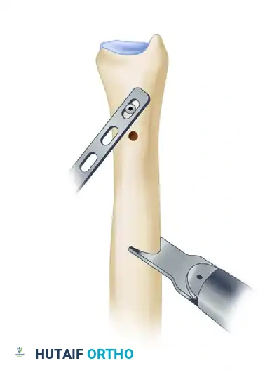
Figure 7: The plate is rotated back into position and secured using dynamic compression technique to close the osteotomy gap.
To maximize the rigidity of the construct and achieve absolute biomechanical stability, a separate, independent interfragmentary lag screw is placed across the osteotomy site. This screw must be inserted at exactly 90 degrees to the plane of the oblique osteotomy to generate maximal interfragmentary compression without inducing shear forces. Depending on the specific geometry of the cut and the plate position, this 3.5-mm or 2.7-mm lag screw may be placed through a designated hole in the plate or independently, outside the plate footprint. The addition of the lag screw neutralizes torsional forces and significantly reduces the risk of hardware failure and nonunion.
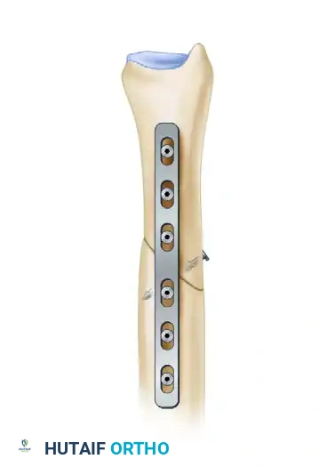
Figure 8: Placement of an independent interfragmentary lag screw perpendicular to the osteotomy plane to provide absolute stability.
Following complete fixation, the wound is thoroughly irrigated with sterile saline to remove bone debris. The tourniquet is deflated, and meticulous hemostasis is achieved using bipolar electrocautery. The deep antebrachial fascia is closed loosely to prevent compartment syndrome, followed by a layered closure of the subcutaneous tissues and skin. A sterile, non-adherent dressing is applied, and the extremity is immobilized in a well-molded short-arm cast or a rigid sugar-tong splint in neutral rotation to protect the fixation during the initial inflammatory phase of healing.
Complications, Incidence Rates, and Salvage Management
Nonunion and Delayed Union
Despite rigorous surgical technique, ulnar shortening osteotomy is associated with a distinct set of complications, the most formidable being delayed union and nonunion. The diaphyseal ulna, particularly at the junction of the middle and distal thirds, is characterized by a relatively precarious endosteal and periosteal blood supply. Historically, when transverse osteotomies were performed without rigid lag screw fixation, nonunion rates approached a staggering 10% to 15%. However, with the advent of the oblique osteotomy technique, the utilization of dedicated compression plating systems, and the strict adherence to minimal periosteal stripping, the incidence of nonunion has been drastically reduced to approximately 2% to 4% in contemporary academic centers.
Risk factors for nonunion include tobacco use (which increases the risk by a factor of four), inadequate interfragmentary compression, thermal necrosis from aggressive saw use without irrigation, and excessive soft-tissue stripping. If a nonunion is identified radiographically (lack of bridging trabeculae at 6 months) and clinically (persistent pain at the osteotomy site), salvage management is required. This typically involves revision surgery with complete hardware removal, debridement of the fibrous pseudoarthrosis down to bleeding, viable bone, application of a larger, more robust plate (e.g., a 3.5-mm locking compression plate), and the mandatory
Clinical & Radiographic Imaging Archive