Perioperative Optimization in Hand Surgery: Antibiotic Stewardship, Operating Room Ergonomics, and Surgical Execution
Key Takeaway
Surgical site infections in hand surgery are rare but potentially devastating. Recent large-scale prospective and retrospective studies demonstrate that routine perioperative antibiotic prophylaxis does not significantly reduce infection rates, even in high-risk patients or complex cases. Optimizing operating room ergonomics, establishing standardized surgical routines, and ensuring precise assistant positioning are far more critical for minimizing intraoperative complications and maximizing surgical efficiency.
Comprehensive Introduction and Patho-Epidemiology
The successful execution of complex hand and upper extremity surgery relies on a foundation of meticulous perioperative planning, strict adherence to evidence-based protocols, and the relentless optimization of the surgical environment. While the technical prowess of the operating surgeon is undeniably paramount, systemic and environmental factors—such as the rational use of perioperative antibiotics, the ergonomic arrangement of the operating theater, and the psychological management of the awake patient—profoundly influence ultimate functional outcomes. Disorganization, physical fatigue, and adherence to outdated clinical dogmas rapidly diminish the efficiency of the operating team, increase cognitive load, and ultimately compromise surgical precision. In the anatomically unforgiving terrain of the human hand, where millimeters dictate the difference between functional restoration and severe iatrogenic impairment, such compromises are unacceptable.
Surgical site infections (SSIs) following elective hand surgery are statistically rare, with reported incidences generally hovering between 0.5% and 2.0% in clean, elective cases. However, a postoperative infection in this anatomically dense and highly specialized region represents a potentially disastrous complication. The complex interplay of contiguous synovial sheaths, potential fascial spaces (such as the midpalmar and thenar spaces), and poorly vascularized structures like tendons and articular cartilage dictates that even a superficial incisional infection can rapidly propagate. This progression can quickly evolve into suppurative flexor tenosynovitis, deep space abscesses, or osteomyelitis. Such catastrophic complications cause severe impairment of hand biomechanics, drastically delay rehabilitation protocols, and frequently preclude a timely return to occupational activities. Severe infections inevitably necessitate multiple surgical debridements, prolonged intravenous antimicrobial therapy, and can result in permanent, irreversible biomechanical damage due to profound fibro-osseous scarring and tendon necrosis.
Despite these catastrophic risks, the routine, ubiquitous use of perioperative prophylactic antibiotics for many orthopaedic procedures in the hand remains highly questionable and is increasingly discouraged by modern antimicrobial stewardship programs. The historical paradigm of administering broad-spectrum cephalosporins for every carpal tunnel release, trigger finger release, or ganglion excision was born from an abundance of caution rather than robust empirical evidence. The patho-epidemiology of hand infections demonstrates that the robust vascularity of the integument in the upper extremity provides a formidable natural defense against transient intraoperative bacterial seeding. Furthermore, the indiscriminate use of antibiotics carries significant population-level and individual risks, including the proliferation of multi-drug resistant organisms (MDROs), anaphylaxis, and Clostridioides difficile colitis.
This comprehensive chapter delineates the current, rigorously tested evidence regarding perioperative antibiotic prophylaxis in hand surgery, establishes gold-standard protocols for operating room macro- and micro-ergonomics, and details the execution of fundamental surgical approaches facilitated by optimal team dynamics. By synthesizing anatomical principles with evidence-based perioperative management, the orthopaedic surgeon can reliably minimize complications while maximizing surgical efficiency and patient safety.
Detailed Surgical Anatomy and Biomechanics
Mastery of hand surgery requires an intimate understanding not only of the patient's micro-anatomy but also of the biomechanics of the surgical team. Microsurgery and intricate hand procedures require the absolute elimination of physiologic tremor, which is achieved through strict adherence to ergonomic principles and a profound understanding of anatomical spatial relationships. The surgeon’s posture directly impacts their ability to perform delicate maneuvers, such as epineurial nerve coaptation or microvascular anastomosis. When the surgeon’s forearms are inadequately supported, the intrinsic musculature of the upper extremity must continuously fire to maintain spatial positioning, leading to rapid lactic acid accumulation, muscle fatigue, and the amplification of resting tremor. Conversely, when the working surface of the operating hand table is positioned exactly at elbow height, the skeletal architecture of the surgeon's upper body supports the weight of the arms, allowing the intrinsic hand muscles to operate with maximal precision and minimal metabolic demand.
The surgical anatomy relevant to the midlateral digital approach—a fundamental exposure technique—is defined by the intricate fascial ligaments that stabilize the digital integument during grip and pinch. The digital neurovascular bundles, comprising the proper digital artery and proper digital nerve, travel longitudinally along the volar-lateral aspect of the digit. These crucial structures are suspended and protected by two distinct fascial systems: Cleland’s ligaments and Grayson’s ligaments. Cleland’s ligaments are robust, relatively avascular fascial bands that originate from the lateral aspect of the phalanges and insert into the dorsal skin, passing dorsal to the neurovascular bundle. They provide critical stability to the skin during digital flexion and extension.
Conversely, Grayson’s ligaments are more delicate, diaphanous fascial structures that originate from the volar aspect of the flexor tendon sheath and insert into the volar skin, passing volar to the neurovascular bundle. A precise understanding of this three-dimensional relationship is mandatory for safe surgical dissection. During a midlateral approach, the surgeon must confidently incise Grayson’s ligaments to mobilize the neurovascular bundle volarly, thereby protecting it from iatrogenic injury while gaining unimpeded access to the underlying flexor tendon sheath and the volar aspect of the phalanges. Failure to recognize these fascial planes often results in inadvertent transection of the digital nerve or artery.
Deep to the neurovascular bundle and the fascial suspensory ligaments lies the fibro-osseous flexor tendon sheath, a highly specialized biomechanical pulley system. This sheath is composed of a series of thickened annular pulleys (A1 through A5) and thinner, pliable cruciform pulleys (C1 through C3). The annular pulleys, particularly the A2 and A4 pulleys located over the proximal and middle phalanges respectively, are biomechanically critical for preventing bowstringing of the flexor tendons during active flexion. The surgical anatomy of this region dictates that any approach to the flexor tendons must meticulously preserve the A2 and A4 pulleys to maintain the moment arm and mechanical advantage of the flexor digitorum superficialis (FDS) and flexor digitorum profundus (FDP) tendons. The midlateral approach provides excellent visualization of this pulley system without placing a longitudinal scar directly over the volar gliding surface of the tendons.
Exhaustive Indications and Contraindications
The decision-making process regarding perioperative protocols, anesthetic choices, and surgical approaches in hand surgery must be highly individualized, yet grounded in established evidence-based guidelines. The shift toward Wide Awake Local Anesthesia No Tourniquet (WALANT) and the strict curtailment of prophylactic antibiotics represent significant evolutions in clinical practice. However, these modern paradigms are not universally applicable. The surgeon must meticulously weigh the specific indications and contraindications for each intervention to optimize patient safety and functional recovery.
The indications for perioperative antibiotic prophylaxis in hand surgery have been drastically narrowed in recent years. While routine, clean, elective procedures lasting less than two hours (such as carpal tunnel release, trigger finger release, De Quervain's release, and simple ganglion excisions) definitively do not require prophylactic antibiotics, specific high-risk scenarios mandate their use. Absolute indications include open fractures of the phalanges, metacarpals, or carpus; traumatic wounds heavily contaminated with organic debris or soil (e.g., farm injuries); human or animal bites (which pose a high risk for Eikenella corrodens and Pasteurella multocida infections, respectively); and procedures involving the implantation of massive foreign bodies, such as total wrist arthroplasty or complex modular endoprostheses.
The choice of surgical approach is similarly governed by strict anatomical indications. The midlateral digital approach is highly indicated for procedures requiring extensive access to the flexor tendon sheath, such as complex flexor tenolysis, repair of zone II flexor tendon lacerations, or the radical debridement of suppurative flexor tenosynovitis. Its primary advantage is the prevention of volar flexion contractures, as the resulting scar does not cross the volar flexion creases. However, it is relatively contraindicated in scenarios requiring bilateral access to both neurovascular bundles simultaneously, or in cases of severe crush injuries where the lateral integument is already severely compromised. In such instances, a volar Bruner (zigzag) incision may be more appropriate, provided the apices of the flaps extend to the midlateral line to prevent scar contracture.
| Clinical Parameter | Primary Indications | Absolute Contraindications | Relative Contraindications |
|---|---|---|---|
| Perioperative Antibiotics | Open fractures, mammalian bites, gross purulence, massive implant arthroplasty, severe crush injuries with devitalized tissue. | Clean, elective soft tissue procedures (e.g., CTR, trigger finger) in immunocompetent hosts. | Clean-contaminated procedures in patients with a history of severe C. difficile or multiple drug allergies. |
| WALANT Anesthesia | Tendon repairs (requires active motion testing), tenolysis, trigger finger, CTR, patients with severe medical comorbidities precluding general anesthesia. | Patient refusal, severe preoperative anxiety/panic disorders, known allergy to amide local anesthetics. | Procedures requiring bone grafting from distant sites, extremely prolonged microvascular reconstructions (>4 hours). |
| Midlateral Approach | Flexor tenolysis, zone II tendon repairs, digital nerve/artery repairs, drainage of flexor tenosynovitis, lateral mass excision. | Procedures requiring simultaneous bilateral digital access, volar skin necrosis requiring flap coverage. | Pre-existing severe lateral scarring from previous trauma or surgery. |
| Bruner (Zigzag) Approach | Extensive volar exposure, Dupuytren's fasciectomy, volar plate arthroplasty. | Incisions crossing the flexion creases at a 90-degree angle (leads to severe contracture). | Severe volar soft tissue loss, active volar skin infection. |
Pre-Operative Planning, Templating, and Patient Positioning
Efficiency on the day of surgery begins weeks in advance through meticulous preoperative planning. If a complex surgical procedure, such as a multi-digit replantation or a vascularized bone graft, is being arranged, the surgeon must communicate specific, detailed requests regarding special needs to the operating room staff well ahead of time. This includes the reservation of the operating microscope, the verification of specific micro-instrument trays (including jeweler's forceps, Castroviejo needle holders, and adventitial scissors), and the procurement of specialized sutures (e.g., 4-0 to 6-0 non-absorbable core sutures for tendon repair, and 8-0 to 10-0 nylon for microvascular anastomosis).
The psychological and auditory environment of the operating room must be carefully managed, particularly in the modern era of WALANT surgery. When the patient is fully conscious and actively participating in the intraoperative assessment of tendon gliding, the operating room must function as a "sterile cockpit." Loud noises, the dropping of heavy instruments, inappropriate bursts of conversation, or any expressions of surgical frustration may severely alarm the patient, inducing tachycardia and hypertension, and must be strictly avoided. The surgeon must act as the leader of the room, setting a calm, focused, and reassuring tone. Background music, specifically chosen by the patient, has been empirically shown to reduce intraoperative anxiety and lower systemic catecholamine release, thereby improving peripheral perfusion.
Surgeon ergonomics and patient positioning are inextricably linked to surgical success. The operating surgeon should sit on a firm, comfortable, and highly stable stool. When sitting, the surgeon’s knees should be almost level with the hips, and the feet must rest flat on the floor without strain. The back should be comfortably erect to prevent the insidious onset of cervical and lumbar radiculopathy—a common occupational hazard that prematurely ends many surgical careers. The working surface of the operating hand table must be exactly at elbow height, providing a solid, unyielding support for the forearms. Illumination is equally critical; the overhead surgical light should be directed from above the surgeon’s left shoulder (for a right-handed surgeon) to shine directly onto the operative field, effectively eliminating shadows cast by the surgeon's own hands or instruments.
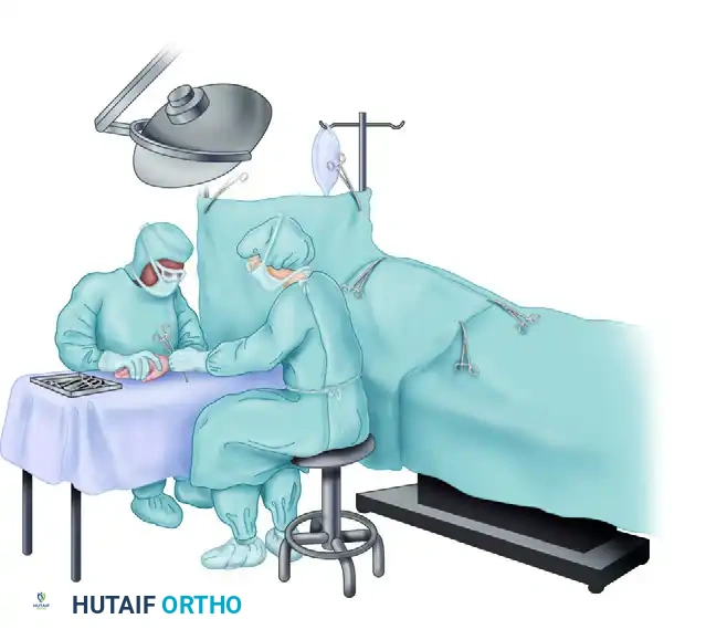
The surgical assistant plays a paramount role in the execution of the procedure. Seated directly opposite the surgeon, the assistant must view the operative field from a vantage point 8 to 10 cm higher than the surgeon. This specific height differential allows the assistant a clear, downward line of vision into the depths of the wound without having to lean forward, which would obstruct the surgeon’s view, bump the microscope, or block the overhead lighting. The assistant's primary duty is dynamic, intelligent retraction. By applying precise counter-tension and securely holding the adjacent digits out of the field, the assistant provides the surgeon with a static, unimpeded target. The basic instrument tray should be placed on a Mayo stand perfectly level with the working surface, and the instruments must always be arranged in the exact same spatial order. This standardized layout allows the surgeon to develop profound muscle memory, reaching for a Senn retractor or a Freer elevator without ever looking away from the microscopic operative field.
Step-by-Step Surgical Approach and Fixation Technique
To illustrate the critical interplay between surgeon ergonomics, assistant positioning, and precise surgical execution, we examine the midlateral digital incision. This approach is a workhorse technique for accessing the flexor tendon sheath, digital nerves, and phalanges without causing the devastating volar scar contractures associated with poorly planned volar incisions. The success of this approach relies heavily on the assistant's ability to manipulate the digit and the surgeon's precise identification of micro-anatomical landmarks.
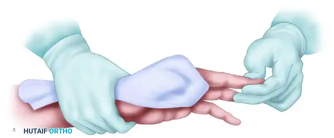
The procedure begins with optimal positioning and dynamic retraction. As demonstrated in the ergonomic setup, the assistant firmly grasps the patient's hand, holding the target digit in extension while gently flexing and retracting the adjacent digits completely out of the surgical field. The assistant's grip must be firm enough to provide absolute stability but atraumatic enough to prevent digital ischemia. Crucially, the assistant must ensure the target digit does not rotate during the incision; any inadvertent pronation or supination of the finger will distort the anatomical landmarks, leading to an improperly placed incision that may inadvertently cross the volar flexion creases.
The surgeon begins by identifying the dorsal and volar flexion creases of the proximal interphalangeal (PIP) and distal interphalangeal (DIP) joints. The theoretical midlateral line connects the apices of these flexion creases when the finger is fully flexed. Using a #15 blade, the surgeon makes a precise longitudinal incision exactly along this midlateral line. It is a critical surgical principle that the incision must remain strictly dorsal to the volar flexion creases. If the incision wanders volarly, the resulting scar will inevitably contract during the remodeling phase of wound healing, resulting in a permanent, functionally limiting flexion contracture of the digit.
Following the skin incision, superficial dissection is carried out through the subcutaneous fat. The surgeon must utilize delicate spreading motions with tenotomy scissors to identify and preserve the dorsal sensory branches of the digital nerve, which provide critical sensation to the dorsal skin and nail fold. As the dissection deepens, the surgeon encounters the fascial suspensory system. Grayson’s ligaments, located volar to the neurovascular bundle, are sharply divided. Once Grayson's ligaments are released, the proper digital artery and nerve are clearly visualized. The assistant then utilizes a small, blunt retractor (such as a Ragnell or Senn retractor) to gently retract the entire neurovascular bundle volarly. With the neurovascular bundle safely protected and retracted, the surgeon has direct, unimpeded access to the underlying fibro-osseous flexor tendon sheath, the critical annular pulleys, and the volar aspect of the phalanges, allowing for subsequent tenolysis, tendon repair, or fracture fixation.
Complications, Incidence Rates, and Salvage Management
Despite meticulous perioperative optimization and precise surgical technique, complications in hand surgery can and do occur. The dense anatomy of the hand means that a single complication can have cascading effects on multiple tissue systems, leading to profound functional impairment. The surgeon must be intimately familiar with the incidence, early recognition, and salvage management of these adverse events. A proactive approach to complication management is the hallmark of an experienced hand surgeon.
Surgical site infections (SSIs), while rare in elective cases (0.5% - 2.0%), represent a significant threat. In the era of antibiotic stewardship, vigilant postoperative monitoring replaces prophylactic drug administration. If a patient presents with disproportionate pain, pathological erythema extending beyond the immediate incision, or purulent drainage, the surgeon must have a low threshold for intervention. Superficial cellulitis may be managed with targeted oral antibiotics based on local antibiograms, but deep space infections or suppurative flexor tenosynovitis (characterized by Kanavel's cardinal signs) demand immediate return to the operating room for radical irrigation and debridement. Delaying surgical intervention in the presence of a closed-space infection leads to rapid tendon necrosis and irreversible articular cartilage destruction.
Iatrogenic neurovascular injury is a devastating complication, most frequently occurring during the initial surgical exposure. The incidence of digital nerve injury during elective approaches such as Dupuytren's fasciectomy or trigger finger release ranges from 1% to 3%. The most common technical error leading to this complication is the failure to maintain strict rotational control of the digit, causing the surgeon to inadvertently place the incision directly over the neurovascular bundle. If a digital nerve is transected intraoperatively, it must be recognized immediately and repaired primarily using microsurgical techniques (8-0 or 9-0 epineurial sutures) without tension. Missed nerve injuries present postoperatively as painful neuromas and profound sensory deficits, requiring complex salvage procedures such as neuroma excision and interpositional nerve grafting.
| Complication | Estimated Incidence | Primary Etiology / Risk Factors | Salvage Management / Intervention |
|---|---|---|---|
| Surgical Site Infection (SSI) | 0.5% - 2.0% (Elective) | Poor sterile technique, hematoma formation, patient comorbidities (uncontrolled diabetes, smoking). | Early recognition; oral antibiotics for superficial cellulitis; emergent operative I&D for deep space/sheath infections. |
| Iatrogenic Digital Nerve Injury | 1.0% - 3.0% | Distorted anatomy (e.g., Dupuytren's spiral cord), poor surgical exposure, aberrant incision placement. | Immediate intraoperative microsurgical primary repair. Late presentation requires neuroma excision and nerve grafting. |
| Postoperative Flexion Contracture | 5.0% - 15.0% | Incisions crossing volar flexion creases, inadequate postoperative splinting, delayed mobilization. | Aggressive hand therapy, dynamic extension orthoses. Refractory cases may require surgical release or Z-plasty. |
| Tendon Adhesions / Rupture | 4.0% - 10.0% (Post-repair) | Poor suture technique, excessive tissue handling, failure to adhere to early active motion protocols. | Intensive therapy for adhesions; tenolysis at 6 months if plateaued. Ruptures require prompt re-exploration and re-repair. |
| Surgeon Ergonomic Injury | > 60% (Career prevalence) | Poor OR posture, un-supported forearms, improper microscope/loupe focal length. | Ergonomic optimization, physical therapy, core strengthening, strict adherence to optimal table/chair height. |
Phased Post-Operative Rehabilitation Protocols
The ultimate success of complex hand surgery extends far beyond the sterile confines of the operating room. The most technically perfect flexor tendon repair or intricate nerve coaptation will inevitably fail if it is not followed by a rigorously standardized, scientifically phased postoperative rehabilitation protocol. The hand surgeon must work in close, continuous collaboration with a certified hand therapist (CHT) to guide the patient through the critical phases of tissue healing. Postoperative protocols must balance the competing demands of protecting the surgical repair from mechanical failure while simultaneously preventing the formation of restrictive fibro-osseous adhesions.
The immediate postoperative phase (Days 0 to 5) is focused entirely on wound protection and aggressive edema control. Edema is the primary enemy of hand rehabilitation; protein-rich interstitial fluid rapidly organizes into dense, restrictive scar tissue if left unchecked. The surgical site is dressed with a non-adherent contact layer (e.g., Adaptic or Xeroform) to prevent disruption of the healing epithelium during dressing changes. This is followed by fluffed gauze to gently accommodate postoperative swelling without causing a tourniquet effect. A well-padded, custom-fabricated orthosis is applied based on the specific structures repaired. For example, following a volar flexor tendon repair, a dorsal blocking splint is applied with the wrist in 20 degrees of flexion, the metacarpophalangeal (MCP) joints in 70 degrees of flexion, and the interphalangeal (IP) joints in neutral extension. The patient is strictly instructed to keep the hand elevated above the level of the heart at all times.
The early protected mobilization phase (Days 5 to 21) marks the initiation of controlled mechanical stress to the healing tissues. Under the direct, one-on-one guidance of the CHT, early active motion (EAM) protocols are initiated. Scientific evidence has conclusively demonstrated that controlled, early gliding of repaired tendons within their sheaths stimulates intrinsic tenocyte healing, increases ultimate tensile strength, and significantly reduces the formation of extrinsic adhesions to the surrounding pulley system. Patients perform controlled "place-and-hold" exercises and active composite flexion within the safe constraints of their protective orthosis. During this phase, despite the omission of routine prophylactic antibiotics, the surgical site is meticulously monitored by both the surgeon and the therapist for any signs of pathological erythema or delayed wound healing.
The intermediate and late rehabilitation phases (Weeks 3 through 12+) focus on progressive load bearing, scar remodeling, and the restoration of normal biomechanics. By week 4 to 6, depending on the robustness of the repair, protective orthoses are gradually weaned. Aggressive scar massage and the application of silicone gel sheeting are utilized to soften the surgical scar and prevent tethering of the underlying tendons. Progressive strengthening exercises, utilizing therapeutic putty and graded hand grippers, are introduced to restore grip and pinch strength. The final phase of rehabilitation involves work-conditioning programs tailored to the patient's specific occupational demands, ensuring a safe, durable return to full functional capacity without the risk of late mechanical failure.
Summary of Landmark Literature and Clinical Guidelines
The modern, evidence-based approach to perioperative optimization in hand surgery is heavily informed by several landmark studies that have fundamentally challenged historical dogmas. The paradigm of administering prophylactic antibiotics for all hand surgeries, once considered a standard of care, has been decisively dismantled by robust, large-scale clinical data. The orthopaedic community relies on these pivotal studies to justify the implementation of strict antibiotic stewardship programs, protecting patients from the hazards of unnecessary antimicrobial exposure.
The most definitive guidance stems from a massive retrospective cohort study encompassing 8,850 patients. This exhaustive analysis evaluated the efficacy of prophylactic antibiotics across a broad, highly representative spectrum of elective hand procedures. The study's results were unequivocal: there was absolutely no statistically significant difference in the frequency of surgical site infections between patients who received perioperative antibiotics and those who did not. Crucially, this lack of efficacy persisted even when the data was stratified to examine designated "high-risk" cohorts. Patients with active tobacco use, poorly controlled diabetes mellitus, and those undergoing procedures with prolonged operative times derived no protective benefit from prophylactic antibiotics.
These retrospective findings were subsequently corroborated by a rigorously controlled prospective randomized trial involving 1,340 patients. This trial provided Level I evidence demonstrating no statistically significant difference in infection rates based on antibiotic administration. The researchers meticulously stratified patients by numerous surgical variables and found no difference in infection rates between elective and emergency surgeries, between operations lasting less than 2 hours and those lasting longer, or even between "clean" wounds and "crush/dirty" wounds (excluding gross contamination or farm injuries).
Supported by this overwhelming body of evidence, the American Society for Surgery of the Hand (ASSH) and modern antibiotic stewardship guidelines conclude that antibiotic prophylaxis should not be routinely administered for standard, clean surgery of the hand. Furthermore, the integration of these stewardship principles with the ergonomic and anesthetic advancements pioneered in WALANT surgery (extensively documented by Lalonde and colleagues) represents the current gold standard. By optimizing the surgical environment, minimizing unnecessary pharmacological interventions, and adhering to strict biomechanical principles, the hand surgeon can consistently deliver superior outcomes while mitigating systemic risks.
