Osteoarthritis of the Wrist: SLAC Wrist Pathoanatomy and Surgical Management
Key Takeaway
Scapholunate advanced collapse (SLAC) is the most common pattern of degenerative wrist arthritis, driven by untreated scapholunate ligament injuries. This predictable progression of articular wear begins at the radioscaphoid joint and advances to the midcarpal joint, characteristically sparing the radiolunate articulation. Surgical management depends on the stage of degeneration and includes motion-preserving procedures such as proximal row carpectomy (PRC) or four-corner arthrodesis with scaphoid excision to alleviate pain while maintaining functional wrist kinematics.
Comprehensive Introduction and Patho-Epidemiology
Degenerative arthritis developing in the wrist is most frequently the result of altered carpal kinematics, classically presenting as Scapholunate Advanced Collapse (SLAC). The SLAC wrist represents a highly predictable, sequential pattern of progressive articular degeneration related to chronic, unaddressed instability around the scaphoid. First described comprehensively by Watson and Ballet in 1984, the SLAC pattern accounts for the vast majority of wrist osteoarthritis cases encountered in clinical practice. While primary degenerative changes or crystalline arthropathies—such as calcium pyrophosphate dihydrate (CPPD) deposition disease (pseudogout)—can occasionally manifest in the carpus, the overwhelming majority of SLAC wrists are the direct sequelae of posttraumatic biomechanical derangement. Specifically, this stems from an untreated, unrecognized, or inadequately managed rupture of the scapholunate interosseous ligament (SLIL). A parallel kinematic failure occurs in the setting of chronic scaphoid nonunion, leading to a nearly identical degenerative cascade known as Scaphoid Nonunion Advanced Collapse (SNAC).
The epidemiology of SLAC wrist is intrinsically linked to the epidemiology of scapholunate ligament injuries, which are the most common carpal instability patterns. These injuries predominantly affect young to middle-aged adult males, often following a fall onto an outstretched hand (FOOSH) with the wrist in extension, ulnar deviation, and intercarpal supination. Because the initial ligamentous injury may be radiographically subtle and clinically misdiagnosed as a routine "wrist sprain," patients frequently present years or even decades later when irreversible articular cartilage loss has already occurred. The natural history of a complete, unhealed scapholunate ligament tear is a relentless progression toward SLAC wrist, though the temporal timeline of this progression is highly variable, ranging from months in high-demand laborers to decades in lower-demand individuals.
Understanding the patho-epidemiology of the SLAC wrist requires an appreciation of the wrist as a complex, intercalated joint system rather than a simple hinge. The carpus lacks direct tendinous insertions to actively control the proximal row; instead, it relies entirely on precise articular geometry and complex intrinsic and extrinsic ligamentous checkreins to maintain stability under compressive loads. When the scapholunate linkage fails, the proximal row loses its synchronous motion, leading to abnormal force transmission across the radiocarpal and midcarpal joints. The resulting exponential increase in focal contact stresses inevitably overwhelms the biological capacity of the articular cartilage, initiating the irreversible cascade of osteoarthritis. The surgical management of this condition has evolved significantly over the past several decades, moving away from high-complication historical procedures like silicone arthroplasty toward highly reliable, motion-preserving biological salvage operations such as Proximal Row Carpectomy (PRC) and Four-Corner Fusion (4CF).
Detailed Surgical Anatomy and Biomechanics
The scaphoid functions as the critical mechanical bridge and primary stabilizing strut between the proximal and distal carpal rows. Normal wrist kinematics rely heavily on the intact scapholunate interosseous ligament (SLIL) to couple the intrinsic flexion tendency of the scaphoid with the intrinsic extension tendency of the lunate. The SLIL itself is a complex, C-shaped structure composed of three distinct regions: the robust dorsal region (the primary restraint to translation and distraction), the volar region (which controls rotation), and the central membranous region (which is biomechanically insignificant but serves as an important landmark during arthroscopy). When the SLIL is completely disrupted, and secondary stabilizers such as the dorsal intercarpal (DIC) and radioscaphocapitate (RSC) ligaments become attenuated over time, this vital kinematic linkage is catastrophically lost.
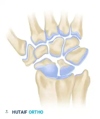
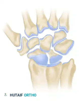
Following this dissociation, the scaphoid flexes and pronates under the compressive loads of the crossing tendons and the distal carpal row. Simultaneously, the lunate—now untethered from the scaphoid but still securely bound to the triquetrum via the intact lunotriquetral ligament—extends. This uncoupling results in the classic Dorsal Intercalated Segment Instability (DISI) deformity. The end result is a wrist with profound biomechanical derangement characterized by several distinct pathological hallmarks. First, there is a widening of the scapholunate gap, clinically recognized as the "Terry Thomas" or "David Letterman" sign on anteroposterior radiographs. Second, the flexed scaphoid shifts its contact area dorsally on the radial styloid. Because the scaphoid is no longer articulating congruently within the elliptical scaphoid fossa of the distal radius, the contact surface area is drastically reduced, leading to an exponential increase in focal articular contact stresses and subsequent cartilage degradation. Third, the capitate migrates proximally into the widened scapholunate interval, acting as a wedge that further drives the scaphoid and lunate apart, leading to narrowing and degeneration of the capitolunate joint.
Remarkably, despite the extensive destruction of the radioscaphoid and midcarpal compartments, the radiolunate joint is almost universally spared in the SLAC wrist progression. This absolute preservation is a function of unique articular geometry. The lunate fossa of the distal radius and the proximal articular surface of the lunate share a concentrically spherical relationship with a nearly identical radius of curvature. Even in the extended, pathological position of a DISI deformity, the radiolunate joint maintains a congruent, load-sharing articulation. It distributes compressive forces evenly without the pathological, edge-loading shear forces seen in the incongruent radioscaphoid compartment. This anatomical quirk is the absolute cornerstone of modern salvage procedures; the preservation of the radiolunate joint allows surgeons to perform motion-preserving procedures like PRC or 4CF. If the radiolunate joint exhibits degenerative changes, these procedures are strictly contraindicated.
Radiographic Staging of SLAC Wrist
The progression of SLAC wrist osteoarthritis follows a strict, sequential pattern originally described by Watson and Ballet. Understanding these stages is critical for preoperative planning, patient counseling, and surgical decision-making.
Stage I SLAC Wrist
In Stage I, the degenerative changes are strictly localized to the articulation between the radial styloid and the distal pole of the scaphoid. The flexed, pronated posture of the scaphoid causes abnormal point-loading against the radial styloid. This leads to localized joint space narrowing, subchondral sclerosis, and characteristic osteophyte formation at the radial styloid. The remainder of the carpus remains radiographically normal, though the underlying DISI deformity is present.
Stage II SLAC Wrist
As the instability and abnormal kinematics persist, the degenerative process extends proximally to involve the entire radioscaphoid fossa. The scaphoid continues to articulate abnormally with the elliptical scaphoid fossa of the distal radius, leading to complete loss of the radioscaphoid articular cartilage. Radiographs demonstrate severe joint space narrowing, subchondral cysts, and sclerosis throughout the entire radioscaphoid articulation. The capitolunate and radiolunate joints remain preserved at this stage.

Stage III SLAC Wrist
In Stage III, the capitate subluxates proximally into the widened scapholunate interval. This proximal migration alters the kinematics of the midcarpal joint, leading to narrowing, sclerosis, and osteophyte formation at the capitolunate joint. The capitate head becomes flattened and irregular. Despite this extensive midcarpal and radioscaphoid destruction, the radiolunate joint remains remarkably preserved due to its spherical congruency.
Exhaustive Indications and Contraindications
The surgical treatment of the SLAC wrist is highly individualized and dictated by the specific stage of arthritis, the patient's chronological and physiological age, occupational demands, functional requirements, and the precise status of the radiolunate and proximal capitate articular surfaces. The primary goal of any surgical intervention in this setting is the reliable alleviation of pain, with a secondary, yet crucial, goal of preserving a functional arc of motion and maximizing grip strength. Historically, the surgical treatment of this problem involved limited intercarpal arthrodesis combined with scaphoid excision and Silastic (silicone) scaphoid replacement. However, due to the high incidence of "silicone synovitis"—a destructive, foreign-body giant cell reaction to particulate silicone wear debris causing massive carpal osteolysis—this practice has been universally abandoned in modern orthopedic surgery.
Today, the algorithmic approach to the SLAC wrist relies primarily on Proximal Row Carpectomy (PRC), Four-Corner Fusion (4CF) with scaphoid excision, Total Wrist Arthrodesis (TWA), and occasionally, palliative procedures such as total wrist denervation. PRC involves the excision of the scaphoid, lunate, and triquetrum, essentially converting the complex carpal articulation into a simple hinge joint where the capitate articulates directly within the lunate fossa of the distal radius. This procedure relies entirely on the presence of pristine cartilage on both the proximal capitate and the lunate fossa. Conversely, 4CF involves the excision of the entire scaphoid to eliminate the painful radioscaphoid arthritis, combined with the decortication and rigid arthrodesis of the capitate, hamate, lunate, and triquetrum. By fusing the midcarpal joint, any pre-existing capitolunate arthritis (Stage III) is effectively neutralized, and forces are transmitted through the fused carpal block into the preserved radiolunate joint.
Total wrist arthrodesis is reserved for pancarpal arthritis (sometimes referred to as Stage IV SLAC, though this terminology is debated), failed salvage procedures, or young, heavy manual laborers who require absolute, indestructible stability at the expense of all radiocarpal motion. Total wrist denervation, involving the surgical transection of the terminal articular branches of the posterior and anterior interosseous nerves, is occasionally utilized as a standalone palliative measure in low-demand, medically frail patients who cannot tolerate prolonged immobilization or extensive surgery, or as an adjunct to PRC or 4CF.
| Procedure | Primary Indications | Absolute Contraindications | Relative Contraindications |
|---|---|---|---|
| Proximal Row Carpectomy (PRC) | Stage I or II SLAC/SNAC; Low to moderate demand patients; Older patients seeking faster recovery. | Stage III SLAC (Capitolunate arthritis); Radiolunate arthritis; Inflammatory arthropathy. | Young, heavy manual laborers (high risk of late radiocapitate arthrosis); Ulnar positive variance. |
| Four-Corner Fusion (4CF) | Stage II or III SLAC/SNAC; High-demand manual laborers; Young patients requiring durability. | Radiolunate arthritis; Severe pancarpal arthrosis; Active infection. | Heavy smokers (high risk of nonunion); Patients unable to tolerate 8-12 weeks of immobilization. |
| Total Wrist Arthrodesis (TWA) | Pancarpal arthritis; Radiolunate arthritis; Failed PRC or 4CF; Paralytic deformities. | Open physes (pediatric patients); Need for functional wrist motion (e.g., certain musicians, personal hygiene needs). | Bilateral total wrist arthrodesis (severely limits daily functional independence). |
| Wrist Denervation (AIN/PIN) | Palliative pain relief in medically frail patients; Adjunct to other procedures. | Need for structural correction; Advanced, rapidly progressive collapse. | Patient expectation of normal grip strength or complete, permanent pain eradication. |
Pre-Operative Planning, Templating, and Patient Positioning
Thorough preoperative planning is essential for achieving optimal outcomes in the surgical management of the SLAC wrist. The clinical evaluation begins with a detailed history, focusing on the duration of symptoms, prior trauma, occupational demands, and the specific functional limitations the patient is experiencing. Physical examination typically reveals localized swelling, marked tenderness over the dorsal radioscaphoid joint and the anatomical snuffbox, and a significant reduction in both grip strength and the radiocarpal arc of motion, particularly in extension and radial deviation. The Watson scaphoid shift test may elicit a painful clunk in earlier stages, though in advanced SLAC wrists with severe stiffness and osteophyte formation, the scaphoid is often rigidly fixed in its flexed position, rendering the test negative or simply painful without a palpable clunk.
Standard radiographic evaluation must include high-quality posteroanterior (PA), true lateral, scaphoid view, and clenched-fist PA views of the wrist. The PA view is scrutinized for the presence of the "Terry Thomas" sign, the loss of radioscaphoid joint space, and the proximal migration of the capitate. The true lateral view is critical for quantifying the DISI deformity, specifically evaluating the radiolunate and scapholunate angles. While standard radiographs are often sufficient for diagnosing and staging the SLAC wrist, advanced imaging is frequently employed for precise surgical planning. Non-contrast Magnetic Resonance Imaging (MRI) is highly sensitive for evaluating the integrity of the interosseous ligaments and the early loss of articular cartilage, though its resolution may sometimes underestimate the true extent of chondral damage. Computed Tomography (CT) scans, particularly with 3D reconstructions, are invaluable for assessing bone stock, evaluating cystic changes within the carpus, and precisely mapping the geometry of the osteophytes prior to surgical excision.
Despite advanced imaging, the absolute gold standard for evaluating the articular cartilage—specifically the critical proximal capitate and lunate fossa surfaces—is direct intraoperative visualization or diagnostic wrist arthroscopy. Many surgeons advocate for a brief diagnostic arthroscopy immediately preceding the open procedure to definitively assess the capitate head. If a PRC is planned but arthroscopy reveals full-thickness eburnation of the proximal capitate, the surgeon must dynamically pivot to a 4CF or total wrist arthrodesis. Proper preoperative templating involves utilizing digital software to overlay various fixation devices, such as circular radiolucent plates or headless compression screws, to ensure optimal sizing and trajectory without impinging on the radiocarpal joint.
For patient positioning, the patient is placed supine on the operating table with the operative extremity extended onto a radiolucent hand table. A well-padded proximal arm pneumatic tourniquet is applied to provide a bloodless surgical field. The arm is prepped and draped in a standard sterile fashion, ensuring that the entire hand and forearm are exposed to allow for intraoperative manipulation and fluoroscopic imaging. The fluoroscopy unit (C-arm) should be positioned parallel to the hand table, entering from the distal or contralateral side, allowing the surgeon to obtain orthogonal views effortlessly throughout the procedure.
Step-by-Step Surgical Approach and Fixation Technique
The surgical execution of limited intercarpal arthrodesis, specifically the Four-Corner Fusion (4CF) with scaphoid excision, demands meticulous soft tissue handling, precise articular decortication, and rigid biomechanical fixation. The following details the exhaustive operative steps for this procedure.
1. Surgical Approach and Exposure
A dorsal longitudinal incision is made, centered precisely over the radiocarpal joint, typically in line with Lister's tubercle and extending distally along the axis of the third metacarpal. Subcutaneous tissues are bluntly dissected, taking great care to identify, mobilize, and protect the sensory branches of the superficial radial nerve radially and the dorsal sensory branch of the ulnar nerve ulnarly. The extensor retinaculum is identified, and the third dorsal compartment is incised. The extensor pollicis longus (EPL) tendon is mobilized and transposed radially into the subcutaneous tissues to prevent late attritional rupture.
The second and fourth dorsal compartments are elevated subperiosteally from the distal radius to expose the dorsal wrist capsule. At this juncture, a prophylactic posterior interosseous nerve (PIN) neurectomy is routinely performed. The PIN is identified at the proximal margin of the dorsal capsule, resting on the interosseous membrane, and a 1-centimeter segment is excised to denervate the dorsal capsule. A ligament-sparing dorsal capsulotomy—such as the Berger ligament-splitting flap or a radially based rectangular flap—is meticulously elevated to expose the radiocarpal and midcarpal joints. Preserving the integrity of the dorsal radiocarpal and dorsal intercarpal ligaments within the capsular flap is paramount for postoperative stability.
2. Joint Preparation and Scaphoid Excision
Upon entering the joint, the articular surfaces are systematically inspected to confirm the preoperative staging. The lunate fossa of the distal radius is critically evaluated; if full-thickness cartilage loss is present, the procedure must be converted to a total wrist arthrodesis. The scaphoid is then excised. This is typically performed piecemeal using a combination of rongeurs, osteotomes, and a high-speed burr. Extreme caution must be exercised during the excision of the volar pole of the scaphoid to avoid iatrogenic injury to the underlying volar radiocarpal ligaments, specifically the radioscaphocapitate (RSC) ligament, which acts as the crucial volar sling preventing ulnar translation of the carpus postoperatively.
Attention is then turned to the midcarpal joint. Using a high-speed burr under continuous saline irrigation (to prevent thermal necrosis of the bone) and sharp surgical curettes, the articular cartilage is meticulously denuded from the four target fusion interfaces: the capitolunate, capitohamate, hamatotriquetral, and lunotriquetral joints. The decortication must proceed down to healthy, bleeding subchondral cancellous bone. The geometry of the carpal bones should be preserved as much as possible to maintain carpal height; excessive burring can lead to a collapsed, unstable fusion mass.
3. Deformity Correction and Bone Grafting
The hallmark of a successful 4CF is the anatomical reduction of the lunate out of its extended DISI posture. A 0.062-inch Kirschner wire (K-wire) is driven dorsally into the lunate to act as a joystick. The lunate is then flexed into a neutral, collinear relationship with the long axis of the radius. A second joystick may be placed in the capitate to aid in reducing the capitolunate articulation. Once reduced, provisional stabilization is achieved by driving K-wires from the radius into the lunate, or from the capitate into the lunate.
Robust osteogenesis requires high-quality cancellous bone graft. This is typically harvested locally from the distal radius. A small cortical window is created at Lister's tubercle, and cancellous bone is harvested using a curette. Alternatively, the excised scaphoid can be meticulously milled to provide autograft, provided it is not excessively sclerotic or cystic. The harvested cancellous bone is tightly packed into the prepared intercarpal spaces, ensuring all gaps are obliterated to maximize the surface area for bony consolidation.
4. Rigid Internal Fixation
Rigid fixation is paramount to prevent micromotion and subsequent nonunion. Modern surgical techniques frequently utilize dorsal circular locking plates specifically designed for four-corner fusions. The plate is positioned centrally over the junction of the four bones. It is critical to countersink the plate slightly into the dorsal cortex of the capitate and lunate using a specialized reamer to prevent dorsal hardware prominence and extensor tendon impingement. Locking screws are then placed into the capitate, hamate, triquetrum, and lunate.
Alternatively, traditional methods utilizing multiple headless compression screws (e.g., Herbert screws) remain highly effective and biomechanically sound, offering superior interfragmentary compression without the risk of dorsal hardware prominence. Screws are typically directed from the capitate into the lunate, from the triquetrum into the lunate, and across the capitohamate joint. Following the application of fixation, the provisional K-wires are removed. Intraoperative fluoroscopy is heavily utilized in multiple planes (PA, true lateral, and semi-pronated oblique) to ascertain the satisfactory position of the carpal bones, the absolute correction of the DISI deformity, and the appropriate length and intraosseous placement of all hardware.
5. Meticulous Closure
The surgical field is irrigated copiously to remove any particulate bone debris, which could cause symptomatic heterotopic ossification. The dorsal capsular flap is meticulously repaired using non-absorbable or slowly absorbable braided sutures. A watertight capsular closure is vital to prevent extensor tendon bowstringing, provide a smooth gliding surface for the overlying tendons, and optimize joint proprioception. The extensor retinaculum is repaired, leaving the EPL transposed in the subcutaneous tissues. The skin is closed with nylon sutures, and a sterile, nonadherent dressing is applied, followed by a well-padded, rigid short-arm volar plaster splint, placing the wrist in approximately 15 to 20 degrees of extension.
Complications, Incidence Rates, and Salvage Management
While motion-preserving salvage procedures for the SLAC wrist are generally highly successful in alleviating pain and restoring functional utility to the hand, they are major reconstructive undertakings associated with a distinct profile of potential complications. Orthopedic surgeons must comprehensively counsel patients on these risks, setting realistic preoperative expectations regarding ultimate range of motion, grip strength, and the potential need for secondary surgeries. The complication profile differs slightly between PRC and 4CF, with 4CF carrying a higher risk of nonunion and hardware-related issues, while PRC carries a higher risk of late progressive radiocapitate arthrosis.
| Complication | Incidence Rate | Etiology / Risk Factors | Prevention and Salvage Management |
|---|---|---|---|
| Nonunion (Pseudarthrosis) | 3% - 10% (4CF) | Smoking, inadequate decortication, thermal necrosis from burring, insufficient rigid fixation, early non-compliant mobilization. Most common at the capitolunate interval. | Prevention: Meticulous cartilage removal to bleeding bone, copious irrigation during burring, rigid compression fixation, strict smoking cessation. Salvage: Revision bone grafting and internal fixation vs. conversion to Total Wrist Arthrodesis (TWA). |
| Hardware Impingement | 5% - 15% (4CF) | Dorsal prominence of circular plates or protruding K-wires irritating the overlying extensor tendons, leading to tenosynovitis or attritional rupture. | Prevention: Countersinking dorsal plates, utilizing low-profile titanium implants, or using buried headless compression screws. Meticulous capsular closure. Salvage: Hardware removal after solid radiographic union is achieved. Extensor tendon transfer if ruptured. |
| Progressive Osteoarthritis | 10% - 20% over 10 years (PRC) | In PRC: Incongruency between the capitate head and lunate fossa. In 4CF: Failure to reduce the lunate out of DISI, leading to abnormal point-loading on the radiolunate joint. | Prevention: Strict adherence to indications (avoiding PRC in Stage III SLAC). Anatomical reduction of the lunate during 4CF. Salvage: Conversion to Total Wrist Arthrodesis. |
| Decreased Range of Motion | Universal (Expected outcome) | Altered carpal kinematics, surgical scarring, prolonged immobilization. Patients typically retain ~50% of the normal flexion-extension arc. | Prevention: Preoperative counseling to set expectations. Precise surgical technique to avoid over-tensioning the capsule. Timely initiation of postoperative therapy. Salvage: Arthroscopic or open capsular release (rarely indicated or successful); aggressive hand therapy. |
| Complex Regional Pain Syndrome (CRPS) | 1% - 5% | Aberrant sympathetic nervous system response to surgical trauma, tight cast application, or unrecognized nerve injury. | Prevention: Gentle soft tissue handling, prophylactic PIN neurectomy, adequate postoperative analgesia, early digital mobilization. Salvage: Aggressive occupational therapy, neuropathic pain modulators (gabapentin), stellate ganglion blocks. |
Phased Post-Operative Rehabilitation Protocols
The postoperative rehabilitation protocol following surgical reconstruction for a SLAC wrist must delicately balance two competing biological imperatives: the absolute need for rigid immobilization to achieve solid bony union (in the case of 4CF) or capsular healing (in the case of PRC), and the critical need to initiate early motion to prevent debilitating, irreversible wrist stiffness and tendon adhesions. The protocol is typically phased and highly dependent on the specific procedure performed, with PRC allowing for slightly earlier mobilization than 4CF.
Phase I: Maximum Protection (0 to 2 Weeks)
Immediately following surgery, the patient remains in the bulky, rigid postoperative splint applied in the operating room. The primary goals during this phase are strict edema control, pain management, and the prevention of digital stiffness. The patient is instructed to keep the operative extremity elevated above the level of the heart at all times. Immediate, aggressive active range of motion of all digits (full composite flexion and extension), the elbow, and the shoulder is mandatory. The patient returns to the clinic at approximately 10 to 14 days postoperatively for wound inspection and removal of skin sutures.
Phase II: Controlled Immobilization (2 to 8 Weeks)
Following suture removal, the rehabilitation paths diverge slightly. For a Four-Corner Fusion, the patient is transitioned into a rigid, short-arm fiberglass cast or a custom-molded thermoplastic splint that must be worn continuously. Because bone healing takes a minimum of 6 to 8 weeks, absolute immobilization of the wrist is maintained to prevent micromotion at the arthrodesis sites. For a Proximal Row Carpectomy, patients are often placed in a custom thermoplastic splint but may begin gentle, therapist-guided active range of motion (AROM) of the wrist at 3 to 4 weeks, as there is no arthrodesis site requiring protection, only capsular healing.
Phase III: Early Mobilization and Weaning (8 to 12 Weeks)
At the 8-week mark, new radiographs are obtained. For the 4CF patient, if clinical exam reveals no tenderness and radiographs demonstrate bridging trabeculae across the fusion sites, the rigid cast is discontinued. The patient is transitioned to a removable splint and begins a formal occupational therapy program. Therapy focuses on active and active-assisted range of motion exercises, emphasizing wrist flexion, extension, pronation, and supination. Passive stretching is generally avoided at this stage to prevent excessive stress on the maturing fusion mass. If K-wires were utilized and left protruding, they are typically removed in the clinic between 8 and 10 weeks.
Phase IV: Strengthening and Return to Function (3 to 6 Months)
At approximately 12 weeks (3 months) postoperatively, assuming solid radiographic union is confirmed, the protective splint is completely discontinued during the day. Progressive strengthening exercises are initiated, utilizing putty, grip strengtheners, and light weights. The focus shifts to restoring functional grip strength and endurance. Patients are counseled that maximal medical improvement, particularly regarding grip strength and the resolution of residual aching, may take up to 12 to 18 months. Return to unrestricted activities, including heavy manual labor, impact sports, and heavy lifting, is generally restricted for at least 4 to 6 months to ensure the fusion mass is robust enough to withstand maximal physiological loads.
Clinical Pitfall: The premature initiation of aggressive passive stretching or the early return to heavy lifting before definitive radiographic union is confirmed is the leading iatrogenic cause of hardware failure and nonunion in intercarpal arthrodesis. Surgeons must always correlate the clinical lack of tenderness with definitive bridging trabeculae on orthogonal radiographs before advancing the rehabilitation protocol.
Summary of Landmark Literature and Clinical Guidelines
