العربية
Part of the Master Guide

AAOS & ABOS Basic Science MCQs (Set 3): Bone Biology, Biomechanics & Anatomy Review

Orthopedic Anatomy 2026 MCQs: Board Review Questions & Answers (Part 1)

23 Apr 2026 52 min read 84 Views
Figure for Anatomy 2002 MCQs - Part 1 - Question 1

Key Takeaway

Your ultimate guide to Orthopedic Anatomy 2026 MCQs: Board Review Questions & Answers (Part 1) starts here. Top-rated Orthopedic Anatomy 2026 MCQs bank. Practice with clinical case questions, orthopedic surgery board review, and evidence-based answers updated for 2026.

Orthopedic Anatomy 2026 MCQs: Board Review Questions & Answers (Part 1)

Comprehensive 100-Question Exam


00:00

Start Quiz

Question 1

A patient has right shoulder pain. Figure 1a shows a gadolinium-enhanced transverse MRI scan at the level of the coracoid. Figure 1b shows an arthroscopic view of the anterior structures from a posterior portal. These images reveal which of the following findings?


Explanation

1b The area shown in the arthroscopic view and MRI scan is referred to as a Buford complex and represents a normal labral variant. It consists of a thickened, cord-like middle glenohumeral ligament, a superior labral attachment of the middle glenohumeral ligament just anterior to the biceps tendon, and absence of the anterosuperior labrum. This combination of findings can be confusing and may simulate labral pathology. Mistaken repair of the lesion back to the glenoid rim can result in significant loss of external rotation. A Bankart lesion would be located at the inferior anterior glenoid rim. The subscapularis is seen anterior to the labrum. Normal variations that occur in the anterosuperior labrum can simulate pathology. Gusmer PB, Potter HG, Schatz JA, et al: Labral injuries: Accuracy of detection with unenhanced MR imaging of the shoulder. Radiology 1996;200:519-524. Griffin LY (ed): Orthopaedic Knowledge Update: Sports Medicine. Rosemont, IL, American Academy of Orthopaedic Surgeons, 1994, pp 47-63.

Question 2

What muscle attaches to the site shown by the arrow in Figure 2?


Explanation

The latissimus dorsi inserts on the humerus metaphysis between the pectoralis major (posterior) and teres major (anterior). Teres minor inserts on the base of the greater tuberosity. Pectoralis minor does not insert on the humerus. Williams PL, Warwick R, Dyson M, Bannister LH: Neurology, in Gray's Anatomy, ed 37. Edinburgh, Scotland, Churchill Livingstone, 1989, pp 1131-1132.

Question 3

Figures 3a and 3b show the inversion stress radiographs of a patient's ankle. What is the most likely ligament injury pattern?


Explanation

3b The radiographic findings show 30 degrees of talar tilt (severe) and 10 mm of anterior translation that typically involves laxity of both of the major lateral ligaments of the ankle (anterior talofibular and calcaneofibular). There is no evidence of deltoid laxity. Harper MC: Stress radiographs in the diagnosis of lateral instability of the ankle and hindfoot. Foot Ankle 1992;13:435-438.

Question 4

Posterior sternoclavicular dislocations are most commonly associated with which of the following complications?


Explanation

Posterior sternoclavicular dislocations are commonly associated with tracheal compression, which can be a life-threatening condition requiring immediate reduction. The other listed complications are less common. Brooks AL, Henning GD: Injury to the proximal clavicular epiphysis, abstracted. J Bone Joint Surg Am 1972;54:1347-1348.

Question 5

An AP radiograph of the pelvis is shown in Figure 4. What muscle attaches to the avulsed fragment of bone identified by the arrow?


Explanation

The radiograph reveals an avulsion of the ischial apophysis, most likely the result of violent contraction of the attached hamstring tendons (semimembranosus, semitendinosus, and long head of the biceps femoris). The short head of the biceps femoris arises from the linea aspera on the posterior femur. The pectineus and adductor longus attach to the pubic portion of the pelvis. The piriformis runs from the sacrum to the femur. Woodburne RT (ed): Essentials of Human Anatomy. New York, NY, Oxford University Press, 1978, pp 542-545.

Question 6

A patient with an acromioclavicular dislocation has a very prominent distal clavicle. Examination reveals that the deformity increases rather than reduces with an isometric shoulder shrug. Which of the following structures is most likely intact?


Explanation

Severely displaced acromioclavicular injuries disrupt the deltotrapezial fascia and muscular origin in addition to the ligaments (acromioclavicular and coracoclavicular or trapezoid and conoid). When the deltoid is still attached to the clavicle, an isometric shoulder shrug will tend to reduce the displacement. When the deltoid is detached but the trapezius is attached, this manuever will increase the deformity and surgery may be indicated.

Question 7

Figures 5a and 5b show axial and coronal MRI images of the left ankle of a patient with lateral ankle pain. What is the most likely diagnosis?


Explanation

5b The figures show a longitudinal split within the peroneus brevis tendon as it courses posterior to the fibula. The peroneus longus tendon has been driven between the medial and lateral components of the peroneus brevis tendon. Peroneal split syndrome is a cause of lateral ankle pain but may be less asymptomatic in the elderly. It may be associated with tendon subluxation following a tear of the superior peroneal retinaculum.

Question 8

Which of the following anatomic structures is often difficult to visualize during elbow arthroscopy?


Explanation

The ulnar collateral ligament is often difficult to visualize during elbow arthroscopy. It can be seen clearly in only 10% to 30% of elbow arthroscopies. All of the other structures should be easily and thoroughly seen and palpated during elbow arthroscopy. Johnson LL: Arthroscopic Surgery: Principles and Practice. St Louis, MO, CV Mosby, 1988.

Question 9

The quadrilateral space in the shoulder contains which of the following structures?


Explanation

The quadrilateral or quadrangular space of the shoulder is formed laterally by the humerus, proximally by the subscapularis (and teres minor viewed from posterior), distally by the teres major, and medially by the long head of triceps. The posterior humeral circumflex artery and axillary nerve pass through it. The axillary artery is more proximal. The radial nerve and profunda brachii pass through a triangular space more inferior. The circumflex scapular artery passes through a triangular space more medial. Hollinshead WH: Textbook of Anatomy, ed 3. Hagerstown, MD, Harper and Row, 1974, pp 205-206.

Question 10

Based on the MRI scan shown in Figure 6, the abnormal signal is seen in what carpal bone?


Explanation

The MRI scan reveals an abnormal signal in the trapezoid, which lies adjacent to the capitate in the distal carpal row. The tumor is a giant cell tumor of bone. Cooney WP, Linscheid RL, Dobyns JH: The Wrist: Diagnosis and Operative Treatment. St Louis, MO, Mosby-Year Book, 1998, vol 1, pp 278-282. Green DP, Hotchkiss RN, Pederson WC (eds): Operative Hand Surgery, ed 4. New York, NY, Churchill Livingstone, 1999, pp 2238-2240. bar based on these measurements is shown in Figure 54d. Initial treatment should consist of 1- bony bar resection and distal fibula epiphysiodesis. 2- bony bar resection and corrective osteotomy. 3- bony bar resection and physiodesis of the opposite distal tibial physis. 4- corrective osteotomy and a limb-lengthening procedure. 5- corrective osteotomy and physiodesis of the opposite distal tibial physis. 2 54a 54b 54c 54d Mapping of a physeal bar from biplane polytomography or CT helps to identify lesions that should be treated surgically and aids in planning the surgical approach and resection. Criteria for surgical excision are at least 2 years of longitudinal growth remaining and involvement of no more than 50% of the physis. Osteotomy is required if angular deformity is greater than 20 degrees. Although this physeal bar is large, it is slightly less than 50% of the total area of the physis. Limb lengthening in this case should be reserved for failure of bar resection. Physiodesis of the opposite distal tibia at this age would result in disproportionate shortening of both tibiae. Carlson WO, Wenger DR: A mapping method to prepare for surgical excision of a partial physeal arrest. J Pediatr Orthop 1984;4:232-238.

Question 11

The recurrent motor branch of the median nerve innervates which of the following muscles?


Explanation

The recurrent motor branch of the median nerve supplies the thenar muscles (abductor pollicis brevis, flexor pollicis brevis, and opponens pollicis) that are primarily responsible for thumb opposition. The nerve can be injured in carpal tunnel release. A branch of the nerve also supplies the first lumbrical. The adductor pollicis and the interossei are supplied by the ulnar nerve. Last RJ: Anatomy: Regional and Applied, ed 6. London, England, Churchill Livingstone, 1978, p 109.

Question 12

Which of the following nerves innervates the muscle that originates from the middle third of the dorsal surface of the lateral border of the scapula, as shown in Figure 7?


Explanation

Teres minor originates from the middle third of the dorsal surface of the lateral border of the scapula. It is supplied by the axillary nerve (C5). Williams PL, Warwick R, Dyson M, Bannister LH: Myology, in Gray's Anatomy, ed 37. Edinburgh, Scotland, Churchill Livingstone, 1989, pp 611-615.

Question 13

Based on the MR arthrogram of the elbow shown in Figure 8, which of the following structures is torn?


Explanation

Based on the MR arthrogram in which gadolinium (bright on T1-weighted images) was injected into the joint space prior to imaging, the study shows a tear of the anterior band of the ulnar collateral ligament (UCL). The disruption in the distal end of the UCL is outlined by contrast. A small collection of contrast extravasation into the flexor musculature further confirms the presence of a tear. The UCL has a broad-based attachment on the medial epicondyle and has a pointed or tapered attachment distally on the ulna. Most UCL tears occur distally at the ulnar (coronoid) attachment. MR arthrography provides improved sensitivity compared to conventional MRI, without contrast, for the detection of UCL pathology, particularly in the subacute or chronic setting. After the soft-tissue edema and joint fluid associated with the injury have resolved, the torn end of the ligament may lie in contact with its adjacent attachment and create a false-negative appearance. In this patient, a noncontrasted MR arthrogram showed no tear, yet the tear is apparent with intra-articular contrast and distention. MR arthrography of the elbow also may be useful in detecting intra-articular bodies or in evaluation for loose osteochondral fragments or flaps. Morrey BF: Acute and chronic instability of the elbow. J Am Acad Orthop Surg 1996;4:117-128.

Question 14

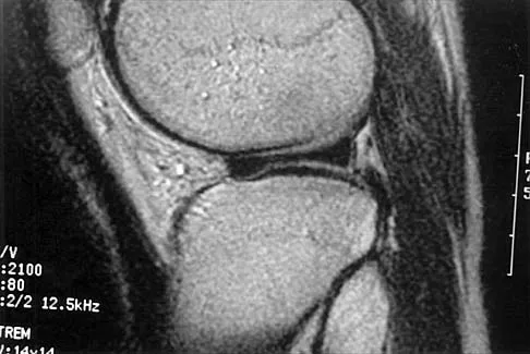
A 26-year-old man has recurrent right knee pain. Figures 9a and 9b show consecutive sagittal T2-weighted MRI scans, and Figure 9c shows a coronal T1-weighted MRI scan. What is the most likely diagnosis?


Explanation

9b 9c A discoid meniscus is a large disk-like meniscus. It is seen in the lateral meniscus in 3% of the population; a discoid medial meniscus is much less common. It can be identified on the coronal view by noting meniscal tissue extending into the tibial spine at the intercondylar notch. The average width of a normal meniscus is less than 11 mm. A bow-tie appearance should not be seen on more than two consecutive sagittal images because the conventional thickness of the sagittal slices is 3 mm and the interval between two consecutive slices is 1.5 mm. Two sagittal slices will cover a 9-mm thickness. A discoid meniscus can be diagnosed on the sagittal views by noting a bow-tie appearance on more than two consecutive images. Helms CA: MR image of the knee, in Fundamentals of Skeletal Radiology, ed 2. Philadelphia, PA, WB Saunders, 1995, pp 172-191.

Question 15

The gluteus maximus is innervated by which of the following nerves?


Explanation

The inferior gluteal nerve supplies the gluteus maximus muscle. The superior gluteal nerve supplies the gluteus medius, gluteus minimus, and tensor fascia lata muscles. The femoral nerve supplies the quadriceps, sartorius, and pectineus muscles. The pudendal nerve is primarily a sensory nerve.

Question 16

The dorsal (Thompson) approach to the proximal forearm uses which of the following intermuscular intervals?


Explanation

The Thompson posterior approach is used in treatment of fractures of the proximal radius. Dissection is carried out through the interval between the extensor carpi radialis brevis (radial nerve) and the extensor digitorum communis (posterior interosseous nerve). To identify this interval, the forearm is pronated and the mobile lateral wad of muscles (the ulnar-most belly is the extensor carpi radialis brevis) is grasped with the thumb and finger and pulled from the much less mobile mass of the extensor digitorum communis. The furrow created is marked with a skin marker for subsequent skin incision. The skin incision follows a line from the lateral epicondyle of the humerus to a point corresponding to the middle of the posterior aspect of the wrist. Distally, the intermuscular plane is between the extensor carpi radialis brevis and the extensor pollicis longus. Crenshaw AH Jr: Surgical techniques and approaches, in Canale ST (ed): Campbell's Operative Orthopaedics, ed 9. St Louis, MO, Mosby-Year Book, 1998, vol 1, pp 128-129. Hoppenfeld S, deBoer P: Posterior approach to the radius, in Surgical Exposures in Orthopaedics: The Anatomic Approach, ed 2. Philadelphia, PA, Lippincott-Raven, 1992, pp 136-146.

Question 17

A 45-year-old man who smokes reports the rapid onset of color changes and coolness in the fingers. Examination shows an abnormal Allen test. Plain radiographs of the hand and wrist are normal. Which of the following studies will best aid in diagnosis?


Explanation

The patient has symptoms typical of Raynaud's phenomenon secondary to underlying vascular disease. The next most appropriate step in the management of this patient should be to perform contrast angiography on the involved upper extremity to look for proximal or distal arterial lesions or insufficiencies. MRI and contrast CT are not as specific as angiography for the identification of vascular lesions of the upper extremity. Although patients with primary Raynaud's vasospastic disease can have normal angiographic findings, they typically are younger than age 40 years, are female, and have normal results on an Allen test. Green DP, Hotchkiss RN, Pederson WC (eds): Operative Hand Surgery, ed 4. New York, NY, Churchill Livingstone, 1999, pp 2288-2290.

Question 18

A purulent flexor tenosynovitis of the thumb may communicate with the small finger flexor through which of the following structures?


Explanation

Only the flexor sheaths of the thumb and small finger are continuous from the digit through the carpal canal and into the distal forearm. If one of the sheaths ruptures from synovitis, it may contaminate the other sheath through Parona's space in the distal forearm. This potential space lies superficial to the pronator quadratus and deep to the flexor tendons. Green DP, Hotchkiss RN, Pederson WC (eds): Operative Hand Surgery, ed 4. New York, NY, Churchill Livingstone, 1999, pp 1044-1045.

Question 19

Which of the following nerves travels with the deep palmar arch?


Explanation

The ulnar nerve divides alongside the pisiform, and the deep branch supplies the three hypothenar muscles and crosses the palm with the deep palmar arch to supply the two ulnar lumbricals, all interossei, and finally the adductor pollicis. The superficial branch supplies the ulnar digital branches to the small and ring fingers. The median nerve branches are more superficial in the palm near the superficial palmar arch. Last RJ: Anatomy: Regional and Applied, ed 6. London, England, Churchill Livingstone, 1978, p 109.

Question 20

Figures 10a through 10c show the plain radiograph and MRI scans of a 41-year-old man who has right hip pain. What is the most likely diagnosis?


Explanation

10b 10c Transient osteoporosis is a self-limited painful but reversible disorder. Although first described in pregnant women, it is more common in young to middle-aged men. The radiograph shows loss of mineralization in the right hip relative to the left side. There is no osseous destruction or cortical expansion typical of metastasis or giant cell tumor. The process is confined to the femoral side of the joint unlike rheumatoid arthritis, which would be centered in the joint. Osteonecrosis is better defined with sharp but irregularly shaped margins, and there is no double-line sign. The MRI scans reveal diffuse edema in the femoral head and neck that is atypical for osteonecrosis. Transient osteoporosis may recur in the same or opposite hip.

Question 21

Figure 11 shows the anatomic dissection of the medial side of the knee joint after removal of the superficial fascia. The arrow is pointing to what structure?


Explanation

The semitendinosus and gracilis tendons lie beneath the superficial fascia and superficial to the medial collateral ligament. The semitendinosus is located more inferior to the gracilis tendon. The sartorius is more posterior and distal as is the medial collateral ligament. The semimembranosus is posterior. Pagnani MJ, Warner JJ, O'Brien SJ, Warren RF: Anatomic considerations in harvesting the semitendinosus and gracilis tendons and a technique of harvest. Am J Sports Med 1993;21:565-571.

Question 22

Figure 12 shows a lateral radiograph of the elbow. What is the most likely diagnosis?


Explanation

The figure shows a supracondylar process, which is a normal anatomic variant. An osteochondroma tends to occur more toward the end of bones, and the medullary space of the underlying bone extends into the base of the osteochondroma. The presence of a supracondylar process is usually asymptomatic. However, the ligament of Struthers that always extends from the supracondylar process to the medial epicondyle can result in median nerve entrapment secondary to trauma. Last RJ: Anatomy: Regional and Applied, ed 6. London, England, Churchill Livingstone, 1978, pp 132-133.

Question 23

Which of the following nerves is most likely responsible for symptoms associated with plantar fasciitis?


Explanation

The first branch of the lateral calcaneal nerve innervates the abductor digiti minimi. It is reported to be trapped at the interval between the abductor hallucis and the quadratus plantae muscles.

Question 24

A 16-year-old cheerleader reports an ache in the right shoulder and arm that is worse after activity. She denies any history of acute trauma. Examination reveals a positive sulcus sign and an AP glide test with a posterior and anterior apprehension sign. To confirm a diagnosis of multidirectional instability, which of the following imaging studies is most appropriate?


Explanation

Multidirectional instability is a common finding in young female athletes. The anatomic structures are all intact but are hypermobile; therefore, CT and bone scans and scapular Y-views are often normal. Obtaining a weighted or AP stress view while applying downward traction on the arm will document instability and hypermobility of the joint. MRI generally is not indicated in this condition. Ultrasound is used primarily for rotator cuff pathology. Neer CS II, Foster CR: Inferior capsular shift for involuntary inferior and multidirectional instability of the shoulder: A preliminary report. J Bone Joint Surg Am 1980;62:897-908.

Question 25

Which of the following findings is seen in the chest radiograph shown in Figure 13?


Explanation

Orthopaedic surgeons are often responsible for interpreting radiographs of general examinations such as the chest radiograph shown. For accurate interpretation, it is important to systematically review all of the information available on the radiograph. Using this approach, the fracture of the left proximal humerus is readily recognized. Linear air soft-tissue density at the lung periphery would suggest a pneumothorax, but this finding is not shown on the radiograph. The upper thoracic spine is well aligned. The sternoclavicular and distal clavicles are normal.

Question 26

In the anterior approach to the hip (Smith-Petersen), the superficial surgical interval relies on an internervous plane. Which of the following describes the innervation of the muscles defining this plane?


Explanation

The superficial interval in the Smith-Petersen approach is between the sartorius (femoral nerve) and tensor fasciae latae (superior gluteal nerve). This provides a true internervous plane for safe access to the anterior hip.

Question 27

A 35-year-old overhead athlete presents with posterior shoulder pain and weakness in external rotation. An MRI shows atrophy of the teres minor. Entrapment of a nerve in the quadrilateral space is suspected. Which of the following structures forms the superior border of this space?


Explanation

The quadrilateral space is bordered superiorly by the teres minor, inferiorly by the teres major, medially by the long head of the triceps, and laterally by the surgical neck of the humerus. It contains the axillary nerve and posterior circumflex humeral artery.

Question 28

During reconstruction of the posterolateral corner (PLC) of the knee, identifying the insertion of the popliteofibular ligament is critical. To which specific anatomical aspect of the fibula does this ligament attach?


Explanation

The popliteofibular ligament originates from the musculotendinous junction of the popliteus and attaches to the posteromedial aspect of the fibular styloid (apex). It is a crucial static stabilizer against external rotation.

Question 29

A patient undergoes open carpal tunnel release. Postoperatively, they exhibit profound weakness in thumb opposition but normal thumb interphalangeal joint flexion. The recurrent motor branch of the median nerve was likely injured. This branch typically enters the thenar musculature via which of the following variations?


Explanation

The extraligamentous course is the most common anatomic variation (around 50-80%) of the recurrent motor branch of the median nerve, curving radially around the distal edge of the transverse carpal ligament. A transligamentous course occurs in about 20% of patients and is at highest risk during release.

Question 30

A patient presents with acquired adult flatfoot deformity resulting from posterior tibial tendon insufficiency. The secondary static stabilizer of the medial longitudinal arch is often attenuated. Which of the following bands of the calcaneonavicular (spring) ligament complex is the strongest and most critical for arch support?


Explanation

The superomedial band of the spring ligament is the thickest and strongest component. It acts as the primary static sling supporting the talar head and is most frequently torn or attenuated in flatfoot deformity.

Question 31

A 24-year-old gymnast sustains a traumatic tear of the triangular fibrocartilage complex (TFCC). Arthroscopy reveals a tear in the central articular disc. What is the healing potential of this specific region, and what is its vascular supply?


Explanation

The central articular disc of the TFCC is avascular, receiving nutrients only through diffusion from synovial fluid. Consequently, tears in the central portion (Palmer 1A) have poor healing potential and are typically treated with debridement rather than repair.

Question 32

When placing C2 pedicle screws during posterior cervical fusion, the trajectory must avoid the vertebral foramen. At the C2 level, what is the typical anatomical relationship of the vertebral artery to the pedicle/pars?


Explanation

In the C2 vertebra, the vertebral artery courses laterally and inferiorly relative to the pedicle and pars interarticularis before turning medially to enter the C1 foramen transversarium. Trajectories must remain medial and superior to avoid vascular injury.

Question 33

A patient develops posterolateral rotatory instability (PLRI) of the elbow following a dislocation. The primary deficient structure originates on the lateral epicondyle and inserts on which of the following structures?


Explanation

PLRI is caused by deficiency of the lateral ulnar collateral ligament (LUCL). The LUCL originates from the lateral epicondyle and inserts on the supinator crest of the ulna, acting as the primary restraint to posterolateral subluxation of the radial head.

Question 34

During an anterolateral approach to the distal tibia, the superficial peroneal nerve is at risk. At what average distance proximal to the lateral malleolus does this nerve pierce the crural fascia to become subcutaneous?


Explanation

The superficial peroneal nerve typically pierces the deep crural fascia to become subcutaneous approximately 10 to 12 cm proximal to the tip of the lateral malleolus. It then bifurcates into the medial and intermediate dorsal cutaneous nerves.

Question 35

During the ilioinguinal approach to the acetabulum, severe bleeding occurs over the superior pubic ramus near the symphysis. This is most likely due to an injury to the "corona mortis," which is an anastomosis between which of the following vessel systems?


Explanation

The corona mortis is a vascular anastomosis between the external iliac (or inferior epigastric) and obturator systems. It crosses the superior pubic ramus and is highly vulnerable during anterior pelvic and acetabular exposures.

Question 36

An elderly patient sustains a displaced femoral neck fracture, risking avascular necrosis. The primary blood supply to the adult femoral head is derived from the lateral epiphyseal artery. This vessel is a terminal branch of which artery?


Explanation

The primary blood supply to the adult femoral head comes from the lateral epiphyseal artery, which is a terminal branch of the medial femoral circumflex artery (MFCA). The MFCA courses posterior to the femoral neck, making it vulnerable in displaced fractures.

Question 37

A weightlifter tears his pectoralis major tendon at its insertion. Surgical repair is planned. Which of the following accurately describes the anatomy of the pectoralis major tendon insertion on the humerus?


Explanation

The pectoralis major tendon inserts in a U-shaped bilaminar fashion. The clavicular head forms the anterior lamina, while the sternal head twists 180 degrees so that its lowest fibers insert highest and most posterior on the lateral lip of the bicipital groove.

Question 38

A patient develops compartment syndrome of the lower leg. The surgeon performs a dual-incision four-compartment fasciotomy. Which of the following nerves is located within the deep posterior compartment?


Explanation

The tibial nerve runs within the deep posterior compartment of the leg, along with the posterior tibial and peroneal vessels. It innervates the tibialis posterior, flexor digitorum longus, and flexor hallucis longus muscles.

Question 39

The scaphoid bone is highly susceptible to nonunion following a fracture due to its unique intraosseous blood supply. The major blood supply enters the scaphoid through its dorsal ridge and is a branch of which artery?


Explanation

The primary blood supply to the scaphoid (approx 70-80%) enters distally via the dorsal ridge from branches of the radial artery (dorsal carpal branch). This retrograde perfusion puts proximal pole fractures at high risk for avascular necrosis.

Question 40

A patient presents with inability to form an "OK" sign with their thumb and index finger, but they have no sensory deficits in the hand. Compression of the anterior interosseous nerve (AIN) is suspected. Which of the following muscles is uniquely innervated by the AIN?


Explanation

The AIN provides motor innervation to the flexor pollicis longus, the radial half of the flexor digitorum profundus (index and middle fingers), and the pronator quadratus. Weakness in FPL and FDP to the index finger causes the classic inability to make an "OK" sign.

Question 41

A patient sustains a midfoot crush injury. Radiographs show widening of the space between the first and second metatarsals. The Lisfranc ligament is likely ruptured. What are the specific osseous attachments of this ligament?


Explanation

The Lisfranc ligament is a strong interosseous ligament that connects the lateral aspect of the medial cuneiform to the medial base of the second metatarsal. It is the primary stabilizer of the second tarsometatarsal joint.

Question 42

During arthroscopic meniscectomy, understanding the differences between the medial and lateral menisci is crucial to avoid complications. Which of the following is a characteristic feature of the lateral meniscus compared to the medial meniscus?


Explanation

The lateral meniscus is more circular (O-shaped), more mobile, and covers a larger area of the tibial plateau than the medial meniscus. It lacks an attachment to the lateral collateral ligament, separated from it by the popliteus tendon.

Question 43

A patient complains of deep gluteal pain radiating down the posterior thigh. MRI shows no lumbar disc herniation. The patient has a known anatomic variation where a portion of the sciatic nerve pierces the piriformis muscle. Which component of the nerve typically pierces the muscle in this variant?


Explanation

In about 15% of the population, the sciatic nerve has an anatomical variation in its relationship with the piriformis. The most common variant (Beaton and Anson type B) involves the common peroneal division piercing the piriformis while the tibial division passes below it.

Question 44

A 45-year-old man presents with severe radicular pain radiating down the anterior aspect of his right thigh to the knee, accompanied by weakness in knee extension. An MRI reveals a far lateral (extraforaminal) disc herniation at the L3-L4 level. Which nerve root is most likely compressed?


Explanation

In the lumbar spine, a far lateral (extraforaminal) disc herniation compresses the exiting nerve root at the same level. Therefore, an L3-L4 far lateral disc herniation compresses the L3 nerve root, presenting with anterior thigh pain and quadriceps weakness.

Question 45

A volleyball player presents with painless weakness in external rotation of the shoulder. Examination reveals atrophy isolated to the infraspinatus fossa. An MRI demonstrates a paralabral cyst. Where is the cyst most likely located to cause this specific presentation?


Explanation

The suprascapular nerve innervates both the supraspinatus and infraspinatus. Compression at the spinoglenoid notch, typically by a paralabral cyst associated with a posterior labral tear, affects only the branch to the infraspinatus, causing isolated external rotation weakness and infraspinatus atrophy.

Question 46

A 28-year-old overhead athlete presents with poorly localized posterior shoulder pain and paresthesias over the lateral deltoid. MRI reveals isolated atrophy of the teres minor. Which anatomic structures form the boundaries of the space where the affected nerve is most likely compressed?


Explanation

The axillary nerve is compressed in quadrilateral space syndrome. The quadrilateral space is bounded by the teres minor (superiorly), teres major (inferiorly), long head of the triceps (medially), and surgical neck of the humerus (laterally).

Question 47

During surgical reconstruction of the posterolateral corner (PLC) of the knee, the surgeon isolates the popliteofibular ligament. Which of the following accurately describes its true anatomic attachments?


Explanation

The popliteofibular ligament (PFL) originates from the musculotendinous junction of the popliteus. It attaches distally to the posteromedial aspect of the fibular head, acting as a crucial stabilizer against external rotation.

Question 48

During a posterior approach to the hip for total hip arthroplasty, the surgeon meticulously manages the short external rotators. Which tendon, if left intact, directly protects the deep branch of the medial femoral circumflex artery from iatrogenic injury?


Explanation

The deep branch of the medial femoral circumflex artery (MFCA) courses posteriorly beneath the obturator externus tendon. Preserving the obturator externus during a posterior approach prevents iatrogenic injury to the primary blood supply of the femoral head.

Question 49

A 45-year-old woman presents with dorsal radial wrist pain. Physical examination reveals tenderness just distal to Lister's tubercle. Ultrasound confirms tenosynovitis of the third extensor compartment. Which of the following muscles acts as the primary motor for the tendon located in this compartment?


Explanation

The third extensor compartment of the wrist contains only the extensor pollicis longus (EPL) tendon. The EPL tendon notably uses Lister's tubercle as a fulcrum to change its line of pull toward the thumb.

Question 50

When placing an iliosacral screw into the S1 vertebral body for pelvic ring fixation, anterior misplacement of the screw out of the sacral ala places which nerve root at greatest risk of direct injury?


Explanation

The L5 nerve root courses directly anterior to the sacral ala after exiting the L5-S1 foramen. Therefore, errant anterior placement of an S1 iliosacral screw places the L5 nerve root in immediate jeopardy.

Question 51

A patient undergoes a tarsal tunnel release. The surgeon makes a curved incision posterior to the medial malleolus. Proceeding strictly from anterior to posterior, what is the correct anatomical order of the structures encountered beneath the flexor retinaculum?


Explanation

The correct anterior-to-posterior order in the tarsal tunnel is Tibialis posterior, Flexor digitorum longus, posterior tibial Artery, posterior tibial Vein, tibial Nerve, and Flexor hallucis longus. This is remembered by the mnemonic "Tom, Dick, AND Very Nervous Harry."

Question 52

During an anterior (Henry) approach to the diaphyseal radius, the supinator is reflected to expose the bone. Deep in the proximal forearm, the anterior interosseous nerve (AIN) is identified running alongside the anterior interosseous artery. From which parent vessel does the anterior interosseous artery directly branch?


Explanation

The anterior interosseous artery is a direct branch of the common interosseous artery. The common interosseous artery itself is a short branch originating from the ulnar artery just distal to the radial tuberosity.

Question 53

A 32-year-old man presents with chronic elbow instability. Examination reveals apprehension during supination, valgus stress, and axial loading of the elbow. Which structure is most likely deficient, and what is its normal anatomic insertion site?


Explanation

The patient's clinical presentation is classic for posterolateral rotatory instability (PLRI), which is caused by deficiency of the lateral ulnar collateral ligament (LUCL). The LUCL originates on the lateral epicondyle and inserts on the supinator crest of the ulna.

Question 54

A 35-year-old professional volleyball player presents with painless shoulder weakness. Examination reveals isolated profound atrophy of the infraspinatus with completely preserved supraspinatus muscle bulk. Where is the most likely anatomic site of nerve entrapment?


Explanation

Compression of the suprascapular nerve at the spinoglenoid notch affects only the motor branches to the infraspinatus. Entrapment at the more proximal suprascapular notch would cause atrophy of both the supraspinatus and infraspinatus.

Question 55

During an ilioinguinal approach for an anterior column acetabular fracture, vigorous arterial bleeding is encountered when dissecting over the superior pubic ramus. This bleeding most likely originates from an anastomosis between which two vascular systems?


Explanation

The bleeding is from the corona mortis, a vascular anastomosis crossing the superior pubic ramus. It connects the external iliac (or inferior epigastric) system with the obturator vessels (internal iliac system).

Question 56

The distal tibiofibular syndesmosis provides critical structural stability to the ankle mortise. Which of its component ligaments is biomechanically the strongest and provides the greatest resistance to lateral displacement of the fibula?


Explanation

The posterior inferior tibiofibular ligament (PITFL) is the strongest component of the syndesmotic complex. It contributes the majority of resistance against lateral translation of the distal fibula.

Question 57

During surgical exploration for a complex flexor tendon laceration in zone II of the index finger, the surgeon must vent some pulleys to facilitate tendon glide. Which annular pulleys are considered the most critical to preserve or reconstruct to prevent significant bowstringing?


Explanation

The A2 and A4 pulleys are the most biomechanically critical annular pulleys in the digits. Their sacrifice predictably leads to significant bowstringing of the flexor tendons and loss of functional excursion.

Question 58

In a 12-year-old boy undergoing in situ pinning for a slipped capital femoral epiphysis (SCFE), the surgeon must avoid the terminal branches of the medial femoral circumflex artery. These crucial retinacular vessels typically penetrate the proximal femur at which anatomic location?


Explanation

The primary blood supply to the femoral head comes from the lateral epiphyseal artery, a terminal branch of the medial femoral circumflex artery. These vessels penetrate the capsule and enter the bone at the posterosuperior aspect of the femoral neck.

Question 59

A 35-year-old male is undergoing open reduction and internal fixation of a capitellum fracture using the Kocher approach. To safely access the joint and protect the posterior interosseous nerve (PIN), the dissection must utilize which of the following internervous planes?


Explanation

The Kocher approach utilizes the internervous plane between the anconeus (radial nerve) and the extensor carpi ulnaris (posterior interosseous nerve). This safely approaches the lateral elbow while protecting the PIN, provided the dissection stays anterior to the anconeus.

Question 60

A 72-year-old female sustains a displaced femoral neck fracture. Which of the following arterial structures provides the primary blood supply to the adult femoral head and is most at risk of disruption in this injury?


Explanation

The deep branch of the medial femoral circumflex artery (MFCA) provides the primary blood supply to the adult femoral head. The artery of the ligamentum teres provides a negligible supply in adults, and the lateral femoral circumflex artery primarily supplies the greater trochanter and anterior neck.

Question 61

A 28-year-old overhead athlete presents with vague posterior shoulder pain and numbness over the lateral deltoid. MRI reveals severe isolated atrophy of the teres minor. Which of the following neurovascular bundles is most likely compressed in the quadrilateral space?


Explanation

Quadrilateral space syndrome involves compression of the axillary nerve and the posterior circumflex humeral artery. The space is bounded by the teres minor (superior), teres major (inferior), long head of the triceps (medial), and the humeral shaft (lateral).

Question 62

During a primary total hip arthroplasty using the direct anterior (Smith-Petersen) approach, the surgeon exploits an internervous plane between two muscles. Which of the following accurately describes this plane?


Explanation

The Smith-Petersen approach utilizes the superficial internervous plane between the sartorius (femoral nerve) and the tensor fasciae latae (superior gluteal nerve). The deep plane is between the rectus femoris and the gluteus medius.

Question 63

A 45-year-old avid cyclist presents with intrinsic muscle weakness and numbness in his ring and small fingers. He is diagnosed with handlebar palsy due to compression in Guyon's canal. Which of the following structures forms the anatomic roof of this canal?


Explanation

The roof of Guyon's canal is formed by the palmar carpal ligament (and palmaris brevis muscle). The floor is formed by the flexor retinaculum (transverse carpal ligament) and the pisohamate ligament.

Question 64

While performing a tarsal tunnel release, the surgeon identifies the structures passing posterior to the medial malleolus. What anatomical structure is located immediately posterior to the flexor digitorum longus (FDL) tendon?


Explanation

The anatomical order of structures behind the medial malleolus from anterior to posterior is: Tibialis posterior, Flexor digitorum longus, posterior tibial Artery, tibial Nerve, and Flexor hallucis longus (Tom, Dick, AND Harry). Therefore, the artery is immediately posterior to the FDL.

Question 65

During ankle arthroscopy, establishment of the posterolateral portal places a specific neurovascular bundle at highest risk of iatrogenic injury. Which of the following structures is most vulnerable?


Explanation

The posterolateral portal in ankle arthroscopy is made just lateral to the Achilles tendon. This location places the sural nerve and the small saphenous vein at significant risk if the incision is strayed too far laterally or made too deeply.

Question 66

A patient suffers a severe forearm crush injury and subsequently demonstrates an inability to make the 'OK' sign, exhibiting extended distal interphalangeal joints of the thumb and index finger. This specific nerve palsy represents denervation to which muscle group?


Explanation

The inability to make the 'OK' sign indicates an anterior interosseous nerve (AIN) palsy. The AIN is a motor branch of the median nerve that innervates the FDP to the index and middle fingers, the FPL, and the pronator quadratus.

Question 67

A spinal surgeon is placing L4 pedicle screws for a lumbar fusion. If the right L4 pedicle screw breaches the medial pedicle wall, which neural structure is at greatest immediate risk of injury?


Explanation

A medial breach of the pedicle puts the traversing nerve root of the level below at risk. At the L4 level, the traversing root is L5. The L4 exiting root passes laterally and inferiorly to the L4 pedicle.

Question 68

A 22-year-old soccer player undergoes arthroscopic meniscal repair for a bucket-handle tear. The success of meniscal repair relies heavily on the vascularity of the peripheral rim. The primary blood supply to the peripheral third of the meniscus arises from which vessels?


Explanation

The peripheral 10-30% of the menisci (the red-red zone) receives its blood supply from the capsular vessels, which are derived from the medial and lateral, superior and inferior geniculate arteries. The middle geniculate artery primarily supplies the cruciate ligaments.

Question 69

Biomechanical understanding of the anterior cruciate ligament (ACL) is critical for anatomic reconstruction. The ACL is divided into the anteromedial (AM) and posterolateral (PL) bundles. During normal knee kinematics, when is the AM bundle tightest?


Explanation

The anteromedial (AM) bundle of the ACL tightens primarily in knee flexion, thereby resisting anterior tibial translation in the flexed position. The posterolateral (PL) bundle is tightest in extension and resists rotatory loads.

Question 70

When evaluating a posterior cruciate ligament (PCL) injury on MRI, a radiologist must closely inspect its origin and insertion. Which of the following describes the correct anatomic femoral origin of the PCL?


Explanation

The PCL originates on the anterolateral aspect of the medial femoral condyle and inserts on the posterior aspect of the tibial plateau, approximately 1 cm below the articular surface.

Question 71

A trauma surgeon is performing an anterolateral approach to the distal humerus for a complex fracture. Knowledge of the radial nerve's course is crucial. At approximately what distance proximal to the lateral epicondyle does the radial nerve pierce the lateral intermuscular septum to enter the anterior compartment?


Explanation

The radial nerve pierces the lateral intermuscular septum to transition from the posterior compartment to the anterior compartment approximately 10 cm proximal to the lateral epicondyle. This is a critical landmark to avoid iatrogenic injury during lateral and anterolateral exposures.

Question 72

During wrist arthroscopy for triangular fibrocartilage complex (TFCC) evaluation, the standard 3-4 portal is established. This portal is placed between which two extensor tendon compartments?


Explanation

The standard 3-4 arthroscopy portal of the wrist is located between the 3rd dorsal compartment (EPL) and the 4th dorsal compartment (EDC). It is placed just distal to Lister's tubercle.

Question 73

During an anterior (Henry) approach to the radius, the surgeon enters the internervous plane in the proximal forearm. Which two nerves innervate the muscles that define this superficial proximal interval?


Explanation

The proximal superficial interval of the Henry approach lies between the brachioradialis (innervated by the radial nerve) and the pronator teres (innervated by the median nerve).

Question 74

A 28-year-old sustains a displaced talar neck fracture. Which of the following blood vessels provides the primary blood supply to the talar body, placing it at high risk for avascular necrosis?


Explanation

The artery of the tarsal canal, a branch of the posterior tibial artery, is the dominant blood supply to the talar body. Disruption of this vessel in talar neck fractures significantly increases the risk of avascular necrosis.

Question 75

When reconstructing the posterolateral corner (PLC) of the knee, a surgeon isolates the primary static restraint to varus opening at 30 degrees of knee flexion. What are the origin and insertion of this structure?


Explanation

The lateral collateral ligament (LCL) is the primary restraint to varus stress at 30 degrees of flexion. It originates on the lateral femoral epicondyle and inserts on the fibular head.

Question 76

A patient presents with vague posterior shoulder pain and isolated weakness of the teres minor and deltoid. MRI reveals a paralabral cyst compressing structures within the quadrilateral space. Which of the following correctly describes the anatomical borders of this space?


Explanation

The quadrilateral space is bordered by the teres minor superiorly, teres major inferiorly, the long head of the triceps medially, and the surgical neck of the humerus laterally. It transmits the axillary nerve and posterior circumflex humeral artery.

Question 77

During flexor tendon repair in Zone II, preservation or reconstruction of the pulley system is crucial to prevent mechanical bowstringing. Which of the following pulleys arise directly from the periosteum of the proximal and middle phalanges, respectively?


Explanation

The A2 pulley arises from the proximal half of the proximal phalanx, and the A4 pulley arises from the middle portion of the middle phalanx. These are the most critical pulleys for preventing bowstringing during finger flexion.

Question 78

A primary total hip arthroplasty is performed using the direct anterior approach (Smith-Petersen). Which internervous plane is utilized during the superficial dissection of this surgical approach?


Explanation

The superficial interval of the direct anterior approach is between the sartorius (femoral nerve) and the tensor fasciae latae (superior gluteal nerve).

Question 79

A 32-year-old elite volleyball player presents with isolated weakness in external rotation of the dominant shoulder. Physical examination reveals isolated atrophy of the infraspinatus with no supraspinatus involvement. Where is the most likely location of nerve compression?


Explanation

Compression of the suprascapular nerve at the spinoglenoid notch affects only the motor branches to the infraspinatus, sparing the supraspinatus. Compression at the suprascapular notch would affect both muscles.

Question 80

A 45-year-old undergoes an anterolateral approach to the distal tibia for a pilon fracture. During deep dissection, which neurovascular bundle is at risk and must be carefully retracted medially along with the anterior compartment musculature?


Explanation

During the anterolateral approach to the distal tibia, the deep peroneal nerve and anterior tibial artery course deep to the anterior compartment muscles and must be protected by retracting them medially.

Question 81

A surgeon is utilizing a direct lateral approach to the fibula for an ORIF of a distal third shaft fracture. The superficial peroneal nerve is at risk as it exits the deep fascia to become subcutaneous. On average, at what distance proximal to the tip of the lateral malleolus does this nerve pierce the crural fascia?


Explanation

The superficial peroneal nerve transitions from the lateral compartment to the subcutaneous layer approximately 10 to 12 cm proximal to the tip of the lateral malleolus, placing it at risk during distal fibular approaches.

Question 82

During a posterior approach to the hip (Kocher-Langenbeck), which two muscles form the interval where the deep branch of the medial circumflex femoral artery is most at risk?


Explanation

The deep branch of the medial circumflex femoral artery runs in the interval between the obturator externus and the quadratus femoris. Protecting the obturator externus and limiting the release of the quadratus femoris during a posterior approach helps preserve the femoral head blood supply.

Question 83

A patient presents with thenar atrophy and inability to oppose the thumb after suffering a distal radius fracture. The affected nerve most commonly branches from the main nerve at which location relative to the transverse carpal ligament?


Explanation

The recurrent motor branch of the median nerve most commonly exhibits an extraligamentous course (about 50-80% of cases). It branches from the median nerve distal to the transverse carpal ligament and curves back to innervate the thenar muscles.

Question 84

When performing an arthroscopic capsular release for adhesive capsulitis, the axillary nerve is most vulnerable at the 6 o'clock position. Approximately how far is the axillary nerve from the inferior glenoid rim in a standard adult shoulder?


Explanation

The axillary nerve passes through the quadrilateral space and lies closest to the joint capsule directly inferiorly. It is located approximately 10 to 15 mm from the inferior glenoid rim at the 6 o'clock position.

Question 85

During a transforaminal endoscopic lumbar discectomy at L4-L5, the surgeon utilizes Kambin's triangle as a safe working zone. Which of the following forms the anterior border of this anatomical safe zone?


Explanation

Kambin's triangle is bordered anteriorly by the exiting nerve root (L4), inferiorly by the superior endplate of the lower vertebral body (L5), and posteriorly by the superior articular process of the lower vertebra (L5).

Question 86

A patient requires reconstruction of the posterolateral corner (PLC) of the knee. Regarding the femoral insertions of these structures, where does the popliteus tendon insert relative to the fibular collateral ligament (FCL)?


Explanation

On the lateral femoral epicondyle, the popliteus tendon inserts distal and anterior to the origin of the fibular collateral ligament. Understanding this footprint relationship is critical for anatomical PLC reconstruction.

Question 87

During a medial approach to the midfoot for an accessory navicular excision, a tendinous intersection known as the Master knot of Henry is identified. Which of the following accurately describes the relationship of the tendons at this knot?


Explanation

At the Master knot of Henry in the plantar midfoot, the flexor hallucis longus (FHL) tendon crosses dorsal (superior or deep) to the flexor digitorum longus (FDL) tendon.

Question 88

A patient presents with an inability to actively extend the fingers at the metacarpophalangeal joints but maintains normal wrist extension with radial deviation. Entrapment of the affected nerve is most likely to occur at the proximal edge of which muscle?


Explanation

The patient has posterior interosseous nerve (PIN) palsy, sparing the extensor carpi radialis longus (which is innervated by the radial nerve proper). The most common site of PIN compression is the Arcade of Frohse, the proximal fibrous edge of the superficial head of the supinator.

None

Clinic OS
Medically Verified Content by
Prof. Clinic OS
Consultant Orthopedic & Spine Surgeon
Chapter Index