العربية
Part of the Master Guide

General Orthopedics 2026 Practice Questions: Set 13 (Solved)

General Orthopedics 2026 Practice Questions: Set 1 (Solved)

27 Apr 2026 130 min read 102 Views
Orthopedic Surgery Board Review 2026 | High-Yield MCQs - Set 1

Key Takeaway

Looking for accurate information on General Orthopedics 2026 Practice Questions: Set 1 (Solved)? Access high-yield General Orthopedics questions for the 2026 board exam. This module (Set 1) covers critical topics including surgical techniques, pathology, and treatment protocols with verified answers.

HY 2026


00:00

Start Quiz

Question 1 High Yield

Cell signaling through the activation of a transmembrane receptor complex formed by serine/threonine kinase receptors occurs with which of the following growth factors?


Detailed Explanation

Cell activation and transcription varies with the target cell, the growth factor-receptor combination, and the biologic state of the cell. The growth factors in the transforming growth factor-beta (TGF-ß) superfamily signal through serine/threonine kinase receptors. Fibroblast growth factors, insulin-like growth factors, and platelet-derived growth factors signal through tyrosine kinase receptors. Growth hormone is released by the pituitary and circulates to the liver where target cells are stimulated to release insulin-like growth factor. Lieberman J, Daluiski A, Einhorn TA: The role of growth factors in the repair of bone: Biology and clinical applications. J Bone Joint Surg Am 2002;84:1032-1044.

References:

  • Schmitt JM, Hwang K, Winn SR, et al: Bone morphogenetic proteins: An update on basic biology and clinical relevance. J Orthop Res 1999;17:269-278.

<span>Question 2</span> <span>High Yield</span>
In the most common condition causing a winged scapula, which of the following nerves is affected?
<img alt="General Orthopedics 2026 Practice Questions: Set 1 (Solved) - Figure 1" class="q-img mcq-img" height="393" loading="lazy" onclick="window.open(this.src)" src="/media/mcq-images/25/general-orthopedics-2026-set-1-mcqs-4056-fig-1.webp" title="Click to enlarge" width="464"/>
<button class="opt-btn" data-qid="2" onclick="handleSelect(this, '2', 0)"> <span class="opt-char">A</span> <span>Long thoracic nerve</span> </button> <button class="opt-btn" data-qid="2" onclick="handleSelect(this, '2', 1)"> <span class="opt-char">B</span> <span>Spinal accessory nerve</span> </button> <button class="opt-btn" data-qid="2" onclick="handleSelect(this, '2', 2)"> <span class="opt-char">C</span> <span>Suprascapular nerve</span> </button> <button class="opt-btn" data-qid="2" onclick="handleSelect(this, '2', 3)"> <span class="opt-char">D</span> <span>Dorsal scapular nerve</span> </button> <button class="opt-btn" data-qid="2" onclick="handleSelect(this, '2', 4)"> <span class="opt-char">E</span> <span>Thoracodorsal nerve</span> </button>
<button onclick="toggleExp('2')" style="background:none; border:none; color:#7f8c8d; text-decoration:underline; cursor:pointer;">Show Explanation</button>
<span class="exp-title">Detailed Explanation</span><div markdown="1">A winged scapula is most often associated with Parsonage-Turner syndrome, a condition thought to be due to an inflammatory or immune-mediated mechanism. Certain muscles are predisposed, particularly the serratus anterior muscle innervated by the long thoracic nerve. Other less common nerve lesions (eg, the spinal accessory and dorsal scapular nerves) may also cause winged scapulae. Kline DG, Hudson AR: Nerve Injuries: Operative Results for Major Nerve Injuries, Entrapments and Tumors. Philadelphia, PA, WB Saunders, 1995.
<strong>References:</strong><ul><li>van Alfen N, van Engelen BG: The clinical spectrum of neuralgic amyotrophy in 246 cases. Brain 2006;129:438-450.</li></ul>

<span>Question 3</span> <span>High Yield</span>
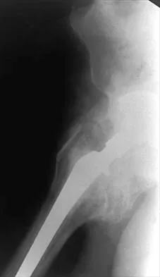
Figure 45 shows the radiograph of a 2-year-old patient who has progressive lumbar scoliosis as the result of hemivertebra. Examination reveals no associated cutaneous lesions, and an MRI scan shows no associated intraspinal anomalies. Treatment should consist of
<img alt="General Orthopedics 2026 Practice Questions: Set 1 (Solved) - Figure 2" class="q-img mcq-img" height="393" loading="lazy" onclick="window.open(this.src)" src="/media/mcq-images/25/general-orthopedics-2026-set-1-mcqs-4056-fig-2.webp" title="Click to enlarge" width="262"/>
<button class="opt-btn" data-qid="3" onclick="handleSelect(this, '3', 0)"> <span class="opt-char">A</span> <span>hemivertebra excision.</span> </button> <button class="opt-btn" data-qid="3" onclick="handleSelect(this, '3', 1)"> <span class="opt-char">B</span> <span>anterior and posterior spinal fusion with instrumentation from T4 to L4.</span> </button> <button class="opt-btn" data-qid="3" onclick="handleSelect(this, '3', 2)"> <span class="opt-char">C</span> <span>convex anterior hemiepiphyseodesis.</span> </button> <button class="opt-btn" data-qid="3" onclick="handleSelect(this, '3', 3)"> <span class="opt-char">D</span> <span>convex posterior hemiarthrodesis.</span> </button> <button class="opt-btn" data-qid="3" onclick="handleSelect(this, '3', 4)"> <span class="opt-char">E</span> <span>an orthosis.</span> </button>
<button onclick="toggleExp('3')" style="background:none; border:none; color:#7f8c8d; text-decoration:underline; cursor:pointer;">Show Explanation</button>
<span class="exp-title">Detailed Explanation</span><div markdown="1">In a retrospective review of 10 patients treated with hemivertebra excision for hemivertebra in the levels of T12 to L3, the procedure was found to be safe and effective. The procedure provided an average curve correction of 67 degrees and was greatest in patients who were younger than age 4 years at the time of surgery. Long anterior and posterior fusion with instrumentation is not the treatment of choice at this age. Either anterior hemiepiphyseodesis or posterior hemiarthrodesis in this isolated hemivertebra setting would be inadequate. Brace treatment is ineffective in management of the primary curvature.
<strong>References:</strong><ul><li>Callahan BC, Georgopoulos G, Eilert RE: Hemivertebral excision for congenital scoliosis. J Pediatr Orthop 1997;17:96-99.</li></ul>

<span>Question 4</span> <span>High Yield</span>
Based on the radiograph shown in Figure 4, the innervation of what muscle is most at risk with total hip arthroplasty?
<img alt="General Orthopedics 2026 Practice Questions: Set 1 (Solved) - Figure 3" class="q-img mcq-img" height="365" loading="lazy" onclick="window.open(this.src)" src="/media/mcq-images/25/general-orthopedics-2026-set-1-mcqs-4056-fig-3.webp" title="Click to enlarge" width="486"/>
<button class="opt-btn" data-qid="4" onclick="handleSelect(this, '4', 0)"> <span class="opt-char">A</span> <span>Quadriceps</span> </button> <button class="opt-btn" data-qid="4" onclick="handleSelect(this, '4', 1)"> <span class="opt-char">B</span> <span>Extensor hallucis longus</span> </button> <button class="opt-btn" data-qid="4" onclick="handleSelect(this, '4', 2)"> <span class="opt-char">C</span> <span>Lateral gastrocnemius</span> </button> <button class="opt-btn" data-qid="4" onclick="handleSelect(this, '4', 3)"> <span class="opt-char">D</span> <span>Adductor magnus</span> </button> <button class="opt-btn" data-qid="4" onclick="handleSelect(this, '4', 4)"> <span class="opt-char">E</span> <span>Semitendinosus</span> </button>
<button onclick="toggleExp('4')" style="background:none; border:none; color:#7f8c8d; text-decoration:underline; cursor:pointer;">Show Explanation</button>
<span class="exp-title">Detailed Explanation</span><div markdown="1">The radiograph reveals a Crowe IV deformity in a patient with developmental dysplasia of the hip. If hip arthroplasty is performed, then some degree of limb lengthening is anticipated. Excessive limb lengthening can result in sciatic nerve palsy in these patients. The peroneal branch of the sciatic nerve is most often affected. Of the muscles listed, only the extensor hallucis longus is innervated by the peroneal branch of the sciatic nerve. Eggli S, Hankemayer S, Muller ME: Nerve palsy after leg lengthening in total replacement arthroplasty for developmental dysplasia of the hip. J Bone Joint Surg Br 1999;81:843-845.
<strong>References:</strong><ul><li>Schmalzried TP, Amstutz HC, Dorey FJ: Nerve palsy associated with total hip replacement: Risk factors and prognosis. J Bone Joint Surg Am 1991;73:1074-1080.</li></ul>

<span>Question 5</span> <span>High Yield</span>
When performing a flexor tendon repair of a digit other than the thumb, what structures of the flexor tendon sheath should be preserved?
<img alt="General Orthopedics 2026 Practice Questions: Set 1 (Solved) - Figure 4" class="q-img mcq-img" height="345" loading="lazy" onclick="window.open(this.src)" src="/media/mcq-images/25/general-orthopedics-2026-set-1-mcqs-4056-fig-4.webp" title="Click to enlarge" width="486"/>
<button class="opt-btn" data-qid="5" onclick="handleSelect(this, '5', 0)"> <span class="opt-char">A</span> <span>A1 and A2 pulleys</span> </button> <button class="opt-btn" data-qid="5" onclick="handleSelect(this, '5', 1)"> <span class="opt-char">B</span> <span>A1 and A3 pulleys</span> </button> <button class="opt-btn" data-qid="5" onclick="handleSelect(this, '5', 2)"> <span class="opt-char">C</span> <span>A2 and A3 pulleys</span> </button> <button class="opt-btn" data-qid="5" onclick="handleSelect(this, '5', 3)"> <span class="opt-char">D</span> <span>A2 and A4 pulleys</span> </button> <button class="opt-btn" data-qid="5" onclick="handleSelect(this, '5', 4)"> <span class="opt-char">E</span> <span>C1 and C2 pulleys</span> </button>
<button onclick="toggleExp('5')" style="background:none; border:none; color:#7f8c8d; text-decoration:underline; cursor:pointer;">Show Explanation</button>
<span class="exp-title">Detailed Explanation</span><div markdown="1">The A2 and A4 pulleys are considered the most important parts of the pulley system. If these two structures are preserved, 80% of finger flexion can be maintained. If the pulley system is not left intact or is not reconstructed, "bow-stringing" of the flexor tendons occurs with loss of full flexion. The A2 pulley is over the proximal phalanx and the A4 pulley is over the middle phalanx. Doyle JR: Anatomy of the finger flexor tendon sheath and pulley system. J Hand Surg Am 1988;13:473-484.
<strong>References:</strong><ul><li>Strickland JW: Flexor tendon injuries: I. Foundations of treatment. J Am Acad Orthop Surg 1995;3:44-54.</li></ul>

<span>Question 6</span> <span>High Yield</span>
A 30-year-old woman sustains a transverse amputation of the distal phalanx of the index finger, leaving exposed bone. What is the most appropriate management of the soft-tissue defect?
<button class="opt-btn" data-qid="6" onclick="handleSelect(this, '6', 0)"> <span class="opt-char">A</span> <span>Dressing changes and healing by secondary intention</span> </button> <button class="opt-btn" data-qid="6" onclick="handleSelect(this, '6', 1)"> <span class="opt-char">B</span> <span>Split-thickness skin grafting</span> </button> <button class="opt-btn" data-qid="6" onclick="handleSelect(this, '6', 2)"> <span class="opt-char">C</span> <span>V-Y advancement flap</span> </button> <button class="opt-btn" data-qid="6" onclick="handleSelect(this, '6', 3)"> <span class="opt-char">D</span> <span>Moberg (volar advancement flap)</span> </button> <button class="opt-btn" data-qid="6" onclick="handleSelect(this, '6', 4)"> <span class="opt-char">E</span> <span>First dorsal metacarpal artery-island pedicled flap</span> </button>
<button onclick="toggleExp('6')" style="background:none; border:none; color:#7f8c8d; text-decoration:underline; cursor:pointer;">Show Explanation</button>
<span class="exp-title">Detailed Explanation</span><div markdown="1">V-Y advancement flaps are ideal for fingertip amputations that are transverse or dorsal oblique in nature. Healing by secondary intention is contraindicated with exposed bone. Shortening of exposed bone to allow primary skin closure is a possible alternative, as long as significant shortening of the index finger is avoided. A Moberg flap is useful only for distal amputations of the thumb. The first dorsal metacarpal artery-island pedicled flap uses tissue from the dorsum of the proximal index finger, and is typically used to resurface defects of the thumb. Fassler PR: Fingertip injuries: Evaluation and treatment. J Am Acad Orthop Surg 1996;4:84-92.
<strong>References:</strong><ul><li>Atasoy E, Ioakimidis E, Kasdan ML, et al: Reconstruction of the amputated fingertip with a triangular volar flap: A new surgical procedure. J Bone Joint Surg Am 1970;52:921-926.</li></ul>

<span>Question 7</span> <span>High Yield</span>
Figure 28 shows the radiograph of a 6-year-old girl who has a right thoracic scoliosis that measures 60 degrees. Examination shows multiple cafe-au-lait spots, and family history reveals that the child's mother has the same disorder. The gene responsible for this disorder codes for
<img alt="General Orthopedics 2026 Practice Questions: Set 1 (Solved) - Figure 5" class="q-img mcq-img" height="393" loading="lazy" onclick="window.open(this.src)" src="/media/mcq-images/25/general-orthopedics-2026-set-1-mcqs-4056-fig-5.webp" title="Click to enlarge" width="267"/>
<button class="opt-btn" data-qid="7" onclick="handleSelect(this, '7', 0)"> <span class="opt-char">A</span> <span>dystrophin.</span> </button> <button class="opt-btn" data-qid="7" onclick="handleSelect(this, '7', 1)"> <span class="opt-char">B</span> <span>frataxin.</span> </button> <button class="opt-btn" data-qid="7" onclick="handleSelect(this, '7', 2)"> <span class="opt-char">C</span> <span>neurofibromin.</span> </button> <button class="opt-btn" data-qid="7" onclick="handleSelect(this, '7', 3)"> <span class="opt-char">D</span> <span>peripheral myelin protein.</span> </button> <button class="opt-btn" data-qid="7" onclick="handleSelect(this, '7', 4)"> <span class="opt-char">E</span> <span>sulfate transport protein.</span> </button>
<button onclick="toggleExp('7')" style="background:none; border:none; color:#7f8c8d; text-decoration:underline; cursor:pointer;">Show Explanation</button>
<span class="exp-title">Detailed Explanation</span><div markdown="1">The patient has the dystrophic type of scoliosis seen in patients with neurofibromatosis type I (NF-1). The NF-1 gene is located on chromosome 17 and codes for neurofibromin, believed to be a tumor-suppresser gene. Abnormalities in the dystrophin gene are seen in Duchenne muscular dystrophy and Becker muscular dystrophy. A mutation in the frataxin gene is responsible for Friedreich ataxia. The most common type of hereditary motor and sensory neuropathy (Charcot-Marie-Tooth), HMSN type IA is caused by a complete duplication of the peripheral myelin protein gene. A defect in the cellular sulfate transport protein results in undersulfation of proteoglycans seen in diastrophic dysplasia.
<strong>References:</strong><ul><li>Beaty JH: Orthopaedic Knowledge Update 6. Rosemont, Ill, American Academy of Orthopaedic Surgeons, 1999, pp 225-234.</li></ul>

<span>Question 8</span> <span>High Yield</span>
A 7-year-old boy sustained a head contusion and small bowel injuries in a motor vehicle accident in which he was wearing a lap belt. He subsequently required a bowel resection. Six weeks after the accident, his parents note a painful mass in his lower back. His neurologic examination is normal. A radiograph and CT scans are shown in Figures 47a through 47c. Definitive management should now consist of
<img class="q-img mcq-img" src="/media/upload/general-orthopedics-2026-set-1-mcqs-4056-figure-1.webp"/>
<button class="opt-btn" data-qid="8" onclick="handleSelect(this, '8', 0)"> <span class="opt-char">A</span> <span>transcutaneous electrical stimulation and a lumbar corset.</span> </button> <button class="opt-btn" data-qid="8" onclick="handleSelect(this, '8', 1)"> <span class="opt-char">B</span> <span>transforaminal interbody fusion.</span> </button> <button class="opt-btn" data-qid="8" onclick="handleSelect(this, '8', 2)"> <span class="opt-char">C</span> <span>posterior instrumented L2-L3 reduction and fusion.</span> </button> <button class="opt-btn" data-qid="8" onclick="handleSelect(this, '8', 3)"> <span class="opt-char">D</span> <span>anterior interbody fusion with a cage.</span> </button> <button class="opt-btn" data-qid="8" onclick="handleSelect(this, '8', 4)"> <span class="opt-char">E</span> <span>spine extension bracing.</span> </button>
<button onclick="toggleExp('8')" style="background:none; border:none; color:#7f8c8d; text-decoration:underline; cursor:pointer;">Show Explanation</button>
<span class="exp-title">Detailed Explanation</span><div markdown="1">The posttraumatic lumbar kyphotic deformity will not remodel and is likely to worsen with time because the central line of gravity lies anterior to the deformity and the ligamentous disruption will not heal. The worsening deformity also puts the patient at some risk for future neurologic damage. Ebraheim NA, Savolain ER, Southworth SR, et al: Pediatric lumbar seat belt injuries. Orthopedics 1991;14:1010-1013.
<strong>References:</strong><ul><li>Taylor JA, Eggli KD: Lap belt inhuries of the lumbar spine in children: A pitfall in CT diagnosis. Am J Rad 1988;150:1355-1358.</li></ul>

<span>Question 9</span> <span>High Yield</span>
Which of the following best describes the mechanism of action of gentamycin?
<button class="opt-btn" data-qid="9" onclick="handleSelect(this, '9', 0)"> <span class="opt-char">A</span> <span>Inhibits cell wall synthesis by inhibiting peptidyl traspeptidase</span> </button> <button class="opt-btn" data-qid="9" onclick="handleSelect(this, '9', 1)"> <span class="opt-char">B</span> <span>Increases cell membrane permeability</span> </button> <button class="opt-btn" data-qid="9" onclick="handleSelect(this, '9', 2)"> <span class="opt-char">C</span> <span>Binds to the 30s ribosome subunit interfering with protein synthesis</span> </button> <button class="opt-btn" data-qid="9" onclick="handleSelect(this, '9', 3)"> <span class="opt-char">D</span> <span>Inhibits DNA gyrase</span> </button> <button class="opt-btn" data-qid="9" onclick="handleSelect(this, '9', 4)"> <span class="opt-char">E</span> <span>Forms oxygen radicals leading to loss of helical structure and breakage of DNA strands</span> </button>
<button onclick="toggleExp('9')" style="background:none; border:none; color:#7f8c8d; text-decoration:underline; cursor:pointer;">Show Explanation</button>
<span class="exp-title">Detailed Explanation</span><div markdown="1">Gentamycin and the aminoglycosides (ie, streptomycin, tobramycin, amikacin, and neomycin) work by binding to the 30s ribosome subunit, leading to the misreading of mRNA. This misreading results in the synthesis of abnormal peptides that accumulate intracellularly and eventually lead to cell death. These antibiotics are bactericidal. Cephalosporins, vancomycin, and penicillins interfere with cell wall synthesis by inhibiting the transpeptidase enzyme. Polymyxin, nystatin, and amphotericin increase cell membrane permeability by disrupting the functional integrity of the cell membrane. The quinolones inhibit the enzyme, DNA gyrase. Lastly, metronidazole forms oxygen radicals that are toxic to anaerobic organisms because they lack the protective enzymes, superoxide dismutase and catalase.
<strong>References:</strong><ul><li>Buckwalter JA, Einhorn TA, Simon SR (eds): Orthopaedic Basic Science: Biology and Biomechanics of the Musculoskeletal System, ed 2. Rosemont, IL, American Academy of Orthopaedic Surgeons, 2000, pp 217-236.</li></ul>

<span>Question 10</span> <span>High Yield</span>
Figures 21a and 21b show the radiographs of a 22-year-old man who was shot through the abdomen the previous evening. An exploratory laparotomy performed at the time of admission revealed a colon injury. Current examination reveals no neurologic deficits. Management for the spinal injury should include
<img class="q-img mcq-img" src="/media/upload/general-orthopedics-2026-set-1-mcqs-4056-figure-13.webp"/>
<button class="opt-btn" data-qid="10" onclick="handleSelect(this, '10', 0)"> <span class="opt-char">A</span> <span>oral antibiotics for staphylococcus for 48 hours.</span> </button> <button class="opt-btn" data-qid="10" onclick="handleSelect(this, '10', 1)"> <span class="opt-char">B</span> <span>oral broad-spectrum antibiotics for 7 days.</span> </button> <button class="opt-btn" data-qid="10" onclick="handleSelect(this, '10', 2)"> <span class="opt-char">C</span> <span>IV antibiotics for staphylococcus for 48 hours.</span> </button> <button class="opt-btn" data-qid="10" onclick="handleSelect(this, '10', 3)"> <span class="opt-char">D</span> <span>IV broad-spectrum antibiotics for 48 hours.</span> </button> <button class="opt-btn" data-qid="10" onclick="handleSelect(this, '10', 4)"> <span class="opt-char">E</span> <span>IV broad-spectrum antibiotics for 7 days.</span> </button>
<button onclick="toggleExp('10')" style="background:none; border:none; color:#7f8c8d; text-decoration:underline; cursor:pointer;">Show Explanation</button>
<span class="exp-title">Detailed Explanation</span><div markdown="1">IV broad-spectrum antibiotics should be administered for 7 days. This regimen, when compared to fragment removal or other antibiotic regimens, has been shown to reduce the incidence of spinal infections and reduce the need for metallic fragment removal with perforation of a viscus. Roffi RP, Waters RL, Adkins RH: Gunshot wounds to the spine associated with a perforated viscus. Spine 1989;14:808-811.
<strong>References:</strong><ul><li>Velmahoos GC, Demetriades D: Gunshot wounds of the spine: Should retained bullets be removed to prevent infection? Ann R Coll Surg Engl 1976;94:85-87.</li></ul>

<span>Question 11</span> <span>High Yield</span>
An adult with a distal humeral fracture underwent open reduction and internal fixation. What is the most common postoperative complication?
<button class="opt-btn" data-qid="11" onclick="handleSelect(this, '11', 0)"> <span class="opt-char">A</span> <span>Loss of elbow range of motion</span> </button> <button class="opt-btn" data-qid="11" onclick="handleSelect(this, '11', 1)"> <span class="opt-char">B</span> <span>Nonunion</span> </button> <button class="opt-btn" data-qid="11" onclick="handleSelect(this, '11', 2)"> <span class="opt-char">C</span> <span>Malunion</span> </button> <button class="opt-btn" data-qid="11" onclick="handleSelect(this, '11', 3)"> <span class="opt-char">D</span> <span>Infection</span> </button> <button class="opt-btn" data-qid="11" onclick="handleSelect(this, '11', 4)"> <span class="opt-char">E</span> <span>Ulnar nerve dysfunction</span> </button>
<button onclick="toggleExp('11')" style="background:none; border:none; color:#7f8c8d; text-decoration:underline; cursor:pointer;">Show Explanation</button>
<span class="exp-title">Detailed Explanation</span><div markdown="1">Most patients lose elbow range of motion after open reduction and internal fixation of a distal humeral fracture. Ulnar nerve dysfunction, nonunion, and infection all occur less commonly. Webb LX: Distal humerus fractures in adults. J Am Acad Orthop Surg 1996;4:336-344.
<strong>References:</strong><ul><li>McKee MD, Wilson TL, Winston L, et al: Functional outcome following surgical treatment of intra-articular distal humeral fractures through a posterior approach. J Bone Joint Surg Am 2000;82:1701-1707.</li></ul>

<span>Question 12</span> <span>High Yield</span>
Which of the following is the most relevant clinical factor in the maturation assessment of an adolescent female athlete contemplating anterior cruciate ligament (ACL) reconstruction?
<button class="opt-btn" data-qid="12" onclick="handleSelect(this, '12', 0)"> <span class="opt-char">A</span> <span>Parental height</span> </button> <button class="opt-btn" data-qid="12" onclick="handleSelect(this, '12', 1)"> <span class="opt-char">B</span> <span>Height of older male sibling</span> </button> <button class="opt-btn" data-qid="12" onclick="handleSelect(this, '12', 2)"> <span class="opt-char">C</span> <span>Age of menarche</span> </button> <button class="opt-btn" data-qid="12" onclick="handleSelect(this, '12', 3)"> <span class="opt-char">D</span> <span>Recent change in shoe size</span> </button> <button class="opt-btn" data-qid="12" onclick="handleSelect(this, '12', 4)"> <span class="opt-char">E</span> <span>Presence of breast buds</span> </button>
<button onclick="toggleExp('12')" style="background:none; border:none; color:#7f8c8d; text-decoration:underline; cursor:pointer;">Show Explanation</button>
<span class="exp-title">Detailed Explanation</span><div markdown="1">Age of menarche is the most accurate clinical factor to assess the degree of skeletal maturity in the female athlete. Such an assessment is necessary prior to ACL reconstruction in a skeletally immature female because of the risk of damage to the distal femoral and proximal tibial physes. Height of an older male sibling is not relevant to the female athlete. Parental height and recent change in shoe size are only moderately useful in predicting final growth, and hence, skeletal maturity. The presence of breast buds occurs early in adolescent development; therefore, its presence suggests a high likelihood of future growth. Micheli LJ, Foster TE: Acute knee injuries in the immature athlete. Instr Course Lect 1993;42:473-481. Stanitski CL: Anterior cruciate ligament injury in the skeletally immature patient: Diagnosis and treatment. J Am Acad Orthop Surg 1995;3:146-158.
<strong>References:</strong><ul><li>Fowler PJ: Anterior cruciate ligament injuries in the child, in Drez D, DeLee JD, Miller MD (eds): Orthopaedic Sports Medicine Principles and Practice, ed 2. Philadelphia, PA, WB Saunders, 2003, pp 2067-2074.</li></ul>

<span>Question 13</span> <span>High Yield</span>
A 13-year-old girl has had pain in her ankle and difficulty with sporting activities for the past 6 months. Nonsteroidal anti-inflammatory drugs and use of a short leg cast have provided minimal relief. A radiograph and MRI scan are shown in Figures 43a and 43b. What is the next most appropriate step in treatment?
<img class="q-img mcq-img" src="/media/upload/general-orthopedics-2026-set-1-mcqs-4056-figure-5.webp"/>
<button class="opt-btn" data-qid="13" onclick="handleSelect(this, '13', 0)"> <span class="opt-char">A</span> <span>Ankle ligament repair</span> </button> <button class="opt-btn" data-qid="13" onclick="handleSelect(this, '13', 1)"> <span class="opt-char">B</span> <span>Resection of the accessory navicular</span> </button> <button class="opt-btn" data-qid="13" onclick="handleSelect(this, '13', 2)"> <span class="opt-char">C</span> <span>Resection of the talocalcaneal coalition</span> </button> <button class="opt-btn" data-qid="13" onclick="handleSelect(this, '13', 3)"> <span class="opt-char">D</span> <span>Subtalar arthrodesis</span> </button> <button class="opt-btn" data-qid="13" onclick="handleSelect(this, '13', 4)"> <span class="opt-char">E</span> <span>Triple arthrodesis</span> </button>
<button onclick="toggleExp('13')" style="background:none; border:none; color:#7f8c8d; text-decoration:underline; cursor:pointer;">Show Explanation</button>
<span class="exp-title">Detailed Explanation</span><div markdown="1">The MRI scan shows an obvious talocalcaneal coalition of the medial facet. Because nonsurgical management has failed, surgical resection of the coalition is indicated. Arthrodesis would be indicated only if resection fails to relieve pain or if advanced degeneration of the hindfoot joints is present. McCormack TJ, Olney B, Asher M: Talocalcaneal coalition resection: A 10-year follow-up. J Pediatr Orthop 1997;17:13-15.
<strong>References:</strong><ul><li>Thometz J: Tarsal coalition. Foot Ankle Clin 2000;5:103-118.</li></ul>

<span>Question 14</span> <span>High Yield</span>
A 42-year-old man sustained a fracture of the distal radius with subsequent stiffness in the ipsilateral shoulder. Despite a 6-month program of range-of-motion exercises, external rotation at the side is limited to 10 degrees. Attempts at closed manipulation are unsuccessful. Treatment should now consist of
<img alt="General Orthopedics 2026 Practice Questions: Set 1 (Solved) - Figure 13" class="q-img mcq-img" height="393" loading="lazy" onclick="window.open(this.src)" src="/media/mcq-images/25/general-orthopedics-2026-set-1-mcqs-4056-fig-13.webp" title="Click to enlarge" width="466"/>
<button class="opt-btn" data-qid="14" onclick="handleSelect(this, '14', 0)"> <span class="opt-char">A</span> <span>open release of the posterior capsule.</span> </button> <button class="opt-btn" data-qid="14" onclick="handleSelect(this, '14', 1)"> <span class="opt-char">B</span> <span>arthroscopic release of the rotator cuff interval.</span> </button> <button class="opt-btn" data-qid="14" onclick="handleSelect(this, '14', 2)"> <span class="opt-char">C</span> <span>arthroscopic release of the anteroinferior capsule.</span> </button> <button class="opt-btn" data-qid="14" onclick="handleSelect(this, '14', 3)"> <span class="opt-char">D</span> <span>open subscapularis lengthening.</span> </button> <button class="opt-btn" data-qid="14" onclick="handleSelect(this, '14', 4)"> <span class="opt-char">E</span> <span>open extra-articular release.</span> </button>
<button onclick="toggleExp('14')" style="background:none; border:none; color:#7f8c8d; text-decoration:underline; cursor:pointer;">Show Explanation</button>
<span class="exp-title">Detailed Explanation</span><div markdown="1">When external rotation at the side is limited, the most likely diagnosis is contracture of the rotator cuff interval, including the superior glenohumeral and coracohumeral ligaments. Therefore, the treatment of choice is arthroscopic release of the rotator cuff interval.
<strong>References:</strong><ul><li>Harryman DT II, Matsen FA III, Sidles JA: Arthroscopic management of refractory shoulder stiffness. Arthroscopy 1997;13:133-147.</li></ul>

<span>Question 15</span> <span>High Yield</span>
A 32-year-old woman has had pain and a visibly growing mass in the shoulder for 3 years but denies any history of trauma. Examination reveals a swollen, boggy shoulder mass. The AP radiograph and MRI scan are shown in Figures 20a and 20b. Figures 20c through 20e show a portion of the excised mass and the photomicrographs of the biopsy specimen. What is the most likely diagnosis?
<img class="q-img mcq-img" src="/media/upload/general-orthopedics-2026-set-1-mcqs-4056-figure-6.webp"/>
<button class="opt-btn" data-qid="15" onclick="handleSelect(this, '15', 0)"> <span class="opt-char">A</span> <span>Synovial chondromatosis</span> </button> <button class="opt-btn" data-qid="15" onclick="handleSelect(this, '15', 1)"> <span class="opt-char">B</span> <span>Pigmented villonodular synovitis</span> </button> <button class="opt-btn" data-qid="15" onclick="handleSelect(this, '15', 2)"> <span class="opt-char">C</span> <span>Synovial cell sarcoma</span> </button> <button class="opt-btn" data-qid="15" onclick="handleSelect(this, '15', 3)"> <span class="opt-char">D</span> <span>Tuberculosis</span> </button> <button class="opt-btn" data-qid="15" onclick="handleSelect(this, '15', 4)"> <span class="opt-char">E</span> <span>Chondrosarcoma</span> </button>
<button onclick="toggleExp('15')" style="background:none; border:none; color:#7f8c8d; text-decoration:underline; cursor:pointer;">Show Explanation</button>
<span class="exp-title">Detailed Explanation</span><div markdown="1">The radiographic findings are classic for synovial chondromatosis because of the small calcified opacities within the joint surrounding the synovium. The histologic findings show cartilaginous foci of metaplasia, which may be markedly cellular. However, unlike low-grade chondrosarcoma, it lacks cellular and nuclear pleomorphism. Murphy FP, Dahlin DC, Sullivan CR: Articular synovial chondromatosis. J Bone Joint Surg Am 1982;44:77-86.
<strong>References:</strong><ul><li>Milgram JW: Synovial osteochondromatosis: A histopathological study of thirty cases. J Bone Joint Surg Am 1977;59:792-801.</li></ul>

<span>Question 16</span> <span>High Yield</span>
When planning scoliosis surgery for a patient with a 50-degree thoracolumbar curve and spinal muscular atrophy, it is most important to include
<img alt="General Orthopedics 2026 Practice Questions: Set 1 (Solved) - Figure 19" class="q-img mcq-img" height="393" loading="lazy" onclick="window.open(this.src)" src="/media/mcq-images/25/general-orthopedics-2026-set-1-mcqs-4056-fig-19.webp" title="Click to enlarge" width="249"/>
<button class="opt-btn" data-qid="16" onclick="handleSelect(this, '16', 0)"> <span class="opt-char">A</span> <span>an anterior release and fusion.</span> </button> <button class="opt-btn" data-qid="16" onclick="handleSelect(this, '16', 1)"> <span class="opt-char">B</span> <span>a diaphragmatic pacer to assist postoperative pulmonary function.</span> </button> <button class="opt-btn" data-qid="16" onclick="handleSelect(this, '16', 2)"> <span class="opt-char">C</span> <span>a preoperative gait analysis.</span> </button> <button class="opt-btn" data-qid="16" onclick="handleSelect(this, '16', 3)"> <span class="opt-char">D</span> <span>an evaluation for lower extremity muscle contractures.</span> </button> <button class="opt-btn" data-qid="16" onclick="handleSelect(this, '16', 4)"> <span class="opt-char">E</span> <span>assessment of muscle biopsy findings obtained within the last 6 months to clarify the patient's life expectancy and thus the value of the surgery.</span> </button>
<button onclick="toggleExp('16')" style="background:none; border:none; color:#7f8c8d; text-decoration:underline; cursor:pointer;">Show Explanation</button>
<span class="exp-title">Detailed Explanation</span><div markdown="1">Typically, posterior spinal fusion to the pelvis is recommended for patients with spinal muscular atrophy and advanced scoliosis. Examination for lower extremity muscle contractures is important because the contractures may interfere with good sitting balance. Anterior release and fusion usually are not advised. Diaphragmatic pacing is not indicated because diaphragm function usually is not affected. Patients with spinal muscular atrophy usually are not ambulatory or only marginally ambulatory at the time of scoliosis surgery; therefore, gait analysis usually is not relevant. While a muscle biopsy may have a role in the diagnosis of this disorder, it plays no subsequent role in determining life expectancy or the value of spinal surgery. Daher YH, Lonstein JE, Winter RB, Bradford DS: Spinal surgery in spinal muscular atrophy. J Pediatr Orthop 1985;5:391-395.
<strong>References:</strong><ul><li>Aprin H, Bowen JR, MacEwen GD, et al: Spinal arthrodesis in patients with spinal muscle atrophy. J Bone Joint Surg Am 1982;64:1179-1187.</li></ul>

<span>Question 17</span> <span>High Yield</span>
A 20-year-old football player has immediate pain in the midfoot and is unable to bear weight after an opposing player lands on the back of his plantar flexed foot. AP and lateral radiographs are shown in Figures 4a and 4b. Management should consist of
<img class="q-img mcq-img" src="/media/upload/general-orthopedics-2026-set-1-mcqs-4056-figure-22.webp"/>
<button class="opt-btn" data-qid="17" onclick="handleSelect(this, '17', 0)"> <span class="opt-char">A</span> <span>closed reduction and a non-weight-bearing cast.</span> </button> <button class="opt-btn" data-qid="17" onclick="handleSelect(this, '17', 1)"> <span class="opt-char">B</span> <span>closed reduction and a weight-bearing cast.</span> </button> <button class="opt-btn" data-qid="17" onclick="handleSelect(this, '17', 2)"> <span class="opt-char">C</span> <span>closed reduction and percutaneous pinning.</span> </button> <button class="opt-btn" data-qid="17" onclick="handleSelect(this, '17', 3)"> <span class="opt-char">D</span> <span>open reduction and casting.</span> </button> <button class="opt-btn" data-qid="17" onclick="handleSelect(this, '17', 4)"> <span class="opt-char">E</span> <span>open reduction and internal fixation.</span> </button>
<button onclick="toggleExp('17')" style="background:none; border:none; color:#7f8c8d; text-decoration:underline; cursor:pointer;">Show Explanation</button>
<span class="exp-title">Detailed Explanation</span><div markdown="1">The history and radiographs indicate a Lisfranc fracture-dislocation of the foot. The radiographs show the classic "fleck sign," which is an avulsion of the Lisfranc ligament from the base of the second metatarsal. Most authors recommend open reduction and internal fixation of this injury. Closed reduction can be attempted, but anatomic reduction is unlikely because of the interposed bone fragments and soft tissues. Standard radiographs are not reliable in identifying 1 to 2 mm of subluxation of the tarsometatarsal joint. The tarsometatarsal joint has a poor tolerance to even mild subluxation, and the resulting decrease in joint contact area increases the likelihood of posttraumatic arthritis. Open reduction with the joint visible allows more anatomic reduction and internal fixation of larger osteochondral fragments or excision of smaller interposed fragments. Bellabarba C, Sanders R: Dislocations of the foot, in Coughlin MJ, Mann RA (eds): Surgery of the Foot and Ankle, ed 7. St Louis, MO, Mosby, 1999, vol 2, pp 1539-1558.
<strong>References:</strong><ul><li>Murphy GA: Fractures and dislocations of the foot, in Canale ST (ed): Campbell's Operative Orthopaedics, ed 9. St Louis, MO, Mosby, 1998, vol 2, pp 1956-1960.</li></ul>

<span>Question 18</span> <span>High Yield</span>
The photomicrograph in Figure 37 shows a repaired dural tear 4 days after surgery. The material interposed between the dural edges (D) is composed of
<img alt="General Orthopedics 2026 Practice Questions: Set 1 (Solved) - Figure 22" class="q-img mcq-img" height="420" loading="lazy" onclick="window.open(this.src)" src="/media/mcq-images/25/general-orthopedics-2026-set-1-mcqs-4056-fig-22.webp" title="Click to enlarge" width="486"/>
<button class="opt-btn" data-qid="18" onclick="handleSelect(this, '18', 0)"> <span class="opt-char">A</span> <span>fibroblasts.</span> </button> <button class="opt-btn" data-qid="18" onclick="handleSelect(this, '18', 1)"> <span class="opt-char">B</span> <span>dural remnants.</span> </button> <button class="opt-btn" data-qid="18" onclick="handleSelect(this, '18', 2)"> <span class="opt-char">C</span> <span>pia-arachnoid membrane.</span> </button> <button class="opt-btn" data-qid="18" onclick="handleSelect(this, '18', 3)"> <span class="opt-char">D</span> <span>scar tissue.</span> </button> <button class="opt-btn" data-qid="18" onclick="handleSelect(this, '18', 4)"> <span class="opt-char">E</span> <span>neural elements.</span> </button>
<button onclick="toggleExp('18')" style="background:none; border:none; color:#7f8c8d; text-decoration:underline; cursor:pointer;">Show Explanation</button>
<span class="exp-title">Detailed Explanation</span><div markdown="1">During the initial healing phases of a dural tear, pia and arachnoid from adjacent nerve roots migrate, fill the dural defect, and create a pia-arachnoid plug. It is this initial plugging of the defect that is believed to prevent further egress of cerebrospinal fluid through the defect. The plug has been shown to develop by the second postoperative day. Fibroblastic proliferation occurs within the dura itself and accounts for the bulbous ends of the dura seen in the photomicrograph. The appearance of the material within the dural edges is inconsistent with the appearance of neural elements, and scar tissue formation occurs later in the healing process. Cain JE Jr, Dryer RF, Barton BR: Evaluation of dural closure techniques: Suture methods, fibrin adhesive sealant, and cyanoacrylate polymer. Spine 1988;13:720-725.
<strong>References:</strong><ul><li>Cain JE Jr, Lauerman WC, Rosenthal HG, Broom MJ, Jacobs RR: The histomorphologic sequence of dural repair: Observations in the canine model. Spine 1991;16:S319-S323.</li></ul>

<span>Question 19</span> <span>High Yield</span>
A 22-month-old girl has cerebral palsy. Which of the following findings is a good prognostic indicator of the child's ability to walk in the future?
<button class="opt-btn" data-qid="19" onclick="handleSelect(this, '19', 0)"> <span class="opt-char">A</span> <span>Asymmetric tonic neck reflex</span> </button> <button class="opt-btn" data-qid="19" onclick="handleSelect(this, '19', 1)"> <span class="opt-char">B</span> <span>Moro reflex</span> </button> <button class="opt-btn" data-qid="19" onclick="handleSelect(this, '19', 2)"> <span class="opt-char">C</span> <span>Extensor thrust</span> </button> <button class="opt-btn" data-qid="19" onclick="handleSelect(this, '19', 3)"> <span class="opt-char">D</span> <span>Positive parachute reaction</span> </button> <button class="opt-btn" data-qid="19" onclick="handleSelect(this, '19', 4)"> <span class="opt-char">E</span> <span>Absent foot placement</span> </button>
<button onclick="toggleExp('19')" style="background:none; border:none; color:#7f8c8d; text-decoration:underline; cursor:pointer;">Show Explanation</button>
<span class="exp-title">Detailed Explanation</span><div markdown="1">For the parachute test, the examiner holds the child prone and then lowers the child rapidly toward the floor. The parachute reaction is normal or positive if the child reaches toward the floor. The Moro or startle reflex should not be present beyond age 6 months. Asymmetric tonic neck reflex, extensor thrust, and absent foot placement are abnormal findings at any age. Bleck EE: Orthopaedic Management in Cerebral Palsy. Lavenham, Suffolk, The Lavenham Press, 1987, pp 121-139.
<strong>References:</strong><ul><li>Tachdjian MO: The neuromuscular system: Cerebral palsy, in Wickland EH Jr (ed): Pediatric Orthopaedics, ed 2. Philadelphia, PA, WB Saunders, 1990, vol 2, p 1621.</li></ul>

<span>Question 20</span> <span>High Yield</span>
A 68-year-old woman with metastatic breast carcinoma is seen in the emergency department. She appears lethargic, and she reports abdominal pain, nausea, and constipation. An EKG reveals a shortened QT interval. The only physical finding on examination is diffuse hyporeflexia. What is the most appropriate step in management?
<button class="opt-btn" data-qid="20" onclick="handleSelect(this, '20', 0)"> <span class="opt-char">A</span> <span>Intravenous fluid administration</span> </button> <button class="opt-btn" data-qid="20" onclick="handleSelect(this, '20', 1)"> <span class="opt-char">B</span> <span>Intravenous bisphosphonates</span> </button> <button class="opt-btn" data-qid="20" onclick="handleSelect(this, '20', 2)"> <span class="opt-char">C</span> <span>Intranasal calcitonin</span> </button> <button class="opt-btn" data-qid="20" onclick="handleSelect(this, '20', 3)"> <span class="opt-char">D</span> <span>Methotrexate</span> </button> <button class="opt-btn" data-qid="20" onclick="handleSelect(this, '20', 4)"> <span class="opt-char">E</span> <span>Mithramycin by oral administration</span> </button>
<button onclick="toggleExp('20')" style="background:none; border:none; color:#7f8c8d; text-decoration:underline; cursor:pointer;">Show Explanation</button>
<span class="exp-title">Detailed Explanation</span><div markdown="1">Intravenous fluid administration is the best first step to treat the hypercalcemia of malignancy. Many of these patients are dehydrated, and the increased serum calcium impairs the ability of the kidney to concentrate the urine. The decreased glomerular filtration rate secondary to the hypovolemia also leads to increased tubular resorption of calcium. The establishment of normovolemia will help promote increased urinary excretion of calcium. Lasix can also be used to help promote calciuria. Mithramycin is an antibiotic derived from Streptomyces plicatus. It is part of a group of drugs referred to as chromomycin antibiotics and is the only one of this group used clinically in the United States. It is rarely used in cancer chemotherapy because of its toxicity. A number of drug-related deaths have occurred from the use of mithramycin. Its use is now limited to the treatment of hypercalcemia associated with malignancy where it is used in lower dosage than that used for the treatment of tumors. Methotrexate has no role in the treatment of hypercalcemia of malignancy. While intravenous bisphosphonates are helpful in slowing progression of metastases and may help lower cerum calcium, they are not appropriate in the emergent treatment of hypercalcemia in the metastatic cancer patient.
<strong>References:</strong><ul><li>Stewart AF: Clinical practice: Hypercalcemia associated with cancer. N Engl J of Med 2005;352:373-379.</li></ul>

<span>Question 21</span> <span>High Yield</span>
What is the most common problem seen following epiphysiodesis for limb-length discrepancy?
<img alt="General Orthopedics 2026 Practice Questions: Set 1 (Solved) - Figure 23" class="q-img mcq-img" height="393" loading="lazy" onclick="window.open(this.src)" src="/media/mcq-images/25/general-orthopedics-2026-set-1-mcqs-4056-fig-23.webp" title="Click to enlarge" width="209"/>
<button class="opt-btn" data-qid="21" onclick="handleSelect(this, '21', 0)"> <span class="opt-char">A</span> <span>Fracture through the site of the physeal resection</span> </button> <button class="opt-btn" data-qid="21" onclick="handleSelect(this, '21', 1)"> <span class="opt-char">B</span> <span>Direct neurovascular injury</span> </button> <button class="opt-btn" data-qid="21" onclick="handleSelect(this, '21', 2)"> <span class="opt-char">C</span> <span>Incomplete growth arrest from inadequate physeal excision</span> </button> <button class="opt-btn" data-qid="21" onclick="handleSelect(this, '21', 3)"> <span class="opt-char">D</span> <span>Persistent discrepancy from an error in the timing of the surgery</span> </button> <button class="opt-btn" data-qid="21" onclick="handleSelect(this, '21', 4)"> <span class="opt-char">E</span> <span>Cartilaginous injury because of inadvertent joint penetration</span> </button>
<button onclick="toggleExp('21')" style="background:none; border:none; color:#7f8c8d; text-decoration:underline; cursor:pointer;">Show Explanation</button>
<span class="exp-title">Detailed Explanation</span><div markdown="1">Errors in timing are by far the most common in this technically safe procedure. Incomplete growth arrest has been reported in up to 15% of patients versus timing errors in 61%. Fracture through the site has been reported rarely. Neurovascular and cartilaginous injury are extremely uncommon but always need to be considered when performing surgery in the vicinity of these structures. Blair VP III, Walker SJ, Sheridan JJ, Schoenecker PL: Epiphysiodesis: A problem of timing. J Pediatr Orthop 1982;2:281-284.
<strong>References:</strong><ul><li>Raney ER: Limb-length discrepancy, in Fitzgerald RH, Kaufer H, Malkani AL (eds): Orthopaedics. St Louis, MO, Mosby, 2002, pp 1519-1526.</li></ul>

<span>Question 22</span> <span>High Yield</span>
Which of the following best characterizes the antigenicity of allograft bone?
<button class="opt-btn" data-qid="22" onclick="handleSelect(this, '22', 0)"> <span class="opt-char">A</span> <span>Cell surface glycoproteins are primarily responsible for the antigenicity of the graft.</span> </button> <button class="opt-btn" data-qid="22" onclick="handleSelect(this, '22', 1)"> <span class="opt-char">B</span> <span>Fresh grafts have less antigenicity than cryopreserved grafts.</span> </button> <button class="opt-btn" data-qid="22" onclick="handleSelect(this, '22', 2)"> <span class="opt-char">C</span> <span>Immunosupression provides little difference in response to allogenic bone.</span> </button> <button class="opt-btn" data-qid="22" onclick="handleSelect(this, '22', 3)"> <span class="opt-char">D</span> <span>Hematopoietic elements are the primary cells causing antigenic response.</span> </button> <button class="opt-btn" data-qid="22" onclick="handleSelect(this, '22', 4)"> <span class="opt-char">E</span> <span>Lyophilization (freeze-drying) or chemical sterilization does not change the antigenicity of the graft.</span> </button>
<button onclick="toggleExp('22')" style="background:none; border:none; color:#7f8c8d; text-decoration:underline; cursor:pointer;">Show Explanation</button>
<span class="exp-title">Detailed Explanation</span><div markdown="1">Cell surface glycoproteins present in the heterogeneous population of the cells within the graft are primarily responsible for the antigenicity. Macromolocules of the matrix have also been implicated. Cryopreserved grafts have less antigenicity than fresh. Freezing, freeze-drying, or chemical sterilization and antigen extraction of the bone allograft have all been shown to reduce the antigenicity of the graft. Freeze-drying of retroviral-infected cortical bone and tendon does not inactivate retrovirus. Immunosuppression has been shown to decrease response. Hematopoietic elements along with osteogenic, chondrogenic, fibrous, and vascular cells have been shown to be antigenic. Crawford MJ, Swenson CL, Arnoczky SP, et al: Lyophilization does not inactivate infectious retrovirus in systemically infected bone and tendon allografts. Am J Sports Med 2004;32:580-586. Stevenson S, Li XQ, Davy DT, et al: Critical biological determinants of incorporation of non-vascularized cortical bone grafts: Quantification of a complex process and structure. J Bone Joint Surg Am 1997;79:1-16.
<strong>References:</strong><ul><li>Simon SR (eds): Orthopaedic Basic Science. Rosemont, IL, American Academy of Orthopaedic Surgeons, 1994, pp 277-320.</li></ul>

<span>Question 23</span> <span>High Yield</span>
Figure 21 shows the radiograph of an 18-year-old man who was brought to the emergency department with shoulder pain following a rollover accident on an all-terrain vehicle. Examination reveals a fracture with massive swelling; however, the skin is intact and not tented over the fracture. Based on these findings, initial management should consist of
<img alt="General Orthopedics 2026 Practice Questions: Set 1 (Solved) - Figure 24" class="q-img mcq-img" height="283" loading="lazy" onclick="window.open(this.src)" src="/media/mcq-images/25/general-orthopedics-2026-set-1-mcqs-4056-fig-24.webp" title="Click to enlarge" width="486"/>
<button class="opt-btn" data-qid="23" onclick="handleSelect(this, '23', 0)"> <span class="opt-char">A</span> <span>closed reduction of the displaced clavicular fracture.</span> </button> <button class="opt-btn" data-qid="23" onclick="handleSelect(this, '23', 1)"> <span class="opt-char">B</span> <span>a figure-of-8 clavicular brace to stabilize the clavicular fracture.</span> </button> <button class="opt-btn" data-qid="23" onclick="handleSelect(this, '23', 2)"> <span class="opt-char">C</span> <span>arteriography to evaluate for vascular injury.</span> </button> <button class="opt-btn" data-qid="23" onclick="handleSelect(this, '23', 3)"> <span class="opt-char">D</span> <span>electromyography to evaluate for a brachial plexus injury.</span> </button> <button class="opt-btn" data-qid="23" onclick="handleSelect(this, '23', 4)"> <span class="opt-char">E</span> <span>CT to evaluate for a scapular fracture.</span> </button>
<button onclick="toggleExp('23')" style="background:none; border:none; color:#7f8c8d; text-decoration:underline; cursor:pointer;">Show Explanation</button>
<span class="exp-title">Detailed Explanation</span><div markdown="1">The radiographic and clinical findings suggest a scapulothoracic dissociation with a widely displaced clavicular fracture and a laterally displaced scapula. These injuries have a high association with neurovascular injuries to the brachial plexus and subclavian artery. Emergent vascular evaluation with arteriography and possible vascular repair are indicated. This repair can be combined with open reduction and internal fixation of the clavicle to improve stability. Delay in treatment of these vascular injuries can be devastating. Iannotti JP, Williams GR (eds): Disorders of the Shoulder. Philadelphia, PA, Lippincott, 1999, pp 632-635.
<strong>References:</strong><ul><li>Ebraheim NA, An HS, Jackson WT, et al: Scapulothoracic dissociation. J Bone Joint Surg Am 1988;70:428-432.</li></ul>

<span>Question 24</span> <span>High Yield</span>
What is the most appropriate treatment for a chordoma involving the sacrum?
<button class="opt-btn" data-qid="24" onclick="handleSelect(this, '24', 0)"> <span class="opt-char">A</span> <span>Chemotherapy</span> </button> <button class="opt-btn" data-qid="24" onclick="handleSelect(this, '24', 1)"> <span class="opt-char">B</span> <span>External beam radiation therapy</span> </button> <button class="opt-btn" data-qid="24" onclick="handleSelect(this, '24', 2)"> <span class="opt-char">C</span> <span>En bloc surgical resection with negative margins</span> </button> <button class="opt-btn" data-qid="24" onclick="handleSelect(this, '24', 3)"> <span class="opt-char">D</span> <span>Intralesional resection followed by radiation therapy</span> </button> <button class="opt-btn" data-qid="24" onclick="handleSelect(this, '24', 4)"> <span class="opt-char">E</span> <span>Intralesional resection followed by chemotherapy</span> </button>
<button onclick="toggleExp('24')" style="background:none; border:none; color:#7f8c8d; text-decoration:underline; cursor:pointer;">Show Explanation</button>
<span class="exp-title">Detailed Explanation</span><div markdown="1">Chordomas are very radio- and chemotherapy resistant; therefore, en bloc resection with a negative margin is the preferred treatment. Lesions at or below S3 can be resected without compromising pelvis stability, and continence usually is maintained. The mean survival rate for patients with sacral chordomas is approximately 7 years. Patients with chordoma of the mobile (cervical, thoracic, or lumbar) spine have a mean survival rate of approximately 5 years. This difference is most likely the result of an earlier diagnosis. Fardin DF, Garfin SR, Abitbol J, et al (eds): Orthopaedic Knowledge Update: Spine 2. Rosemont, IL, American Academy of Orthopaedic Surgeons, 2002, pp 123-133. Stener B, Gunterberg B: High amputation of the sacrum for extirpation of tumors: Principles and technique. Spine 1978;3:351-366.
<strong>References:</strong><ul><li>Stener B: Resection of the sacrum for tumors. Chir Organi Mov 1990;75:S108-S110.</li></ul>

<span>Question 25</span> <span>High Yield</span>
Which of the following benign bone lesions can develop lung metastases?
<button class="opt-btn" data-qid="25" onclick="handleSelect(this, '25', 0)"> <span class="opt-char">A</span> <span>Chondroblastoma</span> </button> <button class="opt-btn" data-qid="25" onclick="handleSelect(this, '25', 1)"> <span class="opt-char">B</span> <span>Chondromyxoid fibroma</span> </button> <button class="opt-btn" data-qid="25" onclick="handleSelect(this, '25', 2)"> <span class="opt-char">C</span> <span>Fibrous dysplasia</span> </button> <button class="opt-btn" data-qid="25" onclick="handleSelect(this, '25', 3)"> <span class="opt-char">D</span> <span>Aneurysmal bone cyst</span> </button> <button class="opt-btn" data-qid="25" onclick="handleSelect(this, '25', 4)"> <span class="opt-char">E</span> <span>Desmoplastic fibroma</span> </button>
<button onclick="toggleExp('25')" style="background:none; border:none; color:#7f8c8d; text-decoration:underline; cursor:pointer;">Show Explanation</button>
<span class="exp-title">Detailed Explanation</span><div markdown="1">Although considered benign bone lesions, lung metastases can develop in giant cell tumors and chondroblastomas. These often can be treated with multiple thoracotomies, resulting in long-term survival. Roberts PF, Taylor JG: Multifocal benign chondroblastomas: Report of a case. Hum Pathol 1980;11:296-298.
<strong>References:</strong><ul><li>Bloem JL, Mulder JD: Chondroblastoma: A clinical and radiological study of 104 cases. Skeletal Radiol 1985;14:1-9.</li></ul>

<span>Question 26</span> <span>High Yield</span>
An 8-year-old boy reports ankle pain after striking the ground with the medial aspect of his foot while attempting to kick a soccer ball. Radiographs reveal slight distal tibial physeal widening but no other abnormalities. In treating this injury, which of the following associated conditions is most likely present but may be missed without careful evaluation?
<img alt="General Orthopedics 2026 Practice Questions: Set 1 (Solved) - Figure 25" class="q-img mcq-img" height="91" loading="lazy" onclick="window.open(this.src)" src="/media/mcq-images/25/general-orthopedics-2026-set-1-mcqs-4056-fig-25.webp" title="Click to enlarge" width="486"/>
<button class="opt-btn" data-qid="26" onclick="handleSelect(this, '26', 0)"> <span class="opt-char">A</span> <span>Malrotation of the foot</span> </button> <button class="opt-btn" data-qid="26" onclick="handleSelect(this, '26', 1)"> <span class="opt-char">B</span> <span>Neurologic injury</span> </button> <button class="opt-btn" data-qid="26" onclick="handleSelect(this, '26', 2)"> <span class="opt-char">C</span> <span>Vascular injury</span> </button> <button class="opt-btn" data-qid="26" onclick="handleSelect(this, '26', 3)"> <span class="opt-char">D</span> <span>Knee meniscal injury</span> </button> <button class="opt-btn" data-qid="26" onclick="handleSelect(this, '26', 4)"> <span class="opt-char">E</span> <span>Hip fracture</span> </button>
<button onclick="toggleExp('26')" style="background:none; border:none; color:#7f8c8d; text-decoration:underline; cursor:pointer;">Show Explanation</button>
<span class="exp-title">Detailed Explanation</span><div markdown="1">Malrotation of the foot is frequently overlooked in this clinical setting. This can be judged by evaluating and comparing the transmalleolar axes of the affected and unaffected legs. The rotation occurs through the physis and frequently is not recognized until the patient has been walking for a few months. The other conditions are not expected to occur in the clinical setting described. Phan VC, Wroten E, Yngve DA: Foot progression angle after distal tibial physeal fractures. J Pediatr Orthop 2002;22:31-35.
<strong>References:</strong><ul><li>Brook GJ, Greer RB: Traumatic rotational displacements of the distal tibial growth plate. J Bone Joint Surg Am 1970;52:1666-1668.</li></ul>

<span>Question 27</span> <span>High Yield</span>
A 57-year-old woman with diabetes mellitus has purulent drainage from a lateral incision after undergoing open reduction and internal fixation of a displaced ankle fracture 10 days ago. Examination reveals moderate erythema and a foul odor coming from the wound. Cultures are obtained. What is the next most appropriate step in management?
<button class="opt-btn" data-qid="27" onclick="handleSelect(this, '27', 0)"> <span class="opt-char">A</span> <span>Oral cephalosporin</span> </button> <button class="opt-btn" data-qid="27" onclick="handleSelect(this, '27', 1)"> <span class="opt-char">B</span> <span>IV cephalexin and dressing changes</span> </button> <button class="opt-btn" data-qid="27" onclick="handleSelect(this, '27', 2)"> <span class="opt-char">C</span> <span>Betadine dressing and a short leg cast</span> </button> <button class="opt-btn" data-qid="27" onclick="handleSelect(this, '27', 3)"> <span class="opt-char">D</span> <span>Debridement of the wound and removal of the hardware</span> </button> <button class="opt-btn" data-qid="27" onclick="handleSelect(this, '27', 4)"> <span class="opt-char">E</span> <span>Debridement of the wound and maintenance of the hardware</span> </button>
<button onclick="toggleExp('27')" style="background:none; border:none; color:#7f8c8d; text-decoration:underline; cursor:pointer;">Show Explanation</button>
<span class="exp-title">Detailed Explanation</span><div markdown="1">Early postoperative wound infections after open reduction and internal fixation should be treated with aggressive debridement and maintenance of stability of the fracture. If infection persists following healing of the fracture, the hardware should be removed. Carragee EJ, Csongradi JJ, Bleck EE: Early complications in the operative treatment of ankle fractures: Influence of delay before operation. J Bone Joint Surg Br 1991;73:79-82.
<strong>References:</strong><ul><li>Blotter RH, Connolly E, Wasan A, Chapman MW: Acute complications in the operative treatment of isolated ankle fractures in patients with diabetes mellitus. Foot Ankle Int 1999;20:687-694.</li></ul>

<span>Question 28</span> <span>High Yield</span>
A full-term newborn has webbing at the knees, rigid clubfeet, a Buddha-like posture of the lower extremities, and no voluntary or involuntary muscle action at and below the knees. Radiographs of the spine and pelvis reveal an absence of the lumbar spine and sacrum. What maternal condition is associated with this diagnosis?
<button class="opt-btn" data-qid="28" onclick="handleSelect(this, '28', 0)"> <span class="opt-char">A</span> <span>Alcoholism</span> </button> <button class="opt-btn" data-qid="28" onclick="handleSelect(this, '28', 1)"> <span class="opt-char">B</span> <span>Drug abuse</span> </button> <button class="opt-btn" data-qid="28" onclick="handleSelect(this, '28', 2)"> <span class="opt-char">C</span> <span>Down syndrome</span> </button> <button class="opt-btn" data-qid="28" onclick="handleSelect(this, '28', 3)"> <span class="opt-char">D</span> <span>Diabetes mellitus</span> </button> <button class="opt-btn" data-qid="28" onclick="handleSelect(this, '28', 4)"> <span class="opt-char">E</span> <span>Idiopathic scoliosis</span> </button>
<button onclick="toggleExp('28')" style="background:none; border:none; color:#7f8c8d; text-decoration:underline; cursor:pointer;">Show Explanation</button>
<span class="exp-title">Detailed Explanation</span><div markdown="1">The history, physical examination, and radiographic findings are consistent with type IV sacral agenesis or caudal regression syndrome. These children are born with no lumbar spine or sacrum. The T12 vertebra is often prominent posteriorly. Popliteal webbing and knee flexion contractures are common with this diagnosis. There is a higher incidence of this diagnosis when the mother has diabetes mellitus. Maternal drug abuse and alcoholism can produce phenotypically unique children but without the findings described here. Maternal idiopathic scoliosis is not associated with caudal regression syndrome. Chan BW, Chan KS, Koide T, et al: Maternal diabetes increases the risk of caudal regression caused by retinoic acid. Diabetes 2002;51:2811-2816.
<strong>References:</strong><ul><li>Zaw W, Stone DG: Caudal regression syndrome in twin pregnancy with type II diabetes. J Perinatol 2002;22:171-174.</li></ul>

<span>Question 29</span> <span>High Yield</span>
You are interested in learning a new technique for minimally invasive total knee arthroplasty. The Keyhole Genuflex system seems appealing to you because the instrumentation comes with wireless controls. Which of the following represents an acceptable arrangement?
<button class="opt-btn" data-qid="29" onclick="handleSelect(this, '29', 0)"> <span class="opt-char">A</span> <span>The local Keyhole representative has invited you and your spouse out to dinner at a local restaurant to discuss your interest in their new minimally invasive total knee system, the Keyhole Genuflex knee.</span> </button> <button class="opt-btn" data-qid="29" onclick="handleSelect(this, '29', 1)"> <span class="opt-char">B</span> <span>Keyhole has offered to pay your tuition to attend a CME course sponsored by the American Association of Hip & Knee Surgeons where both the Genuflex and the competing Styph total knee are discussed and demonstrated.</span> </button> <button class="opt-btn" data-qid="29" onclick="handleSelect(this, '29', 2)"> <span class="opt-char">C</span> <span>Keyhole will pay your expenses to attend a workshop, in Phoenix at their company headquarters, to learn how to implant the Genuflex knee and to see how the implant is manufactured and tested.</span> </button> <button class="opt-btn" data-qid="29" onclick="handleSelect(this, '29', 3)"> <span class="opt-char">D</span> <span>Keyhole will pay you $500 for each knee that you implant if you switch from your current total knee system.</span> </button> <button class="opt-btn" data-qid="29" onclick="handleSelect(this, '29', 4)"> <span class="opt-char">E</span> <span>After you have implanted 25 Genuflex knees, Keyhole will list you on their website as a consultant, pay you a consulting fee of $5,000 per year, and invite you to a golf tournament for their consultants at a resort.</span> </button>
<button onclick="toggleExp('29')" style="background:none; border:none; color:#7f8c8d; text-decoration:underline; cursor:pointer;">Show Explanation</button>
<span class="exp-title">Detailed Explanation</span><div markdown="1">Both the AAOS and AdvaMed, the medical device manufacturer's trade organization, have written guidelines that address potential conflicts of interest regarding interactions between physicians and manufacturer's representatives when it comes to patients' best interest. The AAOS feels that the orthopaedic profession exists for the primary purpose of caring for the patient and that the physician-patient relationship is the central focus of all ethical concerns. When an orthopaedic surgeon receives anything of significant value from industry, a potential conflict of interest exists. The AAOS believes that it is acceptable for industry to provide financial and other support to orthopaedic surgeons if such support has significant educational value and has the purpose of improving patient care. All dealings between orthopaedic surgeons and industry should benefit the patient and be able to withstand public scrutiny. A gift of any kind from industry should in no way influence the orthopaedic surgeon in determining the most appropriate treatment for his or her patient. Orthopaedic surgeons should not accept gifts or other financial support with conditions attached. Subsidies by industry to underwrite the costs of educational events where CME credits are provided can contribute to the improvement of patient care and are acceptable. A corporate subsidy received by the conference's sponsor is acceptable; however, direct industry reimbursement for an orthopaedic surgeon to attend a CME educational event is not appropriate. Special circumstances may arise in which orthopaedic surgeons may be required to learn new surgical techniques demonstrated by an expert or to review new implants or other devices on-site. In these circumstances, reimbursement for expenses may be appropriate. AAOS Standard of Professionalism -Orthopaedist -Industry Conflict of Interest (Adopted 4/18/07), Mandatory Standard numbers 6, 9, 12-15. www3.aaos.org/member/profcomp/SOPConflictsIndustry.pdf The Orthopaedic Surgeon's Relationship with Industry, in Guide to the Ethical Practice of Orthopaedic Surgery, ed 7. Rosemont, IL, American Academy of Orthopaedic Surgeons, 2007. www.aaos.org/about/papers/ethics/1204eth.asp
<strong>References:</strong><ul><li>AdvaMed Code of Ethics on Interactions with Health Care Professionals 2005. www.advamed.org/MemberPortal/searchresults.htm?query=Advamed%20Code%20of%20Ethics%20on%20Interactions%20with%20Health%20Care%20Professionals%202005</li></ul>

<span>Question 30</span> <span>High Yield</span>
Figures 22a and 22b show the radiographs of a patient who reports stiffness of the hip and associated pain. Management should consist of
<img class="q-img mcq-img" src="/media/upload/general-orthopedics-2026-set-1-mcqs-4056-figure-18.webp"/>
<button class="opt-btn" data-qid="30" onclick="handleSelect(this, '30', 0)"> <span class="opt-char">A</span> <span>use of a cane for ambulation.</span> </button> <button class="opt-btn" data-qid="30" onclick="handleSelect(this, '30', 1)"> <span class="opt-char">B</span> <span>diphosphonate therapy.</span> </button> <button class="opt-btn" data-qid="30" onclick="handleSelect(this, '30', 2)"> <span class="opt-char">C</span> <span>physical therapy and indomethacin.</span> </button> <button class="opt-btn" data-qid="30" onclick="handleSelect(this, '30', 3)"> <span class="opt-char">D</span> <span>surgical excision and radiation therapy.</span> </button> <button class="opt-btn" data-qid="30" onclick="handleSelect(this, '30', 4)"> <span class="opt-char">E</span> <span>revision arthroplasty.</span> </button>
<button onclick="toggleExp('30')" style="background:none; border:none; color:#7f8c8d; text-decoration:underline; cursor:pointer;">Show Explanation</button>
<span class="exp-title">Detailed Explanation</span><div markdown="1">The patient has grade IV heterotopic ossification with the limb in an abnormal nonfunctional position. Treatment should consist of excision of the bone to restore hip motion and prophylaxis to prevent recurrent formation. The best time to excise the bone is controversial, with no conclusive evidence supporting early or late excision. Pellegrini VD Jr, Koniski AA, Gastel JA, Rubin P, Evarts CM: Prevention of heterotopic ossification with irradiation after total hip arthroplasty: Radiation therapy with a single dose of eight hundred centigray administered to a limited field. J Bone Joint Surg Am 1992;74:186-200.
<strong>References:</strong><ul><li>Warren SB, Brooker AF Jr: Excision of heterotopic bone followed by irradiation after total hip arthroplasty. J Bone Joint Surg Am 1992;74:201-210.</li></ul>

<span>Question 31</span> <span>High Yield</span>
A 34-year-old man underwent open reduction and internal fixation of a closed both bones forearm fracture 11 months ago. The radiographs shown in Figures 32a and 32b reveal a 3-mm gap and loose screws. What is the best treatment option?
<img class="q-img mcq-img" src="/media/upload/general-orthopedics-2026-set-1-mcqs-4056-figure-17.webp"/>
<button class="opt-btn" data-qid="31" onclick="handleSelect(this, '31', 0)"> <span class="opt-char">A</span> <span>Vascularized fibular graft</span> </button> <button class="opt-btn" data-qid="31" onclick="handleSelect(this, '31', 1)"> <span class="opt-char">B</span> <span>Locked intramedullary rodding</span> </button> <button class="opt-btn" data-qid="31" onclick="handleSelect(this, '31', 2)"> <span class="opt-char">C</span> <span>Tricortical iliac crest grafting and compression plating</span> </button> <button class="opt-btn" data-qid="31" onclick="handleSelect(this, '31', 3)"> <span class="opt-char">D</span> <span>Cancellous autograft and plating</span> </button> <button class="opt-btn" data-qid="31" onclick="handleSelect(this, '31', 4)"> <span class="opt-char">E</span> <span>BMP-7</span> </button>
<button onclick="toggleExp('31')" style="background:none; border:none; color:#7f8c8d; text-decoration:underline; cursor:pointer;">Show Explanation</button>
<span class="exp-title">Detailed Explanation</span><div markdown="1">In an atrophic nonunion with a good soft-tissue envelope, adequate plating with cancellous bone graft can be used to span defects of up to 6 cm. Cortical graft from the fibula or iliac crest is not necessary. BMP-7 is a bone graft substitute and should not be used alone in this patient because the hardware is loose.
<strong>References:</strong><ul><li>Ring D, Allende C, Jafarnia K, et al: Ununited diaphyseal forearm fractures with segmental defects: Plate fixation and autogenous cancellous bone-grafting. J Bone Joint Surg Am 2004;86:2440-2445.</li></ul>

<span>Question 32</span> <span>High Yield</span>
A 47-year-old patient has had persistent pain and weakness after undergoing a reamed intramedullary nailing for a midshaft humerus fracture 8 months ago. There is no evidence of infection. Radiographs are shown in Figures 19a and 19b. Management should consist of
<img class="q-img mcq-img" src="/media/upload/general-orthopedics-2026-set-1-mcqs-4056-figure-10.webp"/>
<button class="opt-btn" data-qid="32" onclick="handleSelect(this, '32', 0)"> <span class="opt-char">A</span> <span>electrical stimulation.</span> </button> <button class="opt-btn" data-qid="32" onclick="handleSelect(this, '32', 1)"> <span class="opt-char">B</span> <span>retrograde nailing with multiple unreamed flexible nails to prevent further loss of shoulder function.</span> </button> <button class="opt-btn" data-qid="32" onclick="handleSelect(this, '32', 2)"> <span class="opt-char">C</span> <span>leaving the same nail in place but adding cancellous bone graft.</span> </button> <button class="opt-btn" data-qid="32" onclick="handleSelect(this, '32', 3)"> <span class="opt-char">D</span> <span>exchange nailing with over-reaming and dynamic locking.</span> </button> <button class="opt-btn" data-qid="32" onclick="handleSelect(this, '32', 4)"> <span class="opt-char">E</span> <span>open reduction and plate fixation with autograft and rod removal.</span> </button>
<button onclick="toggleExp('32')" style="background:none; border:none; color:#7f8c8d; text-decoration:underline; cursor:pointer;">Show Explanation</button>
<span class="exp-title">Detailed Explanation</span><div markdown="1">Compression plating remains the treatment of choice for most established humeral nonunions. Autograft is felt to be superior to allograft. Electrical stimulation has not been found to improve healing rates in patients with nonunion after intramedullary nailing. Retrograde nailing with flexible nails gives inadequate rotational control to promote healing in this patient. Adding cancellous graft alone will not stabilize the nonunion site. Dynamic locking has been successful only in the lower extremity because the bone can be loaded axially. McKee MD, Miranda MA, Riemer BL, et al: Management of humeral nonunion after the failure of locking intramedullary nails. J Orthop Trauma 1996;10:492-499.
<strong>References:</strong><ul><li>Beaty JH (ed): Orthopaedic Knowledge Update 6. Rosemont, IL, American Academy of Orthopaedic Surgeons, 1999, pp 271-286.</li></ul>

<span>Question 33</span> <span>High Yield</span>
Figures 20a through 20c show the radiographs of a 69-year-old woman who has severe pain in her dominant right arm after falling on the ice. History includes arthritis, hypertension, and heart disease. She is neurovascularly intact. Management should consist of
<img class="q-img mcq-img" src="/media/upload/general-orthopedics-2026-set-1-mcqs-4056-figure-8.webp"/>
<button class="opt-btn" data-qid="33" onclick="handleSelect(this, '33', 0)"> <span class="opt-char">A</span> <span>a long arm cast.</span> </button> <button class="opt-btn" data-qid="33" onclick="handleSelect(this, '33', 1)"> <span class="opt-char">B</span> <span>immediate functional bracing.</span> </button> <button class="opt-btn" data-qid="33" onclick="handleSelect(this, '33', 2)"> <span class="opt-char">C</span> <span>closed reduction and percutaneous pin fixation.</span> </button> <button class="opt-btn" data-qid="33" onclick="handleSelect(this, '33', 3)"> <span class="opt-char">D</span> <span>percutaneous olecranon pin traction.</span> </button> <button class="opt-btn" data-qid="33" onclick="handleSelect(this, '33', 4)"> <span class="opt-char">E</span> <span>total elbow arthroplasty.</span> </button>
<button onclick="toggleExp('33')" style="background:none; border:none; color:#7f8c8d; text-decoration:underline; cursor:pointer;">Show Explanation</button>
<span class="exp-title">Detailed Explanation</span><div markdown="1">The radiographs reveal a severely comminuted distal humerus fracture. A long arm cast, functional bracing, and closed reduction and percutaneous pin fixation all have a poor outcome and could result in a nonunion that will be very difficult to treat. Open reduction and internal fixation is indicated in most supracondylar humerus fractures, but total elbow arthroplasty is a good alternative in elderly patients who have multiple medical problems and when the fracture pattern may preclude stable enough internal fixation to allow postoperative motion. Cobb TK, Morrey BF: Total elbow arthroplasty as primary treatment for distal humeral fractures in elderly patients. J Bone Joint Surg Am 1997;79:826-832.
<strong>References:</strong><ul><li>Morrey BF: Fractures of the distal humerus: Role of elbow replacement. Orthop Clin North Am 2001;31:145-155.</li></ul>

<span>Question 34</span> <span>High Yield</span>
In a patient who has rheumatoid arthritis with acetabular protrusion, what is the best biomechanical position for the cup with respect to the preoperative center of rotation?
<img alt="General Orthopedics 2026 Practice Questions: Set 1 (Solved) - Figure 35" class="q-img mcq-img" height="393" loading="lazy" onclick="window.open(this.src)" src="/media/mcq-images/25/general-orthopedics-2026-set-1-mcqs-4056-fig-35.webp" title="Click to enlarge" width="237"/>
<button class="opt-btn" data-qid="34" onclick="handleSelect(this, '34', 0)"> <span class="opt-char">A</span> <span>Medial and superior</span> </button> <button class="opt-btn" data-qid="34" onclick="handleSelect(this, '34', 1)"> <span class="opt-char">B</span> <span>Medial</span> </button> <button class="opt-btn" data-qid="34" onclick="handleSelect(this, '34', 2)"> <span class="opt-char">C</span> <span>Lateral and superior</span> </button> <button class="opt-btn" data-qid="34" onclick="handleSelect(this, '34', 3)"> <span class="opt-char">D</span> <span>Anterior and inferior</span> </button> <button class="opt-btn" data-qid="34" onclick="handleSelect(this, '34', 4)"> <span class="opt-char">E</span> <span>Posterior and lateral</span> </button>
<button onclick="toggleExp('34')" style="background:none; border:none; color:#7f8c8d; text-decoration:underline; cursor:pointer;">Show Explanation</button>
<span class="exp-title">Detailed Explanation</span><div markdown="1">Acetabular protrusion in patients with rheumatoid arthritis moves the center of hip rotation medially and posteriorly. Positioning of the acetabular component in a patient with protrusion is best accomplished in the normal (anterior and inferior) position and not in a protruded position. This has been shown both clinically and in a finite-element analysis. Any medial positioning will produce impingement of the prosthesis neck on the rim, and superior placement produces improper hip mechanics. Crowninshield RD, Brand RA, Pedersen DR: A stress analysis of acetabular reconstruction in protrusio acetabuli. J Bone Joint Surg Am 1983;65:495-499.
<strong>References:</strong><ul><li>Ranawat CS, Dorr LD, Inglis AE: Total hip arthroplasty in protrusio acetabuli of rheumatoid arthritis. J Bone Joint Surg Am 1980;62:1059-1065.</li></ul>

<span>Question 35</span> <span>High Yield</span>
A cord-like middle glenohumeral ligament and absent anterosuperior labrum complex can be a normal anatomic capsulolabral variant. If this normal variation is repaired during arthroscopy, it will cause
<img alt="General Orthopedics 2026 Practice Questions: Set 1 (Solved) - Figure 36" class="q-img mcq-img" height="327" loading="lazy" onclick="window.open(this.src)" src="/media/mcq-images/25/general-orthopedics-2026-set-1-mcqs-4056-fig-36.webp" title="Click to enlarge" width="486"/>
<button class="opt-btn" data-qid="35" onclick="handleSelect(this, '35', 0)"> <span class="opt-char">A</span> <span>anterior translation of the humeral head.</span> </button> <button class="opt-btn" data-qid="35" onclick="handleSelect(this, '35', 1)"> <span class="opt-char">B</span> <span>loss of external rotation.</span> </button> <button class="opt-btn" data-qid="35" onclick="handleSelect(this, '35', 2)"> <span class="opt-char">C</span> <span>excessive tightening of the biceps tendon.</span> </button> <button class="opt-btn" data-qid="35" onclick="handleSelect(this, '35', 3)"> <span class="opt-char">D</span> <span>superior migration of the humeral head.</span> </button> <button class="opt-btn" data-qid="35" onclick="handleSelect(this, '35', 4)"> <span class="opt-char">E</span> <span>no excessive changes.</span> </button>
<button onclick="toggleExp('35')" style="background:none; border:none; color:#7f8c8d; text-decoration:underline; cursor:pointer;">Show Explanation</button>
<span class="exp-title">Detailed Explanation</span><div markdown="1">If the Buford complex is mistakenly reattached to the neck of the glenoid, severe painful restriction of external rotation will occur. Williams MM, Snyder SJ, Buford D Jr: The Buford complex - the "cord-like" middle glenohumeral ligament and absent anterosuperior labrum complex: A normal anatomic capsulolabral variant. Arthroscopy 1994;10:241-247.
<strong>References:</strong><ul><li>Cooper DE, Arnoczky SP, O'Brien SJ, et al: Anatomy, histology, and vascularity of the glenoid labrum: An anatomical study. J Bone Joint Surg Am 1992;74:46-52.</li></ul>

<span>Question 36</span> <span>High Yield</span>
A 63-year-old woman has a femoral neck fracture. A biopsy specimen obtained from the fracture site at the time of her hemiarthroplasty reveals metastatic carcinoma. Seven days after surgery, she becomes confused and lethargic. Which of the following laboratory values is most likely implicated in the patient's symptoms at this time?
<button class="opt-btn" data-qid="36" onclick="handleSelect(this, '36', 0)"> <span class="opt-char">A</span> <span>Hemoglobin level of 9.0 g/dL (normal value 11-15 g/dL)</span> </button> <button class="opt-btn" data-qid="36" onclick="handleSelect(this, '36', 1)"> <span class="opt-char">B</span> <span>Sodium level of 132 mEq/L (normal value 135-145 mEq/L)</span> </button> <button class="opt-btn" data-qid="36" onclick="handleSelect(this, '36', 2)"> <span class="opt-char">C</span> <span>Potassium level of 5.0 mEq/L (normal value 4.0-5.2 mEq/L)</span> </button> <button class="opt-btn" data-qid="36" onclick="handleSelect(this, '36', 3)"> <span class="opt-char">D</span> <span>Calcium level of 15 mg/dL (normal value 8.5-10.5 mg/dL)</span> </button> <button class="opt-btn" data-qid="36" onclick="handleSelect(this, '36', 4)"> <span class="opt-char">E</span> <span>Serum uric acid level of 10 mg/dL (normal value 2.7-7.3 mg/dL)</span> </button>
<button onclick="toggleExp('36')" style="background:none; border:none; color:#7f8c8d; text-decoration:underline; cursor:pointer;">Show Explanation</button>
<span class="exp-title">Detailed Explanation</span><div markdown="1">Although many hematologic and electrolyte abnormalities may be present in a patient with advanced metastatic cancer, an elevated serum calcium level is most commonly associated with confusion. Treatment with hydration, diuretics, and bisphosphonates is recommended. Clohishy D: Management of skeletal metastasis in clinical orthopaedics, in Craig E (ed): Operative Orthopaedics. Philadelphia, PA, Lippincott Williams & Wilkins, 1999, pp 994-997.
<strong>References:</strong><ul><li>Mundy GR: Hypercalcemia of malignancy revisited. J Clin Invest 1988;82:1-6.</li></ul>

<span>Question 37</span> <span>High Yield</span>
What patient factor is predictive of better outcomes for surgical management of a displaced calcaneal fracture compared to nonsurgical management?
<img alt="General Orthopedics 2026 Practice Questions: Set 1 (Solved) - Figure 37" class="q-img mcq-img" height="284" loading="lazy" onclick="window.open(this.src)" src="/media/mcq-images/25/general-orthopedics-2026-set-1-mcqs-4056-fig-37.webp" title="Click to enlarge" width="486"/>
<button class="opt-btn" data-qid="37" onclick="handleSelect(this, '37', 0)"> <span class="opt-char">A</span> <span>Young man injured at the work site</span> </button> <button class="opt-btn" data-qid="37" onclick="handleSelect(this, '37', 1)"> <span class="opt-char">B</span> <span>Young woman injured during recreational activities</span> </button> <button class="opt-btn" data-qid="37" onclick="handleSelect(this, '37', 2)"> <span class="opt-char">C</span> <span>Heavy smoker</span> </button> <button class="opt-btn" data-qid="37" onclick="handleSelect(this, '37', 3)"> <span class="opt-char">D</span> <span>Patient older than age 50 years</span> </button> <button class="opt-btn" data-qid="37" onclick="handleSelect(this, '37', 4)"> <span class="opt-char">E</span> <span>Patient with bilateral fractures</span> </button>
<button onclick="toggleExp('37')" style="background:none; border:none; color:#7f8c8d; text-decoration:underline; cursor:pointer;">Show Explanation</button>
<span class="exp-title">Detailed Explanation</span><div markdown="1">A recent randomized trial of surgical versus nonsurgical management of calcaneal fractures showed that patients who were on workers' compensation did poorly with surgical care. These patients had less favorable outcomes regardless of their initial management. Factors such as age, smoking, and vasculopathies compromise skin healing, leading to greater surgical risks. The best results were obtained in patients who are younger than age 40 years, have unilateral injuries and are injured during noncompensable activities. Women tend to do better with surgery than men. Howard JL, Buckley R, McCormack R, et al: Complications following management of displaced intra-articular calcaneal fractures: A prospective randomized trial comparing open reduction internal fixation with nonoperative management. J Orthop Trauma 2003;17:241-249.
<strong>References:</strong><ul><li>Buckley R, Tough S, McCormack R, et al: Operative compared with nonoperative treatment of displaced intra-articular calcaneal fractures: A prospective, randomized, controlled multicenter trial. J Bone Joint Surg Am 2002;84:1733-1744.</li></ul>

<span>Question 38</span> <span>High Yield</span>
Storage of musculoskeletal allografts by cryopreservation is achieved by
<button class="opt-btn" data-qid="38" onclick="handleSelect(this, '38', 0)"> <span class="opt-char">A</span> <span>replacing water in the tissue with alcohol to a moisture level of 5% and then using a vacuum process to remove the alcohol from the tissue.</span> </button> <button class="opt-btn" data-qid="38" onclick="handleSelect(this, '38', 1)"> <span class="opt-char">B</span> <span>maintaining maximum cellular viability of fresh tissue without long-term storage.</span> </button> <button class="opt-btn" data-qid="38" onclick="handleSelect(this, '38', 2)"> <span class="opt-char">C</span> <span>using chemicals to remove cellular water and controlled rate freezing to prevent ice crystal formation.</span> </button> <button class="opt-btn" data-qid="38" onclick="handleSelect(this, '38', 3)"> <span class="opt-char">D</span> <span>freezing the graft twice and packaging the tissue without solution at minus 80 degrees C.</span> </button> <button class="opt-btn" data-qid="38" onclick="handleSelect(this, '38', 4)"> <span class="opt-char">E</span> <span>freezing the graft in water without an antibiotic solution soak during quarantine, with final storage in liquid nitrogen.</span> </button>
<button onclick="toggleExp('38')" style="background:none; border:none; color:#7f8c8d; text-decoration:underline; cursor:pointer;">Show Explanation</button>
<span class="exp-title">Detailed Explanation</span><div markdown="1">Cryopreservation uses chemicals to remove cellular water and controlled rate freezing to prevent ice crystal formation. The tissue is procured, cooled to wet ice temperature for quarantine, and then stored in a container with cryoprotectant solution of dimethyl sulfoxide or glycerol which displaces the cellular water. The controlled rate freezing is then done to prevent ice crystal formation. Fresh allografts are not frozen in order to maintain maximum cellular viability, and this process limits the shelf life of osteochondral allografts. Freeze-drying involves replacement of water in the tissue with alcohol to a moisture level of 5% and then uses a vacuum process to remove the alcohol from the tissue. Preparation of fresh frozen grafts involves freezing the graft twice and packaging the tissue without solution at minus 80 degrees C. American Association of Tissue Banks: Standards for Tissue Banking. MacLean, VA, American Association of Tissue Banks, 1999. Vangsness CT Jr, Triffon MJ, Joyce MJ, et al: Soft tissue allograft reconstruction of the human knee: A survey of the American Association of Tissue Banks. Am J Sports Med 1996;24:230-234.
<strong>References:</strong><ul><li>Brautigan BE, Johnson DL, Caborn DM, et al: Allograft tissues, in DeLee JC, Drez D Jr (eds): Orthopaedic Sports Medicine: Principles and Practice. Philadelphia, PA, WB Saunders, 2003, pp 205-213.</li></ul>

<span>Question 39</span> <span>High Yield</span>
The preferred surgical approach to the elbow of a child with an irreducible type III supracondylar distal humerus fracture and pulseless extremity is through which of the following muscle intervals?
<button class="opt-btn" data-qid="39" onclick="handleSelect(this, '39', 0)"> <span class="opt-char">A</span> <span>Pronator teres and the brachialis</span> </button> <button class="opt-btn" data-qid="39" onclick="handleSelect(this, '39', 1)"> <span class="opt-char">B</span> <span>Pronator teres and the triceps</span> </button> <button class="opt-btn" data-qid="39" onclick="handleSelect(this, '39', 2)"> <span class="opt-char">C</span> <span>Pronator teres and the biceps</span> </button> <button class="opt-btn" data-qid="39" onclick="handleSelect(this, '39', 3)"> <span class="opt-char">D</span> <span>Brachioradialis and the biceps</span> </button> <button class="opt-btn" data-qid="39" onclick="handleSelect(this, '39', 4)"> <span class="opt-char">E</span> <span>Brachioradialis and the brachialis</span> </button>
<button onclick="toggleExp('39')" style="background:none; border:none; color:#7f8c8d; text-decoration:underline; cursor:pointer;">Show Explanation</button>
<span class="exp-title">Detailed Explanation</span><div markdown="1">In a type III supracondylar distal humerus fracture of the elbow, the brachial artery can become incarcerated, yielding a pulseless extremity. In this situation, closed reduction may not be effective; therefore, open management is often necessary. The preferred surgical approach to the brachial artery and to this fracture is the anterior approach to the cubital fossa. The lacertus fibrosis is incised, and the dissection is carried out between the brachialis (musculocutaneous nerve) and the pronator teres (median nerve), mobilizing the brachial artery. Once the brachial artery is mobilized, the anterior elbow joint capsule may be exposed. The interval between the brachialis and the biceps describes the anterolateral approach to the elbow more commonly used for exposure of the proximal aspect of the posterior interosseous nerve. The dissection interval between the brachioradialis and the pronator teres describes the proximal extent of the anterior approach to the radius. Tubiana R, McCullough CJ, Masquelet AC: An Atlas of Surgical Exposures of the Upper Extremity. Philadelphia, PA, JB Lippincott, 1990, p 115.
<strong>References:</strong><ul><li>Hoppenfeld S, deBoer P: Surgical Exposures in Orthopaedics: The Anatomic Approach, ed 2. Philadelphia, PA, Lippincott-Raven, 1992, p 119.</li></ul>

<span>Question 40</span> <span>High Yield</span>
A 28-year-old woman has a moderate hallux valgus deformity and a prominence of the medial eminence. She can participate in all activities and reports that she could wear 3-inch heels in the past, but she now notes medial eminence pain even while wearing a soft leather flat shoe with a cushioned sole. She requests recommendations regarding surgical correction. Examination reveals a 1-2 intermetatarsal angle of 10 degrees. A clinical photograph and radiograph are shown in Figures 13a and 13b. What is the best course of action?
<img class="q-img mcq-img" src="/media/upload/general-orthopedics-2026-set-1-mcqs-4056-figure-12.webp"/>
<button class="opt-btn" data-qid="40" onclick="handleSelect(this, '40', 0)"> <span class="opt-char">A</span> <span>Chevron osteotomy to correct hallux valgus</span> </button> <button class="opt-btn" data-qid="40" onclick="handleSelect(this, '40', 1)"> <span class="opt-char">B</span> <span>Custom orthosis to prevent further deformity</span> </button> <button class="opt-btn" data-qid="40" onclick="handleSelect(this, '40', 2)"> <span class="opt-char">C</span> <span>Observation only</span> </button> <button class="opt-btn" data-qid="40" onclick="handleSelect(this, '40', 3)"> <span class="opt-char">D</span> <span>Steroid injection to decrease inflammation</span> </button> <button class="opt-btn" data-qid="40" onclick="handleSelect(this, '40', 4)"> <span class="opt-char">E</span> <span>Extra-depth shoes</span> </button>
<button onclick="toggleExp('40')" style="background:none; border:none; color:#7f8c8d; text-decoration:underline; cursor:pointer;">Show Explanation</button>
<span class="exp-title">Detailed Explanation</span><div markdown="1">Based on her symptoms and prior shoe wear modifications, the treatment of choice is surgical correction of the hallux valgus with a chevron osteotomy. There are no data to support the use of a custom orthosis to delay the progression of a hallux valgus deformity. Steroid injection would only risk infection, as well as joint and capsule damage. Extra-depth shoes are an option; however, the patient is interested in surgical options. Chou LB, Mann RA, Casillas MM: Biplanar chevron osteotomy. Foot Ankle Int 1998;19:579-584. Coughlin MJ: Roger A. Mann Award: Juvenile hallux valgus. Etiology and treatment. Foot Ankle Int 1995;16:682-697.
<strong>References:</strong><ul><li>Pochatko DJ, Schlehr FJ, Murphey MD, Hamilton JJ: Distal chevron osteotomy with lateral release for treatment of hallux valgus deformity. Foot Ankle Int 1994;15:457-461.</li></ul>

<span>Question 41</span> <span>High Yield</span>
What is the mechanism of action of bisphosphonates?
<button class="opt-btn" data-qid="41" onclick="handleSelect(this, '41', 0)"> <span class="opt-char">A</span> <span>Directly stimulating osteoblastic new bone formation</span> </button> <button class="opt-btn" data-qid="41" onclick="handleSelect(this, '41', 1)"> <span class="opt-char">B</span> <span>Increasing gut absorption of calcium</span> </button> <button class="opt-btn" data-qid="41" onclick="handleSelect(this, '41', 2)"> <span class="opt-char">C</span> <span>Increasing efficiency of 1,25 dihydroxylation of vitamin D in the kidney</span> </button> <button class="opt-btn" data-qid="41" onclick="handleSelect(this, '41', 3)"> <span class="opt-char">D</span> <span>Inhibiting bone resorption by osteoclasts</span> </button> <button class="opt-btn" data-qid="41" onclick="handleSelect(this, '41', 4)"> <span class="opt-char">E</span> <span>Decreasing release of parathyroid hormone</span> </button>
<button onclick="toggleExp('41')" style="background:none; border:none; color:#7f8c8d; text-decoration:underline; cursor:pointer;">Show Explanation</button>
<span class="exp-title">Detailed Explanation</span><div markdown="1">Bisphosphonates are stable analogues of pyrophosphate that have a strong affinity for bone hydroxyapatite; these agents inhibit bone resorption by reducing the recruitment and activity of osteoclasts and increasing apoptosis. Bone formed while patients are receiving bisphosphonate treatment is histologically normal. Bisphosphonates have been shown to be effective in decreasing pathologic fractures, bone pain, and the need for radiation therapy in patients with multiple myeloma and metastatic carcinoma to bone. The most effective method of administration is via monthly intravenous infusion. Osteonecrosis of the mandible is sometimes a complication of this treatment. Gass M, Dawson-Hughes B: Preventing osteoporosis-related fractures: An overview. Am J Med 2006;119:S3-S11.
<strong>References:</strong><ul><li>Buckwalter JA, Einhorn TA, Simon SR (eds): Orthopaedic Basic Science: Biology and Biomechanics of the Musculoskeletal System, ed 2. Rosemont, IL, American Academy of Orthopaedic Surgeons, 2000, pp 226-227.</li></ul>

<span>Question 42</span> <span>High Yield</span>
A comparison of dural tears repaired with suture alone and those treated by suture with fibrin glue supplementation will reveal which of the following findings?
<button class="opt-btn" data-qid="42" onclick="handleSelect(this, '42', 0)"> <span class="opt-char">A</span> <span>Delayed arachnoid interposition with fibrin glue supplementation</span> </button> <button class="opt-btn" data-qid="42" onclick="handleSelect(this, '42', 1)"> <span class="opt-char">B</span> <span>Delayed primary dural healing with fibrin glue supplementation</span> </button> <button class="opt-btn" data-qid="42" onclick="handleSelect(this, '42', 2)"> <span class="opt-char">C</span> <span>A more marked inflammatory response with fibrin glue supplementation</span> </button> <button class="opt-btn" data-qid="42" onclick="handleSelect(this, '42', 3)"> <span class="opt-char">D</span> <span>Higher infection rates with fibrin glue supplementation</span> </button> <button class="opt-btn" data-qid="42" onclick="handleSelect(this, '42', 4)"> <span class="opt-char">E</span> <span>No significant differences</span> </button>
<button onclick="toggleExp('42')" style="background:none; border:none; color:#7f8c8d; text-decoration:underline; cursor:pointer;">Show Explanation</button>
<span class="exp-title">Detailed Explanation</span><div markdown="1">Animal studies assessing the influence of fibrin glue supplementation have detected a markedly greater inflammatory response at the site of application. An increased incidence of infection and delays in healing were not noted. Cain JE Jr, Rosenthal HG, Broom MJ, Jauch EC, Borek DA, Jacobs RR: Quantification of leakage pressures after durotomy repairs in the canine. Spine 1990;15:969-970.
<strong>References:</strong><ul><li>Cain JE Jr, Dryer RF, Barton BR: Evaluation of dural closure techniques: Suture methods, fibrin adhesive sealant, and cyanoacrylate polymer. Spine 1988;13:720-725.</li></ul>

<span>Question 43</span> <span>High Yield</span>
A relative contraindication for anteromedial tibial tubercle transfer for patellar instability is arthrosis in what portion of the patella?
<button class="opt-btn" data-qid="43" onclick="handleSelect(this, '43', 0)"> <span class="opt-char">A</span> <span>Lateral</span> </button> <button class="opt-btn" data-qid="43" onclick="handleSelect(this, '43', 1)"> <span class="opt-char">B</span> <span>Lateral and inferior</span> </button> <button class="opt-btn" data-qid="43" onclick="handleSelect(this, '43', 2)"> <span class="opt-char">C</span> <span>Central</span> </button> <button class="opt-btn" data-qid="43" onclick="handleSelect(this, '43', 3)"> <span class="opt-char">D</span> <span>Medial</span> </button> <button class="opt-btn" data-qid="43" onclick="handleSelect(this, '43', 4)"> <span class="opt-char">E</span> <span>Medial and proximal</span> </button>
<button onclick="toggleExp('43')" style="background:none; border:none; color:#7f8c8d; text-decoration:underline; cursor:pointer;">Show Explanation</button>
<span class="exp-title">Detailed Explanation</span><div markdown="1">Anteromedial displacement of the tibial tubercle unloads the distal and lateral facets of the patella and shifts the forces to the proximal and medial facets. Therefore, if findings indicate arthrosis predominately in the medial and proximal areas of the patella, this is considered a relative contraindication because it may accentuate arthritic symptoms. Fulkerson JP: Anteromedialization of the tibial tuberosity for patellofemoral malalignment. Clin Orthop 1983;177:176-181. Bellemans J, Cauwenberghs F, Witvrouw E, et al: Anteromedial tibial tubercle transfer in patients with chronic anterior knee pain and a subluxation-type patellar malalignment. Am J Sports Med 1997;25:375-381.
<strong>References:</strong><ul><li>Kuroda R, Kambic H, Valdevit A, et al: Articular cartilage contact pressure after tibial tuberosity transfer: A cadaveric study. Am J Sports Med 2001;29:403-409.</li></ul>

<span>Question 44</span> <span>High Yield</span>
What is the maximum acceptable amount of divergence of the interference screw in the femoral tunnel from the bone plug of a bone-patellar tendon-bone graft in anterior cruciate ligament (ACL) reconstruction before pull-out strength is statistically decreased?
<img alt="General Orthopedics 2026 Practice Questions: Set 1 (Solved) - Figure 40" class="q-img mcq-img" height="353" loading="lazy" onclick="window.open(this.src)" src="/media/mcq-images/25/general-orthopedics-2026-set-1-mcqs-4056-fig-40.webp" title="Click to enlarge" width="486"/>
<button class="opt-btn" data-qid="44" onclick="handleSelect(this, '44', 0)"> <span class="opt-char">A</span> <span>0 degrees</span> </button> <button class="opt-btn" data-qid="44" onclick="handleSelect(this, '44', 1)"> <span class="opt-char">B</span> <span>10 degrees</span> </button> <button class="opt-btn" data-qid="44" onclick="handleSelect(this, '44', 2)"> <span class="opt-char">C</span> <span>15 degrees</span> </button> <button class="opt-btn" data-qid="44" onclick="handleSelect(this, '44', 3)"> <span class="opt-char">D</span> <span>30 degrees</span> </button> <button class="opt-btn" data-qid="44" onclick="handleSelect(this, '44', 4)"> <span class="opt-char">E</span> <span>45 degrees</span> </button>
<button onclick="toggleExp('44')" style="background:none; border:none; color:#7f8c8d; text-decoration:underline; cursor:pointer;">Show Explanation</button>
<span class="exp-title">Detailed Explanation</span><div markdown="1">In the early 1990s, a transition was made from a two-incision ACL reconstruction to a single-incision ACL reconstruction, and there was concern over divergence of the femoral screws. It was shown radiographically that approximately 5% of the time, divergence of the screw was greater than 15 degrees from the bone plug. In a bovine model, there was significant loss of pull-out strength with an increase in divergence from 15 degrees to 30 degrees. Therefore, attempts should be made to minimize divergence to 15 degrees or less. Lemos MJ, Jackson DW, Lee TO, et al: Assessment of initial fixation of endoscopic interference femoral screws with divergent and parallel placement. Arthroscopy 1995;11:37-41.
<strong>References:</strong><ul><li>Lemos MJ, Albert J, Simon T, et al: Radiographic analysis of femoral interference screw placement during ACL reconstruction: Endoscopic versus open technique. Arthroscopy 1993;9:154-158.</li></ul>

<span>Question 45</span> <span>High Yield</span>
A 26-year-old ballet dancer reports posterolateral ankle pain, especially with maximal plantar flexion. Examination reveals maximal tenderness just posterior to the lateral malleolus, and symptoms are heightened with forced passive plantar flexion. Radiographs are shown in Figures 42a and 42b. What is the most likely cause of the patient's symptoms?
<img class="q-img mcq-img" src="/media/upload/general-orthopedics-2026-set-1-mcqs-4056-figure-2.webp"/>
<button class="opt-btn" data-qid="45" onclick="handleSelect(this, '45', 0)"> <span class="opt-char">A</span> <span>Posterior impingement of the os trigonum</span> </button> <button class="opt-btn" data-qid="45" onclick="handleSelect(this, '45', 1)"> <span class="opt-char">B</span> <span>Subluxation of the peroneal tendon</span> </button> <button class="opt-btn" data-qid="45" onclick="handleSelect(this, '45', 2)"> <span class="opt-char">C</span> <span>Posterior tibial stress fracture</span> </button> <button class="opt-btn" data-qid="45" onclick="handleSelect(this, '45', 3)"> <span class="opt-char">D</span> <span>Osteochondritis dissecans of the lateral dome of the talus</span> </button> <button class="opt-btn" data-qid="45" onclick="handleSelect(this, '45', 4)"> <span class="opt-char">E</span> <span>Stenosis of the peroneal tendon sheath</span> </button>
<button onclick="toggleExp('45')" style="background:none; border:none; color:#7f8c8d; text-decoration:underline; cursor:pointer;">Show Explanation</button>
<span class="exp-title">Detailed Explanation</span><div markdown="1">The patient has a symptomatic os trigonum caused by impingement that occurs with maximal plantar flexion of the ankle in the demi-pointe or full-pointe position. Patients frequently report posterolateral pain localized behind the lateral malleolus that may be misinterpreted as a disorder of the peroneal tendon. Pain with passive plantar flexion (the plantar flexion sign) indicates posterior impingement, not a problem with the peroneal tendon. The symptoms are not characteristic of a stress fracture, nor do the radiographs show a stress fracture or an osteochondritis dissecans lesion. The os trigonum is modest in its dimensions. The incidence or magnitude of symptoms does not correlate with the size of the fragment. Large fragments may be asymptomatic, while small lesions may create significant symptoms. Marotta JJ, Micheli LJ: Os trigonum impingement in dancers. Am J Sports Med 1992;20:533-536.
<strong>References:</strong><ul><li>Hamilton WG: Foot and ankle injuries in dancers, in Mann RA, Coughlin MJ (eds): Surgery of the Foot and Ankle, ed 6. St Louis, MO, CV Mosby, 1993, pp 1241-1276.</li></ul>

<span>Question 46</span> <span>High Yield</span>
What is the most likely type of pathology seen in Figure 16?
<img alt="General Orthopedics 2026 Practice Questions: Set 1 (Solved) - Figure 43" class="q-img mcq-img" height="393" loading="lazy" onclick="window.open(this.src)" src="/media/mcq-images/25/general-orthopedics-2026-set-1-mcqs-4056-fig-43.webp" title="Click to enlarge" width="337"/>
<button class="opt-btn" data-qid="46" onclick="handleSelect(this, '46', 0)"> <span class="opt-char">A</span> <span>Tumor</span> </button> <button class="opt-btn" data-qid="46" onclick="handleSelect(this, '46', 1)"> <span class="opt-char">B</span> <span>Infection</span> </button> <button class="opt-btn" data-qid="46" onclick="handleSelect(this, '46', 2)"> <span class="opt-char">C</span> <span>Inflammatory</span> </button> <button class="opt-btn" data-qid="46" onclick="handleSelect(this, '46', 3)"> <span class="opt-char">D</span> <span>Congenital</span> </button> <button class="opt-btn" data-qid="46" onclick="handleSelect(this, '46', 4)"> <span class="opt-char">E</span> <span>Trauma</span> </button>
<button onclick="toggleExp('46')" style="background:none; border:none; color:#7f8c8d; text-decoration:underline; cursor:pointer;">Show Explanation</button>
<span class="exp-title">Detailed Explanation</span><div markdown="1">The figure shows the missing pedicle or "winking owl" sign that is characteristic of tumor involvement of the cortical bone of the pedicle. None of the other pathologic processes commonly gives this radiographic picture. Thinned, but not missing pedicles, have been described as a normal variant. McLain R, Weinstein J: Tumors of the spine, in Herkowitz HH (ed): The Spine, ed 4. Philadelphia, PA, WB Saunders, 1992, p 1173.
<strong>References:</strong><ul><li>Charlton OP, Martinez S, Gehweiler JA Jr: Pedicle thinning at the thoracolumbar junction: A normal variant. Am J Roentgenol 1980;134:825-826.</li></ul>

<span>Question 47</span> <span>High Yield</span>
Which of the following treatment regimens for thromboembolic prophylaxis meets the American College of Chest Physicians Guidelines for 10-day treatment after total hip arthroplasty and total knee arthroplasty?
<img alt="General Orthopedics 2026 Practice Questions: Set 1 (Solved) - Figure 44" class="q-img mcq-img" height="365" loading="lazy" onclick="window.open(this.src)" src="/media/mcq-images/25/general-orthopedics-2026-set-1-mcqs-4056-fig-44.webp" title="Click to enlarge" width="486"/>
<button class="opt-btn" data-qid="47" onclick="handleSelect(this, '47', 0)"> <span class="opt-char">A</span> <span>Low-molecular-weight heparin</span> </button> <button class="opt-btn" data-qid="47" onclick="handleSelect(this, '47', 1)"> <span class="opt-char">B</span> <span>Adjusted dose unfractionated heparin</span> </button> <button class="opt-btn" data-qid="47" onclick="handleSelect(this, '47', 2)"> <span class="opt-char">C</span> <span>Aspirin</span> </button> <button class="opt-btn" data-qid="47" onclick="handleSelect(this, '47', 3)"> <span class="opt-char">D</span> <span>Warfarin, INR 1.5 to 2.0</span> </button> <button class="opt-btn" data-qid="47" onclick="handleSelect(this, '47', 4)"> <span class="opt-char">E</span> <span>Elastic compressive stockings</span> </button>
<button onclick="toggleExp('47')" style="background:none; border:none; color:#7f8c8d; text-decoration:underline; cursor:pointer;">Show Explanation</button>
<span class="exp-title">Detailed Explanation</span><div markdown="1">Only three thromboembolic treatment protocols have reached Grade 1A status for the American College of Chest Physicians Guidelines for thromboembolic prophylaxis after total hip arthroplasty and total knee arthroplasty. Grade 1A evidence shows a clear benefit/risk improvement with supportive data from randomized clinical trials, which are strongly applicable in most clinical circumstances. Warfarin is recommended but at an INR level of 2 to 3. Low-molecular-weight heparin and fondaparinox are also acceptable treatment options. Aspirin, adjusted dose unfractionated heparin, and elastic compressive stockings are not recommended as stand-alone options. Colwell C: Evidence based guidelines for prevention of venous thromboembolism: Symposia. Proceedings of the 2005 AAOS Annual Meeting. Rosemont, IL, American Academy of Orthopaedic Surgeons, 2005, pp 15-18.
<strong>References:</strong><ul><li>Freedman KB, Brookenthal KR, Fitzgerald RH, et al: A meta-analysis of thromboembolic prophylaxis following elective total hip arthroplasty. J Bone Joint Surg Am 2000;82:929-938.</li></ul>

<span>Question 48</span> <span>High Yield</span>
A 22-year-old professional baseball catcher has posterior shoulder pain and severe external rotation weakness with the arm in adduction. Radiographs are normal. MRI scans are shown in Figures 15a through 15c. Management should consist of
<img class="q-img mcq-img" src="/media/upload/general-orthopedics-2026-set-1-mcqs-4056-figure-24.webp"/>
<button class="opt-btn" data-qid="48" onclick="handleSelect(this, '48', 0)"> <span class="opt-char">A</span> <span>aspiration and steroid injection.</span> </button> <button class="opt-btn" data-qid="48" onclick="handleSelect(this, '48', 1)"> <span class="opt-char">B</span> <span>rest.</span> </button> <button class="opt-btn" data-qid="48" onclick="handleSelect(this, '48', 2)"> <span class="opt-char">C</span> <span>acromioplasty.</span> </button> <button class="opt-btn" data-qid="48" onclick="handleSelect(this, '48', 3)"> <span class="opt-char">D</span> <span>arthroscopic repair and decompression.</span> </button> <button class="opt-btn" data-qid="48" onclick="handleSelect(this, '48', 4)"> <span class="opt-char">E</span> <span>rehabilitation.</span> </button>
<button onclick="toggleExp('48')" style="background:none; border:none; color:#7f8c8d; text-decoration:underline; cursor:pointer;">Show Explanation</button>
<span class="exp-title">Detailed Explanation</span><div markdown="1">The MRI scans reveal a large posterior paralabral cyst associated with a posterior-superior labral tear. The cyst appears as a well-defined, smoothly marginated mass with low signal intensity on T1-weighted MRI scans and with high signal intensity on T2-weighted MRI scans. MRI also reveals changes in the supraspinatus and infraspinatus muscles secondary to denervation, including decreased muscle bulk and fatty infiltration. MRI has the added advantage, compared with other imaging modalities, of detecting intra-articular lesions, such as labral tears, which are frequently associated with ganglion cysts of the shoulder. In this case of a professional baseball player with a space-occupying lesion causing nerve compression with an associated labral tear, the treatment of choice is arthroscopic decompression of the cyst and repair of the tear. Acromioplasty would not address the primary pathology in this patient. Cummins CA, Messer TM, Nuber GW: Suprascapular nerve entrapment. J Bone Joint Surg Am 2000;82:415-424.
<strong>References:</strong><ul><li>Martin SD, Warren RF, Martin TL, et al: Suprascapular neuropathy: Results of non-operative treatment. J Bone Joint Surg Am 1997;79:1159-1165.</li></ul>

<span>Question 49</span> <span>High Yield</span>
Which of the following long bone fracture patterns occurs after a pure bending force is exerted to the bone?
<button class="opt-btn" data-qid="49" onclick="handleSelect(this, '49', 0)"> <span class="opt-char">A</span> <span>Spiral</span> </button> <button class="opt-btn" data-qid="49" onclick="handleSelect(this, '49', 1)"> <span class="opt-char">B</span> <span>Oblique</span> </button> <button class="opt-btn" data-qid="49" onclick="handleSelect(this, '49', 2)"> <span class="opt-char">C</span> <span>Transverse</span> </button> <button class="opt-btn" data-qid="49" onclick="handleSelect(this, '49', 3)"> <span class="opt-char">D</span> <span>Segmental</span> </button> <button class="opt-btn" data-qid="49" onclick="handleSelect(this, '49', 4)"> <span class="opt-char">E</span> <span>Comminuted</span> </button>
<button onclick="toggleExp('49')" style="background:none; border:none; color:#7f8c8d; text-decoration:underline; cursor:pointer;">Show Explanation</button>
<span class="exp-title">Detailed Explanation</span><div markdown="1">A pure bending force produces a transverse fracture pattern. Spiral fractures are mainly rotational, oblique are uneven bending, segmental are four-point bending, and comminuted are either a high-speed torsion or crush mechanism. Tencer AF, Johnson KD: Biomechanics in Orthopaedic Trauma: Bone Fracture and Fixation. Philadelphia, PA, JB Lippincott, 1994. Gonza ER: Biomechanical long bone injuries, in Gonza ER, Harrington IJ (eds): Biomechanics of Musculoskeletal Injury. Baltimore, MD, Williams & Wilkins, 1982, pp 1-30.
<strong>References:</strong><ul><li>Simon SR (ed): Orthopaedic Basic Science. Rosemont, IL, American Academy of Orthopaedic Surgeons, 1994, p 297.</li></ul>

<span>Question 50</span> <span>High Yield</span>
A patient with rheumatoid arthritis has an unstable pseudarthrosis after undergoing C1-2 posterior fusion. No neurologic deficits are noted, and repair with posterior transarticular fixation screws and a posterior wiring technique at C1-2 is planned. Which of the following preoperative studies offers the best visualization?
<button class="opt-btn" data-qid="50" onclick="handleSelect(this, '50', 0)"> <span class="opt-char">A</span> <span>Lateral flexion-extension radiographs centered over C1-2</span> </button> <button class="opt-btn" data-qid="50" onclick="handleSelect(this, '50', 1)"> <span class="opt-char">B</span> <span>Cervical MRI</span> </button> <button class="opt-btn" data-qid="50" onclick="handleSelect(this, '50', 2)"> <span class="opt-char">C</span> <span>Thin-cut CT through the C1-2 and C2-3 segments</span> </button> <button class="opt-btn" data-qid="50" onclick="handleSelect(this, '50', 3)"> <span class="opt-char">D</span> <span>Vertebral artery angiography</span> </button> <button class="opt-btn" data-qid="50" onclick="handleSelect(this, '50', 4)"> <span class="opt-char">E</span> <span>Electromyography of the cervical roots and spinal cord</span> </button>
<button onclick="toggleExp('50')" style="background:none; border:none; color:#7f8c8d; text-decoration:underline; cursor:pointer;">Show Explanation</button>
<span class="exp-title">Detailed Explanation</span><div markdown="1">Dickman and associates reported a greater than 10% incidence of vertebral artery anomalies at the C1-2 junction that would preclude the use of either unilateral or bilateral transarticular screw placement. They noted that 13 of 105 patients had a high-riding transverse foramen that precluded bilateral screw placement. In another series, 17 of 94 patients had unilateral high-riding transverse foramina and three had bilateral anomalies. Thin-cut CT with sagittal reconstructions offers the best visualization of the anomalous position of the vertebral artery. They noted that single screw placement in combination with posterior C1-2 fusion was an effective means to secure C1-2 stability. MRI gives excellent visualization of soft tissues and spinal cord compression but is not as clear as thin-cut CT for visualization of the vertebral artery foramina. Vertebral artery angiography is an invasive study with an inherent potential for complications. Electromyography does not correlate with vertebral artery anatomy. Paramore CG, Dickman CA, Sonntag VK: The anatomic suitability of the C1-2 complex for transarticular screw fixation. J Neurosurg 1996;85:221-224. Dickman CA, Sonntag VK: Posterior C1-C2 transarticular screw fixation for atlantoaxial arthrodesis. Neurosurgery 1998;43:275-280.
<strong>References:</strong><ul><li>Song GS, Theodore N, Dickman CA, Sonntag VK: Unilateral posterior atlantoaxial transarticular Screw fixation. J Neurosurg 1997;87:851-855.</li></ul>

<span>Question 51</span> <span>High Yield</span>
Which of the following accurately defines changes in Vitamin D requirements as the result of aging?
<button class="opt-btn" data-qid="51" onclick="handleSelect(this, '51', 0)"> <span class="opt-char">A</span> <span>Increase because of decreased levels of serum 25(OH)D</span> </button> <button class="opt-btn" data-qid="51" onclick="handleSelect(this, '51', 1)"> <span class="opt-char">B</span> <span>Increase, but calcium requirements remain the same</span> </button> <button class="opt-btn" data-qid="51" onclick="handleSelect(this, '51', 2)"> <span class="opt-char">C</span> <span>Remain the same, with a decrease in levels of serum 1,25(OH)D</span> </button> <button class="opt-btn" data-qid="51" onclick="handleSelect(this, '51', 3)"> <span class="opt-char">D</span> <span>Remain the same, but calcium requirements increase.</span> </button> <button class="opt-btn" data-qid="51" onclick="handleSelect(this, '51', 4)"> <span class="opt-char">E</span> <span>Decrease, with decreasing circulating PTH levels</span> </button>
<button onclick="toggleExp('51')" style="background:none; border:none; color:#7f8c8d; text-decoration:underline; cursor:pointer;">Show Explanation</button>
<span class="exp-title">Detailed Explanation</span><div markdown="1">Older individuals ingest less Vitamin D and are unable to generate as much as younger people via the skin in response to ultraviolet exposure; thus, there is a decrease in the levels of serum 25(OH) D. This reduction in 25(OH)D leads to a reduction in calcium absorption. There is also decreased conversion in the kidney of 25(OH)D to 1,25(OH)D. This all leads to an increase in the daily requirements of both calcium and Vitamin D. It also results in a responsive increase in PTH secretion in the elderly, as well as renal function impairment and possible renal resistance to PTH. Dawson-Hughes B, Harris SS, Krall EA, et al: Effect of calcium and vitamin D supplementation on bone density in men and women 65 years of age and older. N Engl J Med 1997;337:670-676. Recker RR, Hinders S, Davies M, et al: Correcting calcium nutritional deficiency prevents spine fractures in elderly women. J Bone Miner Res 1996;11:1961-1966.
<strong>References:</strong><ul><li>Rosen CJ, Kiel DP: The aging skeleton, in Favus, MJ (ed): Primer on Metabolic Bone Diseases and Disorders of Mineral Metabolism, ed 4. Philadelphia, PA, Lippincott Williams & Wilkins, 1999, pp 57-59.</li></ul>

<span>Question 52</span> <span>High Yield</span>
Which of the following factors is responsible for causing the distal femur to pivot about a medial axis as the knee moves from full extension into early flexion?
<img alt="General Orthopedics 2026 Practice Questions: Set 1 (Solved) - Figure 48" class="q-img mcq-img" height="393" loading="lazy" onclick="window.open(this.src)" src="/media/mcq-images/25/general-orthopedics-2026-set-1-mcqs-4056-fig-48.webp" title="Click to enlarge" width="290"/>
<button class="opt-btn" data-qid="52" onclick="handleSelect(this, '52', 0)"> <span class="opt-char">A</span> <span>Differential forces generated from the vastus lateralis and vastus medialis</span> </button> <button class="opt-btn" data-qid="52" onclick="handleSelect(this, '52', 1)"> <span class="opt-char">B</span> <span>Differential tension within the bundles of the posterior cruciate ligament</span> </button> <button class="opt-btn" data-qid="52" onclick="handleSelect(this, '52', 2)"> <span class="opt-char">C</span> <span>Differential radius of curvature between the medial and lateral femoral condyles</span> </button> <button class="opt-btn" data-qid="52" onclick="handleSelect(this, '52', 3)"> <span class="opt-char">D</span> <span>Asymmetry of the tibial tubercle on the anterior surface of the tibia</span> </button> <button class="opt-btn" data-qid="52" onclick="handleSelect(this, '52', 4)"> <span class="opt-char">E</span> <span>Asymmetric forces generated from the uneven patellar facets</span> </button>
<button onclick="toggleExp('52')" style="background:none; border:none; color:#7f8c8d; text-decoration:underline; cursor:pointer;">Show Explanation</button>
<span class="exp-title">Detailed Explanation</span><div markdown="1">The radius of curvature of the distal femur is greater over the distal aspect of the lateral femoral condyle than the distal aspect of the medial femoral condyle. As the femur rolls posteriorly during early knee flexion, both condyles undergo similar angular changes equal to the amount of flexion. With a similar amount of angular rotation, the sphere with the larger radius experiences greater net rollback, producing a pivoting motion. Although the anterior cruciate ligament plays a role in producing tibial rotations, the posterior cruciate ligament does not play a significant role in producing such rotations. Similarly, the tibial tubercle does not play a significant role in producing normal rotations of the femur relative to the tibia. The popliteus may also play a role in producing rotational pivots, as might differential laxity of the medial and lateral collateral ligaments in early knee flexion. Pellicci PM, Tria AJ Jr, Garvin KL (eds): Orthopaedic Knowledge Update: Hip and Knee Reconstruction 2. Rosemont, IL, American Academy of Orthopaedic Surgeons, 2000, pp 239-240.
<strong>References:</strong><ul><li>Insall JN, Windsor RE, Scott WN, et al (eds): Surgery of the Knee, ed 2. New York, Churchill Livingstone, 1993, pp 1-13.</li></ul>

<span>Question 53</span> <span>High Yield</span>
A 77-year-old man with diabetes mellitus has had a nonhealing Wagner grade I ulcer under the medial sesamoid for the past 3 months. He smokes tobacco regularly. He has undergone several debridements and total contact casting. Examination reveals no palpable pulses. He has no erythema or purulence, and he is afebrile. Radiographs reveal no abnormalities. What is the best initial diagnostic test to help determine why the ulcer has failed to heal?
<button class="opt-btn" data-qid="53" onclick="handleSelect(this, '53', 0)"> <span class="opt-char">A</span> <span>5.07 Semmes-Weinstein monofilament</span> </button> <button class="opt-btn" data-qid="53" onclick="handleSelect(this, '53', 1)"> <span class="opt-char">B</span> <span>Bone scan</span> </button> <button class="opt-btn" data-qid="53" onclick="handleSelect(this, '53', 2)"> <span class="opt-char">C</span> <span>Thompson's test</span> </button> <button class="opt-btn" data-qid="53" onclick="handleSelect(this, '53', 3)"> <span class="opt-char">D</span> <span>CT</span> </button> <button class="opt-btn" data-qid="53" onclick="handleSelect(this, '53', 4)"> <span class="opt-char">E</span> <span>Noninvasive vascular studies</span> </button>
<button onclick="toggleExp('53')" style="background:none; border:none; color:#7f8c8d; text-decoration:underline; cursor:pointer;">Show Explanation</button>
<span class="exp-title">Detailed Explanation</span><div markdown="1">The best initial test for this patient is to assess the vascular supply to the foot. An elderly smoker with diabetes mellitus has a high risk of peripheral vascular disease. Decreased weight bearing has not been successful. Although a bone scan might be helpful, it would take secondary consideration to the patient's vascular supply, especially in the absence of any acute infection. Monofilament testing would help diagnosis neuropathy, which is a root cause behind the ulcer forming, but does not prevent it from healing. The Thompson's test is used to diagnosis an Achilles tendon rupture.
<strong>References:</strong><ul><li>Brodsky JW: Evaluation of the diabetic foot. Instr Course Lect 1999;48:289-303.</li></ul>

<span>Question 54</span> <span>High Yield</span>
In the management of an open tibia fracture, what factor is considered most important in preventing deep infection?
<button class="opt-btn" data-qid="54" onclick="handleSelect(this, '54', 0)"> <span class="opt-char">A</span> <span>Size of the skin lesion</span> </button> <button class="opt-btn" data-qid="54" onclick="handleSelect(this, '54', 1)"> <span class="opt-char">B</span> <span>Degree and the completeness of the debridement</span> </button> <button class="opt-btn" data-qid="54" onclick="handleSelect(this, '54', 2)"> <span class="opt-char">C</span> <span>Amount of contamination</span> </button> <button class="opt-btn" data-qid="54" onclick="handleSelect(this, '54', 3)"> <span class="opt-char">D</span> <span>Method of fixation</span> </button> <button class="opt-btn" data-qid="54" onclick="handleSelect(this, '54', 4)"> <span class="opt-char">E</span> <span>Cultures of the wound</span> </button>
<button onclick="toggleExp('54')" style="background:none; border:none; color:#7f8c8d; text-decoration:underline; cursor:pointer;">Show Explanation</button>
<span class="exp-title">Detailed Explanation</span><div markdown="1">The most important aspect of management of any open fracture, and in particular the tibia, is the degree and the completeness of the debridement of the soft tissue and most importantly, the muscle. The ultimate function is determined by the amount of muscle left, as well as the ability to heal. The amount of necrotic muscle left in the wound also determines the predisposition to infection. The method of fixation, the size of the wound, and the amount of contamination are controlled by the surgeon or the injury and have little to do with the long-term outcome. Initial wound cultures have little predictive value. Clifford P: Open fractures, in Ruedi TP, Murphy WM (eds): AO Principles of Fracture Management. Stuttgart, Thieme, 2000, pp 617-638.
<strong>References:</strong><ul><li>Lee J: Efficacy of cultures in the management of open fractures. Clin Orthop 1997;339:71-75.</li></ul>

<span>Question 55</span> <span>High Yield</span>
A 30-year-old woman injured her ankle playing soccer 3 months ago. She now reports popping and pain over the lateral side of her ankle. An MRI scan is shown in Figure 33. What structure needs to be repaired to alleviate the popping?
<img alt="General Orthopedics 2026 Practice Questions: Set 1 (Solved) - Figure 49" class="q-img mcq-img" height="393" loading="lazy" onclick="window.open(this.src)" src="/media/mcq-images/25/general-orthopedics-2026-set-1-mcqs-4056-fig-49.webp" title="Click to enlarge" width="306"/>
<button class="opt-btn" data-qid="55" onclick="handleSelect(this, '55', 0)"> <span class="opt-char">A</span> <span>Peroneal longus tendon</span> </button> <button class="opt-btn" data-qid="55" onclick="handleSelect(this, '55', 1)"> <span class="opt-char">B</span> <span>Peroneal brevis tendon</span> </button> <button class="opt-btn" data-qid="55" onclick="handleSelect(this, '55', 2)"> <span class="opt-char">C</span> <span>Superior peroneal retinaculum</span> </button> <button class="opt-btn" data-qid="55" onclick="handleSelect(this, '55', 3)"> <span class="opt-char">D</span> <span>Anterior talofibular ligament</span> </button> <button class="opt-btn" data-qid="55" onclick="handleSelect(this, '55', 4)"> <span class="opt-char">E</span> <span>Calcaneofibular ligament</span> </button>
<button onclick="toggleExp('55')" style="background:none; border:none; color:#7f8c8d; text-decoration:underline; cursor:pointer;">Show Explanation</button>
<span class="exp-title">Detailed Explanation</span><div markdown="1">The symptoms and MRI scan indicate dislocated peroneal tendons. In this patient, the structure that needs to be repaired is the superior peroneal retinaculum. If the popping was coming from a torn peroneal tendon, repair would involve the peroneal longus or brevis tendon, but this is not shown in the MRI scan. The anterior talofibular ligament or the calcaneofibular ligament would need to be repaired if the patient had ankle instability due to an ankle sprain. Jones DC: Tendon disorders of the foot and ankle. J Am Acad Orthop Surg 1993;1:87-94.
<strong>References:</strong><ul><li>Timins ME: MR imaging of the foot and ankle. Foot Ankle Clin 2000;5:83-101.</li></ul>

<span>Question 56</span> <span>High Yield</span>
A patient with a left-sided C6-7 herniated nucleous pulposis would likely have which of the following constellation of findings?
<button class="opt-btn" data-qid="56" onclick="handleSelect(this, '56', 0)"> <span class="opt-char">A</span> <span>Pain into the thumb, triceps weakness, and loss of triceps reflex</span> </button> <button class="opt-btn" data-qid="56" onclick="handleSelect(this, '56', 1)"> <span class="opt-char">B</span> <span>Middle finger numbness, wrist extensor weakness, diminished brachioradialis reflex</span> </button> <button class="opt-btn" data-qid="56" onclick="handleSelect(this, '56', 2)"> <span class="opt-char">C</span> <span>Thumb numbness, wrist extensor weakness, diminished brachioradialis reflex</span> </button> <button class="opt-btn" data-qid="56" onclick="handleSelect(this, '56', 3)"> <span class="opt-char">D</span> <span>Middle finger numbness, triceps weakness, and loss of biceps reflex</span> </button> <button class="opt-btn" data-qid="56" onclick="handleSelect(this, '56', 4)"> <span class="opt-char">E</span> <span>Middle finger numbness, triceps weakness, and loss of triceps reflex</span> </button>
<button onclick="toggleExp('56')" style="background:none; border:none; color:#7f8c8d; text-decoration:underline; cursor:pointer;">Show Explanation</button>
<span class="exp-title">Detailed Explanation</span><div markdown="1">A C6-7 herniation affects the C7 root. The C7 root has the middle finger as its predominant sensory distribution. Its motor function is the triceps, wrist extension, and finger metacarpophalangeal extension. The reflex is the triceps. Magee D: Principles and concepts, in Orthopedic Physical Assessment, ed 3. Philadelphia, PA, WB Saunders, 1997, pp 1-18.
<strong>References:</strong><ul><li>An H: History and physical examination of the spine, in Principles and Techniques of Spine Surgery. Baltimore, MD, Lippincott Williams & Wilkins, 1998, pp 91-101.</li></ul>

<span>Question 57</span> <span>High Yield</span>
A 67-year-old man who underwent humeral head arthroplasty for a four-part fracture 6 months ago reports that he is still unable to actively elevate his arm. Rehabilitation after surgery consisted of a sling with passive range-of-motion exercises for 2 weeks and then progressed to active-assisted and strengthening exercises at 3 weeks. Radiographs are shown in Figures 28a and 28b. What is the primary cause of his inability to elevate the arm?
<img class="q-img mcq-img" src="/media/upload/general-orthopedics-2026-set-1-mcqs-4056-figure-23.webp"/>
<button class="opt-btn" data-qid="57" onclick="handleSelect(this, '57', 0)"> <span class="opt-char">A</span> <span>Rotator cuff tear</span> </button> <button class="opt-btn" data-qid="57" onclick="handleSelect(this, '57', 1)"> <span class="opt-char">B</span> <span>inadequate strengthening exercises</span> </button> <button class="opt-btn" data-qid="57" onclick="handleSelect(this, '57', 2)"> <span class="opt-char">C</span> <span>Instability</span> </button> <button class="opt-btn" data-qid="57" onclick="handleSelect(this, '57', 3)"> <span class="opt-char">D</span> <span>Tuberosity nonunion</span> </button> <button class="opt-btn" data-qid="57" onclick="handleSelect(this, '57', 4)"> <span class="opt-char">E</span> <span>Prosthetic loosening</span> </button>
<button onclick="toggleExp('57')" style="background:none; border:none; color:#7f8c8d; text-decoration:underline; cursor:pointer;">Show Explanation</button>
<span class="exp-title">Detailed Explanation</span><div markdown="1">The radiographs show nonunion of both the greater and lesser tuberosities. Tuberosity pull-off and nonunion remain among the most common causes of failed humeral head arthroplasty for fracture. Strict attention to securing the tuberosities to each other and to the shaft, and autogenous bone grafting from the excised humeral head will decrease the incidence of pull-off and improve healing rates. Active-assisted range-of-motion and strengthening exercises should be delayed until tuberosity healing is noted radiographically, usually at 6 to 8 weeks postoperatively. Hartsock LA, Estes WJ, Murray CA, et al: Shoulder hemiarthroplasty for proximal humeral fractures. Orthop Clin North Am 1998;29:467-475. Hughes M, Neer CS: Glenohumeral joint replacement and postoperative rehabilitation. Phys Ther 1975;55:850-858.
<strong>References:</strong><ul><li>Compito CA, Self EB, Bigliani LU: Arthroplasty and acute shoulder trauma. Clin Orthop 1994;307:27-36.</li></ul>

<span>Question 58</span> <span>High Yield</span>
A 3-year-old child is referred for evaluation of bowed legs. History reveals no dietary deficiencies; however, family history is significant for several members with bowed legs. Examination reveals genu varum, and the child is in the 5th percentile for height and weight. Laboratory studies show normal renal function, a normal calcium level, a decreased phosphate level, and an elevated alkaline phosphatase level. A plain radiograph of the lower extremities is shown in Figure 22. What is the most likely diagnosis?
<img alt="General Orthopedics 2026 Practice Questions: Set 1 (Solved) - Figure 52" class="q-img mcq-img" height="393" loading="lazy" onclick="window.open(this.src)" src="/media/mcq-images/25/general-orthopedics-2026-set-1-mcqs-4056-fig-52.webp" title="Click to enlarge" width="217"/>
<button class="opt-btn" data-qid="58" onclick="handleSelect(this, '58', 0)"> <span class="opt-char">A</span> <span>Blount's disease</span> </button> <button class="opt-btn" data-qid="58" onclick="handleSelect(this, '58', 1)"> <span class="opt-char">B</span> <span>Chondrometaphyseal dysplasia</span> </button> <button class="opt-btn" data-qid="58" onclick="handleSelect(this, '58', 2)"> <span class="opt-char">C</span> <span>Renal osteodystrophy</span> </button> <button class="opt-btn" data-qid="58" onclick="handleSelect(this, '58', 3)"> <span class="opt-char">D</span> <span>Vitamin D-deficient rickets</span> </button> <button class="opt-btn" data-qid="58" onclick="handleSelect(this, '58', 4)"> <span class="opt-char">E</span> <span>Vitamin D-resistant rickets</span> </button>
<button onclick="toggleExp('58')" style="background:none; border:none; color:#7f8c8d; text-decoration:underline; cursor:pointer;">Show Explanation</button>
<span class="exp-title">Detailed Explanation</span><div markdown="1">The differential diagnosis of genu varum includes physiologic genu varum, Blount's disease, skeletal dysplasia, and metabolic bone disease. Children with Blount's disease are generally in the 95th percentile for height and weight, and usually multiple family members are not affected. The radiographs show widening of the physis and metaphyseal flaring. In Blount's disease, the characteristic radiographic changes involve only the tibia, and at this age, most commonly show beaking of the medial metaphysis. Skeletal dysplasias, such as chondrometaphyseal dysplasia, are associated with short stature, and the radiographic changes are similar to those seen here. However, laboratory studies in these children will be within normal limits. Children with chronic renal disease will often be of short stature, and the radiographic findings are again similar to those shown here. However, BUN and creatinine levels are elevated and phosphate levels are elevated rather than decreased in children with renal disease. The absence of dietary deficiencies and positive family history rules out vitamin D-deficient rickets. There are four types of vitamin D-resistant rickets: failure of production of 1,25-dihydroxy vitamin D, phosphate diabetes (hypophosphatemic rickets), end organ insensitivity to vitamin D, and renal tubular acidosis. All types of vitamin D-resistant rickets are resistant to treatment with physiologic doses of vitamin D. The patient's clinical picture, family history, laboratory studies, and radiographs are most consistent with hypophosphatemic rickets. This entity is inherited as a sex-linked dominant trait. Evans GA, Arulanantham K, Gage JR: Primary hypophosphatemic rickets: Effect of oral phosphate and vitamin D on growth and surgical treatment. J Bone Joint Surg Am 1980;62:1130-1138. Loeffler RD Jr, Sherman FC: The effect of treatment on growth and deformity in hypophosphatemic vitamin D-resistant rickets. Clin Orthop 1982;162:4-10. Loder RT, Johnston CE II: Infantile tibia vara. J Pediatr Orthop 1987;7:639-646.
<strong>References:</strong><ul><li>Bassett GS, Scott CI: The osteochondrodysplasias, in Morrissy RT (ed): Pediatric Orthopaedics, ed 3. Philadelphia, Pa, JB Lippincott, 1990, vol 1, pp 91-142.</li></ul>

<span>Question 59</span> <span>High Yield</span>
Figures 37a and 37b show the clinical photographs of a 43-year-old patient with type I diabetes mellitus who has a stump ulcer after undergoing successful transtibial amputation 1 year ago. Which of the following is considered the most predictable method of healing the ulcer and preventing recurrent ulceration?
<img class="q-img mcq-img" src="/media/upload/general-orthopedics-2026-set-1-mcqs-4056-figure-11.webp"/>
<button class="opt-btn" data-qid="59" onclick="handleSelect(this, '59', 0)"> <span class="opt-char">A</span> <span>Refrain from using the prosthesis until the ulcer heals.</span> </button> <button class="opt-btn" data-qid="59" onclick="handleSelect(this, '59', 1)"> <span class="opt-char">B</span> <span>Refrain from using the prosthesis and apply platelet-derived growth factor daily until the ulcer heals.</span> </button> <button class="opt-btn" data-qid="59" onclick="handleSelect(this, '59', 2)"> <span class="opt-char">C</span> <span>Have a prosthetist relieve the area of the anterior-distal tibia to eliminate pressure and allow the ulcer to heal.</span> </button> <button class="opt-btn" data-qid="59" onclick="handleSelect(this, '59', 3)"> <span class="opt-char">D</span> <span>Replace the prosthetic socket liner with a thick silicone liner.</span> </button> <button class="opt-btn" data-qid="59" onclick="handleSelect(this, '59', 4)"> <span class="opt-char">E</span> <span>Perform a wedge resection of the infected tissue, create a soft-tissue envelope with muscle covering the bone, and allow primary healing of the skin.</span> </button>
<button onclick="toggleExp('59')" style="background:none; border:none; color:#7f8c8d; text-decoration:underline; cursor:pointer;">Show Explanation</button>
<span class="exp-title">Detailed Explanation</span><div markdown="1">The ulcer occurred as the result of a mismatch between the shape of the residual limb and the prosthetic socket. With the mismatch, the residual limb pistoned and the tissue failed because of the applied shear forces. The most predictable short- and long-term solution is reconstruction of the residual limb. Refraining from use of the prosthesis will prevent the patient from walking for months. It is unlikely that prosthetic socket modification will allow resolution of this large ulcer.
<strong>References:</strong><ul><li>Hadden W, Marks R, Murdoch G, et al: Wedge resection of amputation stumps: A valuable salvage procedure. J Bone Joint Surg Br 1987;69:306-308.</li></ul>

<span>Question 60</span> <span>High Yield</span>
Contraindications to cervical laminectomy as a treatment for cervical spondylotic myelopathy include which of the following findings?
<button class="opt-btn" data-qid="60" onclick="handleSelect(this, '60', 0)"> <span class="opt-char">A</span> <span>Multilevel disease with spinal cord compression</span> </button> <button class="opt-btn" data-qid="60" onclick="handleSelect(this, '60', 1)"> <span class="opt-char">B</span> <span>Anterior spinal cord compression</span> </button> <button class="opt-btn" data-qid="60" onclick="handleSelect(this, '60', 2)"> <span class="opt-char">C</span> <span>Posterior spinal cord compression</span> </button> <button class="opt-btn" data-qid="60" onclick="handleSelect(this, '60', 3)"> <span class="opt-char">D</span> <span>Cervical kyphosis</span> </button> <button class="opt-btn" data-qid="60" onclick="handleSelect(this, '60', 4)"> <span class="opt-char">E</span> <span>Ossification of the posterior longitudinal ligament</span> </button>
<button onclick="toggleExp('60')" style="background:none; border:none; color:#7f8c8d; text-decoration:underline; cursor:pointer;">Show Explanation</button>
<span class="exp-title">Detailed Explanation</span><div markdown="1">Cervical laminectomy is an accepted treatment for multilevel cervical spondylotic myelopathy. When the compression is posterior, laminectomy addresses it directly; when the compression is anterior, it is addressed indirectly (the spinal cord floats posteriorly away from the anterior compression). Preexisting kyphosis is a contraindication to laminectomy because the cord is unable to float posteriorly away from the anterior compression, and the risk for increasing kyphosis is significant. Kyphosis after laminectomy is more likely to develop in younger patients who have fewer degenerative changes to stabilize the spine. Malone DG, Benzyl EC: Laminotomy and laminectomy for spinal stenosis causing radiculopathy or myelopathy, in Clark CR (ed.): The Cervical Spine, ed 3. Philadelphia, PA, Lippincott Raven, 1998, pp 817-825.
<strong>References:</strong><ul><li>Beaty JH (ed): Orthopaedic Knowledge Update 6. Rosemont, IL, American Academy of Orthopaedic Surgeons, 1999, pp 673-680.</li></ul>

<span>Question 61</span> <span>High Yield</span>
The first branch of the lateral plantar nerve innervates the
<button class="opt-btn" data-qid="61" onclick="handleSelect(this, '61', 0)"> <span class="opt-char">A</span> <span>interossei.</span> </button> <button class="opt-btn" data-qid="61" onclick="handleSelect(this, '61', 1)"> <span class="opt-char">B</span> <span>quadratus plantae.</span> </button> <button class="opt-btn" data-qid="61" onclick="handleSelect(this, '61', 2)"> <span class="opt-char">C</span> <span>flexor digitorum brevis.</span> </button> <button class="opt-btn" data-qid="61" onclick="handleSelect(this, '61', 3)"> <span class="opt-char">D</span> <span>abductor hallucis brevis.</span> </button> <button class="opt-btn" data-qid="61" onclick="handleSelect(this, '61', 4)"> <span class="opt-char">E</span> <span>abductor digiti quinti.</span> </button>
<button onclick="toggleExp('61')" style="background:none; border:none; color:#7f8c8d; text-decoration:underline; cursor:pointer;">Show Explanation</button>
<span class="exp-title">Detailed Explanation</span><div markdown="1">The first branch of the lateral plantar nerve innervates the abductor digiti quinti, and more distal branches of the lateral plantar nerve supply the quadratus plantae and the interossei. The medial plantar nerve supplies the abductor hallucis brevis and the flexor digitorum brevis. Pansky B, House EH: Review of Gross Anatomy, ed 3. New York, NY, Macmillan, 1975, pp 464-476.
<strong>References:</strong><ul><li>Sarrafian SK: Anatomy of the Foot and Ankle. Philadelphia, PA, JB Lippincott, 1983, pp 325-328.</li></ul>

<span>Question 62</span> <span>High Yield</span>
A 12-year-old girl who plays softball has chronic lateral hindfoot aching pain that is aggravated by weight-bearing activity. She reports that the pain has recurred after initial improvement with cast immobilization, and it continues to limit her overall level of activity. Radiographs are seen in Figures 40a through 40c. What is the most appropriate surgical treatment?
<img class="q-img mcq-img" src="/media/upload/general-orthopedics-2026-set-1-mcqs-4056-figure-15.webp"/>
<button class="opt-btn" data-qid="62" onclick="handleSelect(this, '62', 0)"> <span class="opt-char">A</span> <span>Correction of the flatfoot deformity</span> </button> <button class="opt-btn" data-qid="62" onclick="handleSelect(this, '62', 1)"> <span class="opt-char">B</span> <span>Achilles tendon lengthening followed by orthotic support</span> </button> <button class="opt-btn" data-qid="62" onclick="handleSelect(this, '62', 2)"> <span class="opt-char">C</span> <span>Excision of the tarsal coalition</span> </button> <button class="opt-btn" data-qid="62" onclick="handleSelect(this, '62', 3)"> <span class="opt-char">D</span> <span>Sinus tarsi debridement</span> </button> <button class="opt-btn" data-qid="62" onclick="handleSelect(this, '62', 4)"> <span class="opt-char">E</span> <span>Triple arthrodesis</span> </button>
<button onclick="toggleExp('62')" style="background:none; border:none; color:#7f8c8d; text-decoration:underline; cursor:pointer;">Show Explanation</button>
<span class="exp-title">Detailed Explanation</span><div markdown="1">The patient has a calcaneonavicular tarsal coalition. Symptoms of calcaneonavicular coalitions typically are seen between the ages of 10 and 14 years. The cause of pain has not been clearly established. It has been postulated that the coalition stiffens with maturity and microfractures can result, producing pain. Resection of a calcaneonavicular coalition generally has been associated with a satisfactory result. Soft-tissue interposition, most commonly using the extensor digitorum brevis muscle, appears to be helpful. A hindfoot arthrodesis (usually triple) would be reserved if coalition resection proves to be unsuccessful. Achilles tendon lengthening and orthotic support, as well as debridement of the sinus tarsi, are not expected to result in a satisfactory outcome. The patient does not have a flatfoot deformity. Vaccaro AR (ed): Orthopaedic Knowledge Update 8. Rosemont, IL, American Academy of Orthopaedic Surgeons, 2005, pp 757-765.
<strong>References:</strong><ul><li>Lemley F, Berlet G, Hill K, et al: Current concepts review: Tarsal coalition. Foot Ankle Int 2006;27:1163-1169.</li></ul>

<span>Question 63</span> <span>High Yield</span>
Figures 3a and 3b show the current radiographs of a 58-year-old man who underwent total knee arthroplasty with a cruciate ligament sparing prosthesis 7 years ago. Examination reveals boggy synovitis and moderate pain, particularly anteriorly. Management should consist of
<img class="q-img mcq-img" src="/media/upload/general-orthopedics-2026-set-1-mcqs-4056-figure-3.webp"/>
<button class="opt-btn" data-qid="63" onclick="handleSelect(this, '63', 0)"> <span class="opt-char">A</span> <span>follow-up radiographs.</span> </button> <button class="opt-btn" data-qid="63" onclick="handleSelect(this, '63', 1)"> <span class="opt-char">B</span> <span>alendronate, with follow-up examinations every 6 months.</span> </button> <button class="opt-btn" data-qid="63" onclick="handleSelect(this, '63', 2)"> <span class="opt-char">C</span> <span>revision to a posterior stabilized prosthesis.</span> </button> <button class="opt-btn" data-qid="63" onclick="handleSelect(this, '63', 3)"> <span class="opt-char">D</span> <span>exchange of the tibial insert through a limited incision.</span> </button> <button class="opt-btn" data-qid="63" onclick="handleSelect(this, '63', 4)"> <span class="opt-char">E</span> <span>surgical exploration with revision or exchange based on the findings.</span> </button>
<button onclick="toggleExp('63')" style="background:none; border:none; color:#7f8c8d; text-decoration:underline; cursor:pointer;">Show Explanation</button>
<span class="exp-title">Detailed Explanation</span><div markdown="1">The patient has symptoms of synovitis that are most likely the result of the release of particles from the tibial polyethylene. While observation may be warranted in a completely asymtomatic knee, some intervention is indicated for this patient as there is clear radiographic evidence of lysis in both the tibia and femur. The decision about the extent of the revision should be made at the time of surgery. A limited incision technique is not indicated. Grafting (or using graft substitute) the defect is the most appropriate approach for treating the osteolytic lesions. While a posterior stabilized prosthesis might be the solution, surgical findings might dictate otherwise.
<strong>References:</strong><ul><li>Brassard MF, Insall JN, Scuderi GR: Complications of total knee arthroplasty, in Insall JN, Scott WN (eds): Surgery of the Knee, ed 3. Philadelphia, PA, Churchill Livingstone, 2001, vol 2, pp 1801-1844.</li></ul>

<span>Question 64</span> <span>High Yield</span>
An otherwise healthy 54-year-old man who underwent a successful multilevel lumbar decompression and fusion 4 years ago now reports increasingly severe bilateral thigh claudication with paresthesia and severe back pain for the past 12 months. Physical therapy, bracing, and epidural steroids have failed to provide relief. A radiograph and MRI scans are shown in Figures 15a through 15c. He is afebrile, and laboratory studies show an erythrocyte sedimentation rate of 5 mm/h and a normal WBC count. What is the best course of action?
<img class="q-img mcq-img" src="/media/upload/general-orthopedics-2026-set-1-mcqs-4056-figure-25.webp"/>
<button class="opt-btn" data-qid="64" onclick="handleSelect(this, '64', 0)"> <span class="opt-char">A</span> <span>Referral to the pain clinic to consider insertion of a morphine pump</span> </button> <button class="opt-btn" data-qid="64" onclick="handleSelect(this, '64', 1)"> <span class="opt-char">B</span> <span>L1-2 laminectomy</span> </button> <button class="opt-btn" data-qid="64" onclick="handleSelect(this, '64', 2)"> <span class="opt-char">C</span> <span>L1-2 anterior lumbar interbody fusion via a minimally invasive technique</span> </button> <button class="opt-btn" data-qid="64" onclick="handleSelect(this, '64', 3)"> <span class="opt-char">D</span> <span>Posterior laminectomy and uninstrumented fusion</span> </button> <button class="opt-btn" data-qid="64" onclick="handleSelect(this, '64', 4)"> <span class="opt-char">E</span> <span>Posterior decompression and instrumented fusion</span> </button>
<button onclick="toggleExp('64')" style="background:none; border:none; color:#7f8c8d; text-decoration:underline; cursor:pointer;">Show Explanation</button>
<span class="exp-title">Detailed Explanation</span><div markdown="1">The patient has degeneration of an adjacent segment with resultant kyphosis and stenosis. Because he is healthy, has responded well to previous surgery, and has a potentially correctable lesion, he is not a good candidate for an end-stage failed back procedure such as a morphine pump. The stenosis is exacerbated by the deformity; therefore, a simple decompression will contribute to instability. Because of the kyphosis and the patient's relatively young age, the treatment of choice is restoration of sagittal alignment and posterior decompression.
<strong>References:</strong><ul><li>Eck JC, Humphreys SC, Hodges SD: Adjacent-segment degeneration after lumbar fusion: A review of clinical, biomechanical, and radiographic studies. Am J Orthop 1999;28:336-340.</li></ul>

<span>Question 65</span> <span>High Yield</span>
Figures 29a and 29b show the AP and lateral radiographs of a 30-year-old man who has increasingly worse back pain and stiffness. Examination shows painful, limited spinal range of motion. There is no neurologic deficit. What laboratory study would be most helpful in confirming the diagnosis?
<img class="q-img mcq-img" src="/media/upload/general-orthopedics-2026-set-1-mcqs-4056-figure-16.webp"/>
<button class="opt-btn" data-qid="65" onclick="handleSelect(this, '65', 0)"> <span class="opt-char">A</span> <span>HLA-B27</span> </button> <button class="opt-btn" data-qid="65" onclick="handleSelect(this, '65', 1)"> <span class="opt-char">B</span> <span>Prostate-specific antigen</span> </button> <button class="opt-btn" data-qid="65" onclick="handleSelect(this, '65', 2)"> <span class="opt-char">C</span> <span>Rheumatoid factor</span> </button> <button class="opt-btn" data-qid="65" onclick="handleSelect(this, '65', 3)"> <span class="opt-char">D</span> <span>Antinuclear antibody</span> </button> <button class="opt-btn" data-qid="65" onclick="handleSelect(this, '65', 4)"> <span class="opt-char">E</span> <span>Serum protein electrophoresis</span> </button>
<button onclick="toggleExp('65')" style="background:none; border:none; color:#7f8c8d; text-decoration:underline; cursor:pointer;">Show Explanation</button>
<span class="exp-title">Detailed Explanation</span><div markdown="1">The radiographs show ankylosing spondylitis with sclerosis of the sacroiliac joints and a "bamboo spine" in the lumbar region. HLA-B27 is positive in 80% to 90% of patients with ankylosing spondylitis and in about 8% of the general population. The findings do not represent diffuse idiopathic skeletal hyperostosis (DISH), which is a radiographic diagnosis in which there are three consecutive levels of nonmarginated osteophytes without disk degeneration. Calin A: Ankylosing spondylitis. Clin Rheum Dis 1985;11:41-60. Booth R, Simpson J, Herkowitz H: Arthritis of the spine, in Herkowitz HH (ed): The Spine, ed 4. Philadelphia, PA, WB Saunders, 1992, p 431.
<strong>References:</strong><ul><li>van der Linden S, Valkenburg H, Cats A: The risk of developing ankylosing spondylitis in HLA-B27 positive individuals: A family and population study. Br J Rheumatol 1983;22:18-19.</li></ul>

<span>Question 66</span> <span>High Yield</span>
Following a chevron bunionectomy performed through a dorsal approach, a patient has persistent numbness on the dorsal and medial aspect of the hallux. What nerve has most likely been injured?
<img alt="General Orthopedics 2026 Practice Questions: Set 1 (Solved) - Figure 65" class="q-img mcq-img" height="390" loading="lazy" onclick="window.open(this.src)" src="/media/mcq-images/25/general-orthopedics-2026-set-1-mcqs-4056-fig-65.webp" title="Click to enlarge" width="486"/>
<button class="opt-btn" data-qid="66" onclick="handleSelect(this, '66', 0)"> <span class="opt-char">A</span> <span>Lateral plantar nerve</span> </button> <button class="opt-btn" data-qid="66" onclick="handleSelect(this, '66', 1)"> <span class="opt-char">B</span> <span>Deep peroneal nerve</span> </button> <button class="opt-btn" data-qid="66" onclick="handleSelect(this, '66', 2)"> <span class="opt-char">C</span> <span>Dural nerve</span> </button> <button class="opt-btn" data-qid="66" onclick="handleSelect(this, '66', 3)"> <span class="opt-char">D</span> <span>Medial plantar nerve</span> </button> <button class="opt-btn" data-qid="66" onclick="handleSelect(this, '66', 4)"> <span class="opt-char">E</span> <span>Dorsomedial cutaneous nerve of the hallux</span> </button>
<button onclick="toggleExp('66')" style="background:none; border:none; color:#7f8c8d; text-decoration:underline; cursor:pointer;">Show Explanation</button>
<span class="exp-title">Detailed Explanation</span><div markdown="1">The dorsomedial cutaneous nerve of the hallux, which is a distal branch of the superficial peroneal nerve, supplies sensation to the skin on the dorsal and medial half of the hallux and may be injured during a chevron bunionectomy. Injury to the nerve leads to particularly painful neuromas that directly impinge on the shoe. For this reason, direct medial approaches are typically preferred for access to the medial aspect of the metatarsophalangeal joint.
<strong>References:</strong><ul><li>Miller SD: Dorsomedial cutaneous nerve syndrome: Treatment with nerve transection and burial into bone. Foot Ankle Int 2001;22:198-202.</li></ul>

<span>Question 67</span> <span>High Yield</span>
In performing an opening wedge high tibial osteotomy at the tibial tubercle, the osteotome extends 5 mm posteriorly and centrally out of the bone as shown in Figures 17a and 17b. What is the first structure it enters?
<img class="q-img mcq-img" src="/media/upload/general-orthopedics-2026-set-1-mcqs-4056-figure-4.webp"/>
<button class="opt-btn" data-qid="67" onclick="handleSelect(this, '67', 0)"> <span class="opt-char">A</span> <span>Popliteal artery</span> </button> <button class="opt-btn" data-qid="67" onclick="handleSelect(this, '67', 1)"> <span class="opt-char">B</span> <span>Popliteal vein</span> </button> <button class="opt-btn" data-qid="67" onclick="handleSelect(this, '67', 2)"> <span class="opt-char">C</span> <span>Tibial nerve</span> </button> <button class="opt-btn" data-qid="67" onclick="handleSelect(this, '67', 3)"> <span class="opt-char">D</span> <span>Popliteus muscle</span> </button> <button class="opt-btn" data-qid="67" onclick="handleSelect(this, '67', 4)"> <span class="opt-char">E</span> <span>Soleus muscle</span> </button>
<button onclick="toggleExp('67')" style="background:none; border:none; color:#7f8c8d; text-decoration:underline; cursor:pointer;">Show Explanation</button>
<span class="exp-title">Detailed Explanation</span><div markdown="1">The major risk of performing a high tibial osteotomy is neurovascular injury. The new version of the high tibial osteotomy makes a transverse osteotomy at the level of the tibial tubercle. The osteotome is protected by the oblique belly of the popliteus muscle. The popliteal artery and vein and tibial nerve all lie posterior to the muscle. The soleus muscle originates below this level. Clement CD: Anatomy: A Regional Atlas of Human Anatomy, ed 3. Baltimore, MD, Munich, Germany, Urban and Schwarzberg, 1987, Figure 422.
<strong>References:</strong><ul><li>Netter FH: Atlas of Human Anatomy. Summit, NJ, Ciba-Geigy, 1989, plate 480.</li></ul>

<span>Question 68</span> <span>High Yield</span>
A previously asymptomatic 12-year-old girl sustained a direct blow to the right lateral knee from a baseball bat. Examination reveals an area of ecchymosis and tenderness over the lateral thigh. The patient can walk without pain, but range of motion of the knee causes discomfort. Plain radiographs of the knee are shown in Figures 11a and 11b. To address the bone lesion, management should consist of
<img class="q-img mcq-img" src="/media/upload/general-orthopedics-2026-set-1-mcqs-4056-figure-14.webp"/>
<button class="opt-btn" data-qid="68" onclick="handleSelect(this, '68', 0)"> <span class="opt-char">A</span> <span>a three-phase bone scan.</span> </button> <button class="opt-btn" data-qid="68" onclick="handleSelect(this, '68', 1)"> <span class="opt-char">B</span> <span>CT.</span> </button> <button class="opt-btn" data-qid="68" onclick="handleSelect(this, '68', 2)"> <span class="opt-char">C</span> <span>MRI.</span> </button> <button class="opt-btn" data-qid="68" onclick="handleSelect(this, '68', 3)"> <span class="opt-char">D</span> <span>a repeat examination in 6 weeks.</span> </button> <button class="opt-btn" data-qid="68" onclick="handleSelect(this, '68', 4)"> <span class="opt-char">E</span> <span>a biopsy.</span> </button>
<button onclick="toggleExp('68')" style="background:none; border:none; color:#7f8c8d; text-decoration:underline; cursor:pointer;">Show Explanation</button>
<span class="exp-title">Detailed Explanation</span><div markdown="1">The plain radiographs reveal a pedunculated osteochondroma with a fracture. There is a bony growth in the metaphysis of a long bone, on a stalk that is directed away from the nearby epiphysis. On the AP view, the host cortical and medullary bone are shown as "blending" with lesional bone. There is also a fracture through the lesion. Based on these radiographic findings, the diagnosis is an osteochondroma; therefore, initial management of an acute fracture of an osteochondroma is symptomatic treatment alone. Additional imaging studies are not indicated in this patient. At times it may be difficult to distinguish a sessile osteochondroma from a parosteal osteosarcoma. In the latter case, the host medullary bone and lesion bone are not confluent. A CT scan may be helpful to distinguish if the host medullary and cortical bone are confluent with the lesion.
<strong>References:</strong><ul><li>Davids JR, Glancy GL, Eilert RE: Fracture through the stalk of pedunculated osteochondromas: A report of three cases. Clin Orthop 1991;271:258-264.</li></ul>

<span>Question 69</span> <span>High Yield</span>
A 15-year-old girl reports a 6-month history of activity-related knee pain and swelling. A radiograph, MRI scan, and biopsy specimen are shown in Figures 21a through 21c. What is the most likely diagnosis?
<img class="q-img mcq-img" src="/media/upload/general-orthopedics-2026-set-1-mcqs-4056-figure-19.webp"/>
<button class="opt-btn" data-qid="69" onclick="handleSelect(this, '69', 0)"> <span class="opt-char">A</span> <span>Enchondroma</span> </button> <button class="opt-btn" data-qid="69" onclick="handleSelect(this, '69', 1)"> <span class="opt-char">B</span> <span>Giant cell tumor</span> </button> <button class="opt-btn" data-qid="69" onclick="handleSelect(this, '69', 2)"> <span class="opt-char">C</span> <span>Chondroblastoma</span> </button> <button class="opt-btn" data-qid="69" onclick="handleSelect(this, '69', 3)"> <span class="opt-char">D</span> <span>Osteoblastoma</span> </button> <button class="opt-btn" data-qid="69" onclick="handleSelect(this, '69', 4)"> <span class="opt-char">E</span> <span>Chondromyxoid fibroma</span> </button>
<button onclick="toggleExp('69')" style="background:none; border:none; color:#7f8c8d; text-decoration:underline; cursor:pointer;">Show Explanation</button>
<span class="exp-title">Detailed Explanation</span><div markdown="1">The epiphyseal location on the radiograph and MRI scan and the histologic findings of polyhedral cells separated by a chondroid matrix with pericellular, lattice-like "chicken wire" calcification all suggest chondroblastoma. Although giant cell tumors of bone typically occupy an epiphyseal location, they are rare in children and when present are often metaphyseal in skeletally immature patients. Enchondromas and osteoblastomas are generally metaphyseal and, along with giant cell tumors, have very different histology than seen here. Chondromyxoid fibromas are typically metaphyseal in location. Huvos AG: Bone Tumors: Diagnosis, Treatment, and Prognosis. Philadelphia, PA, WB Saunders, 1991, pp 295-313.
<strong>References:</strong><ul><li>Lin PP, Thenappan A, Deavers MT, et al: Treatment and prognosis of chondroblastoma. Clin Orthop Relat Res 2005;438:103-109.</li></ul>

<span>Question 70</span> <span>High Yield</span>
Figure 37 shows the radiograph of a 23-year-old football player who sustained a blow to the anterior aspect of his shoulder. Examination reveals pain and limited rotation. He is unable to flex the arm above the shoulder. Management should include which of the following studies?
<img alt="General Orthopedics 2026 Practice Questions: Set 1 (Solved) - Figure 73" class="q-img mcq-img" height="393" loading="lazy" onclick="window.open(this.src)" src="/media/mcq-images/25/general-orthopedics-2026-set-1-mcqs-4056-fig-73.webp" title="Click to enlarge" width="398"/>
<button class="opt-btn" data-qid="70" onclick="handleSelect(this, '70', 0)"> <span class="opt-char">A</span> <span>Axillary radiograph</span> </button> <button class="opt-btn" data-qid="70" onclick="handleSelect(this, '70', 1)"> <span class="opt-char">B</span> <span>Arthrogram</span> </button> <button class="opt-btn" data-qid="70" onclick="handleSelect(this, '70', 2)"> <span class="opt-char">C</span> <span>Electromyogram</span> </button> <button class="opt-btn" data-qid="70" onclick="handleSelect(this, '70', 3)"> <span class="opt-char">D</span> <span>Bone scan</span> </button> <button class="opt-btn" data-qid="70" onclick="handleSelect(this, '70', 4)"> <span class="opt-char">E</span> <span>Arteriogram</span> </button>
<button onclick="toggleExp('70')" style="background:none; border:none; color:#7f8c8d; text-decoration:underline; cursor:pointer;">Show Explanation</button>
<span class="exp-title">Detailed Explanation</span><div markdown="1">The patient has a posterior dislocation. The radiograph reveals marked internal rotation, but fails to show whether the humeral head is posteriorly displaced. Therefore, an axillary radiograph should be obtained to help confirm the diagnosis. Transverse view CT or MRI scans also may be useful. The other studies will not help confirm the diagnosis. In addition to a direct posterior blow, a shoulder dislocation may be caused by a seizure disorder or electrocution. Bloom MH, Obata WG: Diagnosis of posterior dislocation of the shoulder with the use of Velpeau axillary and angle-up roentgenographic views. J Bone Joint Surg Am 1967;49:943-949.
<strong>References:</strong><ul><li>Rockwood CA: Subluxations and dislocations about the shoulder, in Rockwood CA, Green DP (eds): Fractures in Adults, ed 2. Philadelphia, PA, JB Lippincott, 1984, vol 1, pp 806-856.</li></ul>

<span>Question 71</span> <span>High Yield</span>
Which of the following pharmacologic agents is most likely to adversely affect the success rate of bony union after lumbar arthrodesis?
<button class="opt-btn" data-qid="71" onclick="handleSelect(this, '71', 0)"> <span class="opt-char">A</span> <span>Oxycodone hydrochloride</span> </button> <button class="opt-btn" data-qid="71" onclick="handleSelect(this, '71', 1)"> <span class="opt-char">B</span> <span>Hydrocodone/acetaminophen</span> </button> <button class="opt-btn" data-qid="71" onclick="handleSelect(this, '71', 2)"> <span class="opt-char">C</span> <span>Tramadol</span> </button> <button class="opt-btn" data-qid="71" onclick="handleSelect(this, '71', 3)"> <span class="opt-char">D</span> <span>Imipramine</span> </button> <button class="opt-btn" data-qid="71" onclick="handleSelect(this, '71', 4)"> <span class="opt-char">E</span> <span>Ketorolac</span> </button>
<button onclick="toggleExp('71')" style="background:none; border:none; color:#7f8c8d; text-decoration:underline; cursor:pointer;">Show Explanation</button>
<span class="exp-title">Detailed Explanation</span><div markdown="1">Glassman and associates reported a significantly higher pseudarthrosis rate when ketorolac was used postoperatively compared to a similar group of patients who were not given ketorolac. Animal studies from the same institution support these clinical findings. To reduce narcotic dosage, nonsteroidal anti-inflammatory drugs (NSAIDs) have been promoted as an adjunct for postoperative analgesia in patients undergoing spinal fusion. However, a high failure rate of arthrodesis has been associated with postoperative use of NSAIDs. The analgesics oxycodone hydrochloride, hydrocodone/acetaminophen, and tramadol, as well as the tricyclic antidepressant imipramine, have not been shown to inhibit fusion. Glassman SD, Rose SM, Dimar JR, et al: The effect of postoperative nonsteroidal anti-inflammatory drug administration on spinal fusion. Spine 1998;23:834-838.
<strong>References:</strong><ul><li>Dimar JR II, Ante WA, Zhang YP, et al: The effects of nonsteroidal anti-inflammatory drugs on posterior spinal fusions in the rat. Spine 1996;21:1870-1876.</li></ul>

<span>Question 72</span> <span>High Yield</span>
A 25-year-old male polytrauma patient undergoes initial temporary external fixation for a femoral shaft fracture. He is converted to a femoral nail at 7 days. This management can be expected to result in
<button class="opt-btn" data-qid="72" onclick="handleSelect(this, '72', 0)"> <span class="opt-char">A</span> <span>higher infection rates.</span> </button> <button class="opt-btn" data-qid="72" onclick="handleSelect(this, '72', 1)"> <span class="opt-char">B</span> <span>higher nonunion rates.</span> </button> <button class="opt-btn" data-qid="72" onclick="handleSelect(this, '72', 2)"> <span class="opt-char">C</span> <span>equal union and infection rates.</span> </button> <button class="opt-btn" data-qid="72" onclick="handleSelect(this, '72', 3)"> <span class="opt-char">D</span> <span>higher rate of ARDS.</span> </button> <button class="opt-btn" data-qid="72" onclick="handleSelect(this, '72', 4)"> <span class="opt-char">E</span> <span>higher mortality rate.</span> </button>
<button onclick="toggleExp('72')" style="background:none; border:none; color:#7f8c8d; text-decoration:underline; cursor:pointer;">Show Explanation</button>
<span class="exp-title">Detailed Explanation</span><div markdown="1">Recently Harwood and associates investigated the principles of damage control orthopaedics (DCO) as they apply to patients with femoral shaft fractures. When they compared those who underwent initial external fixation of femoral shaft fractures with conversion to an intramedullary nail to those who underwent intramedullary nailing as their initial treatment, they found the following: overall infection rates were comparable in patients receiving DCO versus primary intramedullary fixation; open fracture was an independent risk factor for infection regardless of the treatment method; contamination rates in external fixator pin sites rose considerably when left in place more than 2 weeks and logistic regression analysis suggests that infection rates may increase when conversion to an intramedullary nail occurs after 2 weeks following external fixation; and there was no significant difference in time to union among treatment groups. Harwood PJ, Giannoudis PV, Probst C, et al: The risk of local infective complications after damage control procedures for femoral shaft fracture. J Orthop Trauma 2006;20:181-189.
<strong>References:</strong><ul><li>Roberts CS, Pape HC, Jones AL, et al: Damage control orthopaedics: Evolving concepts in the treatment of patients who have sustained orthopaedic trauma. Instr Course Lect 2005;54:447-462.</li></ul>

<span>Question 73</span> <span>High Yield</span>
A 12-year-old girl has scoliosis at T5-T10 that measures 62 degrees. A clinical photograph of the axilla is shown in Figure 56. Management should consist of
<img alt="General Orthopedics 2026 Practice Questions: Set 1 (Solved) - Figure 74" class="q-img mcq-img" height="340" loading="lazy" onclick="window.open(this.src)" src="/media/mcq-images/25/general-orthopedics-2026-set-1-mcqs-4056-fig-74.webp" title="Click to enlarge" width="486"/>
<button class="opt-btn" data-qid="73" onclick="handleSelect(this, '73', 0)"> <span class="opt-char">A</span> <span>a thoracolumbosacral orthosis.</span> </button> <button class="opt-btn" data-qid="73" onclick="handleSelect(this, '73', 1)"> <span class="opt-char">B</span> <span>in situ posterior spinal fusion.</span> </button> <button class="opt-btn" data-qid="73" onclick="handleSelect(this, '73', 2)"> <span class="opt-char">C</span> <span>posterior spinal fusion with segmental instrumentation.</span> </button> <button class="opt-btn" data-qid="73" onclick="handleSelect(this, '73', 3)"> <span class="opt-char">D</span> <span>anterior spinal fusion with instrumentation.</span> </button> <button class="opt-btn" data-qid="73" onclick="handleSelect(this, '73', 4)"> <span class="opt-char">E</span> <span>anterior and posterior spinal fusion with posterior segmental instrumentation.</span> </button>
<button onclick="toggleExp('73')" style="background:none; border:none; color:#7f8c8d; text-decoration:underline; cursor:pointer;">Show Explanation</button>
<span class="exp-title">Detailed Explanation</span><div markdown="1">Neurofibromatosis type 1 (NF-1) is an autosomal-dominant disorder affecting about 1 in 4,000 people. NF-1 causes tumors to grow along various types of nerves and affects the development of non-nervous tissues, such as bone and skin. The gene for NF-1 is located on the long arm of chromosome 17 and codes the protein neurofibromin. Research indicates that NF-1 acts as a tumor-suppressor gene and, as such, plays an important role in the control of cell growth and differentiation. Axillary and inguinal freckling is considered a good diagnostic marker for NF-1. The hyperpigmented spots that measure from 2 mm to 4 mm may be congenital, but these typically appear and increase later in life. Scoliosis is the most common musculoskeletal disorder of NF-1. The curves are frequently dystrophic, kyphotic, and have a high risk of pseudarthrosis following spinal fusion. Anterior and posterior spinal fusion with rigid posterior segmental instrumentation is the treatment of choice. Goldberg Y, Dibbern K, Klein J, Riccardi VM, Graham JM Jr: Neurofibromatosis type 1: An update and review for the primary pediatrician. Clin Pediatr 1996;35:545-561.
<strong>References:</strong><ul><li>Kim HW, Weinstein SL: Spine update: The management of scoliosis in neurofibromatosis. Spine 1997;22:2770-2776.</li></ul>

<span>Question 74</span> <span>High Yield</span>
An otherwise healthy 25-year-old man sustained a wound with a 1-cm by 1.5-cm soft-tissue loss over the volar aspect of the middle phalanx of his middle finger. After appropriate debridement and irrigation, the flexor digitorum profundus tendon and neurovascular bundles are visible. The wound should be treated with a
<img alt="General Orthopedics 2026 Practice Questions: Set 1 (Solved) - Figure 75" class="q-img mcq-img" height="348" loading="lazy" onclick="window.open(this.src)" src="/media/mcq-images/25/general-orthopedics-2026-set-1-mcqs-4056-fig-75.webp" title="Click to enlarge" width="486"/>
<button class="opt-btn" data-qid="74" onclick="handleSelect(this, '74', 0)"> <span class="opt-char">A</span> <span>split-thickness skin graft.</span> </button> <button class="opt-btn" data-qid="74" onclick="handleSelect(this, '74', 1)"> <span class="opt-char">B</span> <span>thenar flap.</span> </button> <button class="opt-btn" data-qid="74" onclick="handleSelect(this, '74', 2)"> <span class="opt-char">C</span> <span>cross-finger flap.</span> </button> <button class="opt-btn" data-qid="74" onclick="handleSelect(this, '74', 3)"> <span class="opt-char">D</span> <span>lateral arm flap.</span> </button> <button class="opt-btn" data-qid="74" onclick="handleSelect(this, '74', 4)"> <span class="opt-char">E</span> <span>Moberg (volar advancement) flap.</span> </button>
<button onclick="toggleExp('74')" style="background:none; border:none; color:#7f8c8d; text-decoration:underline; cursor:pointer;">Show Explanation</button>
<span class="exp-title">Detailed Explanation</span><div markdown="1">The wound described indicates loss of soft tissue directly to the level of the tendon, precluding use of skin grafts if excursion of the tendon is desired. A cross-finger flap is ideal for small wounds on the volar aspect of digits. A thenar flap is suitable for tip injuries. A lateral arm flap will not reach the fingers. A Moberg flap is limited to distal injuries of the thumb. Kappel DA, Burech JG: The cross-finger flap: An established reconstructive procedure. Hand Clin 1985;1:677-683.
<strong>References:</strong><ul><li>Lister GD: Skin flaps, in Green DP, Hotchkiss RN (eds): Operative Hand Surgery, ed 3. New York, NY, Churchill Livingstone, 1993, p 1741.</li></ul>

<span>Question 75</span> <span>High Yield</span>
A 24-year-old man who plays golf noted the immediate onset of pain on the ulnar side of his hand and has been unable to swing a club for the past 6 weeks after striking a tree root with his club during his golf swing. Examination reveals full motion of the wrist, diminished grip strength, and tenderness over the hypothenar region. A CT scan of the hand and wrist is shown in Figure 26. Management should consist of
<img alt="General Orthopedics 2026 Practice Questions: Set 1 (Solved) - Figure 76" class="q-img mcq-img" height="393" loading="lazy" onclick="window.open(this.src)" src="/media/mcq-images/25/general-orthopedics-2026-set-1-mcqs-4056-fig-76.webp" title="Click to enlarge" width="475"/>
<button class="opt-btn" data-qid="75" onclick="handleSelect(this, '75', 0)"> <span class="opt-char">A</span> <span>immobilization of the wrist until the fracture heals.</span> </button> <button class="opt-btn" data-qid="75" onclick="handleSelect(this, '75', 1)"> <span class="opt-char">B</span> <span>excision of the hook of the hamate.</span> </button> <button class="opt-btn" data-qid="75" onclick="handleSelect(this, '75', 2)"> <span class="opt-char">C</span> <span>internal fixation of the fractured hook of the hamate.</span> </button> <button class="opt-btn" data-qid="75" onclick="handleSelect(this, '75', 3)"> <span class="opt-char">D</span> <span>ultrasound therapy to promote fracture healing.</span> </button> <button class="opt-btn" data-qid="75" onclick="handleSelect(this, '75', 4)"> <span class="opt-char">E</span> <span>limited intercarpal arthrodesis.</span> </button>
<button onclick="toggleExp('75')" style="background:none; border:none; color:#7f8c8d; text-decoration:underline; cursor:pointer;">Show Explanation</button>
<span class="exp-title">Detailed Explanation</span><div markdown="1">Fractures of the hook of the hamate frequently are not identified in the acute phase. Because the fracture can be difficult to see on plain radiographs, the lack of findings can lead to a painful nonunion. A carpal tunnel view may show the fracture, but a CT scan will best detect the injury. Immobilization is the treatment of choice and will result in union in most patients unless the diagnosis is delayed. However, excision of the fragment may be necessary for patients who have nonunion, persistent pain, or ulnar nerve palsy. Carroll RE, Lakin JF: Fracture of the hook of the hamate: Acute treatment. J Trauma 1993;34:803-805.
<strong>References:</strong><ul><li>Whalen JL, Bishop AT, Linscheid RL: Nonoperative treatment of acute hamate hook fractures. J Hand Surg Am 1992;17:507-511.</li></ul>

<span>Question 76</span> <span>High Yield</span>
A man sustained the injury shown in Figures 51a and 51b. He underwent closed reduction of the radial head dislocation and open reduction and internal fixation of the ulnar fracture. What is the most common cause of persistent radial head subluxation?
<img class="q-img mcq-img" src="/media/upload/general-orthopedics-2026-set-1-mcqs-4056-figure-21.webp"/>
<button class="opt-btn" data-qid="76" onclick="handleSelect(this, '76', 0)"> <span class="opt-char">A</span> <span>Interosseous ligament disruption</span> </button> <button class="opt-btn" data-qid="76" onclick="handleSelect(this, '76', 1)"> <span class="opt-char">B</span> <span>Annular ligament disruption</span> </button> <button class="opt-btn" data-qid="76" onclick="handleSelect(this, '76', 2)"> <span class="opt-char">C</span> <span>Avulsion of the common extensor origin</span> </button> <button class="opt-btn" data-qid="76" onclick="handleSelect(this, '76', 3)"> <span class="opt-char">D</span> <span>Malreduction of the ulnar fracture</span> </button> <button class="opt-btn" data-qid="76" onclick="handleSelect(this, '76', 4)"> <span class="opt-char">E</span> <span>Intra-articular osteochondral debris</span> </button>
<button onclick="toggleExp('76')" style="background:none; border:none; color:#7f8c8d; text-decoration:underline; cursor:pointer;">Show Explanation</button>
<span class="exp-title">Detailed Explanation</span><div markdown="1">The radiographs reveal a Monteggia injury, with a proximal ulnar shaft fracture and a radial head dislocation. Treatment involves open reduction and internal fixation of the ulnar fracture. With correct reduction of the ulna, the radial head is reducible and remains stable, despite an obvious soft-tissue injury around the elbow. Problems with persistent radial head subluxation are almost always attributed to malreduction of the ulnar fracture. Rare causes of persistent radial head subluxation are interposition of soft tissues in the joint and lateral ligamentous injuries. Jupiter JB, Kellam JF: Diaphyseal fractures of the forearm, in Browner B, Jupiter J, Levine A, Trafton P (eds): Skeletal Trauma, ed 2. Philadelphia, PA, WB Saunders, 1992, pp 1421-1454.
<strong>References:</strong><ul><li>Ring D, Jupiter JB, Simpson NS: Monteggia fractures in adults. J Bone Joint Surg Am 1998;80:1733-1744.</li></ul>

<span>Question 77</span> <span>High Yield</span>
To control most spontaneous bleeding into the knee in children with hemophilia, factor VIII must be replaced to what percentage of normal?
<img alt="General Orthopedics 2026 Practice Questions: Set 1 (Solved) - Figure 79" class="q-img mcq-img" height="393" loading="lazy" onclick="window.open(this.src)" src="/media/mcq-images/25/general-orthopedics-2026-set-1-mcqs-4056-fig-79.webp" title="Click to enlarge" width="248"/>
<button class="opt-btn" data-qid="77" onclick="handleSelect(this, '77', 0)"> <span class="opt-char">A</span> <span>0% to 10%</span> </button> <button class="opt-btn" data-qid="77" onclick="handleSelect(this, '77', 1)"> <span class="opt-char">B</span> <span>20% to 30%</span> </button> <button class="opt-btn" data-qid="77" onclick="handleSelect(this, '77', 2)"> <span class="opt-char">C</span> <span>40% to 50%</span> </button> <button class="opt-btn" data-qid="77" onclick="handleSelect(this, '77', 3)"> <span class="opt-char">D</span> <span>60% to 70%</span> </button> <button class="opt-btn" data-qid="77" onclick="handleSelect(this, '77', 4)"> <span class="opt-char">E</span> <span>80% to 90%</span> </button>
<button onclick="toggleExp('77')" style="background:none; border:none; color:#7f8c8d; text-decoration:underline; cursor:pointer;">Show Explanation</button>
<span class="exp-title">Detailed Explanation</span><div markdown="1">The knee is the most common location of spontaneous bleeding in children with hemophilia. Treatment generally requires replacement to 40% to 50% of normal. For surgery, the replacement should be to 100%. The plasma level generally rises 2% for every unit (per kg body weight) of factor VIII administered. Rodriquez-Merchan EC: Management of the orthopaedic complications of hemophilia. J Bone Joint Surg Br 1998;80:191-196.
<strong>References:</strong><ul><li>Koval KJ (ed): Orthopaedic Knowledge Update 7. Rosemont, IL, American Academy of Orthopaedic Surgeons, 2002, p 235.</li></ul>

<span>Question 78</span> <span>High Yield</span>
A 26-year-old woman sustained a nondisplaced femoral neck fracture and treatment consisted of use of percutaneous cannulated screws. At her 3-month follow-up visit, she reports hip pain and is unable to ambulate. A radiograph is shown in Figure 1. What is the next most appropriate treatment?
<img alt="General Orthopedics 2026 Practice Questions: Set 1 (Solved) - Figure 80" class="q-img mcq-img" height="393" loading="lazy" onclick="window.open(this.src)" src="/media/mcq-images/25/general-orthopedics-2026-set-1-mcqs-4056-fig-80.webp" title="Click to enlarge" width="474"/>
<button class="opt-btn" data-qid="78" onclick="handleSelect(this, '78', 0)"> <span class="opt-char">A</span> <span>Bone grafting and revision open reduction and internal fixation</span> </button> <button class="opt-btn" data-qid="78" onclick="handleSelect(this, '78', 1)"> <span class="opt-char">B</span> <span>Hemiarthroplasty</span> </button> <button class="opt-btn" data-qid="78" onclick="handleSelect(this, '78', 2)"> <span class="opt-char">C</span> <span>Dynamic hip screw without angular correction</span> </button> <button class="opt-btn" data-qid="78" onclick="handleSelect(this, '78', 3)"> <span class="opt-char">D</span> <span>Valgus intertrochanteric osteotomy</span> </button> <button class="opt-btn" data-qid="78" onclick="handleSelect(this, '78', 4)"> <span class="opt-char">E</span> <span>Core decompression</span> </button>
<button onclick="toggleExp('78')" style="background:none; border:none; color:#7f8c8d; text-decoration:underline; cursor:pointer;">Show Explanation</button>
<span class="exp-title">Detailed Explanation</span><div markdown="1">Femoral neck fracture nonunion is a challenging problem for orthopaedic surgeons. Vertical fractures are more prone to nonunion due to shear stress rather than compressive forces across the fracture site. Several authors have suggested these fractures are more common in young adults due to injury type and bone composition. It is widely regarded that an effort should be made to salvage the femoral head if vascularity remains. The most common method to treat this complication is valgus intertrochanteric osteotomy of the femur. This functionally makes a vertical fracture more horizontal, converting shear into compressive forces. It also helps correct the varus position of the fracture nonunion. Hartford JM, Patel A, Powell J: Intertrochanteric osteotomy using a dynamic hip screw for femoral neck nonunion. J Orthop Trauma 2005;19:329-333.
<strong>References:</strong><ul><li>Mathews V, Cabanela ME: Femoral neck nonunion treatment. Clin Orthop Relat Res 2004;419:57-64.</li></ul>

<span>Question 79</span> <span>High Yield</span>
The plate seen in Figure 48a was applied to the fracture seen in Figure 48b, and is functioning in what capacity?
<img class="q-img mcq-img" src="/media/upload/general-orthopedics-2026-set-1-mcqs-4056-figure-9.webp"/>
<button class="opt-btn" data-qid="79" onclick="handleSelect(this, '79', 0)"> <span class="opt-char">A</span> <span>Buttress</span> </button> <button class="opt-btn" data-qid="79" onclick="handleSelect(this, '79', 1)"> <span class="opt-char">B</span> <span>Neutralization</span> </button> <button class="opt-btn" data-qid="79" onclick="handleSelect(this, '79', 2)"> <span class="opt-char">C</span> <span>Tension band</span> </button> <button class="opt-btn" data-qid="79" onclick="handleSelect(this, '79', 3)"> <span class="opt-char">D</span> <span>Compression</span> </button> <button class="opt-btn" data-qid="79" onclick="handleSelect(this, '79', 4)"> <span class="opt-char">E</span> <span>Distraction</span> </button>
<button onclick="toggleExp('79')" style="background:none; border:none; color:#7f8c8d; text-decoration:underline; cursor:pointer;">Show Explanation</button>
<span class="exp-title">Detailed Explanation</span><div markdown="1">A Weber type B ankle fracture occurs with a supination external rotation mechanism of injury. The fibula generally fails with a spiral fracture pattern. The lag screws provide compression, and the plate acts to neutralize rotational and angular bending forces. A buttress plate resists vertical shear forces. A tension band is used over areas that may fail in tension, such as an olecranon fracture. Compression is provided by the lag screws, and distraction is again resisted by the lag screws.
<strong>References:</strong><ul><li>Mazzoca AD: Principles of internal fixation, in Browner BD, Jupiter JB, Levine AM, et al (eds): Skeletal Trauma, ed 2. Philadelphia, PA, WB Saunders, 1998, pp 308-309.</li></ul>

<span>Question 80</span> <span>High Yield</span>
A 78-year-old patient undergoing revision total knee arthroplasty has bone loss throughout the knee at the time of revision. A distal femoral augment is used to restore the joint line. One month after surgery, the patient reports pain and is unable to ambulate. A lateral radiograph is shown in Figure 34. What is the most likely etiology of this problem?
<img alt="General Orthopedics 2026 Practice Questions: Set 1 (Solved) - Figure 83" class="q-img mcq-img" height="393" loading="lazy" onclick="window.open(this.src)" src="/media/mcq-images/25/general-orthopedics-2026-set-1-mcqs-4056-fig-83.webp" title="Click to enlarge" width="360"/>
<button class="opt-btn" data-qid="80" onclick="handleSelect(this, '80', 0)"> <span class="opt-char">A</span> <span>Inadequate restoration of the joint line</span> </button> <button class="opt-btn" data-qid="80" onclick="handleSelect(this, '80', 1)"> <span class="opt-char">B</span> <span>Patellar tendon rupture</span> </button> <button class="opt-btn" data-qid="80" onclick="handleSelect(this, '80', 2)"> <span class="opt-char">C</span> <span>Excessive internal rotation of the tibial component</span> </button> <button class="opt-btn" data-qid="80" onclick="handleSelect(this, '80', 3)"> <span class="opt-char">D</span> <span>Flexion gap instability</span> </button> <button class="opt-btn" data-qid="80" onclick="handleSelect(this, '80', 4)"> <span class="opt-char">E</span> <span>Hyperextension of the femoral component</span> </button>
<button onclick="toggleExp('80')" style="background:none; border:none; color:#7f8c8d; text-decoration:underline; cursor:pointer;">Show Explanation</button>
<span class="exp-title">Detailed Explanation</span><div markdown="1">Instability is a leading cause of failure following total knee arthroplasty. Instability can present as global instability, extension gap (varus/valgus) instability, or flexion gap (anterior/posterior) instability. Treatment options are numerous based on the exact pathology. The radiograph reveals anterior/posterior instability with dislocation consistent with flexion gap instability. A loose flexion gap can allow the femoral component to ride above the tibial cam post mechanism, resulting in dislocation. Distal femoral augments treat extension gap instability, whereas tibial augments can treat both flexion and extension gap instability. Posterior condyle augments at the distal femur can also be used to treat flexion gap instability. Flexion gap instability is further aggravated by extension mechanism incompetence. Note the excessively thin patella on the lateral radiograph. Pagnano MW, Hanssen AD, Lewallen DG, et al: Flexion instability after primary cruciate retaining total knee arthroplasty. Clin Orthop 1998;356:39-46. McAuley J, Engh GA, Ammeen DJ: Treatment of the unstable total knee arthroplasty. Inst Course Lect 2004;53:237-241.
<strong>References:</strong><ul><li>Naudie DD, Rorabeck CH: Managing instability in total knee arthroplasty with constrained and linked implants. Instr Course Lect 2004;53:207-215.</li></ul>

<span>Question 81</span> <span>High Yield</span>
Figure 8a shows the clinical photograph of an 83-year-old woman who has an enlarging left forearm mass. MRI scans are shown in Figures 8b and 8c. What is the next most appropriate step in management?
<img class="q-img mcq-img" src="/media/upload/general-orthopedics-2026-set-1-mcqs-4056-figure-7.webp"/>
<button class="opt-btn" data-qid="81" onclick="handleSelect(this, '81', 0)"> <span class="opt-char">A</span> <span>Radiation therapy</span> </button> <button class="opt-btn" data-qid="81" onclick="handleSelect(this, '81', 1)"> <span class="opt-char">B</span> <span>Needle biopsy</span> </button> <button class="opt-btn" data-qid="81" onclick="handleSelect(this, '81', 2)"> <span class="opt-char">C</span> <span>Marginal resection</span> </button> <button class="opt-btn" data-qid="81" onclick="handleSelect(this, '81', 3)"> <span class="opt-char">D</span> <span>Chemotherapy</span> </button> <button class="opt-btn" data-qid="81" onclick="handleSelect(this, '81', 4)"> <span class="opt-char">E</span> <span>Amputation</span> </button>
<button onclick="toggleExp('81')" style="background:none; border:none; color:#7f8c8d; text-decoration:underline; cursor:pointer;">Show Explanation</button>
<span class="exp-title">Detailed Explanation</span><div markdown="1">Any large (greater than 5 cm), deep, heterogeneous mass in the extremities should be considered a sarcoma until proven otherwise. Sarcomas are rare, and without a high index of suspicion, the lesions may be misdiagnosed or there may be a delay in diagnosis. Needle biopsies can obtain sufficient tissue for diagnosis and are associated with less morbidity than open biopsy. Marginal resections or excisional biopsies should be reserved for a few select benign lesions and locations. Damron TA, Beauchamp CP, Rougraff BT, et al: Soft-tissue lumps and bumps. Instr Course Lect 2004;53:625-637.
<strong>References:</strong><ul><li>Sim FH, Frassica FJ, Frassica DA: Soft-tissue tumors: Diagnosis, evaluation, and management. J Am Acad Orthop Surg 1994;2:202-211.</li></ul>

<span>Question 82</span> <span>High Yield</span>
A 22-year-old professional baseball pitcher has had pain in the axillary region of his dominant shoulder for the past several weeks. While throwing a pitch during a game, he notes a sharp pulling sensation with a "pop" in his shoulder. Examination the following day reveals tenderness along the posterior axillary fold and pain and weakness with resisted extension of the shoulder. What is the most likely cause of his symptoms?
<button class="opt-btn" data-qid="82" onclick="handleSelect(this, '82', 0)"> <span class="opt-char">A</span> <span>Type 2 tear of the superior labrum anterior and posterior</span> </button> <button class="opt-btn" data-qid="82" onclick="handleSelect(this, '82', 1)"> <span class="opt-char">B</span> <span>Tear of the anterior labrum</span> </button> <button class="opt-btn" data-qid="82" onclick="handleSelect(this, '82', 2)"> <span class="opt-char">C</span> <span>Tear of the subscapularis tendon</span> </button> <button class="opt-btn" data-qid="82" onclick="handleSelect(this, '82', 3)"> <span class="opt-char">D</span> <span>Tear of the latissimus dorsi tendon</span> </button> <button class="opt-btn" data-qid="82" onclick="handleSelect(this, '82', 4)"> <span class="opt-char">E</span> <span>Tear of the supraspinatus tendon</span> </button>
<button onclick="toggleExp('82')" style="background:none; border:none; color:#7f8c8d; text-decoration:underline; cursor:pointer;">Show Explanation</button>
<span class="exp-title">Detailed Explanation</span><div markdown="1">Injury to the latissimus dorsi tendon recently has been reported as a cause of pain in the thrower's shoulder. The etiology of this injury is felt to be eccentric overload during the follow-through of the throwing motion. Recommended management for this unusual injury consists of a short period of rest, followed by physical therapy to restore shoulder motion and strength. Throwing is allowed when the athlete demonstrates full, pain-free motion and good strength and balance of the rotator cuff and scapular rotator muscles. Currently there are no defined indications for surgical repair. Schickendantz MS, Ho CP, Keppler L, et al: MR imaging of the thrower's shoulder: Internal impingement, latissimus dorsi/subscapularis strains and related injuries. Magn Reson Imaging Clin N Am 1999;7:39-49.
<strong>References:</strong><ul><li>Livesey JP, Brownson P, Wallace WA: Traumatic latissimus dorsi: Tendon rupture. J Shoulder Elbow Surg 2002;11:642-644.</li></ul>

<span>Question 83</span> <span>High Yield</span>
When using surgery extending to the pelvis to treat long spinal deformity in adults, the addition of anterior interbody structural support at the lumbosacral junction serves what biomechanical function?
<button class="opt-btn" data-qid="83" onclick="handleSelect(this, '83', 0)"> <span class="opt-char">A</span> <span>Improves the bone mineral density of the vertebral bodies</span> </button> <button class="opt-btn" data-qid="83" onclick="handleSelect(this, '83', 1)"> <span class="opt-char">B</span> <span>Reduces the strain at the adjacent intervertebral disk</span> </button> <button class="opt-btn" data-qid="83" onclick="handleSelect(this, '83', 2)"> <span class="opt-char">C</span> <span>Reduces the stiffness of the posterior instrumentation</span> </button> <button class="opt-btn" data-qid="83" onclick="handleSelect(this, '83', 3)"> <span class="opt-char">D</span> <span>Reduces the strain on posterior instrumentation</span> </button> <button class="opt-btn" data-qid="83" onclick="handleSelect(this, '83', 4)"> <span class="opt-char">E</span> <span>Increases the strength of the posterior instrumentation</span> </button>
<button onclick="toggleExp('83')" style="background:none; border:none; color:#7f8c8d; text-decoration:underline; cursor:pointer;">Show Explanation</button>
<span class="exp-title">Detailed Explanation</span><div markdown="1">Shufflebarger and others have reported that the placement of anterior interbody structural support at the lumbosacral junction increases the overall construct stiffness and reduces the strain on posterior instrumentation, thereby reducing the risk of screw pull-out or fracture. The stiffness of the posterior instrumentation actually increases, whereas the actual strength of the instrumentation remains the same. Actual strain measured at an adjacent intervertebral disk to a fusion construct is expected to increase. Shufflebarger HL: Moss-Miami spinal instrumentation system: Methods of fixation of the spondylopelvic junction, in Margulies JI, Floman Y, Farcy JPC, et al (eds): Lumbosacral and Spinal Pelvic Fixation. Philadelphia, PA, Lippincott-Raven, 1996, pp 381-393. Cunningham BW: A biomechanical approach to posterior spinal instrumentation: principles and applications, in DeWald RL (ed): Spinal Deformities: A Comprehensive Text. New York, NY, Thieme, 2003, pp 588-600.
<strong>References:</strong><ul><li>Kostuik JP, Valdevit A, Chang HG, et al: Biomechanical testing of the lumbosacral spine. Spine 1998;23:1721-1728.</li></ul>

<span>Question 84</span> <span>High Yield</span>
The relocation test is most reliable for diagnosing anterior subluxation of the glenohumeral joint when
<img alt="General Orthopedics 2026 Practice Questions: Set 1 (Solved) - Figure 87" class="q-img mcq-img" height="375" loading="lazy" onclick="window.open(this.src)" src="/media/mcq-images/25/general-orthopedics-2026-set-1-mcqs-4056-fig-87.webp" title="Click to enlarge" width="486"/>
<button class="opt-btn" data-qid="84" onclick="handleSelect(this, '84', 0)"> <span class="opt-char">A</span> <span>posterior pressure placed on the humeral head results in increased pain.</span> </button> <button class="opt-btn" data-qid="84" onclick="handleSelect(this, '84', 1)"> <span class="opt-char">B</span> <span>external rotation with the arm in 90 degrees of abduction produces apprehension that is relieved by posterior pressure on the humeral head.</span> </button> <button class="opt-btn" data-qid="84" onclick="handleSelect(this, '84', 2)"> <span class="opt-char">C</span> <span>external rotation with the arm in 90 degrees of abduction produces pain that is relieved by posterior pressure on the humeral head.</span> </button> <button class="opt-btn" data-qid="84" onclick="handleSelect(this, '84', 3)"> <span class="opt-char">D</span> <span>external rotation with the arm in 90 degrees of abduction produces no symptoms, but posterior pressure on the humeral head produces pain and apprehension.</span> </button> <button class="opt-btn" data-qid="84" onclick="handleSelect(this, '84', 4)"> <span class="opt-char">E</span> <span>external rotation with the arm in 90 degrees of abduction produces no symptoms, but posterior pressure on the humeral head produces apprehension.</span> </button>
<button onclick="toggleExp('84')" style="background:none; border:none; color:#7f8c8d; text-decoration:underline; cursor:pointer;">Show Explanation</button>
<span class="exp-title">Detailed Explanation</span><div markdown="1">The relocation test is most accurate when true apprehension is produced with the arm in combined abduction and external rotation and then relieved when posterior pressure is placed on the humeral head. Pain with this test is a less specific response and may occur with other shoulder disorders such as impingement.
<strong>References:</strong><ul><li>Speer KP, Hannafin JA, Altchek DW, Warren RF: An evaluation of the shoulder relocation test. Am J Sports Med 1994;22:177-183.</li></ul>

<span>Question 85</span> <span>High Yield</span>
A 2-week-old infant has been referred for evaluation of nonmovement of the left hip. History reveals that the patient was delivered 6 weeks premature by cesarean section. Examination reveals no fever, and there is mild swelling of the thigh. Passive movement of the hip appears to elicit tenderness and very limited hip motion. A radiograph of the pelvis shows mild subluxation of the left hip. The next step in evaluation should consist of
<button class="opt-btn" data-qid="85" onclick="handleSelect(this, '85', 0)"> <span class="opt-char">A</span> <span>aspiration of the left hip.</span> </button> <button class="opt-btn" data-qid="85" onclick="handleSelect(this, '85', 1)"> <span class="opt-char">B</span> <span>application of a Pavlik harness.</span> </button> <button class="opt-btn" data-qid="85" onclick="handleSelect(this, '85', 2)"> <span class="opt-char">C</span> <span>a gallium scan.</span> </button> <button class="opt-btn" data-qid="85" onclick="handleSelect(this, '85', 3)"> <span class="opt-char">D</span> <span>an MRI scan of the spine.</span> </button> <button class="opt-btn" data-qid="85" onclick="handleSelect(this, '85', 4)"> <span class="opt-char">E</span> <span>modified Bryant traction.</span> </button>
<button onclick="toggleExp('85')" style="background:none; border:none; color:#7f8c8d; text-decoration:underline; cursor:pointer;">Show Explanation</button>
<span class="exp-title">Detailed Explanation</span><div markdown="1">The diagnosis of bone and joint sepsis in a newborn is difficult because of the relative lack of obvious signs and symptoms. Fever is usually absent. A study of 34 newborns with osteomyelitis identified prematurity and delivery by cesarean section as predisposing factors. In that study, the most common clinical findings were pseudoparalysis, local swelling, and pain on passive movement. Because early diagnosis is so important, any infant who exhibits these findings should be suspected as having bone or joint sepsis. Once the area of involvement is identified, aspiration is mandatory. In newborns who have an infection about the hip, radiographs may reveal subluxation. In this patient, septic arthritis must be ruled out by aspiration of the hip. Developmental dysplasia of the hip is not painful and is not accompanied by localized swelling. If no purulent material is obtained at the time of hip aspiration, an arthrogram should be obtained to rule out epiphysiolysis of the proximal femur. Because the area of involvement has been identified by clinical examination, a gallium scan or MRI scan of the spine is not indicated. Knudsen CJ, Hoffman EB: Neonatal osteomyelitis. J Bone Joint Surg Br 1990;72:846-851.
<strong>References:</strong><ul><li>Morrissy RT: Bone and joint sepsis, in Morrissy RT, Weinstein SL (eds): Lovell and Winter's Pediatric Orthopaedics, ed 4. Philadelphia, Pa, Lippincott-Raven, 1996, pp 579-624.</li></ul>

<span>Question 86</span> <span>High Yield</span>
When converting the knee shown in Figure 20 to a total knee arthroplasty, satisfactory outcome can be expected in what percent of patients?
<img alt="General Orthopedics 2026 Practice Questions: Set 1 (Solved) - Figure 88" class="q-img mcq-img" height="393" loading="lazy" onclick="window.open(this.src)" src="/media/mcq-images/25/general-orthopedics-2026-set-1-mcqs-4056-fig-88.webp" title="Click to enlarge" width="235"/>
<button class="opt-btn" data-qid="86" onclick="handleSelect(this, '86', 0)"> <span class="opt-char">A</span> <span>Less than 5%</span> </button> <button class="opt-btn" data-qid="86" onclick="handleSelect(this, '86', 1)"> <span class="opt-char">B</span> <span>Less than 50%</span> </button> <button class="opt-btn" data-qid="86" onclick="handleSelect(this, '86', 2)"> <span class="opt-char">C</span> <span>60%</span> </button> <button class="opt-btn" data-qid="86" onclick="handleSelect(this, '86', 3)"> <span class="opt-char">D</span> <span>80%</span> </button> <button class="opt-btn" data-qid="86" onclick="handleSelect(this, '86', 4)"> <span class="opt-char">E</span> <span>90%</span> </button>
<button onclick="toggleExp('86')" style="background:none; border:none; color:#7f8c8d; text-decoration:underline; cursor:pointer;">Show Explanation</button>
<span class="exp-title">Detailed Explanation</span><div markdown="1">Naranja and associates reviewed 37 knees (35 patients, with 28 women and 7 men) without any motion that were converted to total knee arthroplasties. After an average follow-up of 90 months, the patients lacked an average of 7 degrees of extension and had 62 degrees of flexion. Results showed a short-term complication rate of 24% (stiffness requiring manipulation, delayed wound healing, and recurrent hemarthrosis), a major complication rate of 35% (patellar tendon or tibial tubercle avulsion, persistent pain requiring arthrodesis, loosening, and joint stiffness requiring arthrotomy for excision of scar tissue), and an infection rate of 14%. The total complication rate was 57%. A satisfactory outcome (no pain and an unlimited ambulation distance) was obtained in only 10 patients (29%). There was no relationship between results and the angle at which the knee was ankylosed preoperatively. This study revealed that although success in reconstructing a previously ankylosed or arthrodesed knee is possible, the lack of consistent adequate motion and the complication rate may suggest that the surgeon reconsider the risks and benefits of this difficult procedure.
<strong>References:</strong><ul><li>Naranja RJ Jr, Lotke PA, Pagnano MW, Hanssen AD: Total knee arthroplasty in a previously ankylosed or arthrodesed knee. Clin Orthop 1996;331:234-237.</li></ul>

<span>Question 87</span> <span>High Yield</span>
A 2-year-old boy has complete absence of the sacrum and lower lumbar spine. What is the most likely long-term outcome if no spinal pelvic stabilization is performed?
<img alt="General Orthopedics 2026 Practice Questions: Set 1 (Solved) - Figure 89" class="q-img mcq-img" height="393" loading="lazy" onclick="window.open(this.src)" src="/media/mcq-images/25/general-orthopedics-2026-set-1-mcqs-4056-fig-89.webp" title="Click to enlarge" width="379"/>
<button class="opt-btn" data-qid="87" onclick="handleSelect(this, '87', 0)"> <span class="opt-char">A</span> <span>Progressive paralysis</span> </button> <button class="opt-btn" data-qid="87" onclick="handleSelect(this, '87', 1)"> <span class="opt-char">B</span> <span>Neck extension contracture</span> </button> <button class="opt-btn" data-qid="87" onclick="handleSelect(this, '87', 2)"> <span class="opt-char">C</span> <span>Inability to sit without using the hands for support</span> </button> <button class="opt-btn" data-qid="87" onclick="handleSelect(this, '87', 3)"> <span class="opt-char">D</span> <span>Progressive hip dislocation</span> </button> <button class="opt-btn" data-qid="87" onclick="handleSelect(this, '87', 4)"> <span class="opt-char">E</span> <span>Sexual dysfunction</span> </button>
<button onclick="toggleExp('87')" style="background:none; border:none; color:#7f8c8d; text-decoration:underline; cursor:pointer;">Show Explanation</button>
<span class="exp-title">Detailed Explanation</span><div markdown="1">Without stabilization, progressive kyphosis will develop between the spine and pelvis. The kyphosis progresses to the point that the child must use his or her hands to support the trunk, and therefore is unable to use his or her hands for other activities. Neck extension contracture does not usually develop. Neurologic deficit, including sexual dysfunction, is generally present at birth and static. Tachdjian MO: The spine: Congenital absence of the sacrum and lumbosacral vertebrae (lumbosacral agenesis), in Wickland EH Jr (ed): Pediatric Orthopaedics, ed 2. Philadelphia, PA, WB Saunders, 1990, vol 3, p 2228.
<strong>References:</strong><ul><li>Renshaw TS: Sacral agenesis: A classification and review of twenty-three cases. J Bone Joint Surg Am 1978;60:373-383.</li></ul>

<span>Question 88</span> <span>High Yield</span>
A 42-year-old patient has had painful inferior subluxation of the glenohumeral joint following a recent cerebrovascular accident (CVA). Figure 34 shows the AP radiograph of the shoulder. Management should consist of
<img alt="General Orthopedics 2026 Practice Questions: Set 1 (Solved) - Figure 90" class="q-img mcq-img" height="393" loading="lazy" onclick="window.open(this.src)" src="/media/mcq-images/25/general-orthopedics-2026-set-1-mcqs-4056-fig-90.webp" title="Click to enlarge" width="265"/>
<button class="opt-btn" data-qid="88" onclick="handleSelect(this, '88', 0)"> <span class="opt-char">A</span> <span>closed reduction.</span> </button> <button class="opt-btn" data-qid="88" onclick="handleSelect(this, '88', 1)"> <span class="opt-char">B</span> <span>symptomatic sling support and range-of-motion exercises.</span> </button> <button class="opt-btn" data-qid="88" onclick="handleSelect(this, '88', 2)"> <span class="opt-char">C</span> <span>arthroscopic thermal capsulorrhaphy.</span> </button> <button class="opt-btn" data-qid="88" onclick="handleSelect(this, '88', 3)"> <span class="opt-char">D</span> <span>an open anterior-inferior capsular shift.</span> </button> <button class="opt-btn" data-qid="88" onclick="handleSelect(this, '88', 4)"> <span class="opt-char">E</span> <span>a Laterjet procedure.</span> </button>
<button onclick="toggleExp('88')" style="background:none; border:none; color:#7f8c8d; text-decoration:underline; cursor:pointer;">Show Explanation</button>
<span class="exp-title">Detailed Explanation</span><div markdown="1">Following a CVA and with the resumption of upright posture, downward subluxation of the glenohumeral joint may occur. Although usually painless, some patients may report pain secondary to stretching of the brachial plexus. This is the result of flaccid paralysis of the deltoid muscle, and it will persist until some motor tone or spasticity returns to the shoulder girdle musculature. Early sling support and range-of-motion exercises to prevent contracture will provide the best relief. Surgical procedures are not indicated. Braun RM, Botte MJ: Treatment of shoulder deformity in acquired spasticity. Clin Orthop 1999;368:54-65.
<strong>References:</strong><ul><li>McCollough NC III: Orthopaedic evaluation and treatment of the stroke patient. Instr Course Lect 1975;24:45-55.</li></ul>

<span>Question 89</span> <span>High Yield</span>
A 36-year-old woman is wearing an ankle-foot orthosis for a foot drop secondary to spastic hemiplegia following a postpartum stroke 2 years ago. Knee and hip motion and strength are within normal ranges. She has undergone multiple rounds of physical therapy but has seen no improvement over the past several months. No improvement has been recorded by electromyography (EMG) studies over the past year. Examination reveals a 5-degree plantar flexion contracture with clonus, heel varus, and compensatory knee hyperextension when standing. She has 4/5 power in the tibialis anterior and gastrocnemius soleus complex with resistance testing. Everters are 2/5 to resistance testing. EMG gait studies show that the tibialis anterior demonstrates activity during both swing and stance phase that is increased during swing phase. Premature firing of the triceps surae is noted when positioning the foot in equinus prior to floor contact. What is the most appropriate management?
<img alt="General Orthopedics 2026 Practice Questions: Set 1 (Solved) - Figure 91" class="q-img mcq-img" height="393" loading="lazy" onclick="window.open(this.src)" src="/media/mcq-images/25/general-orthopedics-2026-set-1-mcqs-4056-fig-91.webp" title="Click to enlarge" width="306"/>
<button class="opt-btn" data-qid="89" onclick="handleSelect(this, '89', 0)"> <span class="opt-char">A</span> <span>Percutaneous Achilles tendon lengthening</span> </button> <button class="opt-btn" data-qid="89" onclick="handleSelect(this, '89', 1)"> <span class="opt-char">B</span> <span>Percutaneous Achilles tendon lengthening and split tibialis anterior transfer to the lateral cuneiform</span> </button> <button class="opt-btn" data-qid="89" onclick="handleSelect(this, '89', 2)"> <span class="opt-char">C</span> <span>Percutaneous Achilles tendon lengthening and interosseous posterior tibialis tendon transfer to the peroneus tertius</span> </button> <button class="opt-btn" data-qid="89" onclick="handleSelect(this, '89', 3)"> <span class="opt-char">D</span> <span>Percutaneous Achilles tendon lengthening and tenotomy of the long toe flexor tendons</span> </button> <button class="opt-btn" data-qid="89" onclick="handleSelect(this, '89', 4)"> <span class="opt-char">E</span> <span>Percutaneous Achilles tendon lengthening, tenotomy of the long toe flexors, and Bridle procedure</span> </button>
<button onclick="toggleExp('89')" style="background:none; border:none; color:#7f8c8d; text-decoration:underline; cursor:pointer;">Show Explanation</button>
<span class="exp-title">Detailed Explanation</span><div markdown="1">The patient has a dynamic varus deformity secondary to spasticity of the tibialis anterior during stance phase with inverter/everter imbalance. The patient still has active motion of the tibialis anterior; therefore, an out-of-phase posterior tibial tendon transfer should not be performed. The same is true of the Bridle procedure. Transfer of the posterior tibialis in this patient may also result in subsequent planovalgus deformity. Lengthening of the Achilles tendon through a percutaneous tenotomy will restore dorsiflexion and decrease clonus from the stretch response. If adequate dorsiflexion is not obtained intraoperatively, then posterior tibialis tendon lengthening may be considered. A split tibialis anterior tendon transfer to the lateral cuneiform, or, transfer of the entire tendon to the cuneiform should correct the varus component and compensate for the weakened peroneals. Yamamoto H, Okumura S, Morita S, et al: Surgical correction of foot deformities after stroke. Clin Orthop Relat Res 1992;282:213-218. Piazza SJ, Adamson RL, Moran MF, et al: Effects on tensioning errors in split transfers of tibialis anterior and posterior tendons. J Bone Joint Surg Am 2003;85:858-865.
<strong>References:</strong><ul><li>Morita S, Muneta T, Yamamoto H, et al: Tendon transfers for equinovarus deformed foot caused by cerebrovascular disease. Clin Orthop Relat Res 1998;350:166-173.</li></ul>

<span>Question 90</span> <span>High Yield</span>
The cavovarus deformity associated with Charcot-Marie-Tooth (CMT) disease is caused by which of the following?
<button class="opt-btn" data-qid="90" onclick="handleSelect(this, '90', 0)"> <span class="opt-char">A</span> <span>Streptococcal disease during infancy</span> </button> <button class="opt-btn" data-qid="90" onclick="handleSelect(this, '90', 1)"> <span class="opt-char">B</span> <span>Viral infection of the motor nerves</span> </button> <button class="opt-btn" data-qid="90" onclick="handleSelect(this, '90', 2)"> <span class="opt-char">C</span> <span>Sex-linked selective motor imbalance</span> </button> <button class="opt-btn" data-qid="90" onclick="handleSelect(this, '90', 3)"> <span class="opt-char">D</span> <span>Autosomal-dominant myelin sheath disease</span> </button> <button class="opt-btn" data-qid="90" onclick="handleSelect(this, '90', 4)"> <span class="opt-char">E</span> <span>Germ cell defect leading to asymmetrical growth disturbance</span> </button>
<button onclick="toggleExp('90')" style="background:none; border:none; color:#7f8c8d; text-decoration:underline; cursor:pointer;">Show Explanation</button>
<span class="exp-title">Detailed Explanation</span><div markdown="1">The most common inherited neuromuscular disease seen by orthopaedic surgeons is CMT, which is an inherited autosomal-dominant disease. It is more commonly seen in men due to the nature of the inheritance. Identification of cavus deformity in the foot of a child should arouse suspicion. Richardson EG (ed): Orthopaedic Knowledge Update: Foot and Ankle 3. Rosemont, IL, American Academy of Orthopaedic Surgeons, 2004, pp 135-143. Charcot-Marie-Tooth Disease (CMT) Penn State Hershey Medical Center. www.hmc.psu.edu/healthinfo/c/cmt.htm

<span>Question 91</span> <span>High Yield</span>
A 27-year-old woman sustained a bilateral C5-6 facet subluxation in a motor vehicle accident. Neurologic evaluation reveals normal motor, sensory, and reflex functions. She is awake, alert, and cooperative. Initial management should consist of
<img alt="General Orthopedics 2026 Practice Questions: Set 1 (Solved) - Figure 92" class="q-img mcq-img" height="393" loading="lazy" onclick="window.open(this.src)" src="/media/mcq-images/25/general-orthopedics-2026-set-1-mcqs-4056-fig-92.webp" title="Click to enlarge" width="155"/>
<button class="opt-btn" data-qid="91" onclick="handleSelect(this, '91', 0)"> <span class="opt-char">A</span> <span>halo application.</span> </button> <button class="opt-btn" data-qid="91" onclick="handleSelect(this, '91', 1)"> <span class="opt-char">B</span> <span>skeletal traction and attempted closed reduction.</span> </button> <button class="opt-btn" data-qid="91" onclick="handleSelect(this, '91', 2)"> <span class="opt-char">C</span> <span>a soft cervical collar.</span> </button> <button class="opt-btn" data-qid="91" onclick="handleSelect(this, '91', 3)"> <span class="opt-char">D</span> <span>immediate transfer to the operating room for closed reduction.</span> </button> <button class="opt-btn" data-qid="91" onclick="handleSelect(this, '91', 4)"> <span class="opt-char">E</span> <span>immediate transfer to the operating room for open reduction and stabilization posteriorly.</span> </button>
<button onclick="toggleExp('91')" style="background:none; border:none; color:#7f8c8d; text-decoration:underline; cursor:pointer;">Show Explanation</button>
<span class="exp-title">Detailed Explanation</span><div markdown="1">As long as the patient is alert and cooperative, an attempt can be made to reduce the dislocation. This should not be attempted in a patient who is obtunded, comatose, or uncooperative. If any neurologic changes are noted during the reduction maneuver, the attempt should be stopped, appropriate radiographic studies obtained, and open reduction and stabilization planned in the operating room.
<strong>References:</strong><ul><li>Eismont FJ, Arena MJ, Green BA: Extrusion of an intervertebral disc associated with traumatic subluxation or dislocation of cervical facets: Case reports. J Bone Joint Surg Am 1991;73:1555-1560.</li></ul>

<span>Question 92</span> <span>High Yield</span>
A woman with a neck and chest tumor has weakness in the biceps and paresthesias in the thumb. Brachioradialis and infraspinatus function are normal. The lesion is affecting which of the following structures?
<img alt="General Orthopedics 2026 Practice Questions: Set 1 (Solved) - Figure 93" class="q-img mcq-img" height="393" loading="lazy" onclick="window.open(this.src)" src="/media/mcq-images/25/general-orthopedics-2026-set-1-mcqs-4056-fig-93.webp" title="Click to enlarge" width="254"/>
<button class="opt-btn" data-qid="92" onclick="handleSelect(this, '92', 0)"> <span class="opt-char">A</span> <span>C6</span> </button> <button class="opt-btn" data-qid="92" onclick="handleSelect(this, '92', 1)"> <span class="opt-char">B</span> <span>Upper trunk</span> </button> <button class="opt-btn" data-qid="92" onclick="handleSelect(this, '92', 2)"> <span class="opt-char">C</span> <span>Middle trunk</span> </button> <button class="opt-btn" data-qid="92" onclick="handleSelect(this, '92', 3)"> <span class="opt-char">D</span> <span>Posterior cord</span> </button> <button class="opt-btn" data-qid="92" onclick="handleSelect(this, '92', 4)"> <span class="opt-char">E</span> <span>Lateral cord</span> </button>
<button onclick="toggleExp('92')" style="background:none; border:none; color:#7f8c8d; text-decoration:underline; cursor:pointer;">Show Explanation</button>
<span class="exp-title">Detailed Explanation</span><div markdown="1">The lateral cord terminates as the musculocutaneous nerve and also contributes sensory fibers to the median nerve. Involvement of the C6 root or upper trunk could potentially cause weakness of the infraspinatus and the brachioradialis. The middle trunk and the posterior cord do not contribute motor fibers to the thumb or sensory fibers to the thumb.
<strong>References:</strong><ul><li>Kline DG, Hudson AR: Nerve Injuries: Operative Results for Major Nerve Injuries, Entrapments and Tumors. Philadelphia, PA, WB Saunders, 1995, p 334.</li></ul>

<span>Question 93</span> <span>High Yield</span>
A patient who underwent total knee arthroplasty now reports a loss of sensation in the area circled in Figure 38. This area is innervated by which of the following nerves?
<img alt="General Orthopedics 2026 Practice Questions: Set 1 (Solved) - Figure 94" class="q-img mcq-img" height="393" loading="lazy" onclick="window.open(this.src)" src="/media/mcq-images/25/general-orthopedics-2026-set-1-mcqs-4056-fig-94.webp" title="Click to enlarge" width="357"/>
<button class="opt-btn" data-qid="93" onclick="handleSelect(this, '93', 0)"> <span class="opt-char">A</span> <span>Infrapateller branch of the saphenous</span> </button> <button class="opt-btn" data-qid="93" onclick="handleSelect(this, '93', 1)"> <span class="opt-char">B</span> <span>Ascending branch of the peroneal</span> </button> <button class="opt-btn" data-qid="93" onclick="handleSelect(this, '93', 2)"> <span class="opt-char">C</span> <span>Anterior branch of the femoral</span> </button> <button class="opt-btn" data-qid="93" onclick="handleSelect(this, '93', 3)"> <span class="opt-char">D</span> <span>Posterior cutaneous of the thigh</span> </button> <button class="opt-btn" data-qid="93" onclick="handleSelect(this, '93', 4)"> <span class="opt-char">E</span> <span>Lateral cutaneous of the thigh</span> </button>
<button onclick="toggleExp('93')" style="background:none; border:none; color:#7f8c8d; text-decoration:underline; cursor:pointer;">Show Explanation</button>
<span class="exp-title">Detailed Explanation</span><div markdown="1">The saphenous nerve follows the saphenous vein, giving off the infrapatellar branch that crosses the knee anteriorly to supply the peripatellar skin. A longitudinal incision can interrupt the nerve, leaving the terminal distribution without sensation.
<strong>References:</strong><ul><li>Last RJ: Anatomy: Regional and Applied, ed 6. London, England, Churchill Livingstone, 1978, pp 140-150.</li></ul>

<span>Question 94</span> <span>High Yield</span>
A patient who sustained a knife wound to the axilla 4 months ago now has profound interosseous wasting and generalized hand weakness. A brachial plexus injury is likely at which of the following locations in Figure 29?
<img alt="General Orthopedics 2026 Practice Questions: Set 1 (Solved) - Figure 95" class="q-img mcq-img" height="393" loading="lazy" onclick="window.open(this.src)" src="/media/mcq-images/25/general-orthopedics-2026-set-1-mcqs-4056-fig-95.webp" title="Click to enlarge" width="463"/>
<button class="opt-btn" data-qid="94" onclick="handleSelect(this, '94', 0)"> <span class="opt-char">A</span> <span>B</span> </button> <button class="opt-btn" data-qid="94" onclick="handleSelect(this, '94', 1)"> <span class="opt-char">B</span> <span>C</span> </button> <button class="opt-btn" data-qid="94" onclick="handleSelect(this, '94', 2)"> <span class="opt-char">C</span> <span>K</span> </button> <button class="opt-btn" data-qid="94" onclick="handleSelect(this, '94', 3)"> <span class="opt-char">D</span> <span>L</span> </button> <button class="opt-btn" data-qid="94" onclick="handleSelect(this, '94', 4)"> <span class="opt-char">E</span> <span>O</span> </button>
<button onclick="toggleExp('94')" style="background:none; border:none; color:#7f8c8d; text-decoration:underline; cursor:pointer;">Show Explanation</button>
<span class="exp-title">Detailed Explanation</span><div markdown="1">Penetrating sharp wounds in proximity to major nerve or vascular structures should always be acutely explored. Because this patient did not seek treatment for a potentially treatable injury, interosseous wasting implies injury to the C8 and T1 nerve roots that contribute to ulnar nerve function. The most likely location for the brachial plexus injury is the location marked L or the inferior trunk. A wrist drop that is the result of radial nerve dysfunction would be expected with an injury at K or O. An upper brachial plexus palsy with loss of elbow flexion and shoulder abduction would be expected with an injury at B. A loss of elbow flexion alone would be expected following an injury at C. Netter F: The Ciba Collection of Medical Illustrations: The Musculoskeletal System. Part 1, Anatomy, Physiology and Metabolic Disorders. West Caldwell, NJ, Ciba-Geigy, 1991, vol 8, pp 28-29. Wolock B, Millesi H: Brachial plexus-applied anatomy and operative exposure, in Gelberman RH (ed): Operative Nerve Repair and Reconstruction. Philadelphia, PA, JB Lippincott, 1991, vol 2, pp 1255-1272.
<strong>References:</strong><ul><li>Zimmerman NB, Weiland AJ: Assessment and monitoring of brachial plexus injury in the adult, in Gelberman RH (ed): Operative Nerve Repair and Reconstruction. Philadelphia, PA, JB Lippincott, 1991, vol 2, pp 1273-1283.</li></ul>

<span>Question 95</span> <span>High Yield</span>
Figure 26a shows the radiograph of a 55-year-old woman who has pain in her right leg after falling. Laboratory studies reveal an elevated alkaline phosphatase level. A biopsy specimen from the proximal tibia is shown in Figure 26b. What is the most likely diagnosis?
<img class="q-img mcq-img" src="/media/upload/general-orthopedics-2026-set-1-mcqs-4056-figure-20.webp"/>
<button class="opt-btn" data-qid="95" onclick="handleSelect(this, '95', 0)"> <span class="opt-char">A</span> <span>Metastatic breast cancer</span> </button> <button class="opt-btn" data-qid="95" onclick="handleSelect(this, '95', 1)"> <span class="opt-char">B</span> <span>Fibrous dysplasia</span> </button> <button class="opt-btn" data-qid="95" onclick="handleSelect(this, '95', 2)"> <span class="opt-char">C</span> <span>Paget's disease</span> </button> <button class="opt-btn" data-qid="95" onclick="handleSelect(this, '95', 3)"> <span class="opt-char">D</span> <span>Hyperparathyroidism</span> </button> <button class="opt-btn" data-qid="95" onclick="handleSelect(this, '95', 4)"> <span class="opt-char">E</span> <span>Rheumatoid arthritis</span> </button>
<button onclick="toggleExp('95')" style="background:none; border:none; color:#7f8c8d; text-decoration:underline; cursor:pointer;">Show Explanation</button>
<span class="exp-title">Detailed Explanation</span><div markdown="1">Paget's disease of bone is a metabolic disorder of bone remodeling. The normally coupled process of bone resorption and deposition is lost, resulting in excessive localized bone resorption and compensatory increased bone formation. Pagetic bone tends to be more brittle; therefore, it is susceptible to pathologic fractures and subsequent deformities. Lander PH, Hadjipavlou AG: A dynamic classification of Paget's disease. J Bone Joint Surg Br 1986;68:431-438.
<strong>References:</strong><ul><li>Buckwalter JA, Einhorn TA, Simon SR: Orthopaedic Basic Science, ed 2. Rosemont, IL, American Academy of Orthopaedic Surgeons, 2000, pp 320-369.</li></ul>

<span>Question 96</span> <span>High Yield</span>
What is the most common location for localized pigmented villonodular synovitis (PVNS) to occur?
<img alt="General Orthopedics 2026 Practice Questions: Set 1 (Solved) - Figure 98" class="q-img mcq-img" height="393" loading="lazy" onclick="window.open(this.src)" src="/media/mcq-images/25/general-orthopedics-2026-set-1-mcqs-4056-fig-98.webp" title="Click to enlarge" width="217"/>
<button class="opt-btn" data-qid="96" onclick="handleSelect(this, '96', 0)"> <span class="opt-char">A</span> <span>Ankle</span> </button> <button class="opt-btn" data-qid="96" onclick="handleSelect(this, '96', 1)"> <span class="opt-char">B</span> <span>Anterior knee</span> </button> <button class="opt-btn" data-qid="96" onclick="handleSelect(this, '96', 2)"> <span class="opt-char">C</span> <span>Posterior knee</span> </button> <button class="opt-btn" data-qid="96" onclick="handleSelect(this, '96', 3)"> <span class="opt-char">D</span> <span>Hip</span> </button> <button class="opt-btn" data-qid="96" onclick="handleSelect(this, '96', 4)"> <span class="opt-char">E</span> <span>Elbow</span> </button>
<button onclick="toggleExp('96')" style="background:none; border:none; color:#7f8c8d; text-decoration:underline; cursor:pointer;">Show Explanation</button>
<span class="exp-title">Detailed Explanation</span><div markdown="1">Localized PVNS is a form of the disease in which synovial proliferation is restricted to one area of a joint and causes the formation of a small mass-like lesion. The true incidence of this is unknown but is probably less common than the diffuse form of the disease. PVNS presents as a usually painful discrete mass. The anterior compartment of the knee is the most common location. Tyler WK, Vidal AF, Williams RJ, et al: Pigmented villonodular synovitis. J Am Acad Orthop Surg 2006;14:376-385.
<strong>References:</strong><ul><li>Kim SJ, Shin SJ, Choi NH, et al: Arthroscopic treatment for localized pigmented villonodular synovitis of the knee. Clin Orthop Relat Res 2000;379:224-230.</li></ul>

<span>Question 97</span> <span>High Yield</span>
Which of the following methods is considered effective in decreasing the dislocation rate following a total hip arthroplasty using a posterior approach to the hip?
<button class="opt-btn" data-qid="97" onclick="handleSelect(this, '97', 0)"> <span class="opt-char">A</span> <span>Use of a shorter neck length</span> </button> <button class="opt-btn" data-qid="97" onclick="handleSelect(this, '97', 1)"> <span class="opt-char">B</span> <span>Use of a smaller diameter head with a skirted neck extension</span> </button> <button class="opt-btn" data-qid="97" onclick="handleSelect(this, '97', 2)"> <span class="opt-char">C</span> <span>Reconstruction of the external rotators and capsular attachments during closure</span> </button> <button class="opt-btn" data-qid="97" onclick="handleSelect(this, '97', 3)"> <span class="opt-char">D</span> <span>Placement of the acetabular component in 60 degrees of abduction as opposed to 45 degrees of abduction</span> </button> <button class="opt-btn" data-qid="97" onclick="handleSelect(this, '97', 4)"> <span class="opt-char">E</span> <span>Placement of the acetabular component in neutral (0 degrees) anteversion as opposed to 15 to 20 degrees of anteversion</span> </button>
<button onclick="toggleExp('97')" style="background:none; border:none; color:#7f8c8d; text-decoration:underline; cursor:pointer;">Show Explanation</button>
<span class="exp-title">Detailed Explanation</span><div markdown="1">A total hip arthroplasty using the posterior approach has resulted in hip dislocation under certain circumstances. Reconstruction of the external rotator/capsular complex is recognized as a stability-enhancing mechanism for the posterior approach. During the procedure, the acetabular component should be placed in 15 to 20 degrees of anteversion and approximately 45 degrees of abduction. Relative retroversion is a risk factor for posterior dislocation. High abduction angles result in edge loading of the polyethylene and possible early failure, as well as an increased risk of dislocation. Smaller diameter heads and skirted neck extensions used together decrease the range of motion that is allowed before impingement occurs, and this can result in dislocation. Shorter neck lengths generally result in soft-tissue envelope laxity. If laxity occurs, increased offset, neck length, or both can improve stability. Pellicci PM, Bostrom M, Poss R: Posterior approach to total hip replacement using enhanced posterior soft tissue repair. Clin Orthop 1998;355:224-228.
<strong>References:</strong><ul><li>Morrey BF: Difficult complications after hip joint replacement: Dislocation. Clin Orthop 1997;344:179-187.</li></ul>

<span>Question 98</span> <span>High Yield</span>
Which of the following factors will adversely affect bone ingrowth in a revision porous-coated stem?
<button class="opt-btn" data-qid="98" onclick="handleSelect(this, '98', 0)"> <span class="opt-char">A</span> <span>Pore size of 400 um</span> </button> <button class="opt-btn" data-qid="98" onclick="handleSelect(this, '98', 1)"> <span class="opt-char">B</span> <span>Interface instability of 25 um of micromotion</span> </button> <button class="opt-btn" data-qid="98" onclick="handleSelect(this, '98', 2)"> <span class="opt-char">C</span> <span>Use of a nonmodular implant</span> </button> <button class="opt-btn" data-qid="98" onclick="handleSelect(this, '98', 3)"> <span class="opt-char">D</span> <span>Noncircumferential metaphyseal patch coating</span> </button> <button class="opt-btn" data-qid="98" onclick="handleSelect(this, '98', 4)"> <span class="opt-char">E</span> <span>Failure of ingrowth in the previous stem</span> </button>
<button onclick="toggleExp('98')" style="background:none; border:none; color:#7f8c8d; text-decoration:underline; cursor:pointer;">Show Explanation</button>
<span class="exp-title">Detailed Explanation</span><div markdown="1">The optimal conditions for bony ingrowth include a pore size of 100 to 400 um, interface micromotion of 50 um or less, intimate contact between the bone and the implant, circumferential porous coating of the implant, and use of a biocompatible material. Stem designs with patch coatings have a poor record of bony ingrowth, especially in the revision setting. Failure of ingrowth in the previous stem would be the result of its own mechanical milieu and would not necessarily predict results for the new stem. Berry DJ, Harmsen WS, Ilstrup D, Lewallen DG, Cabanela ME: Survivorship of uncemented proximally porous-coated femoral components. Clin Orthop 1995;319:168-177. Cook SD, Thomas KA, Haddad RJ Jr: Histologic analysis of retrieved human porous-coated total joint components. Clin Orthop 1988;234:90-101.
<strong>References:</strong><ul><li>Spector M: Historical review of porous-coated implants. J Arthroplasty 1987;2:163-177.</li></ul>

<span>Question 99</span> <span>High Yield</span>
A 26-year-old man is brought to the emergency department unresponsive and intubated after being found lying on the side of the road. He has a Glasgow Coma Scale score of 6. A chest tube has been inserted on the right side of the chest for a pneumothorax. An abdominal CT scan reveals a small liver laceration and minimal intraperitoneal hematoma. A pneumatic antishock garment (PASG) is on but not inflated. He has bilateral tibia fractures. A pelvic CT scan shows an anterior minimally displaced left sacral ala fracture and left superior and inferior rami fractures. He has received 2 L of saline solution and 4 units of blood but remains hemodynamically unstable. What is the next most appropriate step in management?
<img alt="General Orthopedics 2026 Practice Questions: Set 1 (Solved) - Figure 99" class="q-img mcq-img" height="393" loading="lazy" onclick="window.open(this.src)" src="/media/mcq-images/25/general-orthopedics-2026-set-1-mcqs-4056-fig-99.webp" title="Click to enlarge" width="308"/>
<button class="opt-btn" data-qid="99" onclick="handleSelect(this, '99', 0)"> <span class="opt-char">A</span> <span>Inflation of the abdominal portion of the PASG</span> </button> <button class="opt-btn" data-qid="99" onclick="handleSelect(this, '99', 1)"> <span class="opt-char">B</span> <span>Application of a pelvic clamp</span> </button> <button class="opt-btn" data-qid="99" onclick="handleSelect(this, '99', 2)"> <span class="opt-char">C</span> <span>Application of a pelvic external fixator</span> </button> <button class="opt-btn" data-qid="99" onclick="handleSelect(this, '99', 3)"> <span class="opt-char">D</span> <span>Rapid infusion of 4 more units of blood</span> </button> <button class="opt-btn" data-qid="99" onclick="handleSelect(this, '99', 4)"> <span class="opt-char">E</span> <span>Angiography and embolization</span> </button>
<button onclick="toggleExp('99')" style="background:none; border:none; color:#7f8c8d; text-decoration:underline; cursor:pointer;">Show Explanation</button>
<span class="exp-title">Detailed Explanation</span><div markdown="1">There is no identifiable thoracic, abdominal, or long bone source of ongoing bleeding. The patient has a lateral compression Burgess-Young type I pelvic ring injury. This injury does not increase the pelvic volume because it is not unstable in external rotation. Application of a PASG, a pelvic clamp, or an external fixator may be helpful if the patient has a pelvic injury that is unstable in external rotation or translation but would be of little use in this injury pattern. Persistent hemodynamic instability after administration of 4 units of blood is the decision point where most authors would recommend angiography and embolization. If the pelvis is unstable in external rotation or translation, inflation of the PASG trousers or application of an external fixator is recommended before angiography. Attributing the hemodynamic instability to the head injury before ruling out the pelvis as a source is not indicated. Burgess AR, Eastridge BJ, Young JW, et al: Pelvic ring disruptions: Effective classification system and treatment protocols. J Trauma 1990;30:848-856. Evers BM, Cryer HM, Miller FB: Pelvic fracture hemorrhage: Priorities in management. Arch Surg 1989;124:422-424.
<strong>References:</strong><ul><li>Flint L, Babikian G, Anders M, Rodriguez J, Steinberg S: Definitive control of mortality from severe pelvic fracture. Ann Surg 1990;211:703-707.</li></ul>

<span>Question 100</span> <span>High Yield</span>
A 40-year-old woman who is an avid tennis player reports the insidious onset of progressive left shoulder pain for the past 2 months. Examination reveals full range of motion with a positive impingement sign. Strength in the supraspinatus and infraspinatus muscles is normal, although stress testing is painful. An earlier subacromial cortisone injection provided good, but only temporary relief. An AP radiograph of the left shoulder is shown in Figure 10. Management should now consist of
<img alt="General Orthopedics 2026 Practice Questions: Set 1 (Solved) - Figure 100" class="q-img mcq-img" height="393" loading="lazy" onclick="window.open(this.src)" src="/media/mcq-images/25/general-orthopedics-2026-set-1-mcqs-4056-fig-100.webp" title="Click to enlarge" width="273"/>
<button class="opt-btn" data-qid="100" onclick="handleSelect(this, '100', 0)"> <span class="opt-char">A</span> <span>a rotator cuff exercise program and anti-inflammatory drugs.</span> </button> <button class="opt-btn" data-qid="100" onclick="handleSelect(this, '100', 1)"> <span class="opt-char">B</span> <span>repeat subacromial cortisone injections as necessary.</span> </button> <button class="opt-btn" data-qid="100" onclick="handleSelect(this, '100', 2)"> <span class="opt-char">C</span> <span>open subacromial decompression.</span> </button> <button class="opt-btn" data-qid="100" onclick="handleSelect(this, '100', 3)"> <span class="opt-char">D</span> <span>arthroscopic evacuation of calcium deposits.</span> </button> <button class="opt-btn" data-qid="100" onclick="handleSelect(this, '100', 4)"> <span class="opt-char">E</span> <span>open rotator cuff repair.</span> </button>
<button onclick="toggleExp('100')" style="background:none; border:none; color:#7f8c8d; text-decoration:underline; cursor:pointer;">Show Explanation</button>
<span class="exp-title">Detailed Explanation</span><div markdown="1">The radiograph shows calcific deposits within the substance of the supraspinatus tendon. Patients with this condition are prone to recurrent bouts of acute inflammation in the shoulder. While the response to cortisone injection is often dramatic, repeated injections are not recommended because of injury to the collagen fibers. Good results have been obtained with arthroscopic evacuation of the calcium deposits. In one study, the addition of a subacromial decompression did not improve the results. Jerosch J, Strauss JM, Schmiel S: Arthroscopic treatment of calcific tendinitis of the shoulder. J Shoulder Elbow Surg 1998;7:30-37.
<strong>References:</strong><ul><li>Ark JW, Flock TJ, Flatow EL, Bigliani LU: Arthroscopic treatment of calcific tendinitis of the shoulder. Arthroscopy 1992;8:183-188.</li></ul>

Previous ChapterOrthopedic Upper Extremity 2026 MCQs: Board Review Question… Next Chapter General Orthopedics Board Review 2026: High-Yield MCQs (Set…
Clinic OS
Medically Verified Content by
Prof. Clinic OS
Consultant Orthopedic & Spine Surgeon
Chapter Index

    Back to Master Guide