العربية
Part of the Master Guide

General Orthopedics 2026 Practice Questions: Set 13 (Solved)

Orthopedic Sports Medicine 2026 MCQs: Board Review Questions & Answers (Part 4)

27 Apr 2026 84 min read 71 Views
Figure for Sports Medicine 2004 MCQs - Part 4 - Question 77

Key Takeaway

In this comprehensive guide, we discuss everything you need to know about Orthopedic Sports Medicine 2026 MCQs: Board Review Questions & Answers (Part 4). Top-rated Orthopedic Sports Medicine 2026 MCQs bank. Practice with clinical case questions, orthopedic surgery board review, and evidence-based answers updated for 2026.

Orthopedic Sports Medicine 2026 MCQs: Board Review Questions & Answers (Part 4)

Comprehensive 100-Question Exam


00:00

Start Quiz

Question 1

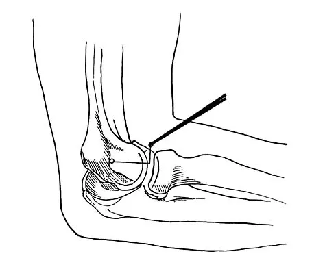
What nerve is most at risk during placement of the anterolateral portal in elbow arthroscopy?


Explanation

The radial nerve is only 4 mm from the anterolateral portal, while the median nerve is 11 mm away from the anteromedial portal. The ulnar nerve is only at risk on the medial side of the elbow. Anterior and posterior interosseous nerves are more distal within the forearm and are not in danger during portal placement. O'Driscoll SW, Morrey BF: Elbow arthroscopy, in Morrey BF (ed): The Elbow. Philadelphia, PA, Lippincott, Williams and Wilkins, 1994, pp 21-34.

Question 2

A 39-year-old man has anterior shoulder pain after landing on his abducted left shoulder while playing softball. Examination reveals a stable glenohumeral joint, pain on passive external rotation of greater than 25 degrees, and pain and weakness on belly press (Napoleon's) test. An MRI scan is shown in Figure 32. To provide maximum pain relief and return of function, management should include


Explanation

The examination and MRI scan confirm a subscapularis rupture and dislocation of the long head of the biceps tendon. The greatest return of function will result from repair of the subscapularis and tenodesis of the biceps tendon. Physical therapy alone will result in inadequate healing of the subscapularis and will not address the biceps tendon. While biceps tenotomy is an option, it will not provide the same level of pain relief and return of function as a tenodesis in a young, active man. There is no evidence for a supraspinatus tear. Deutsch A, Altchek DW, Veltri DM, Potter HG, Warren RF: Traumatic tears of the subscapularis tendon: Clinical diagnosis, magnetic resonance imaging findings, and operative treatment. Am J Sports Med 1997;25:13-22.

Question 3

A 37-year-old racquet player had dominant shoulder pain for 1 year, and cortisone injections provided only temporary relief. Because MRI findings did not reveal a rotator cuff tear, he underwent arthroscopic treatment including subacromial decompression and spur removal below the distal clavicle. Three years following surgery, he now reports that the pain has returned. What is the most likely cause of his pain?


Explanation

Co-planing the distal clavicle may lead to painful acromioclavicular joints in up to 35% of patients; this is felt to be related to destabilizing the distal clavicle. Intra-articular diagnosis of synovitis, degenerative joint disease, and superior labrum anterior and posterior lesions would have been identified at initial arthroscopy (not necessarily seen in open surgery). Ganglions are seen on MRI. Fischer BW, Gross RM, McCarthy JA: Incidence of acromioclavicular joint complications after arthroscopic subacromial decompression. Arthroscopy 1999;15:241-248. Hazel RM, Tasto JP, Klassen J: Arthroscopic subacromial decompression: A 9-year follow-up. Arthroscopy 1998;14:419.

Question 4

Figure 33 shows the radiograph of a 28-year-old avid golfer who has chronic right wrist pain. Management should consist of


Explanation

The patient's chronic symptoms are associated with a fracture of the base of the hook of the hamate; therefore, the treatment of choice is simple excision of the fracture fragment, with reasonable expectations of functional return. Acute fractures may be difficult to treat because of the high incidence of nonunion, but once nonunion is discovered, nonsurgical management usually is unsuccessful. Bone grafting may be a surgical alternative, but successful outcomes with percutaneous fixation or trephination of the fibrous union have not been reported. Geissler WB: Carpal fractures in athletes. Clin Sports Med 2001;20:167-188.

Question 5

A 28-year-old hockey player has a shoulder deformity after being checked into the boards. Examination reveals that swelling has improved, but there is tenderness along the distal clavicle. Radiographs reveal a grade II acromioclavicular joint separation. Initial management should consist of


Explanation

The most common shoulder injury in hockey players is to the acromioclavicular joint. Early rest and control of pain and inflammation is the preferred management. Surgery is reserved for patients with significant coracoclavicular disruption that has failed to respond to nonsurgical management. Cross-chest stretches and overhead exercises may increase symptoms. A cortisone injection within the glenohumeral joint will have little effect. Nuber GW, Bowen MK: Acromioclavicular joint injuries and distal clavicle fractures. J Am Acad Orthop Surg 1997;5:11-18.

Question 6

Which of the following best describes athletic pubalgia?


Explanation

Athletic pubalgia refers to a distinct syndrome of lower abdominal and adductor pain that is mostly commonly seen in high performance male athletes. This condition must be distinguished from others such as painful inflammation of the symphysis pubis, referred to as osteitis pubis. Symptoms attributable to the iliopsoas tendon are most commonly associated with snapping of the tendon. Stress fracture of the pubic ramus may cause symptoms in this area, but it is usually confirmed by imaging studies. Neurapraxia of the pudendal nerve is associated with pressure from the seat in cycling sports and also as a complication associated with traction during surgical procedures. Meyers WC, Foley DP, Garrett WE, Lohnes JH, Mandlebaum BR: Management of severe lower abdominal or inguinal pain in high-performance athletes: PAIN (Performing Athletes with Abdominal or Inguinal Neuromuscular Pain Study Group). Am J Sports Med 2000;28:2-8.

Question 7

Figures 34a and 34b show the radiographs of a 28-year-old man who fell on his outstretched arm with significant force while mountain biking. The nerve deficit most likely to occur would result in weakness of


Explanation

34b The patient has a Monteggia fracture-dislocation (proximal ulnar fracture and radial head dislocation). The posterior interosseous nerve branch of the radial nerve is the most likely to be injured and could result in weakness of thumb extension and finger metacarpal extension. Considerably less likely are injuries to the more proximal radial nerve branches supplying the extensor carpi radialis longus and brevis, resulting in weak wrist extension; the ulnar nerve supplying the digital intrinsics, resulting in weak finger abduction; the anterior interosseous branch of the median nerve, resulting in weakness of the flexor pollicis longus; and the distal median nerve, resulting in weakness of thumb opposition. Bado JL: The Monteggia lesion. Clin Orthop 1967;50:71-86.

Question 8

Which of the following activities can improve posterior capsular contractures?


Explanation

Posterior capsule stretching is performed in the cross-chest and behind the back positions. Stretching in internal rotation in the abducted shoulder will further stretch the posterior capsule. Wide grip stretch, and anterior capsule and strengthening exercises will not necessarily stretch the capsule. Ellenbacher TS: Shoulder internal and external rotation strength and range of motion of highly-skilled junior tennis players. Isokinetic Exercise Sci 1992;2:1-8.

Question 9

Figure 35 shows the radiograph of a 35-year-old weightlifter who has had pain with overhead lifts for the past 7 months. Cortisone injections in the acromioclavicular joint provided only temporary relief. A bone scan reveals increased activity of the acromioclavicular joint. Treatment should now consist of


Explanation

Osteolysis of the distal clavicle is common in weightlifters; therefore, distal clavicle excision is the treatment of choice. A subacromial decompression alone would not alleviate the acromioclavicular joint symptoms. Interval closure, biceps degeneration, and superior labrum anterior and posterior repair would limit superior migration but would not explain the abnormal bone scan. Thermal capsular shrinkage does not have a role here. Flatow EL, Cordasco FA, McCluskey GM, Bigliani LU: Arthroscopic resection of the distal clavicle via a superior portal: A critical quantitative radiographic assessment of bone removal. Arthroscopy 1990;6:153-154.

Question 10

Following reconstruction of the anterior cruciate ligament (ACL), which of the following rehabilitation exercises has the greatest potential to harm the graft?


Explanation

Isometric quadriceps contraction between 15 and 30 degrees of flexion creates significant strain in the ACL and potential damage to the reconstructed graft. Isolated quadriceps contraction with knee flexion of greater than 60 degrees, hamstring contraction at any angle of knee flexion, and active knee motion between 35 and 90 degrees of flexion create substantially less strain in the properly implanted ACL graft. Beynnon BD, Gleming BC, Johnson RL, Nichols CE, Renstrom PA, Pope MH: Anterior cruciate ligament strain behavior during rehabilitation exercises in vivo. Am J Sports Med 1995;23:24-34.

Question 11

A young active patient with a complete isolated posterior cruciate ligament (PCL) tear undergoes a double bundle PCL reconstruction. The tensioning pattern of the anterolateral (AL) and posteromedial (PM) bundles most likely to reproduce the most normal knee kinematics would be to tension


Explanation

During flexion and extension of the normal knee, the AL bundle of the PCL is taut in flexion, and the PM bundle is taut when the knee is near extension. The AL bundle is approximately two times larger at its midsubstance, stiffer, and has a higher ultimate load than the PM bundle. In vitro testing has demonstrated that by tensioning the AL bundle at 90 degrees of flexion and the PM bundle at 0 degrees of flexion, essentially normal knee kinematics are restored. Tensioning the AL bundle at 45 degrees of flexion and the PM bundle at 0 degrees of flexion would result in increased laxity with flexion at 90+ degrees. Tensioning the AL bundle at 90 degrees of flexion and the PM bundle at 45 degrees of flexion would result in increased laxity near extension. Harner CD, Janaushek MA, Kanamori A, Yagi M, Vogrin T, Woo SL: Biomechanical analysis of a double-bundle posterior cruciate ligament reconstruction. Am J Sports Med 2000;28:144-151.

Question 12

Accurate evaluation of the upper portion of the subscapularis muscle is best accomplished with active internal rotation


Explanation

Internal rotators of the shoulder include the subscapularis, pectoralis major, teres major, and latissimus dorsi muscles. The subscapularis has two portions, with the upper portion receiving its innervation from the upper subscapular nerve (C5) and the lower portion from the lower subscapular nerve (C5-6). The two tests commonly performed to isolate the internal rotation to the subscapularis muscle are the lift-off test and the belly press test. Electromyographic findings have shown the lift-off test to be more accurate for the lower portion of the subscapularis and the belly press test to be more sensitive for the upper portion. Hintermeister RA, Lange GW, Schultheis JM, Bey MJ, Hawkins RJ: Electromyographic activity and applied load during shoulder rehabilitation exercises using elastic resistance. Am J Sports Med 1998;26:210-220.

Question 13

During what phase of the throwing motion is the highest torque measured across the glenohumeral joint?


Explanation

Electromyography is used to evaluate muscular firing patterns about the shoulder during the throwing sequence. The rotator cuff muscles and biceps are relatively inactive during the acceleration phase, whereas the pectoralis major, serratus anterior, latissimus dorsi, and subscapularis show highest activity. By contrast, deceleration is accomplished by the rotator cuff musculature and the larger trunk muscles acting in concert to slow down the arm. It is during this phase of follow through that the highest torque is measured secondary to eccentric muscle contraction. Jobe FW, Moynes DR, Tibone JE, Perry J: An EMG analysis of the shoulder in pitching: A second report. Am J Sports Med 1984;12:218-220. Pappas AM, Zawacki RM, Sulliva TJ: Biomechanics of baseball pitching: A preliminary report. Am J Sports Med 1985;13:216-222.

Question 14

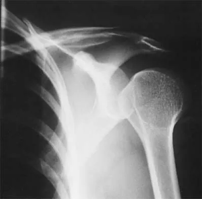
Figure 36 shows the radiograph of a 28-year-old man who injured his shoulder in a motocross race. Management should consist of


Explanation

Fractures of the distal one third of the clavicle have a high incidence of delayed union (45% to 67%) and nonunion (22% to 33%) with nonsurgical management. Surgical stabilization with tension band techniques or a combination of plate and screw techniques is indicated, especially in young, active patients. In this patient, significant displacement of the fracture implies injury to the coracoclavicular ligaments with a higher risk of delayed union or nonunion. Various surgical treatments have been recommended, but the use of smooth wires is not indicated because of the potential for hardware migration. Jupiter JB, Ring D: Fractures of the clavicle, in Ianotti JP, Williams GR (eds): Disorders of the Shoulder: Diagnosis and Management, ed 1. Philadelphia, PA, Lippincott, Williams and Wilkins, 1999, pp 709-736.

Question 15

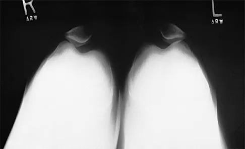
Figure 37 shows the radiograph of a 21-year-old collegiate basketball player who has had mild midfoot aching for the past 4 months. What is the best course of action?


Explanation

A stress fracture of the navicular is considered a high-risk injury because of the incidence of nonunion. If identified early, cast immobilization with no weight bearing is appropriate. However, this patient is a high-caliber athlete who has had symptoms for 4 months. Therefore, surgery is recommended to expedite recovery and optimize the chance of healing. Meyer SA, Saltaman CL, Albright JP: Stress fractures of the foot and ankle. Clin Sports Med 1993;12:395-413. Kahn JM, Fuller PJ, Burkner PD, et al: Outcome of conservative and surgical management of navicular stress fractures in athletes: Eighty-six cases proven with computerized tomography. Am J Sports Med 1992;20:657-666.

Question 16

A 17-year-old high school gymnast who has peripatellar knee pain has been unable to practice on a consistent basis for the past 3 years. She denies any specific injury events. Physical therapy for modalities, quadriceps strengthening, and hamstring stretching provide temporary relief. A trial of patellar taping significantly reduces her pain. Examination reveals an 15-degree Q angle, moderate lateral facet tenderness, negative patellar apprehension, and the inability to evert the patella. Radiographs show a moderate lateral patellar tilt. Treatment should now consist of


Explanation

The patient has patellofemoral stress and a tight lateral retinaculum that has failed to respond to nonsurgical management; therefore, the most appropriate treatment includes an arthroscopic lateral retinacular release. A patellar restraining brace may aggravate the peripatellar pain by increasing pressure on the lateral facet. There is no evidence of patellar instability or significant malalignment; therefore, medial retinacular repair or a tibial tubercle transfer is not indicated. A modified Maquet tibial tubercle elevation would be considered only for significant patellofemoral arthrosis. Gambardella RA: Techical pitfalls of patellofemoral surgery. Clin Sports Med 1999;18:897-903.

Question 17

An active 55-year-old man who felt a sudden pop in the left heel while playing tennis 6 months ago was diagnosed with an ankle sprain around the time of injury. He now reports calf atrophy and severe weakness with running. Examination reveals a palpable defect in the Achilles tendon and only trace passive ankle flexion when the calf is squeezed. At the time of surgery, an Achilles tendon defect of 6 cm cannot be approximated. Surgical management of the Achilles tendon should include


Explanation

Chronic or neglected Achilles tendon ruptures can present a surgical problem. Ideally, end-to-end apposition of tendon should be attempted, but this should be accomplished without placing the foot in marked equinus. A defect of greater than 5 cm requires the use of a tendon transfer either alone or in combination with a V-Y advancement of the gastrocnemius. Because of its proximity to the Achilles tendon and its strength as a plantar flexor, the flexor hallucis longus is an ideal choice for this task. Studies have shown that early active range-of-motion exercises after an Achilles tendon repair is beneficial for tendon healing and improved clinical outcomes. Myerson M: Achilles tendon ruptures. Instr Course Lect 1999;48:219-230.

Question 18

Figures 38a and 38b show the AP and lateral radiographs of a 12-year-old baseball pitcher who has pain in his right dominant elbow. Management should consist of


Explanation

38b The patient has a stress fracture of the olecranon; therefore, the most appropriate management is modification of activities below the threshold of symptoms to allow for healing. Ultrasound provides no benefit, and immobilization is not necessary. MRI is not necessary because there is no associated ligamentous injury. Arthroscopy is not indicated. Cabanela ME, Morrey BF: Fractures of the olecranon, in Morrey BF (ed): The Elbow and Its Disorders. Philadelphia, PA, WB Saunders, 2000, pp 365-379.

Question 19

What is the most reproducible landmark for the accurate anatomic placement of the tibial tunnel for an anterior cruciate ligament (ACL) reconstruction?


Explanation

The anterior border of the PCL is the most accurate and reproducible landmark for appropriate placement of the tibial tunnel for an ACL reconstruction. The central sagittal insertion point of the ACL is consistently 10 to 11 mm anterior to the anterior border of the PCL ligament. The anterior border of the tibia is not well visualized and does not serve as a reference point. While the posterior border of the anterior horn of the lateral meniscus could be used as a reference point, it has twice the variability of the PCL reference point. The posterior border of the tibia is difficult to identify and has greater variability than the PCL relative to the AP dimension of the proximal tibial surface. The anterior horn of the medial meniscus is also more variable than the PCL. Hutchinson MR, Bae TS: Reproducibility of anatomic tibial landmarks for anterior cruciate ligament reconstructions. Am J Sports Med 2001;29:777-780.

Question 20

A 20-year-old football player sustains a dorsiflexion external rotation injury to his right ankle. During sideline evaluation, which of the following findings best indicates a syndesmosis ankle sprain without diastasis?


Explanation

The inability to single leg hop is considered the best indicator of a syndesmosis ankle sprain without diastasis. Tenderness along the syndesmosis, the deltoid, or over the anterior talofibular ligament or anterior distal tibia/fibula may present later, following the initial injury. The squeeze test and tenderness with dorsiflexion and external rotation may be positive but often are not present initially. The best determinant for prediction of return to play is the amount of tenderness along the syndesmosis, measured from the distal fibula up the syndesmosis. Nussbaum ED, Hosea TM, et al: Prospective evaluation of syndesmosis ankle sprains without diastasis. Am J Sports Med 2001;29:31-35. Miller CD, Shelton WR, Barrett GR, et al: Deltoid and syndesmosis ligament injury of the ankle without fracture. Am J Sports Med 1985;23:746-750.

Question 21

A 20-year-old college baseball pitcher reports the insidious onset of medial elbow pain. Examination reveals medial elbow tenderness, a normal neurologic examination, and no obvious valgus laxity. Plain radiographs are normal. MRI scans are shown in Figures 39a and 39b. Management should consist of


Explanation

39b Throwers and in particular, pitchers, are prone to high valgus loads to the elbow. A constellation of medial elbow pathology can develop, including medial epicondylitis, ulnar nerve neuritis, medial ulnar collateral ligament injuries, and posteromedial osteophytes of the olecranon. The MRI scans show significant increases in signal intensing as well as fiber disruption of the medial collateral ligament, indicating a complete tear. The common flexor origin shows a homogeneous signal and normal morphology. Therefore, excision of posterior osteophytes and debridement of the common flexor origin are not indicated. Likewise, this patient's symptoms do not indicate ulnar nerve pathology; therefore ulnar nerve transposition is not indicated. Primary repair of medial collateral ligament tears of the elbow lead to unpredictable results with an unacceptable rate of reoperation. The most predictable result in treating this high-demand athlete is reconstruction of the medial collateral ligament with autogenous tissue. Norris TR (ed): Athletic Injuries of the Elbow. Rosemont, IL, American Academy of Orthopaedic Surgeons, 1997, pp 311-323.

Question 22

What is the most common arthroscopic finding of internal impingement in an overhead athlete?


Explanation

Internal impingement occurs when the articular side of the supraspinatus abrades against the posterior superior glenoid in the cocking position. Damage may include a posterior labral tear where the contact occurs, not anteriorly as in a Bankart lesion. Biceps fraying and acromion spurs are more commonly seen in extrinsic impingement. Loose bodies may occur from multiple lesions associated with instability and articular cartilage disorders but are uncommon in internal impingement. Jobe CM: Posterior superior impingement of the rotator cuff on the glenoid rim as a cause of shoulder pain in the overhead athlete. Arthroscopy 1993;9:697-699.

Question 23

A 16-year-old high school football player who sustained an acute forceful dorsiflexion ankle injury reported that he felt a pop and then noted immediate swelling over the lateral malleolus. Examination 24 hours later reveals moderate swelling and tenderness along the lateral malleolus. The external rotation, squeeze, anterior drawer, and talar tilt tests are negative. Subluxation of the peroneal tendons is palpable over the peroneal groove of the fibula. Radiographs reveal a small cortical avulsion off the distal rim of the fibula. The stress views show no instability. Initial management for this injury should include


Explanation

The patient has an acute peroneal tendon dislocation. The evaluation for syndesmotic injury and lateral ankle instability is negative. The cortical avulsion off the distal tip of the lateral malleolus, a rim fracture, is characteristic of peroneal tendon dislocations. The sensation of apprehension or frank subluxation of the peroneal tendons with active dorsiflexion of the foot while the foot is held in plantar flexion confirms the diagnosis. Based on these findings, initial management should consist of cast immobilization and protected weight bearing. If a recurrent or chronic condition develops, surgery is the most reliable treatment option. Arrowsmith SR, Fleming LL, Allman FL: Traumatic dislocations of the peroneal tendons. Am J Sports Med 1983;11:142-146.

Question 24

The essential lesion responsible for posterolateral rotatory instability of the elbow is disruption of the


Explanation

Posterolateral rotatory instability (PLRI) of the elbow represents a three-dimensional injury pattern of rotational displacement of the ulna from the trochlea and the radius from the capitellum. The ulna supinates (externally rotates) past its normal limit and the radiocapitellar joint subluxates posterolaterally, permitting the coronoid process to slide beneath the trochlea. In cadaver studies, the lateral ulnar collateral ligament has been shown to be the essential lesion responsible for PLRI. The medial collateral ligament (of which the anterior bundle is the most important) is the primary restraint to valgus instability. The posterolateral capsule and radial collateral ligament may be disrupted in a complete posterolateral dislocation but are not essential injuries for PLRI. The primary function of the annular ligament is to stabilize the proximal radioulnar joint. O'Driscoll SW, Jupiter JB, King GJW, Hotchkiss RN, Morrey BF: The unstable elbow. J Bone Joint Surg Am 2000;82:724-738. Olsen BS, Sojbjerg JO, Dalstra M, Sneppen O: Kinematics of the lateral constraints of the elbow. J Shoulder Elbow Surg 1996;5:333-341.

Question 25

When evaluating articular cartilage, what extracellular matrix component is most closely associated with the deep calcified cartilage zone?


Explanation

Collagen type X is produced only by hypertrophic chondrocytes during enchondral ossification (growth plate, fracture callus, heterotopic ossification) and is associated with calcification of cartilage in the deep zone of articular cartilage. Collagen type I is the predominant collagen in bone, ligament, and tendon. Collagen type II is the predominant collagen in articular cartilage. Proteoglycan aggrecan and hyaluronic acid are components of the extracellular matrix and are involved in the compressive strength characteristics of articular cartilage. Buckwalter JA, Mankin HJ: Articular cartilage: Tissue design and chondrocyte matrix interactions. Instr Course Lect 1998;47:477-486.

Question 26

A 19-year-old female presents with recurrent patellar dislocations. Imaging shows a tibial tubercle-trochlear groove (TT-TG) distance of 14 mm and a normal Insall-Salvati ratio. An isolated medial patellofemoral ligament (MPFL) reconstruction is planned.

What is the primary biomechanical consequence of placing the femoral tunnel for the MPFL graft significantly proximal to Schöttle's point?


Explanation

The MPFL is the primary restraint to lateral patellar translation from 0 to 30 degrees of flexion. Proper femoral tunnel placement (Schöttle's point) is critical for graft isometry. If the femoral tunnel is placed too proximal, the distance between the patellar and femoral attachments increases as the knee flexes. This non-isometric placement causes the graft to become excessively tight in flexion, leading to medial patellofemoral cartilage overload, restricted range of motion (loss of flexion), and potentially iatrogenic medial patellar subluxation or increased risk of early osteoarthritis.

Question 27

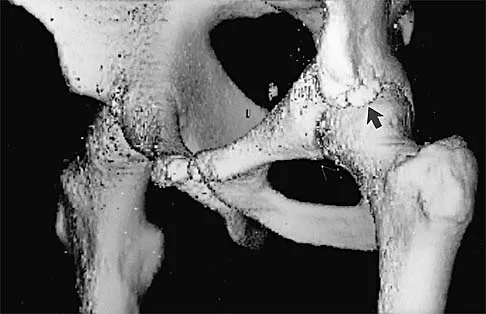
A 28-year-old hockey player undergoes hip arthroscopy for a symptomatic CAM-type femoroacetabular impingement and a focal anterosuperior labral tear. The anterolateral (AL) portal is established first under fluoroscopic guidance. To safely establish the mid-anterior (MAP) portal, the surgeon must remain lateral to the sagittal plane of the anterior superior iliac spine (ASIS). Deviating medial to this plane primarily increases the risk of injury to which of the following structures?


Explanation

During hip arthroscopy, the mid-anterior portal (MAP) is typically placed 5 to 7 cm distal to the anterolateral portal at approximately a 45-degree angle. The lateral femoral cutaneous nerve (LFCN) is at greatest risk during the establishment of the anterior portals. Staying lateral to the sagittal plane extending distally from the anterior superior iliac spine (ASIS) helps minimize the risk of injuring the LFCN, which typically courses medial to this plane. The femoral nerve and artery are located further medially within the femoral triangle.

Question 28

A 21-year-old male football player undergoes primary ACL reconstruction with a bone-patellar tendon-bone autograft. Two years later, he presents with an atraumatic graft rupture. Standing lateral radiographs demonstrate a posterior tibial slope (PTS) of 16 degrees. If a revision ACL reconstruction is performed without addressing the bony anatomy, what biomechanical alteration is most responsible for an increased risk of early graft failure?


Explanation

An increased posterior tibial slope (typically >12-13 degrees is considered abnormal and highly clinically relevant) significantly increases the anterior shear force on the tibia during axial loading (weight-bearing). This increased anterior directed force places higher stress on the native ACL or an ACL graft, predisposing the patient to failure of the reconstruction. In revision scenarios with extreme posterior tibial slope (>12-14 degrees), an anterior closing wedge high tibial osteotomy may be indicated to decrease the slope and protect the revision graft.

Question 29

A 62-year-old laborer presents with chronic, debilitating shoulder pain and pseudoparalysis. MRI reveals a massive, retracted, irreparable tear of the supraspinatus and infraspinatus with Grade 4 fatty infiltration. The subscapularis and teres minor are intact. He undergoes an arthroscopic superior capsular reconstruction (SCR) using a thick dermal allograft.

Biomechanically, how does the SCR primarily restore shoulder kinematics in this specific clinical scenario?


Explanation

Superior capsular reconstruction (SCR) is designed to address massive, irreparable posterosuperior rotator cuff tears. Biomechanically, it functions primarily as a static restraint. By rigidly attaching a graft from the superior glenoid (replacing the native superior capsule) to the greater tuberosity footprint, the SCR acts as a tether that depresses the humeral head and resists superior migration during deltoid activation. This statically restores the coronal plane force couple, allowing the intact deltoid and remaining rotator cuff to elevate the arm more effectively.

Question 30

A 22-year-old collegiate baseball pitcher presents with posteromedial elbow pain and a noticeable decrease in throwing velocity over the last two months. He describes a severe 'catching' and 'locking' sensation specifically in the deceleration phase of throwing. Physical examination reveals a 15-degree flexion contracture and sharp pain in the posteromedial olecranon fossa when forced terminal extension is applied concurrently with a valgus stress. The milking maneuver is negative. What is the most likely diagnosis?


Explanation

Valgus extension overload (VEO) syndrome in overhead athletes results from chronic, repetitive valgus stress and extension forces. This leads to posterior radiocapitellar compression and traction/shear forces on the medial olecranon tip as it impinges within the olecranon fossa. Patients characteristically present with posteromedial pain, a flexion contracture, and pain on forced terminal extension with valgus stress (the moving valgus stress test may also be positive, but specifically terminal extension pain points to impingement/osteophytes). The 'catching' in the deceleration phase is classic for VEO, often secondary to posteromedial olecranon osteophytes or loose bodies.

Question 31

A 55-year-old female presents with acute medial knee pain following a squatting maneuver. MRI reveals a complete radial tear directly adjacent to the posterior root attachment of the medial meniscus, with associated meniscal extrusion of 4 mm.

Which of the following best describes the biomechanical consequence of leaving this specific root injury unaddressed?


Explanation

The posterior roots of the menisci are critical for anchoring the meniscus and allowing it to convert axial joint loads into hoop stresses. A posterior medial meniscal root tear disrupts this structural continuity, resulting in meniscal extrusion and complete loss of hoop stress generation. Biomechanical studies have demonstrated that an unaddressed posterior medial meniscal root tear results in contact areas and peak contact pressures that are virtually indistinguishable from a total medial meniscectomy, rapidly leading to accelerated articular cartilage degeneration.

Question 32

A 24-year-old rugby player presents for management of recurrent anterior shoulder instability. He has had four dislocations. Computed tomography (CT) with 3D sagittal reconstruction demonstrates an anteroinferior glenoid bone loss of 22% and a large, engaging Hill-Sachs lesion. Based on current literature and evidence-based treatment algorithms, which of the following is the most appropriate surgical management?


Explanation

In the setting of recurrent anterior shoulder instability with critical glenoid bone loss (typically cited as >15-20% depending on the functional demands) and an engaging Hill-Sachs lesion (off-track lesion), isolated soft tissue repairs (like arthroscopic Bankart) have an unacceptably high failure rate. The Latarjet procedure (transfer of the coracoid process with the attached conjoined tendon to the anterior glenoid) provides a triple blocking effect (bone augmentation, sling effect of the conjoined tendon on the lower subscapularis, and capsular repair) and is the gold standard for subcritical/critical bone loss in collision athletes.

Question 33

A 14-year-old male gymnast with open physes presents with chronic right knee pain. MRI reveals a 2 x 2 cm osteochondritis dissecans (OCD) lesion on the lateral aspect of the medial femoral condyle.

The articular cartilage is intact, and there is no high T2 signal fluid behind the fragment, indicating a stable lesion. He has failed 6 months of strict non-operative management including restricted weight-bearing. What is the most appropriate next step in management?


Explanation

For a stable juvenile osteochondritis dissecans (JOCD) lesion (intact articular cartilage, no fluid behind the fragment on MRI) that has failed a prolonged course (typically 3-6 months) of non-operative management, arthroscopic drilling is the treatment of choice. Drilling penetrates the sclerotic margin of the lesion to promote vascular ingrowth and healing of the osteochondral fragment. Restorative procedures like OATS or ACI are reserved for unstable lesions, unsalvageable fragments, or large full-thickness defects.

Question 34

During an open subpectoral biceps tenodesis, the surgeon creates a cortical window in the bicipital groove just distal to the transverse humeral ligament. However, due to extensive fraying of the long head of the biceps tendon, the incision and deep dissection are extended further distally along the humeral shaft. If medial retractors are placed too aggressively deep to the conjoined tendon and biceps muscle belly during this distal extension, which neurovascular structure is at greatest risk of iatrogenic injury?


Explanation

During an open subpectoral biceps tenodesis, the musculocutaneous nerve is the primary neural structure at risk. It classically pierces the coracobrachialis approximately 5 to 8 cm distal to the coracoid process and courses distally between the biceps brachii and the brachialis muscles. Aggressive medial retraction, especially when the approach is extended distally beneath the conjoined tendon and biceps muscle belly, places excessive traction on or risks direct injury to the musculocutaneous nerve.

Question 35

A 26-year-old man sustains a dashboard injury resulting in an isolated posterior cruciate ligament (PCL) tear. Following failure of non-operative management, a single-bundle PCL reconstruction is planned. To accurately reproduce the biomechanics of the primary restraint to posterior tibial translation at 90 degrees of flexion, the graft should be placed to reconstruct which specific bundle, and where is its native femoral footprint located?


Explanation

The PCL consists of two main bundles: the larger anterolateral (AL) bundle and the smaller posteromedial (PM) bundle. The AL bundle is tight in flexion (the primary restraint to posterior translation at 90 degrees) and is the bundle reconstructed in a single-bundle PCL reconstruction. Its native femoral footprint is located on the lateral aspect of the medial femoral condyle. Specifically, it is positioned shallow (anterior in the notch) and superior (proximal, near the notch roof) relative to the articular margin.

Question 36

During an anatomic single-bundle anterior cruciate ligament (ACL) reconstruction, the femoral tunnel is drilled through the accessory anteromedial portal. Compared to traditional drilling through a transtibial portal, what is the primary biomechanical advantage of this technique?


Explanation

Anatomic ACL femoral tunnel placement (drilled lower on the lateral notch wall into the native footprint) better restores rotational stability and resists the pivot shift compared to the traditional high, vertical placement often achieved with the transtibial technique. The more horizontal graft orientation obtained via the anteromedial portal significantly improves rotational control.

Question 37

A 25-year-old male sustains a multiligament knee injury. Examination reveals a grade 3 posterior sag and grade 3 varus opening in full extension and at 30 degrees of flexion. The dial test shows increased external rotation at 30 degrees but is symmetric at 90 degrees. He is planned for PCL and posterolateral corner (PLC) reconstruction.

What anatomic structure of the PLC is the primary restraint to varus gapping at 30 degrees of knee flexion?


Explanation

The fibular collateral ligament (LCL) is the primary restraint to varus stress at 30 degrees of knee flexion. The popliteus tendon and the popliteofibular ligament act as the primary restraints to external rotation. In a complete PLC reconstruction, restoring the LCL is critical for coronal plane (varus) stability.

Question 38

A 65-year-old male presents with pseudoparalysis of the shoulder. An MRI reveals an irreparable, chronically retracted tear of the subscapularis tendon with significant fatty infiltration (Goutallier stage 4). The posterosuperior cuff is intact. He is scheduled to undergo a pectoralis major transfer. Which portion of the pectoralis major is typically transferred to best replicate the force vector of the native subscapularis?


Explanation

For irreparable subscapularis tears, transferring the sternal head of the pectoralis major, routed deep (posterior) to the conjoint tendon, most closely recreates the line of pull of the native subscapularis muscle and acts to stabilize the anterior joint and restore internal rotation function.

Question 39

A 28-year-old professional hockey player presents with chronic anterior groin pain exacerbated by hip flexion and internal rotation. Radiographs demonstrate a pistol-grip deformity of the proximal femur.

Which of the following radiographic parameters is most diagnostic of a Cam-type femoroacetabular impingement (FAI)?


Explanation

An alpha angle greater than 50-55 degrees (often measured on a lateral or Dunn view) is indicative of a decreased femoral head-neck offset characteristic of Cam impingement. A lateral center edge angle > 40 degrees and a positive cross-over sign (acetabular retroversion) are findings associated with Pincer-type impingement.

Question 40

A 14-year-old female gymnast presents with progressive lateral elbow pain and mechanical catching. Imaging reveals an unstable 1.5 cm osteochondral defect (OCD) of the capitellum with loose bodies in the joint. The lateral radiocapitellar ligament is intact. Which surgical treatment provides the best long-term outcome and highest rate of return to sport for an unstable defect of this size in a high-demand athlete?


Explanation

In adolescent overhead athletes and gymnasts, large (>1 cm) and unstable capitellar OCD lesions have poor results with microfracture or debridement alone. Osteochondral autograft transfer (OATS), typically harvested from the lateral femoral condyle, provides the highest rate of return to high-demand sports and superior long-term functional outcomes.

Question 41

A 50-year-old female experiences a sudden "pop" in the posterior aspect of her knee while descending stairs. She is diagnosed with a complete medial meniscus posterior root tear with 3 mm of extrusion on MRI. Biomechanically, what is the direct consequence of leaving this tear untreated compared to the native knee?


Explanation

A complete posterior root tear of the medial meniscus leads to an immediate loss of circumferential hoop stresses, which effectively renders the meniscus nonfunctional (meniscal extrusion). Biomechanically, this is equivalent to a total medial meniscectomy, leading to significantly increased peak contact pressures and accelerated onset of osteoarthritis.

Question 42

A 45-year-old recreational weightlifter presents with deep anterior shoulder pain. An MRI arthrogram demonstrates a Type II SLAP tear without rotator cuff pathology. A trial of physical therapy and injections has failed to provide relief. What is the most appropriate surgical management for this patient to minimize postoperative stiffness and maximize return to pre-injury activity?


Explanation

In patients over the age of 35-40, SLAP repair is associated with a significantly higher risk of postoperative stiffness, persistent pain, and lower rates of return to sport compared to biceps tenodesis. Biceps tenodesis is currently the preferred surgical treatment for symptomatic Type II SLAP tears in older or middle-aged patients.

Question 43

A 19-year-old female is undergoing a medial patellofemoral ligament (MPFL) reconstruction for recurrent lateral patellar instability. Correct placement of the femoral tunnel is essential to avoid over-constraining the joint. Which of the following landmarks accurately describes the anatomic femoral footprint of the native MPFL?


Explanation

The anatomic femoral footprint of the MPFL is located in the saddle-shaped depression between the adductor tubercle (proximal) and the medial epicondyle (distal). Non-anatomic placement, particularly too proximal and anterior, increases graft tension in flexion, leading to stiffness, graft failure, or elevated patellofemoral contact pressures.

Question 44

A 25-year-old male cyclist falls directly onto his right shoulder. Clinical examination reveals a prominent clavicle, and radiographs confirm a 120% superior displacement of the distal clavicle relative to the acromion, with an increased coracoclavicular distance. He is diagnosed with a Type III acromioclavicular (AC) joint separation. According to current evidence, what is the recommended initial management for this injury in a non-laboring individual?


Explanation

Type III AC joint separations (100% to 250% superior displacement, though older classifications said 100% displacement) are generally treated nonoperatively initially, especially in non-overhead athletes and non-laborers, as long-term functional outcomes are equivalent to surgery with fewer complications. Surgery is reserved for patients who fail nonoperative treatment or, controversially, for elite overhead athletes and heavy laborers.

Question 45

A 32-year-old male suffers an acute Achilles tendon rupture while playing basketball. He elects to pursue nonoperative management. Compared to traditional prolonged cast immobilization, what is the primary advantage of employing an early functional rehabilitation protocol (early weight-bearing in an orthosis with functional ROM)?


Explanation

Recent high-quality literature demonstrates that nonoperative management of acute Achilles tendon ruptures utilizing an early functional rehabilitation protocol has an equivalent re-rupture rate to operative management. It avoids surgical complications (infection, nerve injury) and provides better early functional outcomes and less muscle atrophy compared to traditional rigid cast immobilization.

Question 46

A 50-year-old female presents with acute-onset medial joint line pain and an effusion after squatting deeply. An MRI shows an extrusion of the medial meniscus on coronal views and an 'empty meniscus' or 'ghost sign' on a single sagittal cut. Figure 1

illustrates a similar pathology. Biomechanically, what is the direct consequence of this specific injury pattern if left untreated?


Explanation

The scenario describes a posterior medial meniscal root tear. The 'ghost sign' on sagittal MRI is classic for this injury. The meniscal roots are critical for anchoring the meniscus and converting axial loads into circumferential 'hoop stresses.' A complete root tear abolishes these hoop stresses, leading to meniscal extrusion. Biomechanical studies have shown that a posterior medial meniscal root tear results in a loss of contact area and an increase in peak contact pressures equivalent to a total medial meniscectomy, predisposing the patient to rapid-onset osteoarthritis.

Question 47

A 28-year-old professional hockey player undergoes hip arthroscopy for femoroacetabular impingement (FAI). The surgeon establishes an anterolateral portal, followed by an anterior portal. Which of the following describes the typical sensory deficit expected if the nerve most at risk during the placement of the anterior portal is iatrogenically injured?


Explanation

The anterior portal in hip arthroscopy places the lateral femoral cutaneous nerve (LFCN) at significant risk. The LFCN provides sensory innervation to the anterolateral thigh. To minimize risk to the LFCN, the anterior portal is typically established under direct arthroscopic visualization from the anterolateral portal, and the capsule is penetrated parallel to the femoral neck. Medial thigh sensation is supplied by the obturator nerve, while the lateral lower leg and foot are innervated by branches of the sciatic nerve.

Question 48

A 22-year-old collegiate baseball pitcher reports severe medial elbow pain and a recent drop in his fast pitch velocity. He experiences sharp pain during the late cocking and early acceleration phases of throwing. The moving valgus stress test is markedly positive. An MRI arthrogram demonstrates a high-grade partial tear of the ulnar collateral ligament (UCL). If conservative management fails and surgical reconstruction is planned, which anatomic bundle of the UCL must be targeted as it is the primary restraint to valgus stress at 90 degrees of flexion?


Explanation

The ulnar collateral ligament (UCL) of the elbow consists of anterior, posterior, and transverse bundles. The anterior bundle is the primary restraint to valgus stress of the elbow between 30 and 120 degrees of flexion, which includes the critical 90-degree position during the throwing motion. Consequently, it is the anterior bundle that is the primary focus of reconstruction during a 'Tommy John' surgery. The posterior bundle is a secondary restraint, and the transverse bundle contributes little to valgus stability.

Question 49

A 32-year-old competitive weightlifter feels a sudden tearing sensation and 'pop' in his anterior axilla while performing a one-rep max bench press. Physical examination reveals an asymmetrical loss of the anterior axillary fold, significant ecchymosis, and weakness in internal rotation and adduction. An MRI confirms a complete rupture of the pectoralis major tendon. During an open surgical repair, the surgeon must accurately reattach the tendon to its anatomic footprint. Which of the following accurately describes the insertion of the sternal head of the pectoralis major relative to the clavicular head?


Explanation

The pectoralis major tendon is composed of a sternal head and a clavicular head that twist 90 degrees before inserting onto the lateral lip of the bicipital groove of the humerus. Because of this twisting mechanism, the lower (sternal) fibers cross deep to the upper (clavicular) fibers and insert proximal to them. Therefore, the sternal head inserts proximal and deep relative to the clavicular head. Understanding this U-shaped footprint is crucial for anatomic repair.

Question 50

A 24-year-old rugby player sustains a blow to the anteromedial aspect of his fully extended right knee. He complains of posterolateral knee pain, giving way, and difficulty descending stairs. On examination, the dial test reveals 20 degrees of increased external rotation compared to the contralateral normal knee at 30 degrees of flexion, but symmetrical external rotation at 90 degrees of flexion. Based on these physical examination findings, which of the following injury patterns is most likely present?


Explanation

The dial test (tibial external rotation test) evaluates the integrity of the posterolateral corner (PLC) and the posterior cruciate ligament (PCL). Increased external rotation of greater than 10 degrees (compared with the normal side) at 30 degrees of knee flexion, but NOT at 90 degrees of flexion, is indicative of an isolated posterolateral corner (PLC) injury. Increased external rotation at both 30 and 90 degrees of flexion indicates a combined PCL and PLC injury.

Question 51

A 26-year-old professional baseball pitcher presents with a dull, aching posterior shoulder pain. Examination reveals 20 degrees of internal rotation and 130 degrees of external rotation of the dominant shoulder at 90 degrees of abduction. The contralateral shoulder has 60 degrees of internal rotation and 100 degrees of external rotation. Radiographs are unremarkable. What is the primary underlying anatomic pathology associated with this athlete's Glenohumeral Internal Rotation Deficit (GIRD)?


Explanation

Glenohumeral internal rotation deficit (GIRD) is a common condition in overhead throwing athletes, characterized by a loss of internal rotation (typically >20 degrees compared to the contralateral side) with a corresponding increase in external rotation. The primary underlying pathology is contracture of the posteroinferior joint capsule, often secondary to repetitive microtrauma during the deceleration phase of throwing. The first-line treatment consists of targeted posterior capsular stretching, such as the 'sleeper stretch'.

Question 52

A 14-year-old male cross-country runner presents with vague, intermittent anterior knee pain and mechanical catching for the past 3 months. Radiographs demonstrate an osteochondral lesion of the lateral aspect of the medial femoral condyle. He has widely open distal femoral physes. Figure 7

demonstrates the MRI findings, which reveal a 1.5 cm by 1.5 cm lesion with intact overlying cartilage and no high T2 fluid signal behind the fragment. What is the most appropriate initial management?


Explanation

The patient has juvenile osteochondritis dissecans (OCD) of the knee, typically located on the lateral aspect of the medial femoral condyle. Because the patient is skeletally immature (open physes) and the MRI demonstrates a stable lesion (no high T2 signal or fluid undermining the fragment, intact cartilage), there is a high potential for spontaneous healing. The most appropriate initial management is non-operative, consisting of rest, activity modification, and restriction from high-impact sports. Surgical intervention (drilling or fixation) is reserved for unstable lesions or failure of prolonged non-operative management.

Question 53

A 28-year-old cyclist sustains a severe type V acromioclavicular (AC) joint separation after being thrown over the handlebars. He fails conservative management and undergoes an anatomic coracoclavicular (CC) ligament reconstruction. To accurately recreate the biomechanics of the native ligaments, the surgeon must drill the clavicular tunnels mimicking the anatomic footprints. Which of the following accurately describes the anatomic orientation of the conoid and trapezoid ligaments?


Explanation

The coracoclavicular (CC) ligament complex consists of the conoid and trapezoid ligaments, which are the primary vertical restraints of the AC joint. The conoid ligament is cone-shaped and attaches to the conoid tubercle on the posteromedial aspect of the inferior clavicle. The trapezoid ligament is broad and attaches anterolaterally to the trapezoid ridge. Therefore, relative to each other, the conoid is medial and posterior, while the trapezoid is lateral and anterior. Recreating this footprint is essential during anatomic CC reconstructions.

Question 54

A 16-year-old female gymnast presents with a history of recurrent lateral patellar dislocations. She is indicated for a medial patellofemoral ligament (MPFL) reconstruction. To ensure proper isometry of the graft, the femoral tunnel must be placed at 'Schöttle/'s point'. Which of the following best describes the anatomic location of the MPFL origin on the femur?


Explanation

The medial patellofemoral ligament (MPFL) provides 50-60% of the restraining force against lateral patellar displacement from 0 to 30 degrees of knee flexion. Its anatomic femoral origin is located in a 'saddle' region situated between the medial epicondyle and the adductor tubercle. Non-anatomic placement of the femoral tunnel during reconstruction (most commonly placed too anterior or too proximal) leads to anisometry, causing increased patellofemoral pressures, graft stretching, or a block to flexion.

Question 55

A 23-year-old male underwent an anterior cruciate ligament (ACL) reconstruction using a bone-patellar tendon-bone autograft 6 months ago. He has aggressively participated in physical therapy but continues to complain of a painful mechanical block to terminal extension, lacking 10 degrees compared to the uninjured side. Figure 11

displays a sagittal MRI of his knee. What is the most likely diagnosis?


Explanation

The clinical presentation of a mechanical block to extension along with pain at terminal extension following ACL reconstruction is highly suspicious for a Cyclops lesion. This is a focal, nodular area of anterior arthrofibrosis that forms anterior to the ACL graft, physically blocking extension. It occurs in 1-10% of ACL reconstructions. Although an excessively anterior tibial tunnel can cause roof impingement and a block to extension, the discrete nodule visualized on MRI (often hyperintense on T2 anterior to the graft) defines a Cyclops lesion. Treatment consists of arthroscopic excision.

Question 56

A 22-year-old soccer player sustains a twisting knee injury. Radiographs demonstrate a small avulsion fracture of the lateral tibial plateau just distal to the articular surface (Segond fracture). Biomechanical studies demonstrate that the structure avulsed in this injury pattern primarily resists which of the following forces?


Explanation

The Segond fracture is a pathognomonic avulsion fracture of the anterolateral complex (specifically the anterolateral ligament [ALL] or anterolateral capsule). This injury is highly associated with an anterior cruciate ligament (ACL) tear. Biomechanically, the ALL acts as an important secondary stabilizer to internal tibial rotation, especially at higher flexion angles.

Question 57

During an open subpectoral biceps tenodesis, the conjoint tendon is retracted medially to optimize exposure of the bicipital groove and pectoralis major tendon. Overzealous medial retraction of the conjoint tendon is most likely to result in injury to which of the following nerves?


Explanation

The musculocutaneous nerve typically enters the coracobrachialis muscle approximately 5 to 8 cm distal to the coracoid process. During open subpectoral biceps tenodesis, retracting the conjoint tendon medially places the musculocutaneous nerve at significant risk of traction injury or direct transection if dissection strays too far medial.

Question 58

A 26-year-old male with symptomatic cam-type femoroacetabular impingement undergoes hip arthroscopy with osteochondroplasty. During resection of the cam lesion on the femoral neck, aggressive resection of the posterolateral aspect is avoided to prevent injury to the retinacular vessels. These critical vessels are terminal branches of which artery?


Explanation

The medial femoral circumflex artery (MFCA) gives off the retinacular vessels, which provide the primary and most critical blood supply to the adult femoral head. These vessels course along the posterosuperior and posteroinferior aspects of the femoral neck. Resection of the posterolateral femoral neck during cam osteochondroplasty must be carefully monitored to avoid injuring these vessels, which could precipitate avascular necrosis of the femoral head.

Question 59

A 52-year-old female undergoes an MRI of the knee for posterior knee pain after a deep squat. The MRI demonstrates a complete radial tear at the posterior root attachment of the medial meniscus. If left untreated, this specific injury pattern alters the joint biomechanics most similarly to which of the following scenarios?


Explanation

A medial meniscus posterior root tear results in a complete loss of circumferential hoop stresses, often leading to meniscal extrusion. Biomechanically, this loss of function decreases the tibiofemoral contact area and significantly increases peak contact pressures to levels that are equivalent to a total medial meniscectomy, leading to rapid development of osteoarthritis.

Question 60

During a classic Latarjet procedure for anterior shoulder instability with significant glenoid bone loss, the coracoid process is transferred to the anterior glenoid rim. To expose the anterior glenoid, how is the subscapularis muscle typically managed in the traditional Latarjet technique described by Walch?


Explanation

The classic Latarjet procedure involves a longitudinal split of the subscapularis muscle (typically at the junction of the superior two-thirds and inferior one-third). This split allows passage of the coracoid graft and the attached conjoint tendon to the anterior glenoid rim. This "sling effect" of the conjoint tendon traversing the subscapularis split provides a dynamic stabilizing effect.

Question 61

A 20-year-old collegiate pitcher undergoes a flexor-pronator splitting approach for ulnar collateral ligament (UCL) reconstruction. During the initial subcutaneous dissection over the medial epicondyle, a nerve is encountered and carefully protected. Injury to this nerve would result in a sensory deficit to which of the following areas?


Explanation

The medial antebrachial cutaneous nerve (MABC) courses superficially over the medial elbow and is highly at risk during the surgical approach to the medial elbow, such as during a UCL reconstruction. It provides sensation to the medial aspect of the forearm. Neuroma formation or transection of the MABC is a common and troublesome complication in elbow surgery.

Question 62

A 16-year-old gymnast requires medial patellofemoral ligament (MPFL) reconstruction for recurrent instability. Intraoperative fluoroscopy is used to identify Schöttle's point for the femoral tunnel.

Which of the following describes the correct fluoroscopic location of this point on a strictly lateral radiograph?


Explanation

Schöttle's point is a reliable radiographic landmark used to identify the anatomic femoral origin of the MPFL. On a true lateral radiograph, it is located 1 mm anterior to the posterior cortical extension line, 2.5 mm distal to the posterior border of the medial femoral condyle articular surface, and strictly proximal to the level of the posterior aspect of Blumensaat's line.

Question 63

A 62-year-old man presents with a chronic, massive, irreparable posterosuperior rotator cuff tear. He has active forward elevation to 130 degrees but severe pain. He is considered for an arthroscopic Superior Capsular Reconstruction (SCR). Which of the following is considered an absolute contraindication for this procedure?


Explanation

Superior capsular reconstruction (SCR) is indicated for massive, irreparable rotator cuff tears in patients who have an intact or repairable subscapularis, preserved deltoid function, and minimal glenohumeral arthritis. Advanced glenohumeral arthritis (Hamada stage 4 or 5) is an absolute contraindication for SCR; a reverse total shoulder arthroplasty (RTSA) is the preferred and most predictable treatment in this setting.

Question 64

A 13-year-old male presents with vague anterior knee pain and occasional catching. Radiographs reveal a lesion consistent with osteochondritis dissecans (OCD) in the most classic location within the knee. Which of the following best describes this anatomical location?


Explanation

The classic and most common location for an osteochondritis dissecans (OCD) lesion in the knee is the lateral aspect of the medial femoral condyle, which accounts for approximately 70% to 80% of all cases. This is best visualized on a notch (tunnel) view radiograph.

Question 65

A 35-year-old male sustains an acute Achilles tendon rupture playing basketball. He elects for non-operative management. According to recent randomized controlled trials and AAOS clinical practice guidelines, which of the following protocols provides rerupture rates most comparable to operative repair?


Explanation

Historically, non-operative management of Achilles tendon ruptures (involving prolonged cast immobilization) had higher rerupture rates than surgical repair. However, modern level I evidence demonstrates that early functional rehabilitation protocols (involving early weight-bearing and functional bracing with range of motion) yield rerupture rates that are equivalent to operative management, while entirely avoiding surgical complications such as wound breakdown or infection.

Question 66

A 22-year-old collegiate soccer player sustains a twisting knee injury. MRI demonstrates a complete anterior cruciate ligament (ACL) tear and a medial meniscus posterior root tear.

Biomechanically, an unrepaired medial meniscus posterior root tear alters knee joint kinematics in a manner most similar to which of the following?


Explanation

Medial meniscus posterior root tears result in the loss of hoop stresses, leading to medial compartment contact pressures and kinematics that are biomechanically equivalent to a total medial meniscectomy. Repair of the root (e.g., via a trans-tibial pull-out technique) is critical during ACL reconstruction to restore hoop stresses, prevent rapid progression of osteoarthritis, and protect the ACL graft from excessive anterior translation forces.

Question 67

A 24-year-old rugby player presents with recurrent anterior shoulder instability. A CT scan of the shoulder reveals 12% anterior glenoid bone loss and a large Hill-Sachs lesion. Applying the glenoid track concept, the Hill-Sachs lesion is calculated to be 'off-track.' Which of the following is the most appropriate surgical management to minimize the risk of recurrent instability?


Explanation

The glenoid track concept determines whether a Hill-Sachs lesion will engage the anterior glenoid rim. An 'off-track' lesion in the setting of subcritical glenoid bone loss (<15-20%) is best managed with an arthroscopic Bankart repair combined with a remplissage (capsulotenodesis of the infraspinatus into the Hill-Sachs defect). If glenoid bone loss is critical (>20%), a bone-block augmentation procedure such as a Latarjet is indicated.

Question 68

A 20-year-old collegiate baseball pitcher undergoes ulnar collateral ligament (UCL) reconstruction using the modified Jobe (figure-of-8) technique.

Compared to the docking technique, the modified Jobe technique is associated with a higher incidence of which of the following postoperative complications?


Explanation

The modified Jobe (figure-of-8) technique requires more extensive dissection and handling of the ulnar nerve, often involving obligatory ulnar nerve transposition. Consequently, it has historically been associated with a higher rate of postoperative ulnar neuropathy compared to the docking technique, which typically allows the ulnar nerve to be left in situ or involves less manipulation.

Question 69

A 28-year-old athlete presents with a hyperextension injury to the knee. On physical examination, the dial test is performed. There is 15 degrees of increased external rotation of the tibia relative to the uninjured contralateral side at 30 degrees of knee flexion. At 90 degrees of knee flexion, the external rotation is symmetric bilaterally. Which structure is most likely injured?


Explanation

The dial test assesses the integrity of the posterolateral corner (PLC) and the posterior cruciate ligament (PCL). Increased external rotation (>10 degrees compared to the normal side) at 30 degrees of flexion but symmetric rotation at 90 degrees indicates an isolated PLC injury. If external rotation is increased at both 30 and 90 degrees, it indicates a combined PCL and PLC injury.

Question 70

During hip arthroscopy for femoroacetabular impingement (FAI), standard portals are established. The anterior portal is typically made under direct visualization. Which of the following neurological structures is at greatest risk of iatrogenic injury during the establishment of the anterior portal?


Explanation

The lateral femoral cutaneous nerve (LFCN) is at greatest risk during placement of the anterior portal in hip arthroscopy. The anterior portal is typically located at the intersection of a sagittal line drawn distally from the ASIS and a transverse line extending from the tip of the greater trochanter. Variations in LFCN anatomy make it susceptible to injury here. The sciatic nerve is at risk with the posterolateral portal, and the superior gluteal nerve is at risk if the anterolateral portal is placed too far proximal.

Question 71

A 19-year-old female undergoes medial patellofemoral ligament (MPFL) reconstruction for recurrent patellar instability.

To avoid non-anatomic graft placement that could result in excessive tension during knee flexion, the surgeon identifies the femoral footprint utilizing Schöttle's point. Anatomically, this footprint is located in relation to which of the following osseous landmarks?


Explanation

The anatomic femoral origin of the MPFL lies in a saddle-like sulcus that is distal to the adductor tubercle, proximal and posterior to the medial epicondyle, and anterior to the gastrocnemius tubercle. Radiographically, Schöttle's point is 1 mm anterior to the posterior cortical line, 2.5 mm distal to the posterior articular border, and proximal to Blumensaat's line. Proper placement is essential; a graft placed too proximal or anterior will overtighten in flexion.

Question 72

A 21-year-old hockey player sustains a forceful external rotation injury to his right ankle. Examination reveals point tenderness over the anterior inferior tibiofibular ligament (AITFL). Radiographs reveal no fractures. The external rotation stress test elicits pain, but fluoroscopic stress views demonstrate no widening of the medial clear space or tibiofibular overlap. MRI confirms an isolated full-thickness tear of the AITFL; the deltoid and posterior inferior tibiofibular ligaments are intact. What is the most appropriate management?


Explanation

This patient has a stable grade I/II syndesmotic injury (isolated AITFL tear with intact deltoid and PITFL, and no dynamic widening on stress imaging). High ankle sprains without dynamic instability are appropriately managed non-operatively with brief immobilization (e.g., walking boot) and early progressive weight-bearing and physical therapy. Operative fixation (screws or suture buttons) is indicated for unstable syndesmotic injuries demonstrating dynamic or static widening.

Question 73

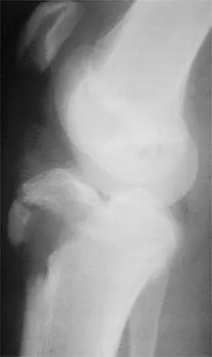
A 45-year-old male presents with acute knee pain and inability to actively extend his knee after a fall while playing basketball.

A lateral radiograph of the knee reveals a significantly low-riding patella (patella baja) compared to the contralateral knee. Which of the following is the most likely diagnosis?


Explanation

A quadriceps tendon rupture leads to unopposed distal pull on the patella by the intact patellar tendon, resulting in an inferiorly displaced patella (patella baja). In contrast, a patellar tendon rupture removes the distal tether, allowing the quadriceps muscle to pull the patella superiorly, resulting in patella alta.

Question 74

A 42-year-old recreational weightlifter presents with deep shoulder pain exacerbated by overhead activities. Physical therapy has failed after 6 months. An MRI arthrogram reveals an isolated Type II superior labrum anterior-posterior (SLAP) tear. Given the patient's age and activity profile, which surgical intervention is statistically associated with the highest rate of return to sport and the lowest rate of revision surgery?


Explanation

In older athletes and patients (typically > 35-40 years old), isolated repair of a Type II SLAP tear is associated with significant postoperative stiffness, continued pain, and a higher revision rate. Evidence strongly supports primary biceps tenodesis in this demographic, which yields higher patient satisfaction, more reliable pain relief, and better return to activity compared to SLAP repair.

Question 75

An 18-year-old athlete undergoes an isolated arthroscopic all-inside repair of a vertical longitudinal tear located in the red-white zone of the medial meniscus. To maximize the biological healing potential of this isolated meniscal repair, which of the following intraoperative adjuncts is most strongly supported by current literature?


Explanation

Meniscal healing is heavily dependent on a robust biological environment. Isolated meniscal repairs have historically lower healing rates compared to repairs performed concomitantly with ACL reconstruction. To mimic the hemarthrosis and release of bone marrow elements (progenitor cells and growth factors) seen in ACL reconstruction, a marrow venting procedure (such as intercondylar notch microfracture or trephination) is highly recommended as a biological adjunct for isolated meniscal repairs to stimulate a 'super clot' and improve healing rates.

Question 76

A 35-year-old male weightlifter presents with chronic anterior shoulder pain that radiates down his arm. He reports pain primarily during the bench press and cross-body adduction. Physical examination reveals point tenderness over the coracoid process, and a positive O'Brien test that is relieved by external rotation. MRI of the shoulder reveals a coracohumeral distance of 4 mm and subscapularis tendinosis without a rotator cuff tear. After failing 6 months of physical therapy and corticosteroid injections, what is the most appropriate surgical management?


Explanation

The clinical presentation (anterior pain, point tenderness over the coracoid, positive cross-body adduction) and MRI findings (decreased coracohumeral distance < 7 mm, subscapularis tendinosis) are classic for subcoracoid impingement. The appropriate management for cases refractory to conservative treatment is an arthroscopic coracoplasty (resection of the posterolateral aspect of the coracoid) to increase the coracohumeral interval.

Question 77

A 22-year-old collegiate football player presents with a recurrent anterior cruciate ligament (ACL) tear after sustaining a contact injury. His primary ACL reconstruction was performed 3 years ago utilizing a bone-patellar tendon-bone (BTB) autograft. Preoperative computed tomography (CT) demonstrates a femoral tunnel diameter of 16 mm and a tibial tunnel diameter of 15 mm. What is the most appropriate next step in surgical management?


Explanation

In the setting of revision ACL reconstruction, significant tunnel widening precludes adequate fixation and biologic incorporation of a new graft. Tunnel widening greater than 14-15 mm is generally an indication for a two-stage revision approach. The first stage consists of hardware removal and bone grafting of the enlarged tunnels. The second stage (ACL reconstruction) is performed 4 to 6 months later once the bone graft has consolidated.

Question 78

A 45-year-old female presents with sudden onset posteromedial knee pain and a palpable 'pop' while squatting. MRI demonstrates a medial meniscus posterior root tear with 4 mm of meniscal extrusion. Biomechanically, a medial meniscus posterior root tear alters knee kinematics and contact pressures equivalently to which of the following?


Explanation

Biomechanical studies have definitively demonstrated that a posterior root tear of the medial meniscus completely disrupts the meniscal hoop stresses. This leads to a loss of load-sharing capability and an increase in peak articular contact pressures that is biomechanically equivalent to a total medial meniscectomy, rapidly predisposing the joint to osteoarthritic degeneration.

Question 79

A 28-year-old offensive lineman complains of recurrent posterior shoulder instability that has failed extensive nonoperative management. Imaging shows a reverse Hill-Sachs lesion involving 25% of the anterior humeral articular surface and an associated posterior labral tear. To prevent engagement and provide the best clinical outcome, what is the most appropriate surgical management?


Explanation

A large reverse Hill-Sachs lesion (involving >20-25% of the anterior articular surface) in the setting of posterior shoulder instability requires addressing the bony defect to prevent anterior joint engagement. The modified McLaughlin procedure (transfer of the subscapularis tendon or lesser tuberosity into the defect) combined with posterior stabilization is the recommended surgical management to restore stability.

Question 80

During reconstruction of the medial patellofemoral ligament (MPFL) for recurrent patellar instability, the surgeon utilizes the Schöttle point to establish the femoral tunnel. If the femoral tunnel is inadvertently placed too proximal and anterior to this isometric point, what is the most likely biomechanical consequence?


Explanation

The Schöttle point describes the anatomic femoral attachment of the MPFL. If the femoral tunnel is placed too proximal and anterior, the distance between the patellar attachment and the femoral attachment increases as the knee goes into flexion. This causes the graft to inappropriately tighten during knee flexion, which can result in medial patellar facet overload, cartilage wear, and a significant loss of knee flexion.

Question 81

A 19-year-old collegiate baseball pitcher is undergoing ulnar collateral ligament (UCL) reconstruction utilizing a palmaris longus autograft. Following the procedure, what is the most commonly reported postoperative complication?


Explanation

Ulnar neuropathy is the most common complication following UCL reconstruction, reported in up to 10-15% of cases. It can manifest as transient numbness, tingling, or weakness in the ulnar nerve distribution. Management of the ulnar nerve during the procedure (e.g., in situ decompression versus subcutaneous or submuscular transposition) remains debated, but neuropathy remains the leading complication regardless of technique.

Question 82

A 25-year-old hockey player is undergoing hip arthroscopy for symptomatic femoroacetabular impingement (FAI) characterized by a prominent cam lesion and an alpha angle of 70 degrees. During the osteochondroplasty, excessive bony resection of the cam lesion at the femoral head-neck junction most significantly increases the risk of which of the following complications?


Explanation

During arthroscopic cam resection (osteochondroplasty), removing more than 30% of the femoral neck diameter significantly alters the biomechanics and load-bearing capacity of the proximal femur. Biomechanical studies have shown that this drastically increases the risk of an iatrogenic femoral neck fracture. Surgeons must carefully template and measure the resection depth to avoid over-resection.

Question 83

A 30-year-old male is evaluated in the clinic following a severe twisting injury to his left knee. Physical examination reveals a positive dial test with 15 degrees of increased external rotation at 30 degrees of flexion compared to the contralateral side. However, at 90 degrees of knee flexion, the external rotation is symmetric bilaterally. Which structure or combination of structures is most likely injured?


Explanation

The dial test evaluates for posterolateral rotatory instability. Increased external rotation (>10 degrees compared to the normal knee) isolated to 30 degrees of flexion with normal symmetry at 90 degrees indicates an isolated injury to the posterolateral corner (PLC). If the test demonstrates increased external rotation at both 30 and 90 degrees, it suggests a combined injury to both the PCL and the PLC.

Question 84

A 62-year-old highly active male presents with chronic pseudoparalysis of his right shoulder. Imaging reveals a massive, retracted, and irreparable supraspinatus and infraspinatus tear. He elects to undergo an arthroscopic superior capsular reconstruction (SCR).

To optimize the biomechanical success of the SCR and effectively centralize the humeral head, the allograft must be securely attached to the superior glenoid and which other structures?


Explanation

In a superior capsular reconstruction (SCR), the graft is anchored medially to the superior glenoid and laterally to the greater tuberosity. To successfully restore the superior restraints and optimize the coronal and sagittal force couples, the anterior and posterior margins of the graft must be sutured side-to-side to the intact subscapularis (anteriorly) and the infraspinatus/teres minor (posteriorly).

Question 85

A 14-year-old male presents with vague knee pain and intermittent mechanical catching. Radiographs reveal a 2 x 2 cm osteochondritis dissecans (OCD) lesion on the lateral aspect of the medial femoral condyle. MRI reveals a high T2 signal rim completely surrounding the osteochondral fragment. His physes remain open.

What is the most appropriate management?


Explanation

While stable osteochondritis dissecans (OCD) lesions in skeletally immature patients are typically managed nonoperatively, an MRI showing a high T2 signal rim completely surrounding the lesion indicates synovial fluid tracking behind the fragment. This defines it as an unstable lesion. Unstable lesions, even in patients with open physes, have a poor healing capacity with conservative care and require surgical intervention, optimally with arthroscopic in situ fixation.

Question 86

A 25-year-old professional football player sustains a contact injury to his right knee. On physical examination, the dial test reveals 20 degrees of increased external rotation at 30 degrees of knee flexion compared to the contralateral knee. However, at 90 degrees of knee flexion, the external rotation is symmetric bilaterally. Based on these examination findings, which of the following structures is most likely injured?


Explanation

The dial test is used to evaluate the posterolateral corner (PLC) and the posterior cruciate ligament (PCL). Increased external rotation (>10-15 degrees compared to the normal side) at 30 degrees of flexion with symmetric rotation at 90 degrees indicates an isolated injury to the PLC (which includes the popliteus tendon, popliteofibular ligament, and LCL). If increased external rotation is present at both 30 and 90 degrees of flexion, it indicates a combined injury to the PLC and the PCL.

Question 87

A 30-year-old competitive weightlifter feels a sudden "pop" and tearing sensation in his anterior axilla while performing a heavy bench press. He presents with extensive ecchymosis and loss of the anterior axillary fold. MRI confirms a complete, full-thickness rupture of the pectoralis major tendon at its insertion. Which of the following best describes the anatomic orientation of the pectoralis major tendon at its insertion onto the humerus?


Explanation

The pectoralis major consists of a clavicular head and a sternal head. As the tendon progresses laterally toward its insertion on the lateral lip of the bicipital groove, the sternal head twists 180 degrees. This results in the sternal (inferior) muscle belly inserting deep and proximal to the clavicular (superior) muscle belly. Most pectoralis major ruptures occur at the sternal head insertion during eccentric loading, such as the descent phase of a bench press.

Question 88

A 19-year-old collegiate wrestler sustains an anterior shoulder dislocation. After successful closed reduction, he complains of numbness over the lateral deltoid and demonstrates weakness with shoulder abduction. Electromyography (EMG) confirms an isolated neurapraxia. What is the expected anatomic distance of the affected nerve from the inferior border of the glenoid at the 6 o'clock position?


Explanation

The patient has an axillary nerve injury, the most commonly injured nerve during anterior shoulder dislocations. The axillary nerve passes through the quadrangular space and courses closely inferior to the glenohumeral joint capsule. Anatomic studies show that the axillary nerve lies, on average, approximately 10 to 15 mm (around 12 mm) inferior to the 6 o'clock position of the glenoid rim. It is highly at risk during procedures that require inferior capsular release.

Question 89

A 22-year-old hockey player presents with chronic groin pain exacerbated by hip flexion and internal rotation. Examination demonstrates a positive anterior impingement test. An anteroposterior pelvis radiograph is obtained.

The radiograph demonstrates a prominent "crossover sign." Measurement of the alpha angle on a Dunn lateral view is 45 degrees. Which of the following is the most likely diagnosis?


Explanation

The crossover sign on an AP pelvis radiograph indicates focal or global acetabular retroversion, which is a classic finding of Pincer-type femoroacetabular impingement (FAI). Pincer FAI is caused by overcoverage of the femoral head by the acetabulum. An alpha angle of 45 degrees is within normal limits (typically < 50-55 degrees), ruling out Cam-type FAI, which is characterized by an abnormal femoral head-neck offset.

Question 90

A 20-year-old collegiate baseball pitcher reports progressive medial elbow pain and decreased pitching velocity. Examination reveals pain with the moving valgus stress test and maximal tenderness slightly distal to the medial epicondyle. MRI shows a high-grade partial tear of the ulnar collateral ligament (UCL). Which of the following bundles of the UCL provides the primary restraint to valgus stress at the elbow between 30 and 120 degrees of flexion?


Explanation

The anterior bundle of the ulnar collateral ligament (UCL) is the primary restraint to valgus stress at the elbow, particularly between 30 and 120 degrees of flexion. It originates on the anteroinferior surface of the medial epicondyle and inserts on the sublime tubercle of the ulna. The posterior bundle acts as a secondary restraint, and the transverse bundle provides no significant stability to valgus stress.

Question 91

A 50-year-old female presents with sudden onset medial-sided knee pain and a "pop" while squatting in her garden. She has no mechanical locking but exhibits an antalgic gait. MRI reveals a 4 mm medial meniscal extrusion and a radial defect directly at the posterior root attachment of the medial meniscus. If left untreated, the biomechanical consequence of this specific injury is most equivalent to which of the following?


Explanation

A posterior medial meniscal root tear completely disrupts the circumferential hoop stresses of the meniscus. Biomechanical studies have demonstrated that this leads to an inability of the meniscus to absorb load, effectively resulting in contact areas and peak contact pressures within the medial compartment that are equivalent to a total medial meniscectomy. This drastically accelerates the progression of medial compartment osteoarthritis.

Question 92

A 45-year-old manual laborer presents with chronic, anterior shoulder pain that has failed 6 months of physical therapy and NSAID management. He describes deep, aching pain and catching when lifting objects away from his body. O'Brien's active compression test is positive. MRI reveals an isolated Type II SLAP (Superior Labrum Anterior to Posterior) tear. Given his age and occupational demands, which of the following is the most appropriate surgical management?


Explanation

In patients older than 40 years, especially those with physically demanding occupations, primary biceps tenodesis is the preferred treatment for symptomatic Type II SLAP tears. Studies have consistently shown that SLAP repairs in this age group are associated with higher rates of postoperative stiffness, persistent pain, and need for revision surgery compared to biceps tenodesis, which offers more reliable pain relief and functional recovery.

Question 93

A 40-year-old water skier sustains a hyperflexion injury to the hip with her knee fully extended. She presents with significant ecchymosis over the posterior thigh, an antalgic gait, and an inability to run. MRI confirms a complete, 3-tendon avulsion of the proximal hamstring complex with 4 cm of retraction. Open surgical repair is planned. Which of the following neurovascular structures is located immediately lateral to the ischial tuberosity and is at highest risk during surgical retrieval and repair?


Explanation

The sciatic nerve lies in close proximity to the proximal hamstring origin, averaging 1.2 to 2.0 cm lateral to the ischial tuberosity. In cases of acute or chronic retracted proximal hamstring avulsions, the sciatic nerve is at significant risk for injury or entrapment in scar tissue. Thorough neurolysis and protection of the sciatic nerve are critical steps during the surgical approach for proximal hamstring repair.

Question 94

A 22-year-old female collegiate soccer player presents with localized anterior knee pain and swelling after matches. She has failed 9 months of conservative management. Knee MRI reveals an isolated, full-thickness chondral defect measuring 3.5 cm² on the weight-bearing surface of the medial femoral condyle. Alignment is normal, and there is no meniscal pathology. Which of the following cartilage restoration procedures is most indicated for this patient?


Explanation

Matrix-induced autologous chondrocyte implantation (MACI) is indicated for symptomatic, full-thickness unipolar chondral defects measuring between 2 cm² and 10 cm² in active patients. Microfracture and osteochondral autograft transfer (OATS) are typically reserved for smaller lesions (< 2 cm²) due to the poor durability of fibrocartilage (microfracture) and donor-site morbidity limitations (OATS). Given her normal alignment, a high tibial osteotomy is not indicated.

Question 95

During diagnostic elbow arthroscopy, the anteromedial portal is frequently established to view the anterior compartment and radiocapitellar joint. Careful creation of this portal is essential to avoid iatrogenic injury. Which of the following nerves is at greatest risk of injury during the placement of the anteromedial portal?


Explanation

The medial antebrachial cutaneous nerve (MABC) is highly superficial and courses in close proximity to the anteromedial elbow portal (usually about 1-2 mm away). It is the most commonly injured cutaneous nerve during elbow arthroscopy. The median nerve is situated deeper and slightly more laterally. The radial nerve is at risk with the anterolateral portal, and the ulnar nerve is at risk with posteromedial portals.

Question 96

A 21-year-old male rugby player presents with recurrent anterior shoulder instability. He has had 5 dislocations in the past year. CT scan reveals 25% anterior glenoid bone loss. He undergoes an open Latarjet procedure. Which of the following describes the "sling effect" provided by the Latarjet procedure?


Explanation

The Latarjet procedure provides stability through a triple-blocking mechanism: 1) the "sling effect" of the conjoined tendon on the inferior subscapularis and anteroinferior capsule when the arm is abducted and externally rotated; 2) the bone block effect of the transferred coracoid extending the glenoid articular arc; and 3) the capsular repair (often using the coracoacromial ligament stump to the capsule).

Question 97

A 45-year-old woman presents with acute onset medial knee pain after a deep knee flexion maneuver. She felt a "pop" in her knee. MRI demonstrates a medial meniscus posterior root tear and a 2 mm extrusion of the medial meniscus on coronal sequences. Which of the following biomechanical consequences is most likely if this injury is treated nonoperatively?


Explanation

A medial meniscus posterior root tear effectively completely disrupts the ability of the meniscus to convert axial loads into hoop stresses. Biomechanical studies have demonstrated that this loss of hoop stresses results in increased peak contact pressures in the medial compartment that are equivalent to those seen after a total medial meniscectomy, accelerating the progression of osteoarthritis.

Question 98

A 55-year-old man presents with anterior shoulder pain and weakness following a fall onto an outstretched hand. On physical examination, he demonstrates a positive bear-hug test and increased passive external rotation compared to the contralateral side. MRI confirms an isolated, full-thickness tear of the subscapularis tendon. During arthroscopic repair, which of the following structures must be carefully evaluated and is most commonly associated with this injury pattern?


Explanation

Isolated subscapularis tears are highly associated with pathology of the biceps pulley (composed of the coracohumeral and superior glenohumeral ligaments) and the long head of the biceps tendon (LHBT). Disruption of the subscapularis and biceps pulley often leads to medial subluxation or dislocation of the LHBT. Therefore, thorough evaluation and concomitant treatment of the LHBT (such as tenodesis or tenotomy) are crucial during surgical management.

Question 99

A 24-year-old male hockey player presents with groin pain exacerbated by hip flexion and internal rotation. Radiographs demonstrate an alpha angle of 70 degrees on the Dunn lateral view and a lateral center edge angle of 35 degrees. He is diagnosed with femoroacetabular impingement (FAI). If left untreated, the pathomechanics of his specific impingement type typically result in which of the following patterns of chondral injury?


Explanation

The patient has a Cam-type FAI, defined by an alpha angle greater than 50-55 degrees, indicating an aspherical femoral head-neck junction. Cam impingement occurs when this prominence is forced into the acetabulum during hip flexion, generating outside-in shear forces. This typically causes delamination of the anterosuperior acetabular cartilage and separation of the labrum from the adjacent cartilage. Conversely, Pincer FAI is more frequently associated with contrecoup lesions in the posterior-inferior acetabulum due to a levering effect.

Question 100

A 28-year-old male suffers a high-energy knee injury resulting in a combined posterior cruciate ligament (PCL) and posterolateral corner (PLC) tear. He undergoes a staged reconstruction. During reconstruction of the posterolateral corner, an anatomic fibular-based technique is planned. Which three primary static stabilizing structures of the PLC are reconstructed in this technique?


Explanation

The anatomic posterolateral corner reconstruction (such as the technique described by LaPrade et al.) aims to recreate the three primary static stabilizers of the PLC: the fibular collateral ligament (FCL), the popliteus tendon (PLT), and the popliteofibular ligament (PFL). Reconstructing these specific structures biomechanically restores varus and external rotatory stability to the knee.

None

Clinic OS
Medically Verified Content by
Prof. Clinic OS
Consultant Orthopedic & Spine Surgeon
Chapter Index