Orthopedic Basic 2026 MCQs: Board Review Questions & Answers (Part 3)
Key Takeaway
This article provides essential research regarding Orthopedic Basic 2026 MCQs: Board Review Questions & Answers (Part 3). Top-rated Orthopedic Basic 2026 MCQs bank. Practice with clinical case questions, orthopedic surgery board review, and evidence-based answers updated for 2026.
Orthopedic Basic 2026 MCQs: Board Review Questions & Answers (Part 3)
Comprehensive 100-Question Exam
00:00
Start Quiz
Question 1
What is the most common location of osteosarcoma?
Explanation
Question 2
A 40-year-old man with an acetabular chondrosarcoma has a small soft-tissue mass. Treatment should consist of
Explanation
Question 3
Figures 29a and 29b show the AP radiograph and CT scan of a 70-year-old man who has left thigh pain. Serum protein electrophoresis shows a monoclonal gammopathy. Additional radiographs of the femur show other lesions. Management should consist of
Explanation
Question 4
What pharmacologic agents are preferred for the treatment of symptomatic active Paget's disease?
Explanation
Question 5
A 7-year-old girl has pain and a mass in the left scapula. A MRI scan and biopsy specimen are shown in Figures 30a and 30b. After staging studies, initial management should consist of
Explanation
Question 6
A 73-year-old man reports increasing back and lower extremity pain. A bone scan is shown in Figure 31. What is the most likely diagnosis?
Explanation
Question 7
A 16-year-old girl has had pain in the left groin for the past 4 months. She notes that the pain is worse at night; however, she denies any history of trauma and has no constitutional symptoms. There is no history of steroid or alcohol use. Examination reveals pain in the left groin with rotation of the hip. There is no associated soft-tissue mass. A radiograph and MRI scan are shown in Figures 32a and 32b, and biopsy specimens are shown in Figures 32c and 32d. What is the most likely diagnosis?
Explanation
Question 8
Ewing's sarcoma of bone most commonly occurs in which of the following locations?
Explanation
Question 9
A previously healthy 14-year-old boy now reports fatigue, and has a bilateral Trendelenburg gait, right hip pain, and bilateral knee and foot pain. Biopsy of a right sacral mass reveals intermediate grade osteosarcoma. There are no metastases. Laboratory studies reveal a serum calcium level of 7.7 mg/dL (normal 8.5 to 10.5), a phosphate level of 2.0 mg/dL (normal 2.7 to 4.5), a 1,25-dihydroxyvitamin D level of less than 10 pg/mL (normal 18 to 62), a parathyroid hormone level of 19 pg/mL (normal 10 to 60), and an alkaline phosphatase level of 428 U/L (normal 15 to 351). What is the most likely cause of the patient's symptoms?
Explanation
Question 10
Which of the following staging studies should be obtained for an adult with an 8-cm deep, high-grade malignant fibrous histiocytoma of the extremity?
Explanation
Question 11
An 18-year-old boy has had pain in the right knee for the past 6 months. Examination reveals some fullness behind the knee but no significant palpable soft-tissue mass. There is no effusion, and he has full knee range of motion. The remainder of the examination is unremarkable. A radiograph and MRI scans are shown in Figures 33a through 33c, and biopsy specimens are shown in Figures 33d and 33e. What is the most likely diagnosis?
Explanation
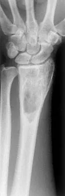
Question 12
A 30-year-old patient has wrist pain. A radiograph and biopsy specimen are shown in Figures 34a and 34b. What is the most likely diagnosis?
Explanation
Question 13
Mutations of what gene are associated with subsequent development of osteosarcoma?
Explanation
Question 14
A 12-year-old girl has painless bowing of the tibia. Radiographs and a biopsy specimen are shown in Figures 35a through 35c. What is the most likely diagnosis?
Explanation
Question 15
A 54-year-old man with metastatic renal cell carcinoma has had increasing pain in the left hip for the past 6 weeks. A radiograph is shown in Figure 36. Prophylactic stabilization will most likely result in
Explanation
Question 16
Which of the following is considered the treatment of choice for a 3-cm chondroblastoma of the distal femoral epiphysis with no intra-articular extension?
Explanation
Question 17
A radiograph, MRI scans, and a biopsy specimen of a 9-year-old boy with thigh pain are shown in Figures 37a through 37d. Management should consist of
Explanation
Question 18
A 47-year-old woman has had a 1-month history of left hip and medial thigh pain that is exacerbated by sitting. Laboratory studies show a total protein level of 8.2 g/dL (normal 6.0 to 8.0) and an immunoglobulin G (IGG) level of 2,130 mg/dL (normal 562 to 1,835). A radiograph, CT scan, and biopsy specimen are shown in Figures 38a through 38c. What is the most likely diagnosis?
Explanation
Question 19
A 14-year-old boy has an anteromedial distal thigh mass. A radiograph and MRI scan are shown in Figures 39a and 39b. An open biopsy of the mass should include
Explanation
Question 20
A 60-year-old man has pain at the tip of the index finger. A radiograph and biopsy specimen are shown in Figures 40a and 40b. Management should consist of
Explanation
Question 21
An infant is born with a mass that involves both the volar and dorsal compartments of the left arm. A clinical photograph and biopsy specimen are shown in Figures 41a and 41b. What is the best initial course of action?
Explanation
Question 22
Which of the following processes does not account for decreased hematopoiesis in patients with metastatic disease?
Explanation
Question 23
A 43-year-old woman has an enlarging mass in the left groin. A radiograph, CT scan, and a biopsy specimen are shown in Figures 42a through 42c. Treatment should consist of
Explanation
Question 24
A 66-year-old man has a high-grade angiosarcoma of the right tibia. A radiograph is shown in Figure 43. Treatment should consist of
Explanation
Question 25
Figures 44a and 44b show the radiographs of a 28-year-old woman who has had progressive hip pain for the past 3 months. What is the most likely diagnosis?
Explanation
Question 26
A 32-year-old woman presents with a lytic lesion in the distal femur that extends to the subchondral bone. Biopsy reveals multinucleated giant cells in a background of mononuclear stromal cells. She is treated with denosumab preoperatively. What is the mechanism of action of this medication?
Explanation
Question 27
In total hip arthroplasty, the use of highly cross-linked polyethylene (HXLPE) has significantly reduced the rate of wear. What is the primary trade-off or disadvantage associated with increasing the cross-linking dose in ultra-high-molecular-weight polyethylene (UHMWPE)?
Explanation
Question 28
Bone morphogenetic proteins (BMPs) play a crucial role in osteoinduction. Following the binding of BMP to its cell surface receptor, which intracellular signaling molecules are primarily phosphorylated to translocate to the nucleus and regulate gene expression?
Explanation
Question 29
A 6-year-old boy is evaluated for multiple fractures with minimal trauma. Radiographs demonstrate diffusely increased bone density, a 'bone-within-bone' appearance in the spine, and Erlenmeyer flask deformities of the distal femora. Which of the following best describes the underlying cellular defect in this condition?
Explanation
Question 30
In periprosthetic joint infections, Staphylococcus epidermidis is a frequent pathogen due to its ability to form a biofilm. Which of the following components is primarily responsible for the structural integrity of the biofilm matrix, shielding the bacteria from host immunity and antibiotics?
Explanation
Question 31
When a continuous, constant load is applied to a ligament over time, the ligament will gradually elongate. This viscoelastic property is best described as:
Explanation
Question 32
A 55-year-old woman with chronic kidney disease presents with diffuse bone pain and muscle weakness. Radiographs reveal Looser zones in the femoral neck and pubic rami. Bone biopsy is performed for histomorphometry. Which of the following parameters is most likely to be increased in this patient?
Explanation
Question 33
During an extensile anterior approach to the humerus for fracture fixation, the surgeon must be cautious to identify and protect a nerve that pierces the lateral intermuscular septum to enter the anterior compartment of the arm. This nerve innervates which of the following muscles?
Explanation
Question 34
The compressive stiffness of normal articular cartilage is primarily dependent on which of the following structural interactions?
Explanation
Question 35
A 45-year-old diabetic patient is undergoing treatment for a chronic lower extremity ulcer. Which of the following phases of wound healing is primarily characterized by the proliferation of fibroblasts and the synthesis of type III collagen?
Explanation
Question 36
A 14-year-old boy presents with a painful mass in his left mid-thigh. Radiographs show a destructive diaphyseal lesion with an 'onion-skin' periosteal reaction. Biopsy reveals uniform small round blue cells. Which of the following genetic translocations is most characteristic of this lesion?
Explanation
Question 37
A 65-year-old man presents with a destructive, lytic lesion in the proximal humerus causing impending fracture. He has a history of a radical nephrectomy for renal cell carcinoma 5 years ago. A prophylactic stabilization is planned. Which of the following should be performed prior to the surgical stabilization?
Explanation
Question 38
A 32-year-old woman presents with a lytic lesion in the distal femur extending to the subchondral bone. Biopsy confirms a giant cell tumor of bone. She is treated with denosumab to downstage the tumor prior to curettage. What is the primary mechanism of action of denosumab in the treatment of this lesion?
Explanation
Question 39
A 50-year-old man presents with a deep, painless, 8-cm soft tissue mass in his anterior thigh. MRI suggests a high-grade soft-tissue sarcoma. An incisional biopsy is planned. Which of the following is a fundamental principle of performing a biopsy for a suspected musculoskeletal sarcoma?
Explanation
Question 40
A 19-year-old man presents with severe night pain in his right shin that is dramatically relieved by nonsteroidal anti-inflammatory drugs (NSAIDs). CT scan shows a 7-mm radiolucent nidus surrounded by dense reactive cortical sclerosis in the tibial diaphysis. If the patient elects for surgical intervention, what is the most appropriate definitive management?
Explanation
Question 41
A 15-year-old boy presents with knee pain. Radiographs reveal a well-circumscribed, eccentrically located lytic lesion in the proximal tibial epiphysis with a thin sclerotic rim. Which of the following histological findings is most characteristic of this lesion?
Explanation
Question 42
A 22-year-old man is diagnosed with an osteosarcoma of the distal femur. Biopsy reveals a high-grade tumor. MRI shows that the tumor has broken through the posterior cortex into the surrounding soft tissues, but whole-body staging reveals no lung or distant bone metastases. According to the Musculoskeletal Tumor Society (Enneking) staging system, what is the stage of this tumor?
Explanation
Question 43
A 68-year-old woman presents with persistent lower back pain. Radiographs reveal a purely lytic 'punched-out' lesion in the L3 vertebral body. Laboratory studies show hypercalcemia, anemia, and a monoclonal spike on serum protein electrophoresis. What is the most appropriate test to definitively confirm the diagnosis and staging of the underlying disease process?
Explanation
Question 44
A 9-year-old girl is evaluated for a severe varus deformity of her proximal femur, described radiographically as a 'shepherd's crook'. She is noted to have large, irregular areas of skin hyperpigmentation on her trunk. Which of the following endocrine abnormalities is most commonly associated with this patient's underlying condition?
Explanation
Question 45
A 28-year-old man presents with a slow-growing, deep-seated, painful mass near his knee joint that has been present for over a year. MRI reveals a well-circumscribed soft-tissue mass with some internal calcifications adjacent to the joint, but not within it. Which of the following is true regarding this specific tumor type?
Explanation
Question 46
A 28-year-old woman presents with persistent, aching knee pain. Radiographs reveal an eccentric, lytic epiphyseal-metaphyseal lesion of the distal femur. Biopsy reveals uniform mononuclear cells interspersed with numerous multinucleated giant cells. Which of the following describes the mechanism of action of the targeted medical therapy most commonly used for this condition?
Explanation
Question 47
A 14-year-old boy presents with progressive thigh pain and swelling, accompanied by low-grade fever. Radiographs show a permeative, diaphyseal lesion with an 'onion skin' periosteal reaction. A biopsy reveals sheets of uniform small, round blue cells. Which of the following chromosomal translocations is most characteristic of this tumor?
Explanation
Question 48
A 60-year-old woman with a history of breast cancer presents with moderate functional pain in her left thigh. Radiographs reveal a purely lytic lesion in the proximal diaphysis of the left femur that involves 50% of the cortical diameter. According to Mirels' criteria, what is her score and the recommended orthopedic management?
Explanation
Question 49
A 35-year-old man presents with a slow-growing, deep-seated, painful mass in the plantar aspect of his foot. MRI demonstrates a well-circumscribed soft-tissue mass adjacent to the plantar fascia. Biopsy reveals a biphasic tumor with both epithelial (glandular) and spindle cell components. What is the most likely cytogenetic abnormality associated with this diagnosis?
Explanation
Question 50
A 16-year-old boy presents with chronic left shoulder pain. Radiographs reveal a well-demarcated, eccentric, lytic lesion in the proximal humeral epiphysis with a thin sclerotic rim. Histological examination shows mononuclear cells with grooved nuclei ('coffee bean' appearance) and areas of pericellular 'chicken-wire' calcification. What is the most likely diagnosis?
Explanation
Question 51
A 55-year-old man complains of vague lower back pain and constipation for the past 6 months. A digital rectal examination reveals a firm presacral mass. MRI shows a large, destructive midline lesion of the sacrum. Biopsy demonstrates lobules of large, vacuolated cells with abundant mucinous cytoplasm (physaliferous cells) set in a myxoid stroma. Which of the following is the most appropriate definitive surgical management?
Explanation
Question 52
A 20-year-old man presents with an aching left thigh pain that is notably worse at night and dramatically relieved by taking ibuprofen. Radiographs show a small radiolucent nidus (< 1.5 cm) surrounded by dense reactive sclerosis in the femoral diaphysis. Which of the following inflammatory mediators is produced in high levels by this lesion and mediates the characteristic pain?
Explanation
Question 53
A 65-year-old man presents with diffuse bone pain and fatigue. Laboratory investigations reveal normocytic anemia, hypercalcemia, and an elevated serum creatinine. Serum protein electrophoresis shows a monoclonal IgG spike. A plain radiograph skeletal survey demonstrates multiple 'punched-out' lytic lesions in the skull and pelvis. Which of the following advanced imaging modalities is considered the most sensitive for assessing the true extent of skeletal involvement in this condition?
Explanation
Question 54
A 9-year-old girl is evaluated for precocious puberty and several large café-au-lait spots with irregular 'coast of Maine' borders on her trunk. Radiographs of her femur demonstrate an expansile medullary lesion with a ground-glass appearance and severe varus bowing. A mutation in which of the following genes is most likely responsible for her condition?
Explanation
Question 55
A 45-year-old man presents with chronic hip pain. Radiographs demonstrate an expansile, lytic lesion in the proximal femoral epiphysis with central calcifications. The physeal plates are closed. Biopsy reveals sheets of large cells with abundant clear cytoplasm and distinct cell membranes, interspersed with trabeculae of woven bone. What is the most likely diagnosis?
Explanation
Question 56
A 12-year-old boy presents with a painful, enlarging mass in the diaphysis of the femur. Radiographs show a permeative lytic lesion with an 'onion skin' periosteal reaction. Biopsy reveals sheets of uniform small round blue cells that strongly express CD99 on immunohistochemistry. Which of the following chromosomal translocations is most characteristic of this patient's diagnosis?
Explanation
Question 57
A 32-year-old woman presents with a large, expansile lytic epiphyseal lesion in her distal femur. Biopsy confirms a giant cell tumor of bone. Due to the extensive size and proximity to the articular surface, the surgeon decides to use neoadjuvant medical therapy to consolidate the tumor and facilitate joint-salvage surgery. Which of the following is the mechanism of action of the most appropriate pharmacological agent?
Explanation
Question 58
A 55-year-old man complains of severe, generalized bone pain and proximal muscle weakness. Laboratory studies reveal severe hypophosphatemia, normal serum calcium, normal parathyroid hormone (PTH), and low 1,25-dihydroxyvitamin D levels. A small soft-tissue mass is discovered on the plantar aspect of his foot. Complete resection of this mass is most likely to reverse his symptoms by eliminating the abnormal paraneoplastic production of which of the following substances?
Explanation
Question 59
A 28-year-old man presents with chronic, dull anterior leg pain. Radiographs reveal a multiloculated, expansile, eccentric lytic lesion involving the anterior cortex of the tibial diaphysis. Histological examination shows islands of epithelial cells interspersed within a fibrous stroma. Immunohistochemistry is strongly positive for cytokeratin. What is the most appropriate definitive management for this lesion?
Explanation
Question 60
A 62-year-old man presents with a destructive lytic bone lesion in his proximal humerus. Further workup reveals a primary renal mass consistent with clear cell renal carcinoma. He is scheduled for wide excision and endoprosthetic reconstruction of the humerus. To minimize severe intraoperative complications, which of the following interventions should be strongly considered 24 to 48 hours prior to the orthopedic surgery?
Explanation
Question 61
According to the American Joint Committee on Cancer (AJCC) staging system for soft tissue sarcomas of the extremities, which of the following tumor characteristics is considered the most critical prognostic factor and the primary determinant of tumor stage?
Explanation
Question 62
A 19-year-old male presents with severe right thigh pain that is distinctly worse at night and dramatically relieved by over-the-counter ibuprofen. Radiographs reveal localized cortical thickening in the proximal femoral diaphysis with a central 7-mm radiolucent nidus. The dramatic pain relief provided by NSAIDs in this condition is directly mediated by the inhibition of which substance produced by the nidus?
Explanation
Question 63
A 15-year-old boy presents with right knee pain of three months duration. Radiographs demonstrate a well-circumscribed, 2-cm lytic lesion with a thin sclerotic margin located entirely within the epiphysis of the distal femur. Fine stippled calcifications are noted internally. Histopathology reveals mononuclear cells with longitudinal nuclear grooves ('coffee bean' nuclei), scattered osteoclast-like giant cells, and areas of 'chicken-wire' calcification. What is the most likely diagnosis?
Explanation
Question 64
A 45-year-old man presents with a painless, deep, 10-cm soft-tissue mass in his posterior thigh. MRI shows a well-defined intramuscular mass that is markedly hyperintense on T2-weighted sequences. Core needle biopsy confirms myxoid liposarcoma. Compared to other high-grade soft tissue sarcomas, this specific subtype is uniquely characterized by which of the following clinical behaviors?
Explanation
Question 65
A 68-year-old woman presents with worsening intractable back pain and generalized fatigue. Investigations reveal normocytic anemia, hypercalcemia, and an elevated monoclonal M-protein spike on serum protein electrophoresis (SPEP). Suspecting multiple myeloma, what is the most sensitive and currently recommended whole-body imaging modality to assess the extent of skeletal osteolytic lesions in this patient?
Explanation
Question 66
A 12-year-old boy presents with a painful mass in his left mid-thigh. Radiographs demonstrate a diaphyseal permeative lytic lesion of the femur with an 'onion skin' periosteal reaction. Core needle biopsy reveals sheets of small, round, blue cells that stain positive for CD99. Which of the following is the most common cytogenetic abnormality associated with this condition?
Explanation
Question 67
A 32-year-old woman presents with worsening knee pain. Radiographs reveal an eccentric, expansile, lytic lesion in the epiphysis of the distal femur extending to the subchondral bone. Biopsy confirms a giant cell tumor of bone. She is initiated on a systemic monoclonal antibody treatment prior to surgical intervention to consolidate the lesion. What is the primary cellular target of this medication?
Explanation
Question 68
When designing an intramedullary nail for a diaphyseal long-bone fracture, minimizing stress shielding while maintaining adequate fatigue strength is a primary biomechanical goal. Which of the following orthopaedic implant materials has a modulus of elasticity closest to that of human cortical bone?
Explanation
Question 69
Articular cartilage is a highly specialized tissue designed to resist compressive and shear forces within the joint. In which zone of articular cartilage are the collagen fibers oriented perpendicular to the articular surface, and which collagen type primarily constitutes these fibers?
Explanation
Question 70
A 68-year-old woman undergoes an uncomplicated total hip arthroplasty. To minimize perioperative blood loss, intravenous tranexamic acid (TXA) is administered. What is the specific mechanism of action of this agent?
Explanation
Question 71
A 28-year-old man presents with a slow-growing, painless mass deep in the plantar aspect of his left foot. Core needle biopsy demonstrates a biphasic histologic pattern consisting of both uniform spindle cells and epithelial cells forming glandular structures.
Which of the following translocations is classically diagnostic for this soft-tissue tumor?

Explanation
Question 72
According to Perren's strain theory of fracture healing, the local mechanical strain environment dictates the specific type of tissue that forms at the fracture site. What is the maximum tissue strain percentage that permits the formation of lamellar bone directly without an intermediate cartilaginous phase (primary bone healing)?
Explanation
Question 73
A 16-year-old girl is diagnosed with a conventional high-grade intramedullary osteosarcoma of the proximal tibia. She undergoes 10 weeks of neoadjuvant chemotherapy, followed by a wide surgical resection and endoprosthetic reconstruction. Assuming no metastases were present at diagnosis, what is the single most important prognostic factor for her long-term survival?
Explanation
Question 74
A 19-year-old man presents with a 6-month history of a dull, aching pain in his right proximal femur that is notably worse at night and dramatically relieved by taking nonsteroidal anti-inflammatory drugs. CT imaging reveals a 7-mm radiolucent nidus surrounded by dense, reactive sclerosis. Histologic examination of the central nidus would most likely reveal which of the following?
Explanation
Question 75
During revision surgery of a modular total hip arthroplasty, the surgeon notes black particulate debris and severe tissue staining around the modular head-neck junction. The implant system consists of a titanium-alloy femoral stem and a cobalt-chromium alloy femoral head. Which combination of corrosive mechanisms is primarily responsible for this specific presentation (trunnionosis)?
Explanation
Question 76
A 30-year-old woman presents with worsening right knee pain. Radiographs reveal an eccentric, lytic epiphyseal lesion of the distal femur that extends to the subchondral bone. A core biopsy confirms the diagnosis of a giant cell tumor of bone. The multidisciplinary tumor board recommends neoadjuvant treatment with denosumab prior to surgical intervention. What is the precise mechanism of action of this medication?
Explanation
Question 77
A 12-year-old boy presents with an enlarging, painful mass over his right thigh. Radiographs demonstrate a large, ill-defined, destructive diaphyseal lesion of the femur with a prominent 'onion-skin' periosteal reaction. Core needle biopsy reveals sheets of small, uniform, round blue cells. Which of the following cytogenetic abnormalities is most likely present in this tumor?
Explanation
Question 78
A 65-year-old woman with metastatic breast cancer presents with diffuse skeletal pain and is found to have multiple osteolytic bone lesions. To prevent skeletal-related events, she is started on intravenous zoledronic acid. Which of the following best describes the intracellular pharmacodynamic mechanism of action of this drug class?
Explanation
Question 79
A 28-year-old man presents with a slowly enlarging, painful mass deep within his plantar foot. Magnetic resonance imaging demonstrates a well-circumscribed, multilobulated mass adjacent to the plantar fascia. Core needle biopsy demonstrates a biphasic tumor with both epithelial and spindle cell components. Which of the following is true regarding this specific soft tissue sarcoma?
Explanation
Question 80
A 19-year-old male complains of a dull, aching pain in his right shin that is consistently worse at night and dramatically relieved by taking ibuprofen. Radiographs reveal a thickened cortical diaphysis with a 7-mm radiolucent nidus. The intense pain experienced by this patient is primarily mediated by local overproduction of which of the following substances?
Explanation
Question 81
A 35-year-old woman undergoes arthroscopic synovectomy for a diffuse, recurrent, and hemorrhagic joint effusion in her left knee. Histopathology shows a hyperplastic synovium with a proliferation of mononuclear cells, multinucleated giant cells, and abundant hemosiderin deposition. The primary molecular driver of this condition is a genetic alteration resulting in the overexpression of which of the following?
Explanation
Question 82
A 55-year-old man is found to have a 6-cm painful lesion in his proximal humerus. Radiographs show a radiolucent lesion with endosteal scalloping greater than two-thirds of the cortical thickness and distinct areas of 'popcorn' calcification. Biopsy reveals hypercellular hyaline cartilage with binucleated cells and a myxoid stroma. Which of the following is the most appropriate definitive management for this lesion?
Explanation
Question 83
A 4-year-old boy presents with a rapidly growing mass in his proximal forearm. An incisional biopsy confirms the diagnosis of rhabdomyosarcoma. Which of the following statements is true regarding the typical staging and biological behavior of this patient's disease?
Explanation
Question 84
A 22-year-old woman sustains a pathologic fracture of her proximal femur. Pre-injury radiographs obtained from an outside hospital reveal a 'ground-glass' appearance of the medullary canal with a severe varus 'shepherd's crook' deformity. Physical examination notes large, irregular café-au-lait macules on her trunk. An activating somatic mutation in which of the following genes is fundamentally responsible for her condition?
Explanation
Question 85
A 34-year-old man presents with chronic, progressive aching pain in his anterior left shin. Radiographs demonstrate a multiloculated, eccentric, lytic lesion in the anterior cortex of the tibial diaphysis. Biopsy reveals a biphasic pattern with clusters of epithelial cells surrounded by a dense, spindle-cell osteofibrous stroma. What is the most appropriate management for this lesion?
Explanation
Question 86
A 19-year-old man presents with right thigh pain that is worse at night and dramatically relieved by ibuprofen. CT scan shows a 9-mm radiolucent nidus surrounded by dense reactive bone in the lesser trochanter. If the patient is intolerant to long-term NSAID use, what is the most appropriate next step in management?
Explanation
Question 87
A 14-year-old boy presents with progressive, activity-related right knee pain. Radiographs demonstrate a well-defined, 2-cm eccentrically located lytic lesion in the proximal tibial epiphysis with a thin sclerotic rim. A biopsy reveals polygonal chondroblasts, multinucleated giant cells, and 'chicken-wire' calcification. What is the most appropriate definitive management?
Explanation
Question 88
A 32-year-old woman presents with worsening knee pain. Radiographs reveal an eccentric, purely lytic lesion in the distal femoral metaphysis extending into the epiphysis up to the subchondral bone, lacking a sclerotic margin. Histology shows mononuclear stromal cells and numerous uniformly distributed multinucleated osteoclast-like giant cells. The stromal cells demonstrate a mutation in the H3F3A gene. What is the molecular target of the preferred medical therapy for unresectable disease?
Explanation
Question 89
A 9-year-old boy presents with progressive diaphyseal pain and a palpable mass in his left femur. Radiographs show an ill-defined permeative lytic lesion with an 'onion-skin' periosteal reaction. A core needle biopsy confirms Ewing sarcoma. Which of the following cytogenetic abnormalities is most characteristically associated with this tumor?
Explanation
Question 90
A 45-year-old woman undergoes a shoulder radiograph after a minor mechanical fall. The film reveals an incidental 3-cm lobulated radiolucent lesion in the proximal humeral metaphysis with internal stippled and 'rings-and-arcs' calcifications. There is no endosteal scalloping, cortical breakthrough, periosteal reaction, or associated soft tissue mass. What is the most appropriate management?
Explanation
Question 91
A 65-year-old man presents with severe lower back pain and fatigue. Laboratory studies show hypercalcemia, anemia, and an elevated serum creatinine. A skeletal survey reveals multiple punched-out lytic lesions in the skull and vertebral bodies. Bone marrow biopsy reveals >10% clonal plasma cells. Which of the following radiographic features best distinguishes this disease from osteolytic bone metastases?
Explanation
Question 92
A 15-year-old girl is diagnosed with high-grade conventional intramedullary osteosarcoma of the distal femur. She completes a standard regimen of neoadjuvant chemotherapy and undergoes a limb-salvage surgical resection. Assuming she has no distant metastases at presentation, which of the following is the most important prognostic factor for her overall survival?
Explanation
Question 93
A 7-year-old boy presents with mild right arm pain after throwing a baseball. Radiographs show a centrally located, well-circumscribed radiolucent lesion in the proximal humeral metaphysis spanning the width of the medullary canal. A small fragment of bone is seen resting in the dependent portion of the cystic lesion. What is the most likely diagnosis?
Explanation
Question 94
A 9-year-old girl is evaluated for a leg length discrepancy and a progressive bowing deformity of her left femur. Radiographs show a 'shepherd's crook' deformity with ground-glass opacities in the proximal femur. Physical examination reveals multiple large café-au-lait spots with irregular 'coast of Maine' borders on her trunk. Which endocrinopathy is most commonly associated with her underlying condition?
Explanation
Question 95
A 28-year-old woman presents with a slow-growing, painful soft-tissue mass near her left knee joint. MRI reveals a deep mass in the popliteal fossa with focal areas of calcification. Core needle biopsy demonstrates a biphasic tumor consisting of both epithelial and spindle cell components. Which of the following is true regarding this specific pathology?
Explanation
Question 96
A 15-year-old boy presents with progressive thigh pain and swelling. Radiographs show a diaphyseal permeative lytic lesion in the femur with a prominent 'onion skin' periosteal reaction. Biopsy confirms sheets of uniform small round blue cells. What is the most common genetic translocation associated with this patient's underlying condition?
Explanation
Question 97
A 32-year-old woman presents with knee pain. Radiographs reveal an eccentric, purely lytic lesion in the distal femoral epiphysis extending to the subchondral bone. Biopsy demonstrates mononuclear stromal cells interspersed with numerous multinucleated giant cells. The patient is prescribed a targeted biologic agent to reduce the tumor size prior to curettage. This agent exerts its effect by directly binding to which of the following?
Explanation
Question 98
A 28-year-old man presents with a slow-growing, painful mass near his knee joint that has been present for two years. MRI reveals a well-circumscribed soft tissue mass adjacent to, but not involving, the joint capsule, with focal calcifications. Biopsy demonstrates a biphasic pattern consisting of both epithelial and spindle cells. Which cytogenetic abnormality is pathognomonic for this tumor?
Explanation
Question 99
A 14-year-old girl undergoes neoadjuvant chemotherapy followed by wide surgical resection of a primary conventional osteosarcoma of the distal femur. Which of the following factors is the most powerful and reliable predictor of long-term overall survival for this patient?
Explanation
Question 100
A 55-year-old man presents with progressive sacral pain and recent onset of bowel and bladder dysfunction. Imaging reveals a large, destructive midline mass centered in the sacrum. Biopsy demonstrates characteristic 'physaliferous' (bubbly) cells set in a myxoid stroma. Which of the following is the most appropriate definitive management strategy for this tumor?
Explanation
None
