Orthopedic Basic 2026 MCQs: Board Review Questions & Answers (Part 1)
Key Takeaway
For anyone wondering about Orthopedic Basic 2026 MCQs: Board Review Questions & Answers (Part 1), Top-rated Orthopedic Basic 2026 MCQs bank. Practice with clinical case questions, orthopedic surgery board review, and evidence-based answers updated for 2026.
Orthopedic Basic 2026 MCQs: Board Review Questions & Answers (Part 1)
Comprehensive 100-Question Exam
00:00
Start Quiz
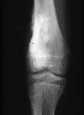
Question 1
A 10-year-old child has leg discomfort with activity. A radiograph, bone scan, and biopsy specimen are shown in Figures 1a through 1c. What is the most likely diagnosis?
Explanation
Question 2
A 13-year-old boy has pain and a firm mass in his left knee. A radiograph and MRI scan are shown in Figures 2a and 2b, and a biopsy specimen is shown in Figure 2c. Based on these findings, what is the most likely diagnosis?
Explanation
Question 3
A 10-year-old boy with a history of retinoblastoma now reports right knee pain. AP and lateral radiographs are shown in Figures 3a and 3b. What is the most likely diagnosis?
Explanation
Question 4
Which of the following tumors have characteristic chromosomal translocations?
Explanation
Question 5
What is the most common MRI appearance of a malignant soft-tissue sarcoma?
Explanation
Question 6
Which of the following factors is associated with the worst prognosis in soft-tissue sarcomas?
Explanation
Question 7
The lesion seen in Figure 4 is most likely the result of metastases from what solid organ?
Explanation
Question 8
A 7-year-old boy has a limp with pain and tenderness over the distal right femur. Radiographs are shown in Figures 5a and 5b. Based on these findings, what is the best course of action?
Explanation
Question 9
A 14-year-old patient has anterior knee pain. Radiographs, an MRI scan, and biopsy specimens are shown in Figures 6a through 6e. What is the most likely diagnosis?
Explanation
Question 10
A 47-year-old woman has an asymptomatic pelvic mass that was discovered on routine gynecologic examination. A radiograph, CT scan, MRI scan, and biopsy specimen are shown in Figures 7a through 7d. Metastatic work-up is negative. Treatment should consist of
Explanation
Question 11
A 20-year-old patient has foot pain. A radiograph and T1-weighted MRI scan are shown in Figures 8a and 8b. A biopsy specimen is shown in Figure 8c. Treatment should consist of
Explanation
Question 12
A 69-year-old man has a painful slow-growing lesion of the distal phalanx of his thumb. History reveals that he has had chronic osteomyelitis of the thumb for the past 12 years. The radiograph and biopsy specimens are seen in Figures 9a through 9c. Treatment should consist of
Explanation
Question 13
A 12-year-old girl has had right knee pain for the past 3 months. Radiographs and a coronal T2-weighted MRI scan are shown in Figures 10a through 10c. A biopsy specimen is shown in Figure 10d. What is the most appropriate treatment for this lesion?
Explanation
Question 14
Which of the following diagnostic studies best distinguishes Ewing's sarcoma from small cell osteosarcoma?
Explanation
Question 15
Figure 11a shows the AP pelvis radiograph of a 25-year-old man who sustained a spinal cord injury 10 years ago. A bone scan and a CT scan are shown in Figures 11b and 11c. To prevent recurrence after resection, management should consist of
Explanation
Question 16
A 40-year-old man has a palpable mass over the dorsum of the ankle. He reports no history of direct trauma but notes that he sustained a laceration to the middle of his leg 6 weeks ago. Examination reveals a 4-cm x 1-cm mass. T1- and T2-weighted MRI scans are shown in Figures 12a and 12b. An intraoperative photograph and biopsy specimen are shown in Figures 12c and 12d. What is the most likely diagnosis?
Explanation
Question 17
What is the most common presentation of a benign bone tumor in childhood?
Explanation
Question 18
Figures 13a and 13b show the MRI scans of a 70-year-old patient who has a posterior calf mass. Examination reveals that the mass extends to the midcalf level. A biopsy specimen reveals a high-grade soft-tissue sarcoma. Metastatic work-up shows no lesions. Management should consist of
Explanation
Question 19
What is the most common secondary malignancy arising in pagetic bone?
Explanation
Question 20
A 15-year-old girl with a midshaft fibular lesion has histologic findings consistent with Ewing's sarcoma. Following induction chemotherapy, local control typically consists of
Explanation
Question 21
Soft-tissue sarcomas most commonly metastasize to the
Explanation
Question 22
The incidence of osteosarcoma is highest in what age group?
Explanation
Question 23
A 9-year-old boy has a painless enlarged mass on the dorsum of his hand. Figures 14a through 14d show the clinical photograph, radiographs, and biopsy specimen. What is the most likely diagnosis?
Explanation
Question 24
What is the most common clinical presentation of a patient with a malignant bone tumor?
Explanation
Question 25
Figure 15a shows the radiograph of a patient who has a chondrosarcoma of the acetabulum. Bone scans are shown in Figures 15b and 15c. Numerous soft subcutaneous masses are present. A clinical photograph of the hand is shown in Figure 15d. What is the most likely diagnosis?
Explanation
Question 26
A 14-year-old boy presents with a painful mass in the diaphysis of his left femur. Biopsy reveals small, round blue cells. Cytogenetic analysis demonstrates a t(11;22) translocation. This chromosomal abnormality results in which of the following fusion proteins?
Explanation
Question 27
Viscoelastic materials, such as articular cartilage and tendons, exhibit time-dependent mechanical behaviors. Which of the following best describes the phenomenon of 'stress relaxation'?
Explanation
Question 28
A 35-year-old man undergoes open reduction and internal fixation of a transverse radial shaft fracture using a dynamic compression plate (DCP) that provides absolute stability. By which of the following mechanisms will the fracture heal?
Explanation
Question 29
In total hip arthroplasty, the use of highly cross-linked polyethylene (HXLPE) has significantly reduced wear rates compared to conventional ultra-high molecular weight polyethylene (UHMWPE). Which of the following trade-offs is most commonly associated with increasing the radiation dose to maximize cross-linking in HXLPE?
Explanation
Question 30
Articular cartilage is highly specialized to resist compressive and shear forces. In which zone of articular cartilage are the collagen fibers oriented parallel to the joint surface, providing the highest tensile strength?
Explanation
Question 31
Following a closed distal humerus fracture, a patient demonstrates complete radial nerve palsy. A nerve conduction study performed at 4 weeks shows absent distal motor responses and the presence of fibrillation potentials on electromyography (EMG). Assuming the nerve trunk is macroscopically intact, this injury is best classified as which of the following?
Explanation
Question 32
Low-molecular-weight heparin (LMWH) is commonly prescribed for deep vein thrombosis (DVT) prophylaxis following total knee arthroplasty. LMWH exerts its primary anticoagulant effect through the selective inhibition of which of the following coagulation cascade components?
Explanation
Question 33
Staphylococcus epidermidis is a common pathogen in periprosthetic joint infections due to its ability to form a robust biofilm on orthopedic implants. Which of the following is the primary constituent of the extracellular polymeric substance (EPS) that protects the bacteria in this biofilm?
Explanation
Question 34
A 25-year-old woman presents with multiple recurrent fragility fractures and a history of delayed dental eruption. Radiographs reveal generalized, diffusely sclerotic, 'bone-within-bone' appearance. This condition is most likely caused by a defect in which of the following cellular mechanisms?
Explanation
Question 35
A 65-year-old man presents with severe lower back pain and fatigue. Radiographs of the spine and pelvis reveal multiple punched-out lytic lesions without reactive sclerosis. Laboratory evaluation is notable for hypercalcemia, anemia, and an elevated serum creatinine. A bone marrow biopsy is expected to show an abnormal proliferation of which cell type?
Explanation
Question 36
A 14-year-old boy presents with progressive knee pain and swelling that awakens him at night. Radiographs show a permeative, destructive lesion in the distal femoral metadiaphysis with an 'onion-skin' periosteal reaction. Histology shows sheets of uniform small round blue cells. Which of the following immunohistochemical markers or genetic translocations is most characteristic of this lesion?
Explanation
Question 37
A 32-year-old woman presents with persistent knee pain. Radiographs demonstrate an eccentric, lytic, epiphyseal lesion in the proximal tibia extending to the subchondral bone without a sclerotic margin. Biopsy reveals multinucleated giant cells intermixed with mononuclear stromal cells. Denosumab is considered for adjuvant treatment prior to surgery. What is the mechanism of action of this medication?
Explanation
Question 38
Which of the following biochemical changes in articular cartilage is most characteristic of the earliest stage of osteoarthritis?
Explanation
Question 39
A 70-year-old man presents with an enlarging, painful mass in his left thigh. He has a long-standing history of Paget's disease of bone affecting his pelvis and left femur. Radiographs show a destructive lytic lesion in the proximal femur with cortical breakthrough and soft tissue extension. Biopsy confirms a secondary malignancy. What is the most common secondary malignancy arising in the setting of Paget's disease?
Explanation
Question 40
During endochondral ossification in fracture healing, which of the following transcription factors is essential for the differentiation of mesenchymal stem cells into chondrocytes?
Explanation
Question 41
Galvanic corrosion is a mode of implant failure that involves the electrochemical destruction of metal. It is most likely to occur in vivo when which of the following two metal alloys are placed in direct contact?
Explanation
Question 42
A 28-year-old woman presents with a slow-growing, painless mass on the posterior aspect of her distal femur. Radiographs reveal a densely mineralized, lobulated mass attached to the posterior cortex by a broad base, with a radiolucent cleft separating the mass from the underlying cortex ('string sign'). What is the characteristic genetic abnormality associated with this tumor?
Explanation
Question 43
A 4-year-old boy is evaluated for short stature and bowing of the legs. Laboratory studies show normal serum calcium, significantly decreased serum phosphate, elevated alkaline phosphatase, and normal parathyroid hormone levels. Genetic testing confirms an X-linked dominant mutation in the PHEX gene. Which of the following is the primary pathophysiologic mechanism of this disease?
Explanation
Question 44
When inserting a fully threaded cortical screw across a fracture to achieve rigid internal fixation without the use of a plate, which of the following represents the primary mechanism by which a screw generates interfragmentary compression (lag technique)?
Explanation
Question 45
A 65-year-old man presents with generalized bone pain, fatigue, and a recent pathological fracture of his left humerus. Laboratory tests reveal anemia, hypercalcemia, and elevated creatinine. A skeletal survey demonstrates multiple 'punched-out' lytic lesions in the skull, spine, and pelvis. A bone marrow biopsy shows >10% clonal plasma cells. Which of the following pathways is most directly responsible for uncoupling bone remodeling and causing the lytic lesions in this disease?
Explanation
Question 46
A 14-year-old boy presents with a destructive diaphyseal lesion of the femur. A biopsy reveals small round blue cells. Cytogenetic analysis demonstrates a t(11;22) translocation. Which of the following fusion proteins is most likely responsible for the pathogenesis of this tumor?
Explanation
Question 47
To maximize the pullout strength of a cortical screw inserted into diaphyseal bone, which of the following design modifications is most effective?
Explanation
Question 48
Which zone of articular cartilage is characterized by having the highest concentration of proteoglycans and the lowest concentration of water?
Explanation
Question 49
Which of the following bone graft substitutes acts primarily via an osteoinductive mechanism?
Explanation
Question 50
A 55-year-old man presents with acute knee pain and swelling. Synovial fluid analysis shows a leukocyte count of 55,000 cells/mm³ with 85% polymorphonuclear leukocytes. Microscopic evaluation demonstrates positively birefringent, rhomboid-shaped crystals. What is the most likely composition of these crystals?
Explanation
Question 51
During the sliding filament mechanism of skeletal muscle contraction, which of the following events directly follows the binding of adenosine triphosphate (ATP) to the myosin head?
Explanation
Question 52
A 35-year-old patient sustained a closed tibia fracture treated with closed reduction and casting. Which of the following best describes the primary source of cells responsible for the formation of the external soft callus during secondary bone healing?
Explanation
Question 53
Rivaroxaban is an oral anticoagulant commonly used for venous thromboembolism prophylaxis following total joint arthroplasty. By which of the following mechanisms does it primarily exert its antithrombotic effect?
Explanation
Question 54
A 45-year-old woman undergoes wide resection of a deep soft tissue sarcoma of the thigh. Histological examination reveals a biphasic pattern consisting of both epithelial and spindle cell components. Cytogenetic testing demonstrates a t(X;18) translocation. What is the most likely diagnosis?
Explanation
Question 55
A 60-year-old man with chronic osteomyelitis of the femur requires targeted antibiotic therapy. Intraoperative cultures grow methicillin-resistant Staphylococcus aureus (MRSA). Which of the following antibiotics is a bactericidal agent that disrupts the bacterial cell membrane by causing rapid depolarization, and requires monitoring for creatine kinase (CK) elevation due to the risk of myopathy?
Explanation
Question 56
A 14-year-old boy presents with a 2-month history of worsening thigh pain, particularly at night. Radiographs reveal a permeative diaphyseal lesion in the femur with an extensive 'onion-skin' periosteal reaction. Biopsy demonstrates sheets of small round blue cells that stain strongly for CD99. Which of the following cytogenetic abnormalities is most characteristic of this patient's diagnosis?
Explanation
Question 57
A 12-year-old girl is diagnosed with a conventional high-grade osteosarcoma of the distal femur. Her family history is notable for a mother who died of premenopausal breast cancer at age 32 and a brother who was treated for an adrenocortical carcinoma. A mutation in which of the following genes is the most likely underlying cause of this patient's susceptibility?
Explanation
Question 58
A 32-year-old woman presents with knee pain. Radiographs reveal an eccentric, expansile lytic lesion in the proximal tibia that extends to the subchondral bone. A biopsy is performed, revealing multinucleated giant cells interspersed within a background of mononuclear stromal cells. If pharmacological treatment is considered to downstage the tumor prior to surgery, what is the primary molecular target of the indicated medication?
Explanation
Question 59
An 18-year-old male presents with a 6-month history of dull, aching back pain that is worsened at night. He reports that the pain is only mildly relieved by ibuprofen. Imaging demonstrates a 2.5 cm radiolucent lesion with a central nidus located in the posterior elements of the L4 vertebra. Histology shows interlacing trabeculae of woven bone surrounded by prominent osteoblasts. What is the most likely diagnosis?
Explanation
Question 60
Which of the following zones of articular cartilage contains the highest concentration of proteoglycans, the lowest concentration of water, and collagen fibers oriented perpendicular to the articular surface?
Explanation
Question 61
A 45-year-old man undergoes closed reduction and intramedullary nailing for a closed, transverse midshaft tibial fracture. Which of the following accurately describes the predominant mechanism of bone healing expected in this scenario?
Explanation
Question 62
A 16-year-old girl presents with a rapidly enlarging, painful mass in her proximal humerus. Radiographs show an expansile, eccentric, multiloculated lytic lesion. Magnetic resonance imaging (MRI) demonstrates multiple fluid-fluid levels. Biopsy reveals blood-filled cystic spaces lacking an endothelial lining. Which of the following genetic alterations is the primary driver of this lesion?
Explanation
Question 63
An 8-year-old girl sustains a minor fall and presents with a proximal femur fracture. Radiographs reveal a 'shepherd's crook' deformity of the proximal femur and a diaphyseal lesion with a 'ground-glass' appearance. The patient has a history of precocious puberty and has large, irregular café-au-lait macules on her trunk. What is the underlying pathophysiological mechanism of her bone disease?
Explanation
Question 64
A 65-year-old active man is undergoing total hip arthroplasty. The surgeon opts to use a highly cross-linked polyethylene (HXLPE) liner. Which of the following best describes the mechanical trade-off when comparing HXLPE to conventional ultra-high-molecular-weight polyethylene (UHMWPE)?
Explanation
Question 65
A 40-year-old man requires bone grafting for an atrophic scaphoid nonunion. The surgeon utilizes a graft source that provides osteoconduction, osteoinduction, and osteogenesis. Which of the following graft materials possesses all three of these properties?
Explanation
Question 66
A 25-year-old female presents with a painless mass on the posterior aspect of her distal thigh. Radiographs reveal a dense, extensively ossified mass attached to the posterior cortex of the distal femur via a broad base. Biopsy shows well-formed woven bone trabeculae within a bland fibrous stroma, with minimal cellular atypia. What is the most likely diagnosis and the most appropriate standard of care?
Explanation
Question 67
A 14-year-old boy presents with a 3-month history of nocturnal thigh pain and low-grade fever. Radiographs demonstrate a permeative, diaphyseal lytic lesion in the femur with a multilamellated 'onion-skin' periosteal reaction. Core needle biopsy reveals uniform small round blue cells. Which of the following cytogenetic abnormalities is most characteristically associated with this patient's diagnosis?
Explanation
Question 68
A 55-year-old man presents with a constant, dull aching pain in his right pelvis that has been progressively worsening over the past 6 months. Radiographs demonstrate a large lytic lesion in the ilium with internal 'ring-and-arc' calcifications and focal cortical breakthrough. Biopsy confirms a grade II (intermediate-grade) chondrosarcoma. Which of the following is the most appropriate management?
Explanation
Question 69
A 32-year-old woman presents with worsening right knee pain. Radiographs reveal a large, eccentric, purely lytic, epiphyseal-metaphyseal lesion in the proximal tibia extending to the subchondral bone. Histology shows numerous multinucleated giant cells in a background of mononuclear spindle cells. The patient is deemed an unresectable surgical candidate and denosumab therapy is initiated. What is the precise mechanism of action of this medication?
Explanation
Question 70
A 65-year-old man presents with intractable lower back pain and fatigue. Radiographs show multiple punched-out lytic lesions in the skull and several vertebral compression fractures. Laboratory tests reveal hypercalcemia and a normocytic anemia. Serum protein electrophoresis shows an M-spike. Which of the following imaging modalities is the most sensitive for assessing the total extent of skeletal involvement in this condition?
Explanation
Question 71
A 19-year-old man presents with localized right leg pain that is significantly worse at night and dramatically relieved within 30 minutes of taking ibuprofen. Imaging demonstrates a 7-mm radiolucent nidus surrounded by dense, reactive sclerotic bone in the anterior cortex of the mid-diaphyseal tibia. If the patient desires definitive treatment but wishes to avoid open surgery, which of the following is considered the standard of care?
Explanation
Question 72
A 28-year-old man presents with a slowly enlarging, relatively painless mass around his left ankle. Radiographs show an eccentric soft tissue mass with stippled calcifications adjacent to, but not involving, the joint space. MRI reveals a heterogeneous mass with a 'triple signal' pattern on T2 sequences. Biopsy confirms a biphasic tumor comprised of epithelial and spindle cell components. Which chromosomal translocation is pathognomonic for this tumor?
Explanation
Question 73
A 9-year-old boy presents with mild shoulder pain after throwing a baseball. Radiographs reveal a centrally located, completely lytic, expansile lesion in the proximal humerus metaphysis that does not breach the cortex. A subtle 'fallen leaf' sign is noted. What biochemical substance is typically found in high concentrations within the fluid of this lesion?
Explanation
Question 74
A 12-year-old girl is evaluated for a prominent leg length discrepancy and a limp. She has a documented history of precocious puberty and has several large, irregular café-au-lait spots with jagged borders on her trunk. Radiographs of her proximal femur demonstrate a 'shepherd's crook' deformity with a ground-glass appearance of the medullary canal. A mutation in which of the following genes is the underlying cause of this patient's syndrome?
Explanation
Question 75
A 45-year-old woman is referred after an incidental finding of a calcified lesion in her proximal humerus during a routine chest radiograph. Subsequent shoulder MRI shows a 3 cm well-circumscribed, lobulated cartilaginous lesion localized entirely within the medullary canal. There is no endosteal scalloping, cortical breakthrough, periosteal reaction, or soft tissue extension. The patient reports absolutely no pain in her shoulder. What is the most appropriate management?
Explanation
Question 76
A 55-year-old man presents with a painful mass in his proximal humerus. A radiograph reveals an intralesional, destructive process with stippled, popcorn-like calcifications. Biopsy confirms the diagnosis of a conventional central chondrosarcoma. Which of the following genetic mutations is most commonly associated with this primary bone tumor?
Explanation
Question 77
You are assisting in the design of a new cancellous screw for metaphyseal fracture fixation. Which of the following modifications to the screw design will most significantly increase its pullout strength?
Explanation
Question 78
Highly cross-linked polyethylene (HXLPE) is widely used in total hip arthroplasty to reduce the incidence of wear and subsequent osteolysis. What is the primary negative biomechanical consequence of increasing the radiation dose used for cross-linking the ultra-high-molecular-weight polyethylene (UHMWPE)?
Explanation
Question 79
A 24-year-old woman presents with a slowly enlarging, painful mass deep within the plantar aspect of her foot. MRI demonstrates a well-circumscribed soft tissue mass near the plantar fascia. Biopsy reveals a biphasic tumor consisting of epithelial cells forming glandular structures and spindle cells in a fascicular pattern. Which cytogenetic abnormality is diagnostic for this tumor?
Explanation
Question 80
A 65-year-old woman with severe osteoporosis is started on denosumab therapy. This medication primarily exerts its mechanism of action by directly inhibiting which of the following cellular interactions?
Explanation
Question 81
A 32-year-old woman presents with worsening knee pain. Radiographs display an eccentric, lytic epiphyseal-metaphyseal lesion of the distal femur extending to the subchondral bone. A biopsy demonstrates numerous multinucleated giant cells uniformly distributed in a background of mononuclear stromal cells. Which of the following statements is true regarding the cellular biology and treatment of this lesion?
Explanation
Question 82
Which of the following represents the earliest biochemical change observed in articular cartilage during the pathogenesis of osteoarthritis?
Explanation
Question 83
A 28-year-old man sustains a closed midshaft humerus fracture and immediately develops a complete radial nerve palsy. At 12 weeks post-injury, he has zero motor recovery. An electromyogram (EMG) shows prominent fibrillation potentials but no motor unit action potentials. What pathophysiologic process best describes what is occurring in the nerve segment distal to the fracture site?
Explanation
Question 84
A 35-year-old man undergoes open reduction and internal fixation of a diaphyseal radius fracture. The surgeon utilizes compression plating to achieve absolute stability. Assuming standard biological conditions, which of the following mechanisms best describes the primary mode of fracture healing expected?
Explanation
Question 85
A 9-year-old boy presents with anterolateral bowing of his left tibia. Physical examination reveals six large cafe-au-lait macules and axillary freckling. The primary underlying genetic defect in this patient results in an abnormality of which of the following cellular signaling pathways?
Explanation
Question 86
A 35-year-old male presents with a slowly enlarging, painless mass around his left knee. MRI demonstrates a soft tissue mass adjacent to the joint capsule but without intra-articular extension. Biopsy reveals a biphasic pattern consisting of spindle cells and epithelial cells. Which of the following chromosomal translocations is most characteristic of this patient's diagnosis?
Explanation
Question 87
A 28-year-old female presents with progressive knee pain. Radiographs reveal an eccentric, purely lytic epiphyseal lesion extending to the subchondral bone of the distal femur. Histological analysis confirms the presence of uniform mononuclear cells mixed with numerous multinucleated giant cells. For unresectable or recurrent disease, systemic therapy can be utilized. Which of the following pathways is the primary target for the medication used in this clinical scenario?
Explanation
Question 88
A 14-year-old boy presents with pain and swelling over his proximal humerus after minor trauma. Radiographs show an expansile, radiolucent, metaphyseal lesion. MRI demonstrates multiple fluid-fluid levels within the lesion. A core biopsy reveals blood-filled cavernous spaces lacking an endothelial lining. A cytogenetic abnormality in which of the following genes is most likely associated with the pathogenesis of this primary lesion?
Explanation
Question 89
A 9-year-old girl is evaluated for a limp and a leg length discrepancy. Radiographs of her proximal femur demonstrate a 'shepherd's crook' deformity with a ground-glass matrix in the medullary canal. She has a history of precocious puberty and café-au-lait macules with irregular, 'coast of Maine' borders. The underlying pathophysiology of her musculoskeletal condition involves a somatic activating mutation in a gene encoding for:
Explanation
Question 90
A 15-year-old boy presents with fever, weight loss, and severe mid-thigh pain. Radiographs demonstrate a permeative diaphyseal lesion of the femur with a prominent 'onion-skin' periosteal reaction. Histology reveals solid sheets of uniform small round blue cells. Immunohistochemistry is strongly positive for CD99. Which of the following fusion gene products is the primary driver of this malignancy?
Explanation
Question 91
A 14-year-old boy presents with right knee pain that wakes him at night. Radiographs show a destructive metaphyseal lesion of the distal femur with a sunburst periosteal reaction and Codman's triangle. A biopsy confirms high-grade osteoblastic osteosarcoma. A germline mutation in which of the following tumor suppressor genes would most strongly predispose this patient to both this primary bone tumor and breast cancer later in life?
Explanation
Question 92
A 55-year-old man presents with a constant, dull ache in his pelvis. Radiographs reveal a large, lytic lesion in the ilium with 'popcorn-like' chondroid calcifications. Biopsy demonstrates hypercellular hyaline cartilage with plump, pleomorphic, and binucleated chondrocytes penetrating host bone trabeculae. Which of the following represents the most appropriate primary treatment modality for the conventional type of this tumor?
Explanation
Question 93
A 19-year-old male complains of severe right thigh pain that is significantly worse at night and promptly relieved by NSAIDs. Computed tomography (CT) displays a 1-cm radiolucent nidus surrounded by dense, sclerotic reactive bone in the femoral diaphysis. The profound pain experienced by this patient is directly mediated by an elevated local concentration of which of the following substances?
Explanation
Question 94
A 45-year-old male presents with a deep, painless, intramuscular mass in his thigh. MRI shows a multi-lobulated mass that is hyperintense on T2-weighted images and contains small nodules of macroscopic fat. Histology reveals primitive non-lipogenic mesenchymal cells, an arborizing 'chicken-wire' capillary network, and signet-ring lipoblasts within a prominent myxoid stroma. Which of the following cytogenetic abnormalities is classically associated with this diagnosis?
Explanation
Question 95
A 40-year-old male presents with a one-year history of chronic, mild right shoulder pain. Radiographs demonstrate a lytic lesion located purely in the epiphysis of the proximal humerus, with a fine sclerotic margin and central calcification. Histology shows sheets of cells with abundant clear cytoplasm and distinct cell membranes, interspersed with areas of hyaline cartilage and reactive trabeculae of woven bone. What is the most likely diagnosis?
Explanation
Question 96
A 15-year-old boy presents with a painful mass in his distal femur. Radiographs reveal a permeative, destructive diaphyseal lesion with a lamellated periosteal reaction. Biopsy reveals sheets of uniform small blue round cells with scant cytoplasm. Cytogenetic analysis is pending. What is the most common translocation associated with this diagnosis, and what fusion protein does it create?
Explanation
Question 97
A 32-year-old woman presents with severe knee pain and a lytic lesion in the proximal tibia extending right to the subchondral bone. Biopsy confirms a giant cell tumor of bone (GCTB). Due to the extensive size and joint proximity precluding immediate joint-salvage surgery, neoadjuvant medical therapy is planned. Which of the following accurately describes the mechanism of the most appropriate pharmacological agent?
Explanation
Question 98
A 14-year-old girl presents with pain and a rapidly expanding mass in her proximal humerus following a minor fall. Radiographs demonstrate an eccentrically located, expansile, purely lytic metaphyseal lesion. MRI exhibits multiple fluid-fluid levels within the lesion. Histological analysis of a core biopsy demonstrates blood-filled cystic spaces separated by fibrous septa containing multinucleated giant cells, with no significant atypia. Which of the following genetic alterations is most characteristic of the primary form of this specific lesion?
Explanation
Question 99
A 19-year-old man reports a 6-month history of right thigh pain that is particularly severe at night and brings him out of sleep. He notes that taking over-the-counter ibuprofen provides dramatic and near-complete relief for several hours. Radiographs show profound diaphyseal cortical thickening with a 7-mm radiolucent nidus. The intense pain and the specific response to nonsteroidal anti-inflammatory drugs (NSAIDs) in this condition are directly related to the high local concentration of which of the following mediators?
Explanation
Question 100
A 28-year-old male presents with a slowly enlarging, deep, tender mass in the plantar aspect of his left foot, which he noticed two years ago. MRI demonstrates a well-circumscribed soft tissue mass intimately associated with the plantar fascia, exhibiting heterogeneous hyperintensity on T2-weighted imaging. A core needle biopsy reveals a biphasic pattern of spindle cells and epithelial cells forming glandular structures. What is the most definitive diagnostic molecular finding for this tumor?
Explanation
None
