العربية
Part of the Master Guide

General Orthopedics 2026 Practice Questions: Set 13 (Solved)

Orthopedic Sports Medicine 2026 MCQs: Board Review Questions & Answers (Part 3)

23 Apr 2026 82 min read 77 Views
Figure for Sports Medicine 2004 MCQs - Part 3 - Question 53

Key Takeaway

We review everything you need to understand about Orthopedic Sports Medicine 2026 MCQs: Board Review Questions & Answers (Part 3). Top-rated Orthopedic Sports Medicine 2026 MCQs bank. Practice with clinical case questions, orthopedic surgery board review, and evidence-based answers updated for 2026.

Orthopedic Sports Medicine 2026 MCQs: Board Review Questions & Answers (Part 3)

Comprehensive 100-Question Exam


00:00

Start Quiz

Question 1

A 32-year-old amateur bowler has progressive pain in the lateral aspect of the proximal forearm and elbow. Nonsurgical management consisting of a tennis elbow brace, nonsteroidal anti-inflammatory drugs, and activity modification has failed to provide relief. Examination reveals tenderness in the lateral aspect of the proximal forearm and exacerbation of symptoms with resisted finger extension. Radiographs of the elbow reveal no abnormalities. Which of the following studies will aid in diagnosis?


Explanation

It is often difficult to accurately discern between lateral epicondylitis and radial tunnel syndrome. Neither MRI nor a bone scan is likely to reveal abnormalities. Electrodiagnostic studies are often inconclusive, and radial tunnel syndrome often presents without motor weakness. The symptoms of radial tunnel syndrome are expected to improve with an injection of lidocaine into the radial tunnel; therefore, this is the test of choice in this clinical scenario. Radiographs of the wrist will not assist in making the diagnosis. Eversmann WW Jr: Entrapment and compression neuropathies, in Green DP (ed): Operative Hand Surgery, ed 3. New York, NY, Churchill Livingston, 1993, pp 1341-1385.

Question 2

What is the most common complication associated with scalene regional anesthesia for shoulder procedures?


Explanation

Failure of the scalene block, necessitating general anesthesia or the immediate administration of narcotic medications, is the most common complication, occurring in 3% to 18% of patients. Cardiac arrest or cardiovascular collapse has been reported in anecdotal occurrences. Seizure that is the result of intravascular injection of local anesthetic is a rare complication, with an incidence reported of 0% to 6%. Neurologic complications, including laryngeal and phrenic nerve injuries, are rare although parathesias lasting up to 2 weeks have been reported in up to 3% of patients. Weber SC, Jain R: Scalene regional anesthesia for shoulder surgery in a community setting: An assessment of risk. J Bone Joint Surg Am 2002;84:775-779.

Question 3

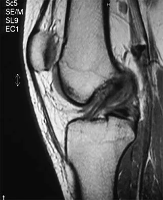
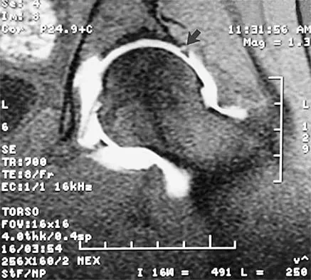
Figure 22 shows the MRI scan of a 20-year-old female basketball player who has pain over the anterior knee that interferes with her performance. Examination reveals phase III Blazina patellar tendinosis. Management should consist of


Explanation

Excision of the affected mucoid degenerative area is considered appropriate management in the Blazina classification system. A finding of phase III indicates persistent pain with or without activities, as well as deterioration of performance. With the appearance of the mucoid degeneration and the vigorous activity level of the intercollegiate basketball player, it is unlikely that nonsurgical management will provide adequate relief. When excising the affected degenerative area, care must be taken to retain normal tendon fibers. The defect in the patellar tendon is closed with absorbable sutures, as is the paratenon. Postoperative rehabilitation involves initial mobilization extension, with progressive range-of-motion and mobilization exercises as tolerated and weight bearing as tolerated. Open chain and isokinetic exercises are delayed until full range of motion and mobility is obtained, generally within 4 weeks. A return to activities is achieved by 80% to 90% of athletes, although there may be occasional activity-related aching for 4 to 6 months after surgery. Blazina ME, et al: Jumper's knee. Orthop Clin North Am 1973;4:665. Kelly DW, Carter VS, Jobe FW, Kerlan RK: Patellar and quadriceps tendon ruptures: Jumper's knee. Am J Sports Med 1984;12:375-380. Krums PE, Ryder B: Operative treatment of patella tendon disorders. Operative Techniques Sports Med 1994;2:303.

Question 4

When comparing the failure load of an evenly tensioned four-stranded hamstring tendon anterior cruciate ligament autograft to a 10-mm bone-patellar tendon-bone autograft, the hamstring graft will fail at a tension


Explanation

The failure load of an evenly tensioned four-stranded hamstring tendon autograft has been reported to be 4,500 Newtons. The failure load of a 10-mm patellar tendon autograft has been estimated at 2,600 Newtons. The intact anterior cruciate ligament failure load has been calculated at 1,725 Newtons. Corry IS, Webb JM, Clingeleffer AJ, Pinczewski LA: Arthroscopic reconstruction of the anterior cruciate ligament: A comparison of patellar tendon autograft and four-strand hamstring tendon autograft. Am J Sports Med 1999;27:448-454. Hamner DL, Brown CH Jr, Steiner ME, et al: Hamstring tendon grafts for reconstruction of the anterior cruciate ligament: Biomechanical evaluation of the use of multiple strands and tensioning techniques. J Bone Joint Surg Am 1999;81:549-557.

Question 5

What pathology is most likely to result in failure of an arthroscopic Bankart repair?


Explanation

Recent studies have documented that an arthroscopic Bankart repair performed with good technique can produce success rates similar to an open repair. However, the results of an arthroscopic repair deteriorate significantly if there is a 25% or greater anterior-inferior glenoid rim defect (inverted pear configuration) or an engaging Hill-Sachs lesion in which the humeral head defect keys onto the glenoid rim in abduction and external rotation. If either of these entities exist or there is multidirectional instability with pathologic hyperextensible tissue laxity, an open repair is recommended. An associated SLAP lesion would not significantly affect the result of the Bankart procedure. Not infrequently, the anterior glenoid labrum is partially or completely disrupted and, in itself, is not a contraindication to arthroscopic Bankart repair. In almost all patients with predominantly unidirectional instability, some degree of capsular/anterior-inferior glenohumeral ligament attenuation is present and can be addressed during the arthroscopic repair. Burkhart SS, De Beer JF: Traumatic glenohumeral bone defects and their relationship to failure of arthroscopic Bankart repairs: Significance of the inverted-pear glenoid and the humeral engaging Hill-Sachs lesion. Arthroscopy 2000;16:677-694.

Question 6

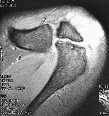
A 37-year-old man has had isolated chronic knee swelling for the past 6 months. He denies any history of specific trauma. Examination reveals a large effusion with a stable knee, but the remainder of the examination is normal. Plain radiographs are unremarkable. An MRI scan reveals a large effusion without meniscal injury. An arthroscopic image of the suprapatellar pouch is shown in Figure 23. What is the most likely diagnosis?


Explanation

The history and physical examination are consistent with a monoarticular joint condition but not typical of joint sepsis. The arthroscopic appearance of brownish proliferative synovium is typical of PVNS. PVNS is a monoarticular synovial disease of unknown etiology and is treated with total synovectomy. The proliferative synovitis is not consistent with chondromalacia. Synovial cell sarcoma is an extracapsular disease. Rheumatoid arthritis typically is polyarticular, and the synovial appearance is not associated with hemosiderin deposition. Flandry FC, Hughston JC, Jacobson KE, Barrack RL, McCann SB, Kurtz DM: Surgical treatment of diffuse pigmented villonodular synovitis of the knee. Clin Orthop 1994;300:183-192.

Question 7

A 35-year-old recreational basketball player reports shoulder pain following a sprawl for a rebound. While examination reveals that he can actively elevate the arm with pain, a subacromial injection fails to provide relief. An MRI scan reveals medial subluxation of the long head of the biceps. Which of the following structures most likely has also been injured?


Explanation

Subscapularis tears can be associated with disruption of the transverse ligament supporting the biceps. The remaining aspects of the rotator cuff, superior labrum, and capsule can be intact with this injury. Petersson CJ: Spontaneous medial dislocation of the tendon of the long biceps brachii. Clin Orthop 1986;211:224-227.

Question 8

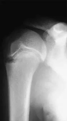
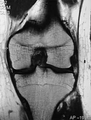
An 18-year-old hockey player sustains an acute anterior shoulder dislocation that requires manual reduction. At arthroscopy, the lesion shown in Figure 24 will be observed in what percent of patients?


Explanation

The clinical photograph shows an acute capsulolabral avulsion from the anterior glenoid, also referred to as a Perthes-Bankart lesion. In patients who sustain an acute dislocation that requires a manual reduction, this pathologic lesion is observed with high frequency. In several research studies, it has been visualized in 80% to 95% of patients at arthroscopy. Taylor DC, Arciero RA: Pathologic changes associated with shoulder dislocations: Arthroscopic and physical examination findings in first-time, traumatic anterior dislocations. Am J Sports Med 1997;25:306-311.

Question 9

A 22-year-old competitive volleyball player has shoulder pain, and rest and a cortisone injection have failed to provide relief. Examination reveals atrophy along the posterior scapula, but an MRI scan does not reveal a rotator cuff tear or labral cyst. What is the most likely cause for the shoulder weakness?


Explanation

Repetitive overhead slams and serves may produce a traction injury to the distal branch of the suprascapular nerve. Bankart, biceps, and superior labrum anterior and posterior injuries can occur but usually do not produce visible atrophy. Muscle avulsion is uncommon. Ferretti A, Cerullo G, Russo G: Suprascapular neuropathy in volleyball players. J Bone Joint Surg Am 1987;69:260-263.

Question 10

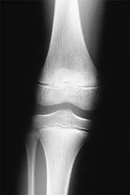
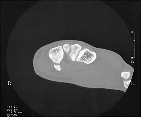
An 11-year-old female gymnast has had gradually increasing right wrist pain for the past 6 months. Examination reveals normal range of motion and strength. Moderate tenderness is present over the distal radius. AP radiographs will most likely show


Explanation

Distal radial physeal stress syndrome has been reported in up to 25% of nonelite gymnasts showing premature closure of the distal radial physis and distal ulnar overgrowth, producing positive ulnar variance. The diagnosis should be suspected when there is tenderness at the distal radial physis in a young gymnast. The pathology is thought to be the result of repetitive compressive stresses caused by upper extremity weight-bearing forces. The recommended treatment is 3 to 6 months of rest. Salter-Harris fractures with a distal radial epiphyseal slip are unlikely, especially in the absence of a specific traumatic event. Mandelbaum BR, Bartolozzi AR, Davis CA, Teurlings L, Bragonier B: Wrist pain syndrome in the gymnast: Pathogenetic, diagnostic, and therapeutic consideration. Am J Sports Med 1989;17:305-317.

Question 11

A 22-year-old wrestler who underwent an open anterior shoulder reconstruction to repair a dislocated shoulder 6 months ago now reports shoulder pain after attempting a takedown. Examination reveals external rotation that is 15 degrees greater than the contralateral side. He has pain associated with abduction and external rotation but no apprehension. Which of the following tests would most likely reveal positive findings?


Explanation

Postoperative subscapularis detachment can be identified with a positive lift-off test that reveals weakness in internal rotation. This complication does not necessarily compromise the anterior capsule repair. The load-and-sift maneuver and articular contrast studies may be normal. Supraspinatus tests for impingement and weakness should be negative. Gerber C, Krushell RJ: Isolated ruptures of the tendon of the subscapularis muscle: Clinical fractures in 16 cases. J Bone Joint Surg Br 1991;73:389-394.

Question 12

Figures 25a and 25b show the clinical photographs of a 19-year-old baseball outfielder who has shoulder pain after sliding headfirst into second base. He reports pain while batting, sliding, and catching. Examination reveals a posterior prominence during midranges of forward elevation, which then disappears with a palpable clunk during terminal elevation and abduction. What is the most likely diagnosis?


Explanation

25b A headfirst slide with the arm extended can injure the posterior shoulder. Winging of the scapula is dynamic and is considered a compensatory effort to prevent subluxation; it is not related to nerve injury. Posterior glenohumeral subluxation can be present during the initiation of a bat swing. Rotator cuff function, interval tears, and superior labrum tears can be painful but do not produce winging. Kuhn JE, Plancher KD, Hawkins RJ: Scapular winging. J Am Acad Orthop Surg 1995;3:319-325.

Question 13

A soccer player who sustained a twisting injury to the right ankle while making a cut is unable to bear weight and has diffuse tenderness over the anterior and lateral aspects of the ankle. Examination also shows a positive squeeze test. Plain radiographs and a stress radiograph are shown in Figures 26a through 26c. Radiographs of the leg and knee are normal. What is the most appropriate management?


Explanation

26b 26c The mechanism of injury, physical examination, and radiographs indicate a "high" ankle sprain with disruption of the distal tibiofibular ligaments and interosseous membrane. These injuries typically involve pronation and external rotation forces. In addition, recovery is significantly delayed, often requiring 6 to 8 weeks to heal. Radiographs obtained months after recovery often show calcification within the distal syndesmosis, which is not typically symptomatic. This patient has gross instability, resulting in a high incidence of chronic diastasis and subluxation leading to impaired function. Treatment should consist of reduction and stabilization with a transsyndesmotic screw because this injury demonstrates a widened syndesmosis. Boytim MJ, Fisher DA, Neumann L: Syndesmotic ankle sprains. Am J Sports Med 1991;19:294-298.

Question 14

When compared with the normal anterior cruciate ligament (ACL), placement of an anterior cruciate ligament graft in the over-the-top position on the femoral side has what effect on its function?


Explanation

The placement of ACL graft with respect to its femoral and tibial attachments has a significant effect on its function. Evidence has shown that if the graft is placed in the over-the-top position, the graft will become lax in flexion and more taut with extension. Conversely, if the graft is placed too anterior on the femoral side, it will tighten in flexion and become lax in extension. Azar FM: Revision anterior cruciate ligament reconstruction. Instr Course Lect 2002;51:335-342. Draganich LF, Hsieh YF, Sherwin SH, et al: Intra-articular anterior cruciate ligament graft placement on the average most isometric line on the femur: Does it reproducibly restore knee kinematics? Am J Sports Med 1999;27:329-334.

Question 15

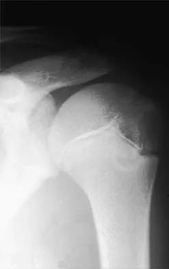
An 11-year-old boy has right shoulder pain and has been unwilling to use the arm after throwing a baseball in a Little League game 3 weeks ago. Examination reveals upper arm and shoulder tenderness with swelling. A radiograph and MRI scan are shown in Figures 27a and 27b. Management should consist of


Explanation

27b The radiograph is consistent with a unicameral (simple) bone cyst. The MRI scan reveals that the cyst is juxtaposed to the physis and therefore can be classified as active (latent cysts are more than 1 cm away from the physis). Active cysts are treated with aspiration and steroid injection, although repeated injections may be necessary. Curettage and bone grafting results in more reliable healing but may lead to growth arrest in active cysts. Iannotti JP, Williams GR: Disorders of the Shoulder: Diagnosis and Management, ed 1. Philadelphia, PA, Lippincott Williams & Wilkins, 1999, pp 945-946.

Question 16

A 37-year-old recreational tennis player undergoes surgery for tennis elbow. Following surgery, she describes clicking and popping on the lateral aspect of the elbow. A lateral pivot shift test is positive. What is the most likely cause of her symptoms?


Explanation

The patient has a posterolateral rotatory instability (PLRI) of the elbow that is most likely the result of iatrogenic injury to the lateral ulnar collateral ligament, the main ligament implicated in PLRI. The anterior band of the medial collateral ligament is implicated in valgus instability. Injury to the radial nerve is unlikely, and the lateral radial collateral ligament makes less of a contribution to elbow stability than does the ulnar component. While the origin of the extensor carpi radialis brevis may contribute to elbow stability, it is not as important a stabilizer as the lateral ulnohumeral ligament. O'Driscoll SW, Morrey BF: Surgical reconstruction of the lateral collateral ligament, in Morrey BF (ed): The Elbow. Philadelphia, PA, Lippincott, Williams and Wilkins, 1994, pp 169-182.

Question 17

An 18-year-old football halfback reports that he had immediate right knee pain after being tackled 1 week ago. Examination now reveals moderate tenderness over the proximal medial tibia and lateral joint and normal cruciate stability. In evaluating the integrity of the posterolateral knee structures, what is the most reliable examination finding?


Explanation

The most reliable test for a relatively isolated posterolateral complex (PLC) injury is the asymmetric tibial external rotation or "dial test." It can be performed with the patient prone or supine. When greater than 10 degrees of external rotation at 30 degrees of flexion is present when compared with the opposite knee, it indicates significant damage to the posterolateral structures. Asymmetric external rotation, which is also present at 90 degrees of flexion, indicates injury to the posterior cruciate ligament (PCL) as well. Varus laxity may indicate significant damage to both the PLC and PCL. Approximately 35% of the normal population may have a reverse pivot shift when examined under anesthesia; therefore, it is considered a less specific test. The external rotation/recurvatum and posterolateral drawer tests are adjunctive in assessing isolated posterolateral laxity but are not thought to be as reliable. Veltri DM, Warren RF: Isolated and combined posterior cruciate injuries. J Am Acad Orthop Surg 1993;1:67-75.

Question 18

Figures 28a through 28d show the radiographs and MRI scans of a 20-year-old basketball player who sustained an inversion injury to his right ankle. Management should consist of


Explanation

28b 28c 28d Osteochondral fractures involving the talar dome have been classified based on radiographic and MRI findings. A nondisplaced and incomplete fracture may be treated effectively with a short leg cast and no weight bearing for 6 weeks. This patient has a complete, separated, and displaced osteochondral fragment involving the midlateral talar dome that will most likely cause pain, mechanical symptoms, and effusion if treated nonsurgically. In addition, there is very little bone remaining on the fragment, making the likelihood of healing with open reduction and internal fixation problematic. The treatment of choice includes arthroscopy, removal of the loose fragment, curettage or drilling of the base, and a rehabilitation program that emphasizes peroneal strengthening, range of motion, and proprioceptive training. Lutter LD, Mizel MS, Pfeffer GB (eds): Orthopaedic Knowledge Update: Foot and Ankle. Rosemont, IL, American Academy of Orthopaedic Surgeons, 1994, pp 205-226.

Question 19

A 19-year-old rugby player has severe knee pain after being injured in a game 2 weeks ago. Examination reveals a knee effusion, limited motion, and increased 3+ Lachman's test and anterior drawer. There is also increased external rotation at 30 degrees of knee flexion when the patient is placed in the prone position. Based on these findings, which of the following actions would most likely increase the risk of anterior cruciate ligament (ACL) reconstruction failure?


Explanation

The patient has a combined ACL and posterolateral corner injury. Failure to diagnose and treat an injury of the posterolateral corner in a patient who has a tear of the anterior or posterior cruciate ligament can result in failure of the reconstructed cruciate ligament. The tibial external rotation test is best performed with the patient in the prone position. A 10-degree side-to-side difference of external rotation at 30 degrees of knee flexion indicates injury to the posterolateral corner. Acute grade III isolated or combined injuries of the posterolateral corner are best treated early by direct repair or by augmentation or reconstruction of all injured ligaments. Postoperative arthrofibrosis after an ACL reconstruction has been observed with preoperative deficiencies of knee motion. Veltri DM, Warren RF: Posterolateral instability of the knee. J Bone Joint Surg Am 1994;76:460-472.

Question 20

The primary function of structure "A" in Figure 29 is to limit


Explanation

The primary function of the popliteofibular ligament is to resist posterolateral rotation of the tibia on the femur, although it also secondarily resists varus angulation and posterior displacement of the tibia on the femur. The posterior cruciate ligament resists posterior tibial displacement, especially at 90 degrees of flexion. The lateral collateral ligament primarily resists varus displacement at 30 degrees of flexion but also resists posterolateral rotatory displacement with flexion that is less than approximately 50 degrees. The anterior and posterior cruciate ligaments resist varus displacement (along with the lateral collateral ligament) at 0 degrees of flexion. The anterior cruciate ligament primarily resists anterolateral displacement of the tibia on the femur. Sugita T, Amis AA: Anatomic and biomechanical study of the lateral collateral and popliteofibular ligaments. Am J Sports Med 2001;29:466-472.

Question 21

While lifting weights, a patient feels a pop in his arm. He has the deformity shown in Figure 30. If left untreated, the patient will have the greatest deficiency in


Explanation

The patient has a distal biceps rupture. While the distal biceps contributes to elbow flexion, its main function is forearm supination. Baker BE, Bierwagen D: Rupture of the distal tendon of the biceps brachii: Operative versus non-operative treatment. J Bone Joint Surg Am 1985;67:414-417. D'Arco P, Sitler M, Kelly J, et al: Clinical, functional, and radiographic assessments of the conventional and modified Boyd-Anderson surgical procedures for repair of distal biceps tendon ruptures. Am J Sports Med 1998;26:254-261.

Question 22

Myositis ossificans is a recognized complication of contusion to the quadriceps muscle. During early rehabilitation, this condition is most likely to be exacerbated by


Explanation

Passive stretching is contraindicated during rehabilitation as it may potentiate the severity of the myositis ossificans. Electrical stimulation, iontophoresis, isometric exercise, and ice/heat contrast are not known to exacerbate this process. Brunet ME, Hontas RB: The thigh, in DeLee JC, Drez D (eds): Orthopaedic Sports Medicine. Philadelphia, PA, WB Saunders, 1994, pp 1086-1112.

Question 23

Which of the following symptoms are most commonly associated with piriformis syndrome?


Explanation

Piriformis syndrome is best characterized by localized posterior hip pain and radicular symptoms in the sciatic distribution because of compression of the piriformis muscle on the sciatic nerve. Weakness in hip extension is not a characteristic finding, nor is pain with hip abduction or flexion. Hypesthesia of the lateral thigh would be more characteristic of a lesion of the lateral femoral cutaneous nerve. Radiating medial thigh pain would suggest hip joint pathology or upper lumbar nerve root irritation. Weakness in internal rotation is not a characteristic feature, and hypesthesia of the perineum would suggest possible involvement of the pudendal nerve. Byrd JWT: Thigh, hip, and pelvis, in Miller MD, Cooper DE, Warner JJP (eds): Review of Sports Medicine and Arthroscopy, ed 2. Philadelphia, PA, WB Saunders, 2002, pp 114-139.

Question 24

A 19-year-old football player who sustained three traumatic anterior shoulder dislocations underwent surgery to repair a Bankart lesion. Nine months after surgery, examination reveals stability, elevation to 150 degrees, external rotation to 0 degrees with the elbow at his side and to 50 degrees at 90 degrees of abduction, and internal rotation to T12. If his range of motion does not improve, he is at most risk for


Explanation

Loss of external rotation can lead to degenerative joint disease following an anterior stabilization procedure. A tight anterior capsule will prevent internal impingement. Risk of thoracic outlet syndrome should not be increased. Subscapularis detachment is a risk following open anterior repair; however, a gain in external rotation would be noted. In time, this patient's shoulder may show increased posterior glenohumeral wear but should not have symptoms of recurrent subluxation unless multidirectional instability is present. Hawkins RJ, Angelo RL: Glenohumeral osteoarthrosis: A late complication of the Putti-Platt repair. J Bone Joint Surg Am 1990;72:1193-1197.

Question 25

A 30-year-old man underwent an open Bankart repair with capsulorrhaphy for recurrent anterior instability 6 months ago. In a recent fall, he described a hyperabduction and external rotation mechanism of injury. He denies dislocating his shoulder. He now has anterior shoulder pain, weakness, and the sensation of instability. Examination reveals tenderness just lateral to the coracoid and bicipital groove. An MRI scan is shown in Figure 31. Management should now consist of


Explanation

Subscapularis tendon tears are being recognized with increasing frequency, and the mechanism of injury involves hyperabduction and external rotation. The patient will have anterior shoulder pain and may report a sensation of instability. Examination will reveal anterior shoulder tenderness over the lesser tuberosity and bicipital groove, and the Gerber lift-off test usually is positive. The MRI scan shown here reveals an intact anterior labrum. The subscapularis tendon is avulsed and retracted, with no evidence of the biceps tendon within the groove; this implies dislocation of the biceps, a common accompanying feature of a subscapularis tear. This injury is also recognized as a complication after open anterior shoulder stabilizations where the subscapularis has been incised as part of the approach. Therefore, the appropriate management involves repair of the subscapularis. The injury does not represent a recurrence so immobilization or revision stabilization, which may be reasonable treatment for recurrent instability, is not indicated. The findings are not consistent with a superior labral tear. Deutsch A, Altchek DW, Veltri DM, et al: Traumatic tears of the subscapularis tendon: Clinical diagnosis, magnetic resonance imaging findings, and operative treatment. Am J Sports Med 1997;25:13-22.

Question 26

When performing an anatomic reconstruction of the posterolateral corner (PLC) of the knee, accurate identification of the femoral attachments of the lateral collateral ligament (LCL) and the popliteus tendon is critical for restoring normal kinematics. What is the correct anatomic relationship of the LCL femoral attachment relative to the popliteus tendon attachment?


Explanation

The anatomic footprint of the lateral collateral ligament (LCL) on the lateral femoral epicondyle is consistently located proximal and posterior to the attachment of the popliteus tendon. Misplacement of these tunnels during PLC reconstruction alters graft isometry and leads to failure.

Question 27

A 24-year-old male presents with persistent knee stiffness 7 months following primary anterior cruciate ligament (ACL) reconstruction using a bone-patellar tendon-bone autograft. Physical examination reveals full knee extension, but flexion is limited to 110 degrees (contralateral side flexes to 140 degrees). Sagittal MRI indicates improper femoral tunnel placement. Which of the following tunnel placement errors is the most likely biomechanical cause of this specific range of motion deficit?


Explanation

A femoral tunnel placed too anteriorly in the intercondylar notch results in the graft tensioning excessively as the knee moves into flexion, leading to a loss of maximal knee flexion. Conversely, an anteriorly placed tibial tunnel typically causes loss of extension due to roof impingement.

Question 28

A 19-year-old collegiate baseball pitcher reports medial elbow pain and a noticeable decline in throwing velocity over the past month. Physical examination reveals tenderness just distal to the medial epicondyle and localized pain with valgus stress testing at 30 degrees of elbow flexion. Which structure is the primary restraint to valgus stress during the late cocking and early acceleration phases of throwing, and what is its correct anatomic distal insertion?


Explanation

The anterior bundle of the ulnar collateral ligament (UCL) is the primary restraint to valgus stress between 30 and 120 degrees of elbow flexion, which corresponds to the late cocking and early acceleration phases of throwing. It originates from the anteroinferior surface of the medial epicondyle and inserts distally on the sublime tubercle of the anteromedial coronoid process of the ulna.

Question 29

A 26-year-old recreational volleyball player undergoes shoulder arthroscopy for persistent anterior shoulder pain. Diagnostic arthroscopy reveals an absent anterosuperior labrum and a thickened, cord-like structure extending from the superior labrum to the anterior margin of the glenoid. If the surgeon mistakenly identifies this structure as a pathological Bankart lesion and surgically secures it to the anterior glenoid rim, what is the most likely postoperative complication?


Explanation

The anatomic variant described is a Buford complex, occurring in about 1.5% of shoulders. It consists of an absent anterosuperior labrum and a thickened, cord-like middle glenohumeral ligament (MGHL). Mistakenly repairing (tenodesing) this normal variant to the anterior glenoid will severely restrict external rotation and cause significant postoperative pain and iatrogenic stiffness.

Question 30

A 30-year-old competitive weightlifter feels a sudden 'pop' in his anterior chest while performing a heavy bench press, followed by immediate weakness and ecchymosis over the anterior axillary fold. He is diagnosed with a complete rupture of the pectoralis major tendon. During surgical repair, a thorough understanding of the insertional anatomy is essential. How does the sternocostal head insert relative to the clavicular head on the lateral lip of the bicipital groove?


Explanation

The pectoralis major has a complex twisted insertion on the proximal humerus. The clavicular head inserts anteriorly and distally, while the sternocostal head twists 180 degrees upon itself such that its inferior-most fibers insert most proximally and posteriorly (deep) to the clavicular head. The sternocostal head is placed under maximum tension during the eccentric phase of a bench press and is the most commonly ruptured segment.

Question 31

A 55-year-old woman experiences a sudden onset of posterior medial knee pain while descending stairs. An MRI confirms a complete posterior root tear of the medial meniscus with 4 mm of medial meniscal extrusion. Biomechanically, what is the direct consequence of this specific tear pattern if left untreated?


Explanation

A complete radial tear or root avulsion of the medial meniscus disrupts the circumferential continuity of the meniscus, completely eliminating its ability to convert axial loads into hoop stresses. Biomechanically, this is functionally equivalent to a total meniscectomy, leading to significantly decreased tibiofemoral contact area and drastically increased peak contact pressures in the medial compartment, which rapidly predisposes the joint to osteoarthritis.

Question 32

A 22-year-old hockey player presents with chronic, deep groin pain exacerbated by hip flexion and internal rotation. Radiographs demonstrate an aspherical femoral head with an alpha angle of 68 degrees. If the patient undergoes hip arthroscopy, what is the most typical pattern of associated intra-articular cartilage damage expected?


Explanation

This patient has Cam-type femoroacetabular impingement (FAI), characterized by a nonspherical femoral head and decreased head-neck offset (high alpha angle). During hip flexion and internal rotation, the cam lesion engages the acetabulum, creating shear forces that classically lead to delamination of the articular cartilage in the anterosuperior quadrant of the acetabulum (frequently causing the 'carpet delamination' sign), often while the overlying labrum remains relatively intact initially.

Question 33

During a medial patellofemoral ligament (MPFL) reconstruction for recurrent patellar instability, the surgeon utilizes fluoroscopy to verify the anatomic femoral origin point (Schöttle's point). On a strictly lateral radiograph of the knee, where is this point correctly located?


Explanation

Schöttle's point serves as the radiographic landmark for the isometric femoral origin of the MPFL. On a perfect lateral radiograph, it is located 1 mm anterior to a line extending the posterior femoral cortex, 2.5 mm distal to the posterior articular border of the medial femoral condyle, and proximal to Blumensaat's line.

Question 34

A 9-year-old male baseball pitcher complains of lateral right elbow pain and stiffness that worsens with throwing. Examination shows a 15-degree flexion contracture. Radiographs reveal diffuse sclerosis and fragmentation involving the entire capitellum, with no loose bodies or localized subchondral bone defects. What is the most appropriate management?


Explanation

The clinical presentation and radiographic findings in a child under 10 years old are characteristic of Panner's disease (osteochondrosis of the capitellum). Unlike osteochondritis dissecans (OCD) of the capitellum, which typically occurs in older adolescents (12-15 years), involves a focal defect, and may lead to loose bodies, Panner's disease is self-limiting, involves the entire ossific nucleus, and uniformly responds to nonoperative treatment consisting of rest and avoidance of throwing until symptoms resolve and radiographic healing occurs.

Question 35

A 17-year-old female swimmer presents with bilateral shoulder pain and a sensation that her shoulders frequently 'slip out of place.' Examination reveals positive sulcus signs bilaterally, positive apprehension and relocation tests, and a Beighton score of 7/9. There is no history of a distinct traumatic dislocation. She has undergone standard rotator cuff strengthening for 3 months with minimal improvement. What is the most appropriate next step in management?


Explanation

This patient's presentation is classic for multidirectional instability (MDI). The hallmark of MDI treatment is a prolonged, focused rehabilitation program. While she had standard rotator cuff strengthening, the rehabilitation for MDI must specifically emphasize periscapular muscle strengthening (serratus anterior, rhomboids, trapezius) and dynamic proprioceptive control. Operative interventions (such as capsular shifts) are strictly reserved for patients who fail at least 6 months of a dedicated, MDI-specific therapy program.

Question 36

A 16-year-old female soccer player undergoes anterior cruciate ligament (ACL) reconstruction using a bone-patellar tendon-bone (BTB) autograft. Which of the following is the most common postoperative complication specific to this graft choice when compared to hamstring autograft?


Explanation

Bone-patellar tendon-bone (BTB) autografts are considered a gold standard for ACL reconstruction but are consistently associated with a higher incidence of anterior knee pain and difficulty kneeling compared to hamstring autografts. Hamstring autografts, conversely, are associated with slightly decreased peak flexion strength and internal rotation strength. Tunnel widening is typically more common with suspensory fixation of soft tissue grafts.

Question 37

A 19-year-old collegiate swimmer presents with bilateral shoulder pain and a sensation of 'slipping'. Physical examination reveals a sulcus sign of 2 cm bilaterally, positive apprehension and relocation tests, and generalized ligamentous laxity with a Beighton score of 7/9. Initial management should consist of:


Explanation

The patient has multidirectional instability (MDI), characterized by generalized laxity and instability in more than one plane (inferior, anterior, posterior). The cornerstone of initial management for MDI is a prolonged, structured physical therapy program emphasizing periscapular stabilizers and rotator cuff strengthening (often for a minimum of 6 months). Surgery is strictly reserved for those who fail extensive nonoperative treatment.

Question 38

A 45-year-old male feels a pop in the posterior aspect of his right knee while squatting to lift a heavy box. He develops a mild effusion and posterior joint line tenderness.

Coronal T2-weighted MRI of the affected knee reveals a 'ghost sign' and >3 mm extrusion of the medial meniscus. What is the most likely diagnosis?


Explanation

Meniscal root tears frequently occur in middle-aged patients during deep flexion activities, such as squatting. The classic MRI findings on coronal T2 imaging include the 'ghost sign' (absence of identifiable meniscal tissue at the root attachment) and meniscal extrusion >3 mm. Biomechanically, a root tear results in a loss of hoop stresses, effectively functioning like a total meniscectomy if left untreated.

Question 39

A 28-year-old male weightlifter presents with a tearing sensation in his anterior chest wall while performing a heavy bench press. Examination reveals ecchymosis over the anterior axillary fold and a palpable defect. If surgical repair is planned, which portion of the pectoralis major tendon is most commonly ruptured and where does it normally insert?


Explanation

Pectoralis major ruptures typically occur at the musculotendinous junction or the tendinous insertion during eccentric loading (e.g., bench press). The sternal head is tensioned maximally when the arm is extended and externally rotated, making it the most vulnerable segment. Anatomically, the sternal head tendon twists 180 degrees to insert superior and deep to the clavicular head on the lateral lip of the bicipital groove.

Question 40

A 24-year-old baseball pitcher presents with deep shoulder pain and a 'dead arm' sensation. An MRI arthrogram reveals a SLAP tear with detachment of the superior labrum and biceps anchor from the glenoid (Type II SLAP tear). Which of the following physical examination tests is designed to evaluate this pathology by utilizing active compression?


Explanation

The O'Brien test (active compression test) is performed with the arm flexed to 90 degrees, adducted 10 to 15 degrees, and internally rotated (thumb pointing down). Pain elicited in this position that is relieved when the arm is externally rotated (thumb pointing up) suggests a SLAP lesion. Neer and Hawkins-Kennedy tests evaluate subacromial impingement; Hornblower's sign tests teres minor pathology; Speed's test assesses long head of biceps pathology.

Question 41

A 26-year-old professional volleyball player presents with insidious onset of posterior shoulder pain and paresthesias over the lateral aspect of the shoulder. She also reports weakness with external rotation. An MRI demonstrates fatty infiltration and atrophy isolated to the teres minor with no rotator cuff tear. What is the most likely cause of her symptoms?


Explanation

Quadrilateral space syndrome is caused by compression of the axillary nerve and the posterior humeral circumflex artery within the quadrilateral space. Clinical presentation includes poorly localized posterior shoulder pain, paresthesias over the lateral deltoid, and weakness in external rotation. MRI characteristically shows isolated atrophy and fatty infiltration of the teres minor (and occasionally the deltoid). Suprascapular nerve entrapment at the spinoglenoid notch causes isolated infraspinatus atrophy.

Question 42

A 30-year-old male sustains a severe varus blow to his anteromedial tibia. Clinical examination (Dial test) reveals a 15-degree increase in external rotation of the affected tibia at 30 degrees of knee flexion compared to the contralateral side. At 90 degrees of knee flexion, the external rotation is symmetric bilaterally. This finding indicates an isolated injury to which of the following structures?


Explanation

The Dial test evaluates for posterolateral corner (PLC) and posterior cruciate ligament (PCL) injuries. An increase in external rotation of greater than 10 degrees (compared to the normal knee) at 30 degrees of flexion, but symmetric rotation at 90 degrees, is pathognomonic for an isolated PLC injury. If external rotation is increased at both 30 and 90 degrees, it indicates a combined injury to the PLC and PCL.

Question 43

A 20-year-old collegiate baseball pitcher complains of medial elbow pain and diminished pitching velocity for 6 weeks. A moving valgus stress test reproduces his pain.

An MR arthrogram confirms a partial tear of the anterior bundle of the ulnar collateral ligament (UCL). What is the most appropriate initial management?


Explanation

The standard of care for an initial partial tear of the UCL in a throwing athlete is nonoperative management. This involves a period of absolute rest from throwing (typically 6-12 weeks), followed by a progressive rehabilitation program focused on strengthening the dynamic medial stabilizers (flexor-pronator mass) and optimizing throwing mechanics. Corticosteroid injections are contraindicated due to the risk of inducing complete ligament rupture. Surgery is reserved for complete tears or failed prolonged nonoperative treatment.

Question 44

A 25-year-old cyclist falls directly onto his shoulder. Radiographs demonstrate 150% superior displacement of the distal clavicle relative to the acromion. Physical examination notes a severe clinical deformity and the MRI shows disruption of the deltotrapezial fascia along with torn acromioclavicular and coracoclavicular ligaments. According to the Rockwood classification, what type of injury is this and what is the generally recommended treatment?


Explanation

This is a Rockwood Type V acromioclavicular (AC) joint injury. It is characterized by 100-300% superior displacement of the clavicle, complete rupture of the AC and CC ligaments, and gross disruption of the deltotrapezial fascia resulting in severe soft tissue stripping. Type V injuries are generally treated with surgical reconstruction to restore shoulder biomechanics. Type III injuries have up to 100% displacement and are often managed conservatively, whereas Type IV injuries involve posterior displacement of the clavicle into or through the trapezius.

Question 45

A 15-year-old female gymnast complains of persistent, insidious low back pain that is exacerbated by spinal extension maneuvers. Her neurological examination is completely normal. Plain AP and lateral radiographs of the lumbar spine are unremarkable. Which of the following imaging modalities is the most appropriate next step to diagnose an early, radiographically occult active pars interarticularis stress reaction?


Explanation

Spondylolysis (a pars interarticularis stress fracture) is common in adolescent athletes involved in extension sports like gymnastics. While SPECT was historically the gold standard for early active lesions, MRI (particularly T2 fat-suppressed or STIR sequences) has largely replaced it as the preferred imaging modality. MRI accurately detects marrow edema representing an active stress reaction without exposing the pediatric patient to the ionizing radiation associated with SPECT or CT scans. CT is excellent for bony detail in chronic or complete nonunions but does not show early marrow edema well.

Question 46

A 25-year-old female undergoes an uncomplicated anterior cruciate ligament (ACL) reconstruction using a bone-patellar tendon-bone autograft. Postoperatively, she reports a well-demarcated area of numbness over the anterolateral aspect of her proximal leg. Motor function is intact. Which of the following nerves was most likely injured during the surgical approach or graft harvest?


Explanation

The infrapatellar branch of the saphenous nerve courses transversely across the anterior aspect of the proximal tibia. It is highly susceptible to injury during the vertical incision used for harvesting a patellar tendon autograft or during anteromedial portal placement. Injury results in sensory loss over the anterolateral aspect of the proximal leg, but motor function remains unaffected.

Question 47

A 19-year-old female collegiate swimmer presents with bilateral shoulder pain and a sensation of 'slipping' with overhead activities. Examination demonstrates a positive sulcus sign, generalized ligamentous laxity with a Beighton score of 6/9, and scapular dyskinesia. Radiographs and an MRI arthrogram (Figure 25) reveal a voluminous capsule but no frank labral tear. What is the most appropriate initial management for this patient?


Explanation

The patient has multidirectional instability (MDI) of the shoulder, characterized by symptomatic instability in more than one direction (anterior, posterior, inferior) and often associated with generalized ligamentous laxity. The mainstay and initial treatment of choice for MDI is a supervised, prolonged physical therapy program (typically 6 months) emphasizing dynamic stabilizer strengthening (rotator cuff and periscapular muscles). Surgery is reserved for patients who fail extensive conservative management.

Question 48

A 28-year-old male sustains a bucket-handle tear of the medial meniscus. During arthroscopy, the tear is localized to the peripheral 3 mm of the meniscal body. To optimize healing after a planned inside-out meniscal repair, the surgeon relies on the primary source of vascularity to this region. Which of the following arterial structures is primarily responsible for the blood supply to this area?


Explanation

The peripheral 10% to 30% of the meniscus (the 'red-red' zone) is highly vascularized and highly amenable to repair. This blood supply originates primarily from the perimeniscal capillary plexus, which is fed by the medial and lateral inferior genicular arteries. The middle genicular artery supplies the cruciate ligaments, whereas synovial diffusion provides nutrition to the avascular central ('white-white') zones of the meniscus.

Question 49

A 30-year-old male presents with knee pain following a dashboard injury during a motor vehicle collision. On examination, a posterior sag sign is present. To confirm a posterior cruciate ligament (PCL) injury, the examiner performs a quadriceps active test. Which of the following correctly describes a positive finding for this test in a PCL-deficient knee?


Explanation

In a PCL-deficient knee, the tibia rests in a posteriorly subluxated position due to gravity when the knee is flexed to 90 degrees. The quadriceps active test is performed by asking the patient to slide their foot anteriorly against resistance (firing the quadriceps). The pull of the patellar tendon pulls the tibia anteriorly to its reduced anatomical position. This anterior shift is a positive quadriceps active test, diagnostic of PCL deficiency.

Question 50

A 27-year-old elite volleyball player complains of vague posterior shoulder pain and progressive weakness in external rotation. Examination reveals isolated atrophy of the infraspinatus muscle with normal supraspinatus bulk and strength. An MRI (Figure 12) demonstrates a paralabral cyst. At which of the following anatomical locations is the suprascapular nerve compression most likely occurring?


Explanation

Isolated weakness and atrophy of the infraspinatus muscle point to entrapment of the suprascapular nerve at the spinoglenoid notch, typically caused by a paralabral cyst associated with a posterior superior labral tear. The suprascapular nerve innervates the supraspinatus muscle prior to passing through the spinoglenoid notch; therefore, compression at the suprascapular notch would affect both the supraspinatus and the infraspinatus muscles.

Question 51

A 22-year-old collegiate baseball pitcher reports medial elbow pain during the late cocking and early acceleration phases of throwing. On examination, he has localized tenderness slightly distal to the medial epicondyle and a positive moving valgus stress test. An MRI of the elbow (Figure 8) demonstrates a full-thickness tear of the ulnar collateral ligament (UCL). Which of the following components represents the primary restraint to valgus stress at the elbow between 30 and 120 degrees of flexion?


Explanation

The anterior bundle of the ulnar collateral ligament (UCL) is the primary static restraint to valgus stress at the elbow from roughly 30 to 120 degrees of flexion. It originates on the anterior inferior surface of the medial epicondyle and inserts on the sublime tubercle of the ulna. The posterior bundle is a secondary restraint, and the transverse ligament provides negligible stability.

Question 52

A 14-year-old female gymnast complains of insidious onset lateral elbow pain, clicking, and a 15-degree extension deficit. Radiographs (Figure 3) demonstrate a radiolucent lesion on the capitellum. MRI confirms an unstable osteochondral defect measuring 14 mm x 12 mm with subchondral fluid and a loose body in the joint space. Six months of nonoperative management has failed. What is the most appropriate surgical management for this athlete?


Explanation

Osteochondritis dissecans (OCD) of the capitellum typically affects young athletes subjected to repetitive valgus compression (gymnasts, pitchers). For unstable lesions with subchondral fluid or loose bodies that fail conservative care, surgery is indicated. For lesions larger than 10 mm, osteochondral autograft transfer (OATS) has been shown to provide superior clinical outcomes and higher rates of return to competitive sports compared to marrow stimulation techniques like microfracture.

Question 53

A 24-year-old male hockey player presents with gradual onset of groin pain that worsens with deep flexion and internal rotation of the hip. A diagnostic intra-articular injection completely relieves his pain temporarily. Radiographs demonstrate an alpha angle of 75 degrees and normal acetabular version. Which of the following best describes the pathophysiologic mechanism of his condition?


Explanation

The patient has Cam-type femoroacetabular impingement (FAI), characterized by an abnormally elevated alpha angle (>50-55 degrees) denoting an aspherical femoral head-neck junction. During hip flexion and internal rotation, this cam lesion is forced into the acetabulum, generating significant shear forces. This mechanism classically causes anterosuperior acetabular cartilage delamination and 'inside-out' tearing of the labrum. Linear crushing of the labrum is characteristic of Pincer impingement.

Question 54

A 31-year-old male cyclist falls directly onto his right shoulder. Clinical examination reveals a prominent distal clavicle. Radiographs (Figure 19) demonstrate 150% superior displacement of the distal clavicle relative to the acromion, with a significantly increased coracoclavicular distance. Which of the following structures must be completely disrupted to result in this radiographic appearance?


Explanation

Superior displacement of the distal clavicle between 100% and 300% relative to the acromion represents a Rockwood Type V acromioclavicular (AC) joint separation. This severe degree of displacement is only biomechanically possible when there is a complete disruption of the acromioclavicular ligaments, the coracoclavicular ligaments (conoid and trapezoid), and the deltotrapezial fascia. A Type III injury (up to 100% displacement) retains an intact deltotrapezial fascia.

Question 55

A 45-year-old recreational tennis player feels a sudden 'pop' in the posterior aspect of his right ankle during a match. Examination reveals a positive Thompson test and a palpable gap 4 cm proximal to the calcaneal insertion. He chooses to undergo non-operative management utilizing a functional rehabilitation protocol. Based on recent high-level evidence, how do the outcomes of functional non-operative management compare to surgical repair for acute Achilles tendon ruptures?


Explanation

Recent multi-center randomized controlled trials (e.g., Willits et al.) have demonstrated that non-operative management of acute Achilles tendon ruptures utilizing a functional rehabilitation protocol (early weight-bearing and range of motion in an orthosis) results in re-rupture rates that are equivalent to surgical repair. However, non-operative management significantly decreases the risk of soft-tissue and wound complications (including infections and nerve injuries) associated with surgery.

Question 56

A 19-year-old collegiate football player undergoes anterior cruciate ligament (ACL) reconstruction using a bone-patellar tendon-bone (BTB) autograft. What is the most common cause of early graft failure (occurring within 6 months) in this patient population?


Explanation

While all listed options can lead to graft failure, technical error (specifically non-anatomic tunnel placement) remains the single most common cause of early ACL graft failure overall. A femoral tunnel placed too anteriorly or vertically is a classic technical error leading to abnormal graft kinematics, stretching, and eventual early failure.

Question 57

Figure 8 shows the MRI of a 45-year-old female who felt a pop in her posterior knee while squatting.

Imaging confirms a medial meniscus posterior root tear with 4 mm of meniscal extrusion. Which of the following biomechanical scenarios most closely mimics the contact mechanics of the knee resulting from this injury?


Explanation

A medial meniscus posterior root tear disrupts the circumferential fibers of the meniscus, leading to a complete loss of hoop stresses. Biomechanically, this renders the meniscus functionally incompetent. The resultant increase in peak contact pressures and decrease in contact area are nearly equivalent to those seen following a total medial meniscectomy, predisposing the joint to rapid articular cartilage degeneration.

Question 58

A 24-year-old male rugby player undergoes an open Latarjet procedure for recurrent anterior shoulder instability associated with 25% glenoid bone loss. During the anterior approach, the conjoined tendon is aggressively retracted medially to obtain adequate exposure of the anterior glenoid neck. Which of the following nerves is at greatest risk of neuropraxia due to this maneuver?


Explanation

The musculocutaneous nerve typically enters the coracobrachialis muscle 3 to 8 cm distal to the tip of the coracoid process. Overly vigorous medial retraction of the conjoined tendon puts direct traction on this nerve, making it the most commonly injured nerve (usually neuropraxia) during the Latarjet procedure.

Question 59

A 28-year-old hockey player undergoes hip arthroscopy for cam-type femoroacetabular impingement. Postoperatively, he reports isolated burning pain and numbness over the anterolateral aspect of his operative thigh. He denies any motor weakness, groin numbness, or perineal symptoms. Injury to which of the following structures is the most likely cause of his symptoms?


Explanation

The lateral femoral cutaneous nerve (LFCN) provides sensation to the anterolateral thigh. It is the most commonly injured nerve during hip arthroscopy, usually due to direct trauma during anterior portal placement. Pudendal nerve neurapraxia is associated with perineal post compression from excessive traction time, which typically causes perineal numbness.

Question 60

A 22-year-old collegiate baseball pitcher presents with posterior elbow pain during the deceleration phase of throwing and a progressive loss of terminal extension. Exam shows posteromedial tenderness and pain with forced elbow extension while valgus stress is applied.

Radiographs demonstrate posteromedial olecranon osteophytes. If operative management is chosen, what is a critical technical consideration?


Explanation

Valgus extension overload (VEO) results in posteromedial olecranon impingement and osteophyte formation, often coexisting with chronic ulnar collateral ligament (UCL) insufficiency. Resecting more than 3 mm of the posteromedial olecranon removes a secondary bony constraint to valgus stress, which significantly increases strain on the UCL and can unmask or worsen frank valgus instability.

Question 61

A 30-year-old male powerlifter feels a tearing sensation in his anterior chest wall while performing a heavy bench press. Clinical examination reveals a palpable defect over the anterior axillary fold and weakness with adduction and internal rotation. During surgical repair, the surgeon isolates the torn sternal head of the pectoralis major. Which of the following accurately describes the native anatomic insertion of the sternal head relative to the clavicular head?


Explanation

The pectoralis major tendon undergoes a 180-degree twist before inserting onto the lateral lip of the bicipital groove. Because of this twist, the lower originating fibers (sternal head) cross under the upper fibers (clavicular head) to insert superiorly and deep to the clavicular head on the humerus.

Question 62

A 52-year-old recreational tennis player presents with chronic right shoulder pain. MRI shows a massive, retracted, and irreparable tear of the supraspinatus and infraspinatus with Goutallier grade 4 fatty infiltration. The subscapularis and teres minor are completely intact.

Active forward elevation is 150 degrees. The surgeon plans an arthroscopic superior capsular reconstruction (SCR). What are the correct anatomic medial and lateral fixation sites for the graft?


Explanation

Superior capsular reconstruction (SCR) is designed to restore the superior restraints of the glenohumeral joint to prevent superior migration of the humeral head. The graft is anchored medially to the superior glenoid neck (just medial to the superior labrum) and laterally to the superior footprint of the greater tuberosity.

Question 63

A 21-year-old female soccer player with a symptomatic 4.5 cm² focal full-thickness chondral defect on the medial femoral condyle has failed prior microfracture. She is scheduled for matrix-induced autologous chondrocyte implantation (MACI). Which of the following describes the correct procedural methodology for MACI compared to first-generation ACI?


Explanation

MACI is a third-generation technique that is performed in two stages. Stage one involves an arthroscopic cartilage biopsy. The chondrocytes are expanded in vitro and seeded onto a type I/III porcine collagen bilayer membrane. Stage two involves securing this cell-seeded membrane into the defect. This contrasts with first-generation ACI, which involved injecting a liquid cell suspension under a sutured periosteal patch (highly prone to periosteal hypertrophy).

Question 64

A 27-year-old male is brought to the emergency department following a high-speed motorcycle accident. Clinical and radiographic evaluation confirms a Schenck KD-IV knee dislocation. After closed reduction, the dorsalis pedis and posterior tibial pulses are palpable but slightly weaker compared to the contralateral limb. The ankle-brachial index (ABI) is measured at 0.85. What is the most appropriate next step in management?


Explanation

In the setting of a knee dislocation, vascular status must be carefully assessed. An ABI < 0.90 or asymmetric pulses indicate a high suspicion for a vascular injury (specifically the popliteal artery). The most appropriate next step is advanced imaging with a CT angiogram to definitively diagnose the injury. Immediate surgical exploration is indicated only if 'hard' signs of ischemia are present (e.g., absent pulses, active hemorrhage, expanding hematoma, pulseless cold limb).

Question 65

A 26-year-old mountain biker sustains a fall onto his shoulder. Clinical examination reveals a prominent distal clavicle.

Radiographs confirm a Type V acromioclavicular (AC) joint separation. The surgeon plans an anatomic coracoclavicular (CC) ligament reconstruction. To accurately recreate the native anatomy, where should the clavicular tunnel for the conoid ligament be placed?


Explanation

Anatomic reconstruction of the CC ligaments requires precise tunnel placement. The native conoid ligament inserts approximately 4.5 cm medial to the distal end of the clavicle and slightly posterior to its midline. The trapezoid ligament inserts approximately 3.0 cm medial to the distal end and slightly anterior. Correct placement optimizes the biomechanical stability of the construct.

Question 66

A 22-year-old football player undergoes an anterior cruciate ligament (ACL) reconstruction using a bone-patellar tendon-bone (BPTB) autograft. Which of the following is the most frequently reported complication specific to this graft choice compared to hamstring autografts?


Explanation

Anterior knee pain, or donor site morbidity, is the most common complication following BPTB autograft ACL reconstruction, reported in 10% to 30% of patients. While patellar fracture and patellar tendon rupture are severe complications specific to this graft, they are relatively rare (typically <1%). Arthrofibrosis and DVT can occur with any graft type.

Question 67

A 28-year-old female sustains an ultra-low velocity knee dislocation during a gymnastics routine. Her knee spontaneously reduces before arrival at the emergency department. The pedal pulses are palpable and symmetric, but her Ankle-Brachial Index (ABI) is 0.85. What is the most appropriate next step in management?


Explanation

An Ankle-Brachial Index (ABI) < 0.9 in the setting of a knee dislocation is highly sensitive for an occult vascular injury, even if palpable pulses are present. CT angiography is the gold standard next step to definitively evaluate for popliteal artery intimal tears or occlusion. Normal pulses do not rule out intimal injury, which can thrombose later, leading to limb ischemia.

Question 68

A 24-year-old rugby player undergoes an open Latarjet procedure for recurrent anterior shoulder instability with 25% glenoid bone loss. Postoperatively, the patient reports numbness over the lateral forearm and weakness in elbow flexion. Which nerve was most likely injured during the procedure?


Explanation

The musculocutaneous nerve typically enters the coracobrachialis 3-8 cm distal to the coracoid process tip. Vigorous medial retraction of the conjoint tendon during the Latarjet procedure places this nerve at high risk of a traction neuropraxia. Clinical signs include lateral forearm numbness (via the lateral antebrachial cutaneous nerve) and weakness in the biceps and brachialis muscles.

Question 69

A 21-year-old hockey player presents with chronic groin pain exacerbated by hip flexion and internal rotation. Radiographs demonstrate a pistol grip deformity and an alpha angle of 75 degrees. Which of the following is the most likely intra-articular finding associated with this specific morphological variant?


Explanation

The patient has a Cam-type femoroacetabular impingement (FAI), indicated by the pistol grip deformity and elevated alpha angle (>50-55 degrees). Cam impingement characteristically causes shear stress on the anterosuperior acetabular cartilage during flexion and internal rotation, leading to chondral delamination from the subchondral bone (the 'peel-back' lesion), often with a relatively intact overlying labrum.

Question 70

A 20-year-old collegiate baseball pitcher has medial elbow pain during the late cocking and early acceleration phases of throwing. MRI shows a high-grade tear of the ulnar collateral ligament (UCL) anterior bundle. Which of the following best describes the biomechanical property of the anterior bundle of the UCL?


Explanation

The anterior bundle of the UCL is the primary restraint to valgus stress at the elbow between 30 and 120 degrees of flexion. It consists of an anterior band (taut in extension up to roughly 90 degrees) and a posterior band (taut from 60 to 120 degrees). The anterior bundle is the most critical ligamentous stabilizer for the throwing athlete.

Question 71

A 45-year-old recreational weightlifter presents with deep shoulder pain and clicking. Physical examination reveals positive O'Brien and Crank tests. MRI arthrogram demonstrates a Type II SLAP tear. Six months of physical therapy and injections have failed. What is the most appropriate surgical management for this patient?


Explanation

In patients older than 40 years with a symptomatic Type II SLAP tear, primary biceps tenodesis has been shown to yield better clinical outcomes, higher rates of return to previous activity levels, and significantly lower complication and stiffness rates compared to primary SLAP repair. SLAP repair in this older demographic is associated with a high rate of postoperative stiffness and clinical failure.

Question 72

A 52-year-old female presents with acute onset medial knee pain after deep squatting. MRI demonstrates a medial meniscus posterior root tear with 4 mm of medial meniscal extrusion. If left completely untreated, the biomechanical consequence of this injury to the knee joint is most equivalent to which of the following?


Explanation

A complete medial meniscus posterior root tear disrupts the crucial hoop stresses of the meniscus. Biomechanical studies have unequivocally shown that this leads to altered contact pressures, decreased contact area, and load distributions that are biomechanically equivalent to a total medial meniscectomy. This severely predisposes the patient to rapid progression of osteoarthritis and spontaneous osteonecrosis of the knee (SONK).

Question 73

A 35-year-old recreational athlete sustains an acute closed Achilles tendon rupture. He is discussing operative versus nonoperative management with his surgeon. Based on current high-level evidence utilizing an accelerated functional rehabilitation protocol, which of the following statements is true?


Explanation

Recent high-quality Level I evidence (such as the Willits et al. trial) demonstrates that when an accelerated functional rehabilitation protocol (involving early weight-bearing and early range of motion) is used, there is no clinically significant difference in the re-rupture rate between operative and nonoperative management of acute Achilles tendon ruptures. However, operative management carries a higher risk of complications such as wound infection and nerve injury.

Question 74

A 28-year-old male cyclist falls directly onto his right shoulder. Radiographs show a 150% superior displacement of the distal clavicle relative to the acromion. Physical exam reveals severe soft tissue tenting and pain. According to the Rockwood classification, what is the injury type and most appropriate management?


Explanation

This is a Rockwood Type V acromioclavicular (AC) joint separation, characterized by >100% (typically 100-300%) superior displacement of the distal clavicle into the trapezius fascia, presenting with gross deformity and soft tissue tenting. Surgical reconstruction of the coracoclavicular (CC) ligaments is the indicated treatment for Type V injuries, whereas Type III injuries (up to 100% displacement) often undergo an initial trial of nonoperative management.

Question 75

A 26-year-old soccer player sustains a twisting injury to his right knee. On examination, the Dial test reveals 15 degrees of increased external rotation at 30 degrees of knee flexion compared to the contralateral side. At 90 degrees of knee flexion, the external rotation is equal bilaterally. What is the most likely diagnosis?


Explanation

The Dial test evaluates external rotation asymmetry of the tibia on the femur. An increase of >10 degrees of external rotation compared to the normal knee is considered positive. If the test is positive at 30 degrees but negative (equalizes) at 90 degrees, it indicates an isolated posterolateral corner (PLC) injury. If it is positive at both 30 and 90 degrees, it indicates a combined PCL and PLC injury.

Question 76

A 24-year-old competitive rugby player with recurrent anterior shoulder dislocations undergoes imaging which demonstrates 25% anteroinferior glenoid bone loss and an engaging Hill-Sachs lesion. He is scheduled for a Latarjet procedure.

The conjoint tendon provides a dynamic sling effect. Which nerve is most at risk during the coracoid osteotomy and transfer?


Explanation

The musculocutaneous nerve typically penetrates the coracobrachialis muscle 3 to 8 cm distal to the tip of the coracoid process, making it highly vulnerable during the Latarjet procedure, particularly during coracoid preparation and retraction of the conjoint tendon.

Question 77

A 55-year-old active female experiences a sudden 'pop' in her posterior knee while squatting. She presents with posterior knee pain but no mechanical locking. MRI

reveals a medial meniscus posterior root tear with a 3-mm extrusion of the medial meniscus. If left untreated, which of the following biomechanical consequences most closely mimics this injury?


Explanation

A medial meniscus posterior root tear disrupts the crucial hoop stresses of the meniscus, rendering it biomechanically equivalent to a total meniscectomy. This leads to a significant increase in peak contact pressures and a decreased contact area in the medial compartment, strongly predisposing the joint to rapid-onset osteoarthritis.

Question 78

A 17-year-old female presents with recurrent lateral patellar dislocations. Nonoperative management has failed. Imaging demonstrates a Caton-Deschamps index of 1.1, a sulcus angle of 135 degrees, and a tibial tubercle-trochlear groove (TT-TG) distance of 24 mm on MRI. Which of the following surgical interventions is most appropriate?


Explanation

A TT-TG distance greater than 20 mm is a standard indication for a medializing tibial tubercle osteotomy in the setting of recurrent patellar instability. Because the MPFL is virtually always ruptured or incompetent in lateral patellar dislocations, it should be reconstructed concurrently to restore soft-tissue balance.

Question 79

A 26-year-old male ice hockey player presents with deep anterior groin pain exacerbated by hip flexion and internal rotation. Radiographs demonstrate an alpha angle of 65 degrees and no crossover sign. Arthroscopy is planned. During hip arthroscopy, establishing the anterior portal places which of the following structures at greatest risk?


Explanation

The anterior portal is located at the intersection of a sagittal line drawn distally from the anterior superior iliac spine (ASIS) and a transverse line from the greater trochanter. The lateral femoral cutaneous nerve and terminal branches of the femoral nerve are at greatest risk when establishing this portal.

Question 80

A 30-year-old male fell from a height, sustaining a multiligamentous knee injury. Physical examination

demonstrates an abnormal dial test at 30 degrees of knee flexion but symmetric external rotation at 90 degrees. These findings indicate an isolated injury to which of the following structures?


Explanation

The dial test evaluates external rotation of the tibia relative to the femur. Increased external rotation (>10 degrees compared to the contralateral side) at 30 degrees of flexion, but not at 90 degrees, indicates an isolated injury to the posterolateral corner (PLC). Increased external rotation at both 30 and 90 degrees indicates a combined PLC and PCL injury.

Question 81

A 20-year-old collegiate baseball pitcher reports progressive medial elbow pain and decreased pitching velocity. The moving valgus stress test is positive. MRI shows a high-grade partial tear of the anterior bundle of the ulnar collateral ligament (UCL). During surgical reconstruction, where is the most appropriate anatomical insertion site on the ulna for the graft?


Explanation

The anterior bundle of the ulnar collateral ligament (UCL) is the primary restraint to valgus stress at the elbow during the throwing motion. It originates on the anteroinferior aspect of the medial epicondyle and inserts on the sublime tubercle of the proximal ulna. Reconstruction grafts must be routed through or anchored at this anatomic footprint to restore proper biomechanics.

Question 82

A 68-year-old male presents with persistent right shoulder pain and pseudoparalysis. Radiographs

show superior migration of the humeral head with an acromiohumeral distance of 4 mm. MRI reveals a massive, retracted tear of the supraspinatus and infraspinatus with Goutallier grade 4 fatty infiltration. He has preserved deltoid function and an intact teres minor. Which of the following is the most appropriate surgical treatment?


Explanation

In an older patient with a massive, irreparable rotator cuff tear (indicated by pseudoparalysis, proximal humeral migration, and severe Goutallier grade 4 fatty infiltration), reverse total shoulder arthroplasty (RTSA) is the gold standard. RTSA alters the center of rotation and relies on the deltoid muscle to elevate the arm, overcoming the superior migration and reliably restoring function.

Question 83

A 22-year-old female undergoes an uncomplicated anterior cruciate ligament (ACL) reconstruction using a bone-patellar tendon-bone (BPTB) autograft. Postoperatively, she reports a localized area of numbness over the lateral aspect of her proximal leg. Which of the following nerves was most likely injured during the procedure?


Explanation

The infrapatellar branch of the saphenous nerve runs transversely across the anterior knee, distal to the patella. It is highly susceptible to iatrogenic injury during the anterior longitudinal incision used for harvesting the patellar tendon autograft. Injury results in paresthesia or numbness over the lateral aspect of the proximal tibia.

Question 84

A 28-year-old male overhead athlete presents with deep shoulder pain and clicking. The 'peel-back' mechanism is suspected to be the cause of his symptoms. Which of the following best describes the pathophysiology of this mechanism?


Explanation

The 'peel-back' mechanism occurs in overhead throwing athletes during the late cocking phase (maximal abduction and external rotation). The biceps vector shifts posteriorly, creating a torsional force at the superior labrum-biceps anchor, which can predictably result in a type II SLAP tear.

Question 85

The anterolateral (AL) and posteromedial (PM) bundles of the posterior cruciate ligament (PCL) exhibit distinct tension patterns during knee motion. Which of the following accurately describes the biomechanical behavior of these bundles?


Explanation

The PCL is composed of two main bundles: the anterolateral (AL) bundle and the posteromedial (PM) bundle. The larger AL bundle is taut in knee flexion and lax in extension, whereas the smaller PM bundle is taut in extension and lax in flexion. This reciprocal tension pattern ensures continuous posterior translational stability throughout the entire arc of motion.

Question 86

A 24-year-old elite baseball pitcher complains of vague, deep right shoulder pain and a recent decrease in throwing velocity. Physical examination demonstrates normal forward elevation, internal rotation of 25 degrees, and external rotation of 130 degrees. The contralateral shoulder has internal rotation of 60 degrees and external rotation of 95 degrees. MRI arthrogram reveals a type II SLAP tear. What is the most appropriate initial management?


Explanation

The patient demonstrates Glenohumeral Internal Rotation Deficit (GIRD) with an essentially normal total arc of motion (155 degrees on the right vs. 155 degrees on the left). In overhead throwers, a type II SLAP tear is often the result of the 'peel-back' mechanism exacerbated by a tight posterior capsule. Initial management must focus on non-operative rehabilitation, specifically utilizing sleeper stretches to address the posterior capsular contracture. Surgical intervention, such as SLAP repair or biceps tenodesis, is reserved for patients who fail an exhaustive trial of targeted physical therapy, as return to prior performance levels after surgery in elite throwers can be unpredictable.

Question 87

A 16-year-old female presents with recurrent lateral patellar instability. She has failed a 6-month trial of physical therapy. Imaging reveals normal patellar height (Caton-Deschamps index 1.0), normal trochlear morphology, and a tibial tubercle-trochlear groove (TT-TG) distance of 23 mm on MRI. What is the most appropriate surgical intervention?


Explanation

A TT-TG distance greater than 20 mm is generally considered pathologic and a primary risk factor for patellar instability. In patients with recurrent lateral patellar instability and a TT-TG distance > 20 mm, an isolated MPFL reconstruction is at high risk of failure due to the excessive lateralizing vector forces. Therefore, the addition of a tibial tubercle osteotomy (anteromedialization, such as the Fulkerson osteotomy) is indicated to correct the bony malalignment and offload the reconstructed MPFL.

Question 88

A 30-year-old competitive weightlifter feels a sudden 'pop' and tearing sensation in his anterior axilla while performing a heavy bench press. Examination reveals extensive ecchymosis over the medial arm and a loss of the normal anterior axillary fold contour. Weakness is most pronounced with internal rotation and adduction of the arm. Which of the following describes the most common anatomical location of this specific injury?


Explanation

The patient has sustained a pectoralis major rupture, classically occurring during the eccentric phase of a bench press. The most common site of injury is an avulsion of the sternal head from its insertion on the proximal humerus, lateral to the bicipital groove. Because the sternal head inserts deep and proximal to the clavicular head, extreme tension is placed selectively on the inferior (sternal) fibers when the arm is extended and externally rotated, making it highly susceptible to isolated tearing or avulsion.

Question 89

A 52-year-old previously active female experiences a sudden pop in the posterior aspect of her knee while descending into a deep squat. Over the next month, she develops significant medial joint line pain and an effusion. MRI demonstrates a medial meniscus posterior root tear. If left untreated, what is the primary biomechanical consequence of this injury?


Explanation

The meniscal roots are critical for anchoring the meniscus and converting axial loads into circumferential hoop stresses. A posterior root tear of the medial meniscus completely disrupts this ability to generate hoop stresses, leading to radial extrusion of the meniscus. Biomechanical studies have shown that a medial meniscus root tear results in a significant decrease in contact area and an increase in peak contact pressures that are essentially equivalent to those seen following a total meniscectomy. This leads to rapid progression of osteoarthritis if not surgically repaired.

Question 90

A 21-year-old collegiate distance runner complains of bilateral, dull, aching anterolateral leg pain that reliably begins 15 minutes into a run and resolves after 30 minutes of rest. Intracompartmental pressure testing is performed to evaluate for chronic exertional compartment syndrome (CECS). According to the Pedowitz criteria, which of the following compartment pressure measurements confirms the diagnosis?


Explanation

The diagnosis of chronic exertional compartment syndrome (CECS) is typically confirmed using the Pedowitz criteria for intracompartmental pressure measurements. The criteria are: 1) a pre-exercise (resting) pressure >= 15 mm Hg, 2) a 1-minute post-exercise pressure >= 30 mm Hg, or 3) a 5-minute post-exercise pressure >= 20 mm Hg. Meeting any one of these criteria is considered diagnostic for CECS.

Question 91

A 6-month post-operative anterior cruciate ligament (ACL) reconstruction patient presents with an inability to achieve terminal extension. The patient describes an anterior knee 'clunk' when attempting to fully extend the knee passively. Sagittal MRI shows a nodular soft tissue mass located anterior to the ACL graft in the intercondylar notch. What is the most appropriate management of this condition?


Explanation

The patient has a 'cyclops lesion' (localized anterior arthrofibrosis), which classically presents as an extension block with a painful clunk at terminal extension several months after ACL reconstruction. The MRI finding of a soft tissue nodule anterior to the tibial insertion of the ACL graft confirms the diagnosis. While physical therapy is utilized initially, established symptomatic cyclops lesions typically do not resolve with conservative management and require arthroscopic excision to restore full, pain-free extension.

Question 92

A 25-year-old rugby player undergoes an MRI arthrogram of the shoulder following a traumatic anterior dislocation. The images demonstrate extravasation of contrast material inferiorly down the humeral shaft, producing a characteristic 'J' sign. Which of the following is the most likely diagnosis?


Explanation

A Humeral Avulsion of the Glenohumeral Ligament (HAGL) occurs when the inferior glenohumeral ligament is torn from its humeral attachment rather than its glenoid attachment. On an MRI arthrogram, contrast leaks inferiorly through the defect into the axillary pouch and tracks down the humeral neck, forming the characteristic 'J' sign. Identifying a HAGL lesion is critical, as it requires specific surgical repair; addressing only a presumed Bankart lesion will result in persistent instability.

Question 93

A 14-year-old female gymnast complains of lateral elbow pain, clicking, and a 15-degree flexion contracture. Radiographs reveal a radiolucency in the capitellum. MRI demonstrates a 12 mm osteochondral defect of the capitellum. Which of the following MRI findings is the strongest absolute indication for surgical intervention?


Explanation

Osteochondritis dissecans (OCD) of the capitellum is common in young gymnasts and overhead athletes. The stability of the lesion dictates treatment. Non-operative management is appropriate for stable lesions with open physes. However, high T2 signal (synovial fluid) interposing behind the osteochondral fragment or a break in the articular cartilage signifies an unstable lesion. Unstable lesions are unlikely to heal with rest alone and are an absolute indication for surgical intervention (e.g., drilling, fixation, or fragment excision with microfracture).

Question 94

During a physical examination of a football player who sustained a direct blow to the anteromedial aspect of the tibia, you perform the dial test. The patient exhibits 20 degrees of increased external rotation of the tibia compared to the uninjured leg at 30 degrees of knee flexion. However, at 90 degrees of knee flexion, the external rotation is symmetric to the uninjured side. This examination finding is most indicative of an isolated injury to which of the following structures?


Explanation

The dial test is used to evaluate the posterolateral corner (PLC) and posterior cruciate ligament (PCL). More than 10 degrees of side-to-side difference in external rotation is considered positive. Increased external rotation at 30 degrees of flexion with normal (symmetric) rotation at 90 degrees of flexion indicates an isolated injury to the PLC. If the external rotation was increased at both 30 degrees and 90 degrees, it would suggest a combined injury to both the PLC and the PCL.

Question 95

A 28-year-old male is brought to the emergency department after a high-velocity knee dislocation involving disruption of the ACL, PCL, and posterolateral corner (KD-III). On physical examination, he demonstrates a profound foot drop and inability to extend his toes. Vascular exam is normal. What is the most common mechanism of injury for the neurological deficit described in this specific clinical scenario?


Explanation

In the setting of a multi-ligament knee injury (especially those involving the posterolateral corner and a varus/hyperextension mechanism), the common peroneal nerve is highly susceptible to injury. Due to its rigid tethering as it wraps around the fibular neck, the nerve undergoes severe stretch (traction injury) during the dislocation event. This classically presents as a foot drop and weakness in ankle dorsiflexion and eversion. Direct transection is rare compared to a severe traction neuropraxia or axonotmesis.

Question 96

A 22-year-old collegiate football player sustains a recurrent anterior shoulder dislocation. Imaging (Figure 15) shows a significant anteroinferior glenoid bone loss estimated at 27%. Which of the following is the most appropriate surgical management?


Explanation

A Latarjet procedure (coracoid transfer) is indicated for recurrent anterior shoulder instability in the presence of critical glenoid bone loss, generally accepted as >20-25%. An arthroscopic Bankart repair alone in this setting has an unacceptably high recurrence rate. Remplissage is used to address large engaging Hill-Sachs lesions (humeral bone loss), not critical glenoid bone loss.

Question 97

A 28-year-old female recreational skier injured her knee. MRI (Figure 8) demonstrates an acute anterior cruciate ligament (ACL) tear and an extrusion of the medial meniscus on the coronal sequence with a missing meniscal bow tie sign on the sagittal sequence. Which of the following is the most appropriate management for the meniscus during ACL reconstruction?


Explanation

The MRI findings of meniscal extrusion and a 'ghost sign' (missing bow tie) on sagittal images are classic for a meniscal root tear. A medial meniscal root tear eliminates the hoop stresses of the meniscus, altering knee biomechanics similarly to a total meniscectomy. The gold standard treatment, particularly in young patients undergoing concurrent ACL reconstruction, is a transosseous pull-out suture or suture anchor repair of the meniscal root.

Question 98

A 30-year-old man sustains a direct blow to the anteromedial aspect of his knee. Examination reveals a positive dial test with 15 degrees of increased external rotation at both 30 and 90 degrees of knee flexion compared to the contralateral side. He also has a positive posterior drawer test. What is the most likely combination of injured structures?


Explanation

The dial test is used to evaluate the posterolateral corner (PLC) and the posterior cruciate ligament (PCL). Increased external rotation (>10 degrees compared to the normal side) at 30 degrees of flexion with normal rotation at 90 degrees suggests an isolated PLC injury. Increased external rotation at both 30 and 90 degrees indicates a combined PLC and PCL injury. The positive posterior drawer test further confirms the PCL injury.

Question 99

A 21-year-old collegiate baseball pitcher complains of medial elbow pain during the late cocking and early acceleration phases of throwing. He reports feeling a 'pop' during his last outing. The moving valgus stress test is positive. An MRI (Figure 10) confirms a high-grade tear of the anterior bundle of the ulnar collateral ligament (UCL) at its insertion on the sublime tubercle. Which of the following graft choices is most commonly used for UCL reconstruction in this scenario?


Explanation

The palmaris longus autograft is the most frequently utilized graft for ulnar collateral ligament (UCL) reconstruction (Tommy John surgery). If the palmaris longus is absent (which occurs in about 15% of the population), the gracilis tendon autograft is an excellent alternative.

Question 100

A 19-year-old female gymnast complains of bilateral shoulder pain and a feeling of her shoulders 'slipping out of place' during routines. Physical examination reveals a positive sulcus sign bilaterally that does not obliterate with external rotation, a positive Beighton score of 7/9, and symmetric voluntary posterior subluxation. What is the most appropriate initial management?


Explanation

This patient presents with classic signs of multidirectional instability (MDI) associated with generalized ligamentous laxity. The initial treatment for MDI is always conservative, consisting of a prolonged course (often 6 months or more) of supervised physical therapy emphasizing strengthening of the periscapular musculature and rotator cuff to provide dynamic shoulder stability. Surgery (such as capsular plication or open inferior capsular shift) is reserved for patients who fail extensive conservative management.

None

Clinic OS
Medically Verified Content by
Prof. Clinic OS
Consultant Orthopedic & Spine Surgeon
Chapter Index