العربية
Part of the Master Guide

General Orthopedics 2026 Practice Questions: Set 13 (Solved)

Orthopedic Basic 2026 MCQs: Board Review Questions & Answers (Part 4)

23 Apr 2026 74 min read 91 Views
Figure for Mtd 2005 MCQs - Part 4 - Question 76

Key Takeaway

This topic focuses on Orthopedic Basic 2026 MCQs: Board Review Questions & Answers (Part 4), Top-rated Orthopedic Basic 2026 MCQs bank. Practice with clinical case questions, orthopedic surgery board review, and evidence-based answers updated for 2026.

Orthopedic Basic 2026 MCQs: Board Review Questions & Answers (Part 4)

Comprehensive 100-Question Exam


00:00

Start Quiz

Question 1

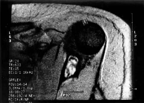
A 19-year-old girl has had pain and swelling in the right ankle for the past 4 months. She denies any history of trauma. Examination reveals a small soft-tissue mass over the anterior aspect of the ankle and slight pain with range of motion of the ankle joint. The examination is otherwise unremarkable. A radiograph and MRI scan are shown in Figures 45a and 45b, and biopsy specimens are shown in Figures 45c and 45d. What is the most likely diagnosis?


Explanation

45b 45c 45d Giant cell tumors typically occur in a juxta-articular location involving the epiphysis and metaphysis of long bones, usually eccentric in the bone. The radiographs show a destructive process within the distal tibia and an associated soft-tissue mass. The histology shows multinucleated giant cells in a bland matrix with a few scattered mitoses. Osteosarcoma can have a similar destructive appearance but a very different histologic pattern with osteoid production. Ewing's sarcoma also can have a diffuse destructive process in the bone. The histologic pattern of Ewing's sarcoma is diffuse round blue cells. Aneurysmal bone cysts typically are seen as a fluid-filled lesion on imaging studies and have only a scant amount of giant cells histologically. Metastatic adenocarcinoma does not demonstrate the pattern shown in the patient's histology specimen. Wold LA, et al: Atlas of Orthopaedic Pathology. Philadelphia, PA, WB Saunders, 1990, pp 198-199.

Question 2

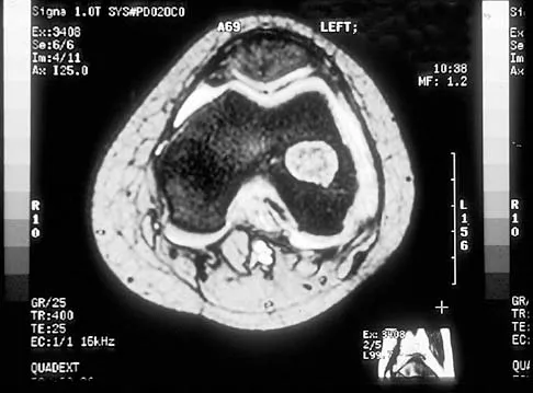
A 13-year-old boy has had a painless mass in the arm for the past 2 months. An MRI scan and biopsy specimens are shown in Figures 46a through 46c. What is the most likely diagnosis?


Explanation

46b 46c Nodular fasciitis is a benign soft-tissue lesion that usually arises from the fascia and is often misdiagnosed as a sarcoma. Desmoid tumors (aggressive fibromatosis) are also benign tumors with a greater tendency for local recurrence. Desmoid tumors have more spindle-shaped fibroblasts in an abundant collagenous matrix. Malignant fibrous histiocytoma is a hypercellular pleomorphic sarcoma more commonly found in adults. The histology is not consistant with a fatty tumor.

Question 3

A 20-year-old man has a symptomatic lesion of fibrous dysplasia in the femoral neck. Management should consist of


Explanation

Fibrous dysplasia in the femoral neck frequently warrants treatment because of the risk of pathologic fracture. Cortical strut grafts reduce the risk of local recurrence compared with cancellous bone grafting. Because of the consequences associated with fracture in this location, prophylactic fixation is recommended. Radiation therapy and chemotherapy are not used for this benign condition. Simon M, et al: Surgery for Bone and Soft Tissue Tumors. Philadelphia, PA, Lippincott Raven, 1998, pp 197.

Question 4

In addition to radiographs of the primary lesion and chest, MRI of the primary lesion, and CT of the chest, staging studies for Ewing's sarcoma should include which of the following?


Explanation

A bone scan and bone marrow biopsy are part of the staging studies for Ewing's sarcoma. Whole body MRI and PET scans are investigational and show promise of greater sensitivity than a bone scan. Schleiermacher G, Peter M, Oberlin O, Philip T, Rubie H, Mechinaud F, et al: Increased risk of systemic relapses associated with bone marrow micrometastasis and circulating tumor cells in localized ewing tumor. J Clin Oncol 2003;21:85-91.

Question 5

Which of the following conditions is transmitted by an autosomal dominant trait?


Explanation

Multiple hereditary exostosis is transmitted by an autosomal dominant trait. Li-Fraumeni syndrome and retinoblastoma are autosomal recessive or associated with autosomal recessive mutations. No genetic predisposition to Ollier's disease or Maffucci's syndrome has been identified. Mirra J (ed): Bone Tumors: Clinical, Radiologic and Pathologic Correlations. Philadelphia, PA, Lea and Febiger, 1989, p 1627.

Question 6

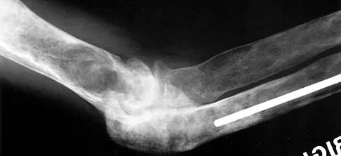
A previously healthy 13-year-old girl has had thigh pain for the past 3 weeks. The radiograph shown in Figure 47a reveals a lesion in the right femur. A bone scan and CT scan of the chest show no evidence of other lesions. A biopsy specimen is shown in Figure 47b. What is the most likely diagnosis?


Explanation

47b The patient has Langerhans cell histiocytosis that may be solitary (eosinophilic granuloma) or associated with systemic illness (Hand-Schuller-Christian disease and Letterer-Siwe disease). The solitary form of the disease, eosinophilic granuloma, typically affects patients in the first three decades of life. Radiographically, it is characterized as a well-defined, lytic, "punched out" intramedullary lesion. Histologically, two cell types, eosinophils and Langerhans cells, are seen. The Langerhans cells are seen as mononuclear histiocyte-like cells with oval nuclei with well-defined round or oval cytoplasm. A prominent nuclear groove can be seen in most of the nuclei (coffee bean nuclei). A mixture of inflammatory cells and lipid-laden foam cells with nuclear debris may be present as well. The lack of nuclear atypia and atypical mitoses excludes malignant conditions such as Ewing's sarcoma, lymphoma of bone, and metastatic neuroblastoma. The lack of acute inflammatory cells excludes the diagnosis of osteomyelitis. The eosinophils have bi-lobed nuclei and granular eosinophilic cytoplasm. Dorfman H, Czerniak B: Bone Tumors. St Louis, MO, Mosby, 1988.

Question 7

A 50-year-old man with metastatic renal cell carcinoma has right hip pain. A radiograph and CT scan are shown in Figures 48a and 48b. The first step in management should consist of


Explanation

48b These lesions are extremely vascular and can cause uncontrolled intraoperative bleeding; therefore embolization is the appropriate first treatment. Because the radiograph and CT scan show a lytic lesion in the supra-acetabular region that affects the weight-bearing dome and medial wall, the next step in treatment would most likely be a total hip arthroplasty and acetabular reconstruction. Treatment with bisphosphonates and radiation therapy will not prevent an acetabular fracture. Cementoplasty is an emerging technique in which cement is injected percutaneously into a lesion, but no long-term results have been reported. Radiofrequency ablation of bone metastases is also an emerging technique that provides palliative pain control. Layalle I, Flandroy P, Trotteur G, Dondelinger RF: Arterial embolization of bone metastases: Is it worthwhile? J Belge Radiol 1998;81:223-225.

Question 8

What is the most common benign bone tumor in childhood?


Explanation

The most common benign bone tumor in childhood is a nonossifying fibroma. It is estimated that 30% of children have a nonossifying fibroma. In most patients, the lesion is not identified until a radiograph is obtained for unrelated reasons. Similarly, most identified cases of fibrous cortical defect are not biopsied because the radiographic and clinical presentations are diagnostic. Aboulafia AJ, Kennon RE, Jelinek JS: Benign bone tumors of childhood. J Am Acad Orthop Surg 1999;7:377-388.

Question 9

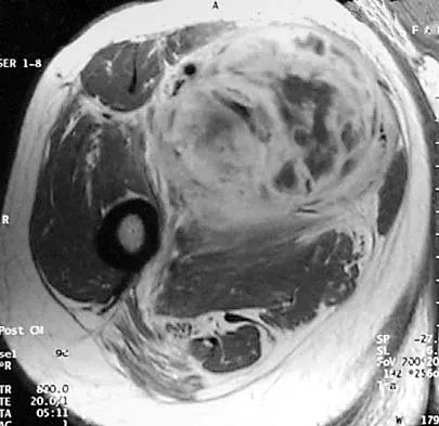
A 13-year-old patient has foot drop and lateral knee pain. AP and lateral radiographs and an MRI scan are shown in Figures 49a through 49c. A biopsy specimen is shown in Figure 49d. What is the preferred method of treatment?


Explanation

49b 49c 49d The "sunburst" radiographic appearance suggests an osteosarcoma, and the histologic findings confirm the diagnosis with malignant cells surrounded by pink osteoid. MRI scans are not particularly helpful in the diagnosis of osteosarcoma but are mandatory for surgical planning. Osteosarcomas are high-grade sarcomas that are best treated with chemotherapy and wide resection. Even though the peroneal nerve is involved, limb salvage is indicated. Survival after limb salvage is equivalent to amputation, with better function. Goorin AM, Abelson HT, Frei E: Osteosarcoma: Fifteen years later. N Engl J Med 1985;313:1637. Link MP, Goorin AM, Miser AW, et al: The effect of adjuvant chemotherapy on relapse-free survival in patients with osteosarcoma of the extremity. N Engl J Med 1986;314:1600.

Question 10

A 13-year-old girl is diagnosed with a stage IIB osteosarcoma of the proximal tibia. Following neoadjuvant chemotherapy, local control should consist of


Explanation

Local control of osteosarcoma consists of wide resection and reconstruction. Radiation therapy is not recommended except in unresectable lesions or for palliation. Curettage and bone grafting result in intralesional resection with an unacceptable high rate of local recurrence. Chemotherapy alone is not adequate for local control. Simon M, Springfield D, et al: Osteogenic Sarcoma: Surgery for Bone and Soft Tissue. Philadelphia, PA, Lippincott Raven, 1998, p 274.

Question 11

A 23-year-old woman has had vague left knee pain for the past 6 months. A radiograph and CT scan are shown in Figures 50a and 50b. What is the most likely diagnosis?


Explanation

50b The radiographic appearance of the lesion emanating from the posterior cortex of the left distal femur is consistent with a surface bone-producing lesion; therefore, the most likely diagnosis is a parosteal osteosarcoma. In an osteochondroma, the cortex and medullary cavity of the lesion are in continuity with that of the native bone. A dedifferentiated chondrosarcoma has histologic components of a high-grade sarcoma plus a benign or low-grade malignant cartilage tumor. Tumoral calcinosis is characterized by amorphous calcium in the soft tissues and does not emanate from the bone itself. While often confused with parosteal osteosarcoma, myositis ossificans is usually more mature at the periphery of the lesion rather than the center. In addition, myositis ossificans does not involve the underlying cortex but remains separate from the bone. Unni KK: Dahlin's Bone Tumors: General Aspects and Data on 11,087 Cases, ed 5. Philadelphia, PA, Lippincott-Raven, 1996, pp 185-196.

Question 12

What is the preferred treatment of a patient with breast cancer and a pathologic fracture of the clavicle in her dominant arm?


Explanation

Closed management should be attempted for upper extremity pathologic fractures, particularly the clavicle. If nonunion or pain persists, surgery may be indicated. Radiofrequency ablation is not indicated for subcutaneous bones. Early motion is likely to cause increased pain and disability. Weber KC, Lewis VO, Randall RL, Lee AK, Springfield D: An approach to the management of the patient with metastatic bone disease. Instr Course Lect 2004;53:663-676.

Question 13

A 14-year-old boy has an asymptomatic mass on the right arm. MRI scans and biopsy specimens are shown in Figures 51a through 51d. Immunostaining is positive for desmin. Additional staging studies should include


Explanation

51b 51c 51d The patient has rhabdomyosarcoma. Axilliary node and bone marrow biopsy are part of the staging because about 12% of patients with rhabdomyosarcoma of the extremity have evidence of lymph nodes metastases at presentation. Bone marrow metastases have been shown to portend a worse prognosis. Lawrence W, Jr., Hays DM, Heyn R, Tefft M, Crist W, Beltangady M, et al: Lymphatic metastases with childhood rhabdomyosarcoma: A report from the Intergroup Rhabdomyosarcoma Study. Cancer 1987;60:910-915.

Question 14

A 15-year-old girl has left knee pain and an enlarging mass in the distal thigh. AP and lateral radiographs are shown in Figures 52a and 52b, and a biopsy specimen is shown in Figure 52c. What is the most likely diagnosis?


Explanation

52b 52c A bone-producing lesion in the metaphysis of an adolescent is most likely an osteosarcoma. The radiographs show a distal femoral bone-producing lesion extending into the surrounding soft tissues. The histologic appearance consists of pleomorphic cells producing osteoid. Ewing's sarcoma and metastatic neuroblastoma do not produce a matrix. Chondrosarcoma is a radiographically destructive lesion with calcification and cartilage cells on histologic section. An osteochondroma is a benign cartilage lesion that is continuous with the medullary cavity of the underlying bone and extends into a bony lesion and covered by a cartilage cap. Unni KK: Dahlin's Bone Tumors: General Aspects and Data on 11,087 Cases, ed 5. Philadelphia, PA, Lippincott-Raven, 1996, pp 143-160.

Question 15

Survival rates for children with soft-tissue sarcoma other than rhabdomyosarcoma are best correlated with


Explanation

In review of 154 patients with nonrhabdomyosarcoma, Rao reported that histologic grade, tumor invasiveness, and adequate surgical margin were the most important prognostic factors. Histologic subtype, use of adjuvant chemotherapy, and patient age were not as important. Size related to degree of invasiveness was not statistically significant. Rao BN: Nonrhabdomyosarcoma in children: Prognostic factors influencing survival. Semin Surg Oncol 1993;9:524-531.

Question 16

A 25-year-old woman has had pain and stiffness in her knee following a motor vehicle accident 9 months ago. The radiograph, CT scan, MRI scan, and biopsy specimen are shown in Figures 53a through 53d. What is the most likely diagnosis?


Explanation

53b 53c 53d Heterotopic ossification may occur spontaneously or following trauma. The imaging studies and histology reveal mature fatty bone marrow and trabecular bone. Osteochondromas are cortically based with the medullary canal extending into the lesion. This is not evident in this patient. Also, no obvious cartilage cap is present. Parosteal osteosarcoma commonly occurs in the posterior distal femoral cortex but is ruled out by the lack of the typical fibrous stromal cells forming the low-grade malignant osteoid. The histology and clinical presentation eliminate osteomyelitis and osteoblastoma. Horne LT, Blue BA: Intra-articular heterotopic ossification in the knee following intramedullary nailing of the fractured femur using a retrograde method. J Orthop Trauma 1999;13:385-388. Stannard JP, Wilson TC, Sheils TM, McGwin G Jr, Volgas DA, Alonso JE: Heterotopic ossification associated with knee dislocation. Arthroscopy 2002;18:835-839.

Question 17

Which of the following lesions most closely resembles Ewing's sarcoma histologically?


Explanation

Ewing's sarcoma is characterized by small round blue cells. Lesions with a similar appearance include lymphoma, primitive neuroectodermal tumor, rhabdomyosarcoma, small cell lung tumor, and metastatic neuroblastoma. Karyotyping, immunohistochemistry, and electron microscopy can help differentiate these lesions.

Question 18

Primary chondrosarcoma of bone most commonly occurs in which of the following locations?


Explanation

The most common location of chondrosarcoma is the pelvis (30%), followed by the proximal femur (20%) and shoulder girdle (15%). Chondrosarcoma rarely affects the spine or hand. Lee FY, Mankin HJ, Fondren G, et al: Chondrosarcoma of bone: An assessment of outcome. J Bone Joint Surg Am 1999;81:326-338.

Question 19

An 18-year-old man has had an enlarging mass in his hand for the past 3 months. Radiographs, an MRI scan, and biopsy specimens are shown in Figures 54a through 54d. What is the most likely diagnosis?


Explanation

54b 54c 54d Also known as Nora's lesion, BPOP is a benign osteocartilaginous tumor that almost always occurs in the hands and feet; one occurrence each in the femur and tibia has been reported. Although local recurrence is common after excision, metastases have not been reported. Abramovici L, Steiner GC: Bizarre parosteal osteochondromatous proliferation (Nora's lesion): A retrospective study of 12 cases, 2 arising in long bones. Hum Pathol 2002;33:1205-1210.

Question 20

A 55-year-old man has had a mass in his right thigh for the past 2 months. An MRI scan and biopsy specimens are shown in Figures 55a through 55c. What is the most likely diagnosis?


Explanation

55b 55c The histology shows extraskeletal myxoid chondrosarcoma, characterized by abundant blue myxoid matrix with cords and nests of small tumor cells. Treatment consists of wide resection. Despite the name, hyaline cartilage is not a common component of these tumors. Adult rhabdomyosarcoma and malignant fibrous histiocytoma are highly pleomorphic sarcomas often containing multinucleated giant cells. Myxoid liposarcoma contains a prominent capillary network and lipoblasts. Myxoma is less cellular than extraskeletal myxoid chondrosarcoma and does not have a cord-like arrangement of tumor cells.

Question 21

Figures 56a through 56c show the radiograph, CT scan, and biopsy specimen of a 44-year-old man who underwent chemotherapy and radiation therapy for lymphoma of the distal femur 20 years ago. His current problem is most likely related to


Explanation

56b 56c The patient has changes consistent with radiation therapy to the femur, including osteopenia and an aggressive appearing neoplasm. The tumor is most likely a radiation-induced sarcoma. This is more likely than recurrent lymphoma at this late date. It is not related to steroid use or a primary lung tumor. Mirra J (ed): Bone Tumors: Clinical, Radiologic and Pathologic Correlations. Philadelphia, PA, Lea and Febiger, 1989, p 353.

Question 22

What is the most common reason an individual with a malignant soft-tissue tumor in the extremities seeks medical attention?


Explanation

Unlike malignant bone tumors, malignant soft-tissue tumors usually are asymptomatic and present with the presence of a mass. Malignant soft-tissue tumors enlarge by centrifugal growth, creating a mass while compressing surrounding tissue. Symptoms may develop as the result of direct compression on neurovascular structures as the tumor enlarges. This is especially true in the pelvis where the tumor can enlarge appreciably without being noticed. However, in the extremities, the tumor is most often apparent before neurologic symptoms develop. An asymptomatic mass is not necessarily benign; therefore, biopsy should not be delayed. It is uncommon for a malignant soft-tissue mass to be discovered incidentally. Soft-tissue tumors are not typically apparent on radiographs; they are best identified with MRI. Brouns F, Stas M, De Wever I: Delay in diagnosis of soft tissue sarcomas. Eur J Surg Oncol 2003;29:440-445. Rougraff B: The diagnosis and management of soft tissue sarcomas of the extremities in the adult. Curr Probl Cancer 1999;23:1-50.

Question 23

Exostoses in which of the following anatomic locations is the most likely to undergo malignant transformation in a patient with multiple hereditary exostosis (MHE)?


Explanation

Although osteochondromas can occur in almost every bone in patients with MHE, proximally located lesions are more likely to undergo malignant transformation. Annual radiographs of the shoulder girdles and pelvis are indicated in patients with MHE. Any enlarging osteochondromas are a concern as possible malignancies. Peterson HA: Multiple hereditary osteochondromata. Clin Orthop 1989;239:222.

Question 24

Initial management of a pathologic fracture of the humerus secondary to a unicameral bone cyst should include


Explanation

Most pathologic humeral fractures secondary to a unicameral bone cyst are minimally displaced and should be immobilized and allowed to heal. Persistent and/or progressive lesions may require treatment. Various treatments of unicameral bone cysts have been described. Acceptable treatment options include curettage and bone grafting, intralesional steroid injection, and percutaneous grafting with bone graft substitutes. MRI is not indicated when the diagnosis of unicameral bone cyst is known. Wilkins RM: Unicameral bone cysts. J Am Acad Orthop Surg 2000;8:217-224.

Question 25

An 11-year-old child has Ewing's sarcoma of the femoral diaphysis with a small soft-tissue mass. Staging studies show no evidence of metastases. Treatment should consist of


Explanation

The use of chemotherapy has dramatically improved survival rates of patients with Ewing's sarcoma. Local disease is best handled with wide resection to decrease local recurrence and to avoid the complications of radiation therapy (ie, secondary sarcomas). Radiation therapy alone is reserved for unresectable lesions or poor surgical margins. Amputation generally is not necessary. Toni A, Neff JR, Sudanese A, et al: The role of surgical therapy in patients with non-metastatic Ewing's sarcoma of the limbs. Clin Orthop 1991;286:225. Picci P, Rougraff BT, Bacci G, et al: Prognostic significance of histopathologic response to chemotherapy in non-metastatic Ewing's sarcoma of the extremities. J Clin Oncol 1993;11:1763.

Question 26

A 15-year-old boy presents with progressive knee pain and a destructive, bone-forming lesion in the distal femoral metaphysis. A core biopsy confirms the diagnosis of high-grade, conventional osteosarcoma. Which of the following genetic alterations is most consistently associated with the underlying pathogenesis of this tumor?


Explanation

Conventional high-grade osteosarcoma is characterized by profound genomic instability. The most consistent alterations involve the inactivation of the tumor suppressor genes RB1 (Retinoblastoma) and TP53. Patients with hereditary retinoblastoma (RB1 mutation) and Li-Fraumeni syndrome (TP53 mutation) have a significantly elevated risk of developing osteosarcoma. MDM2/CDK4 amplification is characteristic of parosteal and low-grade central osteosarcoma. t(X;18) is seen in synovial sarcoma. GNAS mutations are seen in fibrous dysplasia.

Question 27

A 12-year-old girl is evaluated for thigh pain and a low-grade fever. Radiographs reveal a permeative, diaphyseal lesion in the femur with a prominent 'onion skin' periosteal reaction. A biopsy shows sheets of monotonous small round blue cells. The primary translocation associated with this condition results in a fusion protein that acts as an aberrant transcription factor. Immunohistochemistry is most likely to show strong, diffuse membranous staining for which of the following markers?


Explanation

The clinical and radiographic presentation, along with the small round blue cell histology, is highly characteristic of Ewing sarcoma. Ewing sarcoma is classically driven by the t(11;22) translocation, creating the EWS-FLI1 fusion protein. Strong, diffuse, and uniform membranous staining for CD99 (MIC2) is a hallmark of Ewing sarcoma, though it must be interpreted in context as it can occasionally be positive in other tumors. Cytokeratin marks epithelial tissues (carcinomas), S-100 marks neural crest and cartilaginous tumors, SMA marks smooth muscle differentiation, and MyoD1 is a marker for rhabdomyosarcoma.

Question 28

A 14-year-old boy presents with a rapidly expanding, painful lesion in the proximal tibia. Radiographs show an eccentric, expansile, lytic lesion with a thin cortical shell. MRI reveals multiple fluid-fluid levels. Biopsy demonstrates blood-filled spaces lacking endothelial lining, separated by cellular fibrous septa containing multinucleated giant cells. Which of the following molecular abnormalities is considered the primary driver of this lesion?


Explanation

The vignette describes a primary Aneurysmal Bone Cyst (ABC). Primary ABCs are true neoplasms driven by specific genetic rearrangements, most commonly involving the USP6 gene on chromosome 17p13 (e.g., t(16;17) fusing CDH11 to USP6). This leads to USP6 transcriptional upregulation, driving the formation of the cyst. H3F3A mutations are pathognomonic for Giant Cell Tumor of bone and chondroblastoma. GNAS mutations are found in fibrous dysplasia.

Question 29

A 35-year-old woman is diagnosed with a large, destructive, recurrent giant cell tumor of the sacrum with impending neurologic compromise. Surgical resection would result in significant morbidity, so her multidisciplinary oncology team initiates targeted medical therapy. What is the mechanism of action of the most appropriate pharmacological agent?


Explanation

The most appropriate targeted medical therapy for an unresectable or highly morbid Giant Cell Tumor of Bone (GCTB) is denosumab. Denosumab is a fully human monoclonal antibody that specifically binds to and neutralizes RANK Ligand (RANKL). In GCTB, the neoplastic mononuclear stromal cells secrete high levels of RANKL, which recruits and activates the reactive, multinucleated osteoclast-like giant cells that cause bone destruction. By neutralizing RANKL, denosumab prevents giant cell formation and halts osteolysis.

Question 30

A 28-year-old woman complains of a painless, slow-growing mass at the posterior aspect of her knee. Radiographs reveal a dense, heavily ossified mass arising from the posterior surface of the distal femur. A radiolucent cleft separates part of the tumor from the underlying cortex. Histology shows low-grade malignant spindle cells between well-formed, thick bone trabeculae. What is the typical genetic amplification found in this condition?


Explanation

The lesion described is a parosteal osteosarcoma, the most common type of surface osteosarcoma. It typically arises on the posterior aspect of the distal femur and is characterized radiographically by a 'string sign' (a radiolucent cleft separating the tumor from the cortex) and histologically by well-differentiated bone trabeculae within a low-grade fibroblastic stroma. At the molecular level, parosteal osteosarcomas are characterized by supernumerary ring chromosomes containing amplifications of the MDM2 and CDK4 genes (12q13-15 region).

Question 31

A 55-year-old man presents with dull, aching shoulder pain. Radiographs reveal a large medullary cartilaginous lesion in the proximal humerus with 'rings and arcs' calcification and significant endosteal scalloping. Core needle biopsy shows increased cellularity, binucleate chondrocytes, and myxoid changes in the hyaline stroma. Recent molecular studies show that this malignancy shares a specific genetic mutation with benign solitary enchondromas. Which of the following genes is most likely mutated?


Explanation

Point mutations in the isocitrate dehydrogenase genes, IDH1 or IDH2, are found in approximately 50-70% of solitary enchondromas and conventional central chondrosarcomas. This mutation leads to the production of an oncometabolite, D-2-hydroxyglutarate (D-2-HG), which disrupts normal DNA and histone methylation, promoting tumorigenesis. EXT1/EXT2 mutations are characteristic of multiple hereditary exostoses (osteochondromas).

Question 32

A 10-year-old boy presents for orthopedic evaluation. He has multiple palpable bony protuberances around his knees, ankles, and shoulders, short stature, and a progressive valgus deformity of his left knee. A skeletal survey confirms multiple osteochondromas. The genetic mutation most likely responsible for his condition leads to a direct defect in which of the following biological processes?


Explanation

Multiple Hereditary Exostoses (MHE), also known as diaphyseal aclasis, is an autosomal dominant disorder caused by mutations in the EXT1 or EXT2 genes. These genes act as tumor suppressors and encode glycosyltransferases essential for the biosynthesis and elongation of heparan sulfate chains. A deficiency in heparan sulfate proteoglycans alters the diffusion of Indian Hedgehog (Ihh) at the growth plate, leading to abnormal chondrocyte proliferation and the formation of osteochondromas.

Question 33

A 20-year-old woman has an impending pathologic fracture of the proximal femur due to a large, ground-glass, radiolucent lesion. She also has a history of precocious puberty and café-au-lait spots with irregular borders ('coast of Maine'). The underlying cellular defect involves:


Explanation

The clinical triad of polyostotic fibrous dysplasia, precocious puberty (or other endocrinopathies), and café-au-lait spots with irregular borders is diagnostic of McCune-Albright syndrome. This syndrome is caused by a somatic, postzygotic activating mutation in the GNAS gene, which encodes the alpha subunit of the stimulatory G-protein (Gs-alpha). This mutation causes constitutive activation of adenylate cyclase, leading to continuously elevated intracellular cyclic AMP (cAMP) levels, driving abnormal cellular proliferation and endocrine hyperfunction.

Question 34

A 24-year-old man presents with a deep, slow-growing soft tissue mass in his thigh. MRI shows a heterogeneous mass adjacent to, but not within, the knee joint capsule. Biopsy reveals a biphasic tumor containing both epithelial and spindle cell components. Which of the following chromosomal translocations is considered diagnostic for this sarcoma?


Explanation

The vignette describes a synovial sarcoma. Despite its name, synovial sarcoma rarely arises from within a joint cavity; it typically occurs in deep soft tissues near joints in young adults. It can have a biphasic (epithelial and spindle cells) or monophasic (spindle cells only) histologic pattern. Synovial sarcoma is characterized by the hallmark chromosomal translocation t(X;18)(p11;q11), which results in the fusion of the SS18 (formerly SYT) gene on chromosome 18 with one of the SSX genes on the X chromosome.

Question 35

A 16-year-old boy experiences severe, throbbing leg pain that is consistently worse at night and dramatically relieved by taking ibuprofen. Radiographs demonstrate a diaphyseal cortical thickening in the anterior tibia with a 5-mm radiolucent nidus. The intense pain associated with this specific bone lesion is primarily mediated by the local overproduction of which of the following substances?


Explanation

The clinical scenario is a classic presentation of an osteoid osteoma: a small (<1.5 cm) radiolucent nidus surrounded by reactive sclerotic bone, causing intense, unrelenting night pain that is exquisitely sensitive to nonsteroidal anti-inflammatory drugs (NSAIDs). The nidus of an osteoid osteoma produces massive amounts of Prostaglandin E2 (PGE2) and prostacyclin (often 100 to 1000 times the level of normal bone), along with increased expression of cyclooxygenase-2 (COX-2). PGE2 mediates the intense pain and local vasodilation.

Question 36

A 32-year-old woman presents with persistent knee pain. Radiographs show an eccentric, lytic epiphyseal lesion in the distal femur. A biopsy demonstrates mononuclear cells and multinucleated giant cells. Which of the following is the specific molecular target of the monoclonal antibody denosumab, which may be used in the medical management of this condition?


Explanation

Denosumab is a fully human monoclonal antibody that binds to and inhibits RANKL. In giant cell tumors of bone, the neoplastic mononuclear stromal cells express RANKL, which recruits and activates the reactive multinucleated giant cells (osteoclast-like cells) responsible for aggressive bone resorption.

Question 37

A 15-year-old boy is diagnosed with conventional high-grade osteosarcoma of the distal femur. He undergoes neoadjuvant chemotherapy followed by wide surgical resection. Which of the following factors is considered the most significant prognostic indicator for long-term survival in this patient?


Explanation

The most critical prognostic factor for overall survival in patients with localized high-grade osteosarcoma is the histologic response to neoadjuvant chemotherapy. This is defined by the percentage of tumor necrosis in the resected specimen. Greater than 90% necrosis (good responder) is associated with significantly better overall survival compared to less than 90% necrosis (poor responder).

Question 38

A 12-year-old boy presents with progressive mid-thigh pain and swelling. Radiographs show a permeative diaphyseal lesion with an 'onion-skin' periosteal reaction. Biopsy reveals uniform small round blue cells. Cytogenetic analysis of this tumor is most likely to show which of the following translocations?


Explanation

Ewing sarcoma is classically associated with the t(11;22)(q24;q12) chromosomal translocation, which results in the EWSR1-FLI1 fusion gene. t(X;18) is seen in synovial sarcoma; t(9;22) in myxoid chondrosarcoma; t(12;16) in myxoid liposarcoma; and t(2;13) in alveolar rhabdomyosarcoma.

Question 39

A 65-year-old man presents with generalized bone pain and a recent pathologic fracture of the proximal humerus. Laboratory studies show anemia, hypercalcemia, and renal insufficiency. Serum protein electrophoresis reveals a monoclonal spike. Which of the following is the most appropriate initial diagnostic imaging modality for staging his skeletal disease?


Explanation

Whole-body low-dose CT (WBLDCT) is now considered the standard of care and preferred initial imaging modality for detecting osteolytic bone lesions in multiple myeloma. Technetium-99m bone scans depend on osteoblastic activity, which is severely suppressed in myeloma, frequently leading to false-negative results.

Question 40

A 14-year-old girl presents with pain and swelling in her right shoulder. Radiographs demonstrate an expansile, eccentric lytic lesion in the proximal humerus metaphysis with a thin sclerotic rim. MRI shows multiple fluid-fluid levels. The pathogenesis of this lesion is most strongly associated with a rearrangement involving which of the following genes?


Explanation

Primary aneurysmal bone cysts (ABCs) are true neoplasms characterized by translocations causing the upregulation of the USP6 gene on chromosome 17p13. GNAS mutations are seen in fibrous dysplasia; EXT1/EXT2 are associated with multiple hereditary exostoses; RUNX2 is associated with cleidocranial dysplasia; and TP53 is associated with Li-Fraumeni syndrome.

Question 41

A 55-year-old man presents with a constant, dull ache in his right hip that worsens at night. Radiographs reveal a radiolucent lesion in the right ilium with intralesional stippled calcifications and endosteal scalloping. Biopsy confirms a grade II (intermediate-grade) conventional chondrosarcoma. What is the most appropriate management?


Explanation

Conventional chondrosarcomas are generally highly resistant to both chemotherapy and radiation therapy. The mainstay of treatment for intermediate (grade II) and high-grade (grade III) chondrosarcomas, as well as pelvic chondrosarcomas, is wide surgical resection. Intralesional curettage is generally reserved for benign cartilaginous lesions or select peripheral low-grade (grade I) atypical cartilaginous tumors in the appendicular skeleton.

Question 42

A 19-year-old man complains of severe right thigh pain that is worse at night and dramatically relieved by NSAIDs. Radiographs show a small radiolucent nidus surrounded by dense reactive sclerosis in the femoral diaphysis. If a biopsy of the nidus were performed, what would be the expected histologic finding?


Explanation

Osteoid osteoma is a benign bone-forming tumor. Histologically, the central nidus is highly vascular and consists of an anastomosing network of osteoid and woven bone trabeculae lined by prominent, single layers of plump, active osteoblasts.

Question 43

A 9-year-old girl is evaluated for a limp and a discrepancy in leg length. Radiographs reveal a 'shepherd's crook' deformity of the proximal femur with a 'ground-glass' appearance. She also has irregularly bordered hyperpigmented skin macules and a history of precocious puberty. This condition is caused by a somatic activating mutation resulting in the overproduction of which intracellular signaling molecule?


Explanation

McCune-Albright syndrome consists of polyostotic fibrous dysplasia, café-au-lait spots, and endocrine abnormalities. It is caused by a somatic mutation in the GNAS gene, which encodes the stimulatory G protein alpha subunit (Gsα). This mutation results in constitutive activation of adenylyl cyclase and unregulated overproduction of intracellular cAMP.

Question 44

An 8-year-old boy presents to the emergency department with arm pain after throwing a baseball. Radiographs show a minimally displaced pathologic fracture through a centrally located, completely lytic lesion in the proximal humerus metaphysis. A 'fallen leaf' sign is visible. What is the most appropriate initial management?


Explanation

The clinical scenario and 'fallen leaf' sign are diagnostic of a unicameral (simple) bone cyst (UBC). When a patient presents with a pathologic fracture through a UBC in the upper extremity, initial management is non-operative (e.g., sling immobilization) to allow the fracture to heal. Surgical intervention or injections are typically reserved for persistent cysts after fracture healing or lesions with a high risk of recurrent fracture.

Question 45

A 24-year-old woman presents with a slowly enlarging, painful mass around her left knee. MRI demonstrates a deeply seated, multilobulated soft-tissue mass in the popliteal fossa. Radiographs reveal focal stippled calcifications. Biopsy reveals a biphasic pattern of spindle cells and epithelial cells. What is the most common site of distant metastasis for this specific type of sarcoma?


Explanation

The diagnosis is synovial sarcoma, which frequently presents in periarticular regions of young adults, often shows calcifications on radiographs, and has a characteristic biphasic histologic pattern. The most common site of distant metastasis for synovial sarcoma, like most soft-tissue sarcomas, is the lungs. Although synovial sarcoma has a higher rate of regional lymph node metastasis than other sarcomas, the lungs remain the most common overall site.

Question 46

A 14-year-old boy presents with a 3-month history of progressive thigh pain and swelling. Radiographs show a destructive diaphyseal lesion of the femur with a lamellated 'onion skin' periosteal reaction. A core needle biopsy reveals uniform sheets of small round blue cells with scant cytoplasm. Cytogenetic analysis of this tumor is most likely to show which of the following translocations?


Explanation

The clinical and radiographic presentation, along with the 'small round blue cell' histology, is classic for Ewing sarcoma. Ewing sarcoma is characterized by the t(11;22)(q24;q12) translocation, which results in the EWSR1-FLI1 fusion protein. t(9;22) is associated with chronic myelogenous leukemia or extraskeletal myxoid chondrosarcoma. t(X;18) is pathognomonic for synovial sarcoma. t(12;16) is seen in myxoid liposarcoma, and t(2;13) is characteristic of alveolar rhabdomyosarcoma.

Question 47

A 45-year-old man presents with a painful mass in his proximal humerus. Radiographs demonstrate a lytic lesion with intralesional 'popcorn' calcifications and endosteal scalloping involving greater than 2/3 of the cortical thickness. Biopsy confirms a grade II chondrosarcoma. Which of the following represents the most appropriate and definitive management?


Explanation

Intermediate (grade II) and high-grade (grade III) chondrosarcomas are generally resistant to both chemotherapy and radiation therapy. The standard of care for these malignant cartilage tumors is wide surgical resection with negative margins. Intralesional curettage is reserved for benign cartilage lesions (like enchondromas) or select atypical cartilaginous tumors (grade I chondrosarcoma in the appendicular skeleton).

Question 48

A 35-year-old woman is diagnosed with a giant cell tumor of the distal femur. Due to the proximity of the lesion to the articular surface, she is started on a course of denosumab therapy prior to intralesional curettage. Which of the following best describes the specific mechanism of action of this medication?


Explanation

Denosumab is a fully human monoclonal antibody that binds directly to RANK Ligand (RANKL). In Giant Cell Tumor of bone, the neoplastic mononuclear stromal cells overexpress RANKL. This overexpression recruits and activates reactive, non-neoplastic multinucleated giant cells (osteoclast-like cells) which cause bone destruction. By binding RANKL, denosumab prevents it from activating the RANK receptor on osteoclasts and their precursors, thereby halting bone resorption.

Question 49

A 22-year-old man presents with a 6-month history of left knee pain that is significantly worse at night. The pain is dramatically relieved within 30 minutes of taking ibuprofen. Radiographs reveal a 1-cm radiolucent nidus surrounded by dense, reactive sclerotic bone in the proximal tibial diaphysis. Which of the following inflammatory mediators is most responsible for this classic pain pattern?


Explanation

The clinical scenario is classic for an osteoid osteoma. The intense night pain associated with osteoid osteomas is due to the local production of exceptionally high levels of prostaglandins, particularly Prostaglandin E2 (PGE2), by the nidus. Nonsteroidal anti-inflammatory drugs (NSAIDs) inhibit cyclooxygenase, effectively blocking PGE2 production and providing dramatic symptomatic relief.

Question 50

A 65-year-old man presents with new-onset severe lower back pain and generalized fatigue. Laboratory studies reveal a normocytic anemia, elevated serum creatinine, and hypercalcemia. A skeletal survey shows multiple 'punched-out' lytic lesions in the skull and pelvis with no surrounding reactive sclerosis. Which of the following factors is most directly responsible for the uncoupled bone remodeling that leads to these purely lytic lesions?


Explanation

Multiple myeloma is characterized by purely lytic bone lesions resulting from uncoupled bone remodeling (increased osteoclast activity and suppressed osteoblast activity). Myeloma cells secrete DKK1, which inhibits the Wnt signaling pathway, severely suppressing osteoblast differentiation and preventing reactive bone formation. Additionally, they secrete MIP-1a and RANKL, which strongly activate osteoclasts, leading to unchecked bone resorption.

Question 51

A 12-year-old boy is found to have an incidental lesion in the proximal humerus during a workup for shoulder stiffness. Radiographs show a centrally located, well-circumscribed radiolucent lesion in the metaphysis with mild cortical thinning. If a diagnostic aspiration of this intact lesion is performed, which of the following fluid characteristics is most classically expected?


Explanation

The radiographic description strongly suggests a unicameral (simple) bone cyst (UBC). Aspiration of an intact UBC typically yields clear or serosanguineous fluid. Biochemical analysis of this cyst fluid reliably demonstrates significantly elevated levels of prostaglandins (especially PGE2), alkaline phosphatase, interleukins, and nitrogen. Frank blood would be more characteristic of an aneurysmal bone cyst.

Question 52

A 28-year-old woman complains of a slowly enlarging, painful mass in her left foot over the past year. MRI reveals an indeterminate soft tissue mass, and plain radiographs demonstrate stippled calcifications within the substance of the mass. A core biopsy confirms synovial sarcoma. Which of the following represents the characteristic cytogenetic abnormality for this neoplasm?


Explanation

Synovial sarcoma classically harbors the t(X;18)(p11;q11) translocation, which results in the SYT-SSX fusion gene. This tumor often occurs in the extremities of young adults, particularly around the knee or in the foot/ankle. Despite its name, it rarely arises within the joint itself. Punctate calcifications on plain radiographs are seen in up to 30% of cases.

Question 53

A 68-year-old man with a known 15-year history of polyostotic Paget's disease presents with a 2-month history of rapidly worsening pain and swelling in his right thigh, a symptom notably different from his baseline. Radiographs demonstrate a destructive, mixed lytic and sclerotic lesion in the femoral diaphysis with a wide zone of transition, cortical breakthrough, and a soft tissue mass. What is the most likely diagnosis?


Explanation

Malignant transformation occurs in less than 1% of patients with Paget's disease, but the risk increases with widespread polyostotic disease. The most common secondary malignancy arising in Pagetic bone is osteosarcoma (Paget's sarcoma). It typically presents in older adults with new, unrelenting pain and destructive radiographic changes. The prognosis is generally poor compared to primary osteosarcoma in young patients.

Question 54

A 9-year-old girl undergoes a biopsy of an expansile, eccentrically located, lytic metaphyseal lesion in her proximal tibia. The MRI demonstrates multiple fluid-fluid levels. Histopathology describes cavernous, blood-filled spaces lacking a true endothelial lining, separated by fibrous septa containing multinucleated giant cells. Which of the following genetic alterations is most specific to the primary pathogenesis of this lesion?


Explanation

The clinical, radiographic, and histologic findings are diagnostic of an aneurysmal bone cyst (ABC). Primary ABCs are now known to be true neoplasms, driven by translocations involving the USP6 gene on chromosome 17p13 (most commonly t(16;17)). The GNAS1 mutation is associated with fibrous dysplasia; EXT1 with osteochondromas; and H3F3A with giant cell tumors of bone.

Question 55

A 15-year-old boy completes a standard course of neoadjuvant multidrug chemotherapy (MAP: methotrexate, doxorubicin, cisplatin) for high-grade conventional osteosarcoma of the distal femur. He subsequently undergoes a wide surgical resection. The pathology report notes 95% tumor necrosis in the resected specimen. Which of the following best describes the clinical significance of this finding?


Explanation

In the management of high-grade osteosarcoma, the degree of histologic tumor necrosis following neoadjuvant chemotherapy is widely recognized as the single most important prognostic factor for disease-free and overall survival. A 'good response' is classically defined as >90% tumor necrosis (Huvos grade III/IV), which correlates with a significantly higher survival rate compared to patients with a poor response (<90% necrosis).

Question 56

A 55-year-old man presents with an enlarging, painful mass in his proximal humerus. Radiographs show a lytic lesion with 'ring and arc' calcifications in the metadiaphysis. Biopsy demonstrates a hypercellular cartilaginous matrix with plump, binucleated chondrocytes and permeation into the surrounding lamellar bone. Which of the following gene mutations is most commonly associated with this primary bone tumor?


Explanation

The clinical, radiographic, and histologic presentation is classic for a conventional chondrosarcoma. Mutations in isocitrate dehydrogenase 1 and 2 (IDH1 and IDH2) are found in over 50% of central chondrosarcomas. EXT1/EXT2 mutations are associated with multiple hereditary exostoses (osteochondromas). H3F3A mutations are characteristically seen in giant cell tumors of bone and chondroblastomas. USP6 rearrangements are the hallmark of aneurysmal bone cysts. GNAS mutations are seen in fibrous dysplasia.

Question 57

A 14-year-old boy presents with progressive knee pain and swelling for 3 months, accompanied by low-grade fevers. Radiographs of the distal femur reveal a permeative destructive lesion in the metadiaphysis with a prominent 'onion skin' periosteal reaction. Biopsy shows sheets of uniform small round blue cells. Immunohistochemistry is strongly positive for CD99. Which of the following chromosomal translocations is most characteristic of this tumor?


Explanation

The diagnosis is Ewing sarcoma, characterized by the clinical presentation in a young patient, permeative bone destruction with aggressive periostitis, and CD99-positive small round blue cells. The most common translocation is t(11;22)(q24;q12), which fuses the EWSR1 gene to the FLI1 gene, occurring in about 85% of cases. t(X;18) is pathognomonic for synovial sarcoma. t(2;13) is associated with alveolar rhabdomyosarcoma. t(12;16) is seen in myxoid liposarcoma. t(9;22) is the Philadelphia chromosome seen in chronic myeloid leukemia and occasionally extraskeletal myxoid chondrosarcoma.

Question 58

A 32-year-old woman presents with worsening knee pain. Radiographs reveal an eccentric, purely lytic lesion in the distal femur that extends directly to the subchondral bone without a sclerotic margin. Biopsy demonstrates numerous multinucleated giant cells in a background of mononuclear stromal cells. Due to the proximity of the lesion to the joint surface and its size, she is treated with a targeted monoclonal antibody to downstage the tumor prior to surgery. What is the precise mechanism of action of this medication?


Explanation

The lesion is a Giant Cell Tumor (GCT) of bone. Denosumab is a fully human monoclonal antibody frequently used for locally advanced or unresectable GCTs. It binds and inhibits RANK Ligand (RANKL). The neoplastic mononuclear stromal cells in GCT express high levels of RANKL, which inappropriately recruits and activates the reactive multinucleated osteoclast-like giant cells that cause the characteristic massive osteolysis.

Question 59

A 19-year-old man complains of a dull, aching pain in his right thigh that consistently worsens at night but is profoundly relieved by ibuprofen. CT imaging reveals a 0.8-cm radiolucent nidus surrounded by dense reactive cortical sclerosis in the femoral diaphysis. If a surgical resection is performed, histological analysis of the central nidus would most likely reveal which of the following?


Explanation

The clinical history and CT findings are pathognomonic for an osteoid osteoma. Histologically, the central nidus is composed of highly vascularized connective tissue containing interlacing trabeculae of osteoid (woven bone) lined by prominent, benign-appearing osteoblasts. Option A describes an osteochondroma. Option B is characteristic of an aneurysmal bone cyst. Option C describes a myxoid chondrosarcoma. Option D is typical of a benign fibrous lesion such as a non-ossifying fibroma.

Question 60

A 28-year-old woman presents with a slow-growing, painful mass near her right knee that has been present for over two years. Radiographs show a soft-tissue mass with stippled calcifications. An MRI demonstrates a heterogeneous soft-tissue mass adjacent to, but distinct from, the joint space. A biopsy reveals a biphasic pattern consisting of epithelial-like glandular structures and a spindle cell stromal component. Which of the following genetic abnormalities is most specifically associated with this tumor?


Explanation

The clinical presentation (a slow-growing soft tissue mass in a young adult, often near a joint but extra-articular, with calcifications in about 30% of cases) and biphasic histology are classic for synovial sarcoma. Synovial sarcoma is characterized by the t(X;18)(p11;q11) translocation, fusing the SS18 (formerly SYT) gene with an SSX gene (SSX1, SSX2, or SSX4). MDM2 amplification is characteristic of well-differentiated and dedifferentiated liposarcomas. PAX3-FOXO1 is seen in alveolar rhabdomyosarcoma. COL1A1-PDGFB is found in dermatofibrosarcoma protuberans.

Question 61

A 60-year-old man presents with chronic low back pain and a recent onset of bowel and bladder dysfunction. MRI of the sacrum reveals a midline destructive lesion with a large pre-sacral soft-tissue component, displaying high signal intensity on T2-weighted images. A core needle biopsy reveals lobules of cells with abundant, bubbly, vacuolated cytoplasm arranged in cords. Immunohistochemistry for which of the following markers is most specific to confirm the diagnosis of this neoplasm?


Explanation

The clinical scenario of a destructive sacral mass in an older adult causing neurologic compromise, combined with the histological finding of physaliferous (bubbly) cells, is highly characteristic of a chordoma. Chordomas are malignant tumors arising from embryonic notochord remnants. While they typically express S-100, EMA, and Cytokeratin, the nuclear expression of Brachyury (a transcription factor crucial for notochord development) is highly sensitive and specific for chordoma and serves as the diagnostic hallmark.

Question 62

During fracture healing, a complex molecular cascade dictates the differentiation of multipotent mesenchymal stem cells into the specific cell lineages necessary for bone repair. Which of the following transcription factors serves as the critical 'master regulator' for committing mesenchymal stem cells to the osteoblastic lineage?


Explanation

Runx2 (Runt-related transcription factor 2, also known as Cbfa1) is the master transcription factor responsible for the commitment and differentiation of multipotent mesenchymal stem cells into the osteoblastic lineage. SOX9 is the primary regulator for chondrogenic differentiation (cartilage). MyoD regulates myogenic differentiation (muscle). PPAR-gamma is the master regulator for adipogenic differentiation (fat). HIF-1 alpha regulates the cellular response to hypoxia.

Question 63

A 65-year-old woman is being evaluated for generalized bone pain, profound fatigue, and recent onset of hypercalcemia. Laboratory studies confirm a normocytic anemia, elevated serum creatinine, and a monoclonal spike on serum protein electrophoresis. Plain radiographs show multiple sharply marginated 'punched-out' lytic lesions in her skull and pelvis. If a technetium-99m bone scan is performed, which of the following findings is most likely?


Explanation

The patient's presentation meets the CRAB criteria (Hypercalcemia, Renal failure, Anemia, Bone lesions) diagnostic of multiple myeloma. The lytic bone lesions in myeloma result from profound osteoclast activation mediated by myeloma cells, coupled with a striking suppression of osteoblast activity. Because technetium-99m methylene diphosphonate (Tc-99m MDP) bone scans rely on osteoblastic activity (new bone formation) to accumulate radiotracer, the purely osteolytic, osteoblast-suppressed lesions of multiple myeloma typically appear normal or 'cold' on a bone scan. Skeletal surveys or whole-body low-dose CT/MRI/PET are the preferred imaging modalities.

Question 64

A 70-year-old woman with a history of postmenopausal osteoporosis sustains a fragility fracture of her distal radius. She has been on oral alendronate therapy for the past three years. Nitrogen-containing bisphosphonates, such as alendronate, inhibit osteoclast-mediated bone resorption primarily through which of the following molecular mechanisms?


Explanation

Nitrogen-containing bisphosphonates (e.g., alendronate, zoledronic acid, risedronate) primarily exert their antiresorptive effects by inhibiting the enzyme farnesyl pyrophosphate (FPP) synthase within the mevalonate pathway in osteoclasts. This inhibition prevents the prenylation of small GTPase proteins (like Ras, Rho, and Rab) that are essential for osteoclast function, ruffled border formation, and survival, ultimately inducing osteoclast apoptosis. Denosumab binds RANKL (Option B). Odanacatib inhibits cathepsin K (Option E).

Question 65

A 12-year-old boy presents with multiple palpable, painless, bony prominences around his knees, ankles, and shoulders. Radiographs confirm multiple pedunculated and sessile bony outgrowths projecting away from the adjacent joints in the metaphyses of the long bones, continuous with the medullary cavity of the native bone. He is diagnosed with Multiple Hereditary Exostoses (MHE). What is the primary underlying biochemical defect associated with the genetic mutations in this disorder?


Explanation

Multiple Hereditary Exostoses (MHE), also known as diaphyseal aclasis, is an autosomal dominant condition caused by loss-of-function mutations in the EXT1 or EXT2 genes. These genes encode glycosyltransferases essential for the biosynthesis of heparan sulfate proteoglycans. Defective heparan sulfate in the growth plate disrupts the normal diffusion and signaling of critical growth factors (such as Indian Hedgehog and PTHrP), leading to premature and disorganized chondrocyte proliferation that escapes the longitudinal axis, forming osteochondromas. FGFR3 overactivation causes achondroplasia. Defective osteoclast resorption leads to osteopetrosis. Abnormal collagen type I synthesis causes osteogenesis imperfecta.

Question 66

What is the most significant prognostic factor for overall survival in a patient with localized, high-grade intramedullary osteosarcoma of the distal femur following neoadjuvant chemotherapy and surgical resection?


Explanation

The most significant prognostic factor for survival in patients with high-grade osteosarcoma is the degree of histologic tumor necrosis following neoadjuvant chemotherapy. A necrosis rate of >90% (good responder) is strongly correlated with improved disease-free and overall survival rates.

Question 67

A 25-year-old male presents with a slowly growing, painful mass deep in the thigh. MRI shows a well-circumscribed heterogeneous soft tissue mass adjacent to the femur but not invading the bone. Biopsy reveals a biphasic tumor with both epithelial and spindle cell components. Which of the following genetic translocations is most characteristic of this lesion?


Explanation

The clinical presentation and biphasic histology (epithelial and spindle cells) are classic for synovial sarcoma. Synovial sarcoma is characterized by the t(X;18)(p11;q11) translocation, which results in the SYT-SSX fusion gene. This is found in over 90% of cases.

Question 68

A 30-year-old female presents with knee pain. Radiographs reveal an eccentric, purely lytic lesion in the distal femur extending to the subchondral bone. Biopsy confirms a giant cell tumor of bone. Denosumab is considered for medical management to facilitate surgery. What is the primary cellular target of this medication?


Explanation

Giant cell tumor of bone is composed of neoplastic mononuclear stromal cells and reactive multinucleated giant cells. The mononuclear cells express high levels of Receptor Activator of Nuclear factor Kappa-B Ligand (RANKL), which recruits and activates the osteoclast-like giant cells. Denosumab is a monoclonal antibody that binds to RANKL, inhibiting this process.

Question 69

A 60-year-old man presents with progressive, deep aching pain in his right pelvis. Radiographs show a destructive lytic lesion with 'ring and arc' calcifications in the ilium. Biopsy reveals a grade II chondrosarcoma. What is the most appropriate definitive management?


Explanation

Chondrosarcoma (grade II and III) is managed primarily with wide surgical resection. These tumors are generally resistant to both conventional chemotherapy and radiation therapy due to their poor vascularity and slow growth rate, making surgical excision with negative margins the only reliable curative option.

Question 70

A 12-year-old boy presents with pain and swelling in the midshaft of his tibia. Radiographs demonstrate a permeative, diaphyseal lytic lesion with an 'onion skin' periosteal reaction. Histopathology reveals sheets of small round blue cells. Immunohistochemistry is strongly positive for CD99. What is the most appropriate initial treatment sequence?


Explanation

The clinical, radiographic, and histological findings describe Ewing sarcoma. Ewing sarcoma is a systemic disease at presentation, even if metastases are not clinically evident. The standard of care is systemic neoadjuvant chemotherapy followed by local control (which may be wide surgical resection or radiation, depending on the location and resectability) and subsequent adjuvant chemotherapy.

Question 71

A 15-year-old boy reports severe, throbbing right thigh pain that is worse at night and completely relieved by ibuprofen. A CT scan shows a 7-mm radiolucent nidus surrounded by dense sclerotic bone in the femoral diaphysis. What is the primary mediator responsible for the severe pain associated with this lesion?


Explanation

The presentation is classic for an osteoid osteoma. The severe, nocturnal pain that is characteristically relieved by NSAIDs is caused by the high levels of Prostaglandin E2 (PGE2) produced by the nidus, which contains high concentrations of cyclooxygenase-2 (COX-2).

Question 72

A 22-year-old male with known multiple hereditary exostoses (MHE) presents with a rapidly enlarging, painful mass over the posterior aspect of his distal femur. An MRI shows an osteochondroma with a cartilage cap thickness of 2.5 cm. Which of the following gene mutations is most strongly associated with his underlying condition?


Explanation

Multiple hereditary exostoses (MHE) is an autosomal dominant disorder caused by mutations in the EXT1 or EXT2 genes, which are responsible for heparan sulfate synthesis. A cartilage cap thicker than 2 cm in a skeletally mature patient is highly suspicious for malignant transformation to secondary chondrosarcoma.

Question 73

A 16-year-old female presents with mild knee pain. Radiographs reveal an expansile, eccentric lytic lesion in the proximal tibial metaphysis. MRI demonstrates multiple fluid-fluid levels within the lesion. Biopsy shows blood-filled spaces lacking an endothelial lining. Which genetic rearrangement is considered the primary driver of this lesion?


Explanation

The lesion is an aneurysmal bone cyst (ABC). Primary ABCs are now known to be true neoplasms driven by translocations involving the USP6 gene on chromosome 17p13. This genetic rearrangement leads to upregulation of matrix metalloproteinases, driving the cystic destruction of bone.

Question 74

An 8-year-old boy sustains a pathologic fracture of the proximal humerus after a minor fall. Radiographs show a centrally located, completely lytic lesion in the metadiaphysis with a 'fallen leaf' sign. What is the most appropriate initial management for this presentation?


Explanation

The presence of a centrally located lytic lesion with a 'fallen leaf' sign (a cortical fragment that falls to the dependent portion of a fluid-filled cyst) in a child is pathognomonic for a unicameral bone cyst (UBC). When a UBC presents with a pathologic fracture, the initial treatment is nonoperative immobilization (sling and swathe) to allow the fracture to heal. Fracture healing can sometimes lead to spontaneous resolution of the cyst.

Question 75

Which of the following bone morphogenetic proteins (BMPs) is FDA-approved specifically for use in acute, open tibial shaft fractures treated with an intramedullary nail?


Explanation

Recombinant human BMP-2 (rhBMP-2) is FDA-approved for the treatment of acute, open tibial shaft fractures when used in conjunction with an intramedullary nail. RhBMP-7 (osteogenic protein-1) was previously approved under a Humanitarian Device Exemption for recalcitrant long bone nonunions but not specifically for acute open tibial fractures.

Question 76

A 25-year-old man presents with a slowly enlarging, painless mass on the posterior aspect of his distal thigh. Radiographs demonstrate a dense, heavily ossified mass attached to the posterior cortex of the distal femur with a broad base, and a subtle radiolucent line separating a portion of the tumor from the underlying bone (a 'string sign'). Biopsy reveals a spindle cell proliferation with low-grade atypia and well-formed bone trabeculae. Which of the following is the most characteristic genetic alteration associated with this patient's condition?


Explanation

The clinical presentation and radiographic findings (posterior distal femur location, dense ossification, 'string sign') are classic for parosteal osteosarcoma, a low-grade surface osteosarcoma. Cytogenetically, parosteal osteosarcomas are uniquely characterized by supernumerary ring chromosomes that result in the amplification of the MDM2 and CDK4 genes on chromosome 12q13-15. Translocation t(11;22) is seen in Ewing sarcoma, t(X;18) in synovial sarcoma, EXT1 mutations in hereditary multiple exostoses, and GNAS1 in fibrous dysplasia.

Question 77

A 35-year-old woman presents with persistent knee pain. Radiographs reveal an eccentric, purely lytic lesion in the proximal tibial epiphysis that extends to the subchondral bone. A core needle biopsy demonstrates a proliferation of mononuclear stromal cells interspersed with numerous multinucleated giant cells. For cases of this disease that are deemed unresectable or recurrent, medical therapy is often employed. What is the primary mechanism of action of the most commonly used targeted biologic agent for this condition?


Explanation

The patient has a Giant Cell Tumor (GCT) of bone, which typically presents as an eccentric, lytic epiphyseal lesion in a skeletally mature patient. The neoplastic cells in GCT are the mononuclear stromal cells, which express high levels of RANKL. This recruits and activates the reactive multinucleated giant cells (osteoclast-like cells) that cause the characteristic osteolysis. Denosumab, a fully human monoclonal antibody, specifically binds to RANKL, preventing RANK activation. This halts osteoclastogenesis and bone destruction, and is highly effective in treating recurrent or unresectable GCT of bone.

Question 78

A 14-year-old boy presents with progressive thigh pain, low-grade fevers, and an elevated erythrocyte sedimentation rate. Radiographs show a permeative diaphyseal lesion in the femur with a multi-layered, 'onion skin' periosteal reaction. Histologic examination of a biopsy specimen reveals sheets of uniform, small, round blue cells that strongly express CD99 on immunohistochemistry. The most common chromosomal translocation associated with this diagnosis results in which of the following fusion genes?


Explanation

The clinical scenario describes Ewing sarcoma, characterized by a permeative diaphyseal lesion, 'onion skin' periosteal reaction, and small round blue cells expressing CD99 (MIC2). Approximately 85% to 90% of Ewing sarcomas are characterized by a t(11;22)(q24;q12) chromosomal translocation, which fuses the EWS gene on chromosome 22 with the FLI1 gene on chromosome 11, resulting in the EWS-FLI1 fusion protein. The SYT-SSX1 fusion is seen in synovial sarcoma. TLS-CHOP is associated with myxoid liposarcoma. PAX3-FKHR is found in alveolar rhabdomyosarcoma. EWS-ERG is the second most common fusion in Ewing sarcoma (t(21;22)), occurring in about 10% of cases.

Question 79

A 55-year-old man complains of worsening right shoulder pain over several months. Radiographs show a 6-cm radiolucent lesion in the proximal humerus with intralesional stippled 'rings and arcs' calcifications and endosteal scalloping involving 80% of the cortical thickness. Biopsy confirms a conventional Grade II chondrosarcoma. What is the most appropriate definitive management for this patient?


Explanation

Conventional chondrosarcomas are notably resistant to both standard chemotherapy and radiation therapy due to their poor vascularity, slow growth rate, and high extracellular matrix content. Therefore, neoadjuvant or adjuvant systemic therapies are generally not indicated for localized disease. Grade II (intermediate-grade) and Grade III (high-grade) conventional chondrosarcomas require wide surgical resection with negative margins to minimize the risk of local recurrence and metastasis. Intralesional curettage with adjuvants is reserved for benign or low-grade cartilaginous lesions (like enchondromas or selected Grade I chondrosarcomas in the appendicular skeleton).

Question 80

A 16-year-old girl is evaluated for rapid onset of pain and swelling over the distal fibula. Radiographs show an eccentric, expansile, lytic lesion with cortical thinning. An MRI reveals multiple cysts with fluid-fluid levels. Core biopsy is consistent with an aneurysmal bone cyst (ABC). Primary aneurysmal bone cysts are distinct neoplastic entities frequently characterized by a translocation involving which of the following genes?


Explanation

Historically considered reactive lesions, primary aneurysmal bone cysts (ABCs) are now recognized as true neoplasms driven by a specific genetic rearrangement. Up to 70% of primary ABCs demonstrate a translocation involving the USP6 (ubiquitin-specific protease 6) gene on chromosome 17p13, most commonly t(16;17)(q22;p13). This rearrangement leads to USP6 overexpression, driving the neoplastic process. Secondary ABCs (arising in the setting of giant cell tumor, chondroblastoma, or osteoblastoma) do not harbor this USP6 mutation.

Question 81

A 65-year-old man presents to the clinic with severe, unrelenting mid-back pain and progressive fatigue. Laboratory studies reveal hypercalcemia and a normocytic anemia. Plain radiographs demonstrate multiple sharply demarcated, 'punched-out' lytic lesions throughout the calvarium and thoracolumbar spine. Which of the following diagnostic findings is most specific for confirming the primary underlying etiology?


Explanation

The patient's presentation of bone pain, fatigue, hypercalcemia, anemia, and 'punched-out' lytic lesions (without a sclerotic rim) is classic for multiple myeloma. Multiple myeloma is a malignant proliferation of plasma cells. The diagnosis is strongly supported and often confirmed by demonstrating a monoclonal paraprotein (M-protein) via serum protein electrophoresis (SPEP) or urine protein electrophoresis (UPEP), alongside bone marrow biopsy showing clonal plasma cells. Elevated alkaline phosphatase and PSA would be more indicative of metastatic prostate cancer, which typically produces osteoblastic (sclerotic) rather than purely lytic 'punched-out' lesions.

Question 82

An 8-year-old boy presents with localized severe back pain. Plain radiographs of the thoracic spine reveal complete collapse of the T8 vertebral body (vertebra plana) with relative preservation of the adjacent intervertebral disc spaces. If a biopsy of the lesion were performed, immunohistochemical staining would most likely be positive for which of the following pairs of markers?


Explanation

Vertebra plana (uniform collapse of a single vertebral body) in a child is highly characteristic of Eosinophilic Granuloma, the localized form of Langerhans Cell Histiocytosis (LCH). The pathognomonic cells in LCH are Langerhans cells, which are characterized microscopically by folded, 'coffee-bean' shaped nuclei. On immunohistochemistry, these cells consistently stain positive for S-100, CD1a, and Langerin (CD207). Electron microscopy may reveal Birbeck granules (tennis-racket shaped cytoplasmic organelles). CD99 is positive in Ewing sarcoma. Cytokeratin/EMA are markers for carcinomas and some sarcomas (e.g., synovial sarcoma). SMA/desmin mark muscle differentiation. CD34/Factor VIII mark vascular lesions.

Question 83

A 25-year-old woman is evaluated for a limp and dull aching pain in her right hip. She has a history of precocious puberty and notes multiple hyperpigmented macules with irregular borders on her torso. Radiographs of the proximal right femur show an expansile, medullary, ground-glass lesion with a 'shepherd's crook' deformity. The underlying genetic mutation responsible for this patient's condition results in which of the following cellular derangements?


Explanation

The patient has McCune-Albright syndrome, which is characterized by the triad of polyostotic fibrous dysplasia, café-au-lait spots with irregular 'coast of Maine' borders, and endocrine abnormalities (most commonly precocious puberty). The underlying pathogenesis is a post-zygotic somatic missense mutation in the GNAS1 gene. This mutation causes a loss of GTPase activity, leading to the constitutive activation of the Gs-alpha protein. This results in the overproduction of intracellular cyclic AMP (cAMP), which disrupts normal osteoblast differentiation and leads to the formation of immature, woven bone (fibrous dysplasia).

Question 84

A 30-year-old man notes a slowly growing, deeply seated, painful mass on the plantar aspect of his foot that he first noticed over a year ago. MRI reveals a 4-cm soft-tissue mass with multi-lobular, heterogeneous signal adjacent to the plantar fascia. Biopsy reveals a biphasic tumor consisting of both epithelial-like gland structures and a spindle cell stroma. Which of the following cytogenetic abnormalities is pathognomonic for this tumor?


Explanation

The clinical presentation (a slow-growing, painful mass in the foot/ankle of a young adult) and histology (biphasic pattern of epithelial and spindle cells) are highly characteristic of Synovial Sarcoma. Despite its name, it rarely arises within a joint space but typically occurs in periarticular soft tissues. Synovial sarcoma is cytogenetically defined by the t(X;18)(p11;q11) translocation, resulting in the fusion of the SYT gene on chromosome 18 with one of the SSX genes (SSX1, SSX2, or SSX4) on the X chromosome. t(11;22) is seen in Ewing sarcoma; t(12;16) in myxoid liposarcoma; t(2;13) in alveolar rhabdomyosarcoma; and t(9;22) in extraskeletal myxoid chondrosarcoma.

Question 85

An 18-year-old man complains of persistent mid-back pain that is worse at night. He reports that the pain is only partially relieved by ibuprofen. A CT scan of the thoracic spine demonstrates a well-circumscribed, 2.8-cm radiolucent nidus in the posterior elements of T12, surrounded by moderate reactive sclerosis. Based on the size and clinical characteristics of the lesion, what is the most likely diagnosis?


Explanation

Osteoid osteoma and osteoblastoma are histologically identical benign bone-forming tumors. The primary distinction between the two is based on size and clinical behavior. Osteoblastomas are defined by a nidus that is larger than 1.5 to 2.0 cm (typically >2 cm). Clinically, osteoblastomas have a predilection for the posterior elements of the spine (similar to osteoid osteomas) but tend to be progressively enlarging and cause pain that is less consistently relieved by NSAIDs compared to the classic presentation of osteoid osteoma. Given the 2.8-cm nidus size, the correct diagnosis is osteoblastoma.

Question 86

A 15-year-old boy presents with a painful, enlarging mass about the right knee. Imaging reveals a mixed lytic and sclerotic lesion in the distal femoral metaphysis with an aggressive periosteal reaction. Biopsy confirms high-grade intramedullary osteosarcoma. Which of the following genetic alterations is most frequently associated with the pathogenesis of this disease?


Explanation

High-grade intramedullary osteosarcoma is commonly associated with mutations in tumor suppressor genes, particularly p53 (as seen in Li-Fraumeni syndrome) and Rb (retinoblastoma gene). The t(11;22) translocation is characteristic of Ewing sarcoma. MDM2 amplification is a hallmark of low-grade parosteal osteosarcoma and atypical lipomatous tumors. IDH1 mutations are found in enchondromas and central chondrosarcomas. USP6 translocations are associated with aneurysmal bone cysts.

Question 87

A 32-year-old woman presents with a lytic, eccentrically located lesion in the distal radius extending into the subchondral bone. Biopsy demonstrates mononuclear cells intermixed with numerous large, multinucleated osteoclast-like giant cells. The patient is prescribed a targeted medical therapy for her condition. Which of the following best describes the mechanism of action of this drug?


Explanation

The clinical and histologic description is classic for a giant cell tumor of bone (GCT). The neoplastic cells are the mononuclear cells, which express high levels of RANKL. This recruits and activates reactive osteoclast-like giant cells, leading to bone destruction. Denosumab is a monoclonal antibody that targets and binds to RANKL, preventing it from binding to the RANK receptor on the surface of osteoclast precursors, thereby profoundly inhibiting osteoclast formation and function.

Question 88

A 12-year-old boy presents with fever, weight loss, and a painful mass in the diaphysis of the left femur. Radiographs show a permeative lytic lesion with an 'onion-skin' periosteal reaction. A biopsy is performed. Which of the following immunohistochemical markers or genetic findings is most specific to confirm the likely diagnosis?


Explanation

The clinical presentation (mimicking osteomyelitis with systemic symptoms) and radiographic findings (permeative diaphyseal lesion with lamellated periosteal reaction) are classic for Ewing sarcoma. Ewing sarcoma is characterized by the t(11;22)(q24;q12) translocation, which results in the EWS-FLI1 fusion protein. t(X;18) is seen in synovial sarcoma. SATB2 is an osteoblastic marker used to identify osteosarcoma. MUC4 is a marker for low-grade fibromyxoid sarcoma.

Question 89

A 55-year-old man undergoes curettage and bone grafting for an expansile lytic lesion in the proximal phalanx of his ring finger, which has been growing slowly. Histology shows a well-circumscribed cartilaginous tumor with mild cellular atypia. This tumor is most commonly associated with somatic mutations in which of the following genes?


Explanation

The lesion described is an enchondroma, the most common primary bone tumor of the hand. Somatic mutations in the isocitrate dehydrogenase 1 and 2 (IDH1 and IDH2) genes are frequently found in sporadic enchondromas and central chondrosarcomas. GNAS is mutated in fibrous dysplasia. EXT1 and EXT2 are mutated in multiple hereditary exostoses (osteochondromas). SQSTM1 is mutated in Paget's disease of bone, and SH3BP2 is mutated in cherubism.

Question 90

A 20-year-old man presents with deep, aching night pain in his thigh that is dramatically relieved by oral ibuprofen. Radiographs reveal a small radiolucent nidus (<1.5 cm) surrounded by dense reactive sclerosis in the femoral diaphysis. The profound pain relief provided by NSAIDs is primarily due to the lesion's high local production of:


Explanation

Osteoid osteoma is a benign bone-forming tumor characterized by a central nidus that secretes exceptionally high levels of Prostaglandin E2 (PGE2) and cyclooxygenase-2 (COX-2) enzymes. This high concentration of PGE2 causes profound local vasodilation and nerve ending sensitization, resulting in severe night pain. NSAIDs provide dramatic relief by inhibiting COX enzymes and subsequent prostaglandin synthesis.

Question 91

A 14-year-old girl sustains a minor fall and reports persistent shoulder pain. Radiographs of the proximal humerus show a central, purely lytic lesion with cortical thinning. A 'fallen leaf' sign is noted. A needle is advanced into the lesion for aspiration and injection. What type of fluid is typically aspirated from this cyst?


Explanation

The clinical presentation and 'fallen leaf' (or fallen fragment) sign are pathognomonic for a unicameral bone cyst (UBC) or simple bone cyst, which has fractured. Fluid aspirated from an active UBC is typically clear or straw-colored (yellowish) and contains highly elevated levels of prostaglandins, particularly PGE2, which are believed to contribute to cyst expansion by stimulating bone resorption. Aneurysmal bone cysts typically yield dark blood under moderate pressure.

Question 92

A 60-year-old man presents with persistent sacral pain, recent-onset bowel and bladder dysfunction, and a palpable pre-sacral soft-tissue mass. Imaging demonstrates a destructive, midline sacral lesion with a large anterior soft tissue component. Histopathologic examination of the biopsy reveals lobules of vacuolated cells with bubbly cytoplasm set in a myxoid stroma. Which of the following immunohistochemical markers is most highly specific for this tumor?


Explanation

The tumor described is a chordoma, which characteristically arises in the midline axis (most commonly sacrococcygeal or spheno-occipital). Histologically, it features physaliferous ('bubbly') cells in an abundant myxoid stroma. Chordomas arise from notochord remnants. Brachyury is a transcription factor specific to notochordal differentiation and is the most sensitive and specific immunohistochemical marker for distinguishing chordoma from other myxoid or epithelial tumors (like myxoid chondrosarcoma or metastatic carcinoma).

Question 93

A 45-year-old woman is evaluated for a slowly enlarging, painless mass in the deep posterior compartment of her thigh. MRI reveals a well-circumscribed, lobulated mass with heterogeneous high T2 signal and a 'triple signal' pattern. Biopsy shows a biphasic tumor containing both epithelial (glandular) and spindle cell components. Which of the following cytogenetic abnormalities is diagnostic for this tumor?


Explanation

The diagnosis is synovial sarcoma, which frequently presents as a slow-growing, deep-seated soft tissue mass in the lower extremities of young to middle-aged adults. It can present with a biphasic histologic pattern (epithelial and spindle cells) or a monophasic spindle cell pattern. It is genetically characterized by the t(X;18)(p11;q11) translocation, creating the pathognomonic SS18-SSX fusion gene. t(12;16) is seen in myxoid liposarcoma, t(2;13) in alveolar rhabdomyosarcoma, and t(9;22) in extraskeletal myxoid chondrosarcoma.

Question 94

A 65-year-old man is incidentally found to have a substantially elevated serum alkaline phosphatase level on routine laboratory work. His serum calcium, phosphorus, and parathyroid hormone levels are strictly normal. Radiographs of his pelvis show thickened trabeculae, cortical thickening, and overall bone enlargement. A defect in which of the following cell types is considered the primary initiating event in the pathogenesis of this condition?


Explanation

The patient's clinical and radiographic profile is classic for Paget's disease of bone. The disease is characterized by an isolated elevation of alkaline phosphatase with normal calcium and phosphate profiles. The primary pathologic cellular defect in Paget's disease lies in the osteoclasts, which are greatly increased in number, abnormally large, and possess numerous nuclei. This leads to a primary intense osteolytic phase, which is subsequently followed by chaotic, excessive, and structurally unsound osteoblastic bone formation.

Question 95

A 68-year-old man presents with severe generalized lower back pain, persistent fatigue, and recurrent respiratory infections. Laboratory testing reveals normocytic anemia, hypercalcemia, and an elevated serum creatinine. Radiographs of the spine and skull demonstrate multiple 'punched-out' lytic lesions lacking reactive sclerotic margins. A bone marrow aspirate and biopsy are most likely to show an abnormal, clonal proliferation of which of the following cell types?


Explanation

The clinical presentation of anemia, hypercalcemia, renal failure, and punched-out lytic bone lesions is the hallmark of multiple myeloma (CRAB criteria). The diagnosis of multiple myeloma is confirmed by demonstrating greater than 10% clonal plasma cells on a bone marrow biopsy or the presence of a biopsy-proven plasmacytoma, typically accompanied by monoclonal proteins in the serum or urine (M-spike).

Question 96

A 15-year-old boy presents with knee pain and a mixed lytic/sclerotic lesion in the metaphysis of the distal femur. Biopsy confirms high-grade osteosarcoma. Which of the following genetic alterations is most strongly associated with the pathogenesis of this disease?


Explanation

Osteosarcoma is frequently associated with mutations in tumor suppressor genes, particularly p53 (associated with Li-Fraumeni syndrome) and Rb (associated with Retinoblastoma). The t(11;22) translocation is characteristic of Ewing sarcoma; t(X;18) is pathognomonic for synovial sarcoma; GNAS mutations are seen in fibrous dysplasia; and EXT1 gene mutations are the cause of multiple hereditary exostoses.

Question 97

A 32-year-old woman presents with progressive pain and swelling in her left knee. Radiographs reveal an eccentric, completely lytic lesion in the proximal tibia extending to the subchondral bone without a sclerotic margin. Biopsy shows multinucleated giant cells in a background of mononuclear stromal cells. If targeted pharmacological therapy is considered to reduce tumor burden, what is the mechanism of action of the most appropriate agent?


Explanation

The clinical and histologic picture describes a Giant Cell Tumor (GCT) of bone. In GCT, the mononuclear stromal cells are the true neoplastic cells, and they express high levels of RANKL. Denosumab is a monoclonal antibody that targets and binds to RANKL, preventing it from interacting with RANK on the surface of osteoclast precursors. This inhibits osteoclast-mediated bone resorption and effectively reduces tumor size and progression.

Question 98

A 10-year-old boy presents with fever, weight loss, and mid-thigh pain. Imaging shows a permeative, diaphyseal lesion with an 'onion-skin' periosteal reaction. Histology reveals sheets of small, round blue cells that stain strongly positive for CD99. What represents the most significant prognostic factor for overall survival in this patient?


Explanation

The patient has Ewing sarcoma, characterized by classic small round blue cells, CD99 positivity, and a typical diaphyseal location. The most critical prognostic factor for overall survival in Ewing sarcoma is the presence of distant metastases at the time of diagnosis, which drops survival rates significantly. Because all Ewing sarcomas are considered high-grade tumors by definition, histologic grade is not a variable prognostic factor.

Question 99

A 24-year-old man sustains a pathological fracture of the proximal femur. Radiographs prior to the fracture revealed a well-circumscribed, intramedullary lesion with a 'ground-glass' appearance and a 'shepherd's crook' deformity. A biopsy demonstrates irregular trabeculae of woven bone without prominent osteoblastic rimming in a fibrous stroma. Which of the following best describes the underlying molecular pathogenesis of this condition?


Explanation

The clinical and radiographic presentation is classic for fibrous dysplasia. The underlying pathophysiology involves a somatic, activating missense mutation in the GNAS gene, which encodes the alpha subunit of the stimulatory G-protein (Gs-alpha). This mutation leads to constitutive activation of adenylate cyclase, resulting in elevated intracellular cyclic AMP (cAMP) levels that cause abnormal proliferation and differentiation of bone marrow stromal cells.

Question 100

A 19-year-old male complains of severe, right-sided thigh pain that worsens at night and is characteristically relieved by ibuprofen within 30 minutes. Plain radiographs of the femur show a 1-cm radiolucent nidus surrounded by dense reactive sclerosis in the diaphyseal cortex. Which of the following is most accurate regarding the pathophysiology and characteristics of this lesion?


Explanation

The presentation is classic for an osteoid osteoma, which is characterized by nocturnal pain dramatically relieved by NSAIDs. The pain is mediated by high levels of prostaglandins, particularly PGE2, produced by robust COX-2 expression within the nidus. Osteoid osteomas are defined as being less than 1.5 to 2 cm in size (larger lesions are classified as osteoblastomas), have no malignant potential, are typically treated with radiofrequency ablation rather than wide resection, and display uniform, benign osteoblasts on histology.

None

Clinic OS
Medically Verified Content by
Prof. Clinic OS
Consultant Orthopedic & Spine Surgeon
Chapter Index