العربية
Part of the Master Guide

General Orthopedics 2026 Practice Questions: Set 13 (Solved)

Orthopedic Upper Extremity 2026 MCQs: Board Review Questions & Answers (Part 5)

23 Apr 2026 80 min read 71 Views
Figure for Upper Extremity 2005 MCQs - Part 5 - Question 110

Key Takeaway

Looking for accurate information on Orthopedic Upper Extremity 2026 MCQs: Board Review Questions & Answers (Part 5)? Top-rated Orthopedic Upper Extremity 2026 MCQs bank. Practice with clinical case questions, orthopedic surgery board review, and evidence-based answers updated for 2026.

Orthopedic Upper Extremity 2026 MCQs: Board Review Questions & Answers (Part 5)

Comprehensive 100-Question Exam


00:00

Start Quiz

Question 1

A 45-year-old woman who recently underwent biopsy of a lymph node in the right posterior cervical triangle now finds it difficult to hold objects overhead and has diffuse aching in the right shoulder region. What is the most likely diagnosis?


Explanation

The trapezius is innervated by the spinal accessory nerve. The nerve is superficial in the area of the posterior cervical triangle and is prone to injury during dissection. Paralysis of the trapezius causes loss of scapular stability when forward flexion or abduction of the shoulder is attempted. Vastamaki M, Solonen KA: Accessory nerve injury. Acta Orthop Scand 1984;55:296-299.

Question 2

The posterior cord of the brachial plexus terminates into what two main branches?


Explanation

The posterior cord of the brachial plexus terminates into the radial and axillary nerves. The lateral cord terminates in branches to the musculocutaneous and the lateral root of the median nerve. The medial cord terminates in branches to the ulnar and medial roots of the median nerve.

Question 3

Atraumatic neuropathy of the suprascapular nerve usually occurs at what anatomic location?


Explanation

The suprascapular nerve passes through the suprascapular notch and the spinoglenoid notch before innervating the infraspinatus muscle. At both locations, the suprascapular nerve is prone to nerve compression, which often results from a ganglion cyst. The other anatomic locations are not associated with suprascapular nerve impingement. Romeo AA, Rotenberg DD, Bach BR: Suprascapular neuropathy. J Am Acad Orthop Surg 1999;7:358-367.

Question 4

A 22-year-old patient underwent successful reduction of a posterolateral elbow dislocation. Management should now consist of


Explanation

The elbow usually is stable after reduction in most elbow dislocations. Ross and associates reported that supervised motion begun immediately after reduction was effective in uncomplicated dislocations. The elbow will become stiff if immobilization is applied for an extended period of time. Immediate open treatment is not indicated for a simple elbow dislocation. Ross G, McDevitt ER, Chronister R, et al: Treatment of simple elbow dislocation using an immediate motion protocol. Am J Sports Med 1999;27:308-311.

Question 5

A 56-year-old woman who underwent axillary node dissection 4 months ago now reports shoulder pain, weakness of forward elevation, and obvious winging of the scapula. What structure has been injured?


Explanation

The long thoracic nerve, which innervates the serratus anterior, is prone to injury because of its superficial location along the chest wall. The long thoracic nerve is derived from the roots of C5, C6, and C7. The spinal accessory nerve innervates the trapezius, and the thoracodorsal nerve innervates the latissimus dorsi. The posterior cord of the brachial plexus provides the axillary and the radial nerves. Hollinshead WH: Anatomy for Surgeons: The Back and Limbs, ed 3. Philadelphia, PA, Harper and Row, 1982, pp 259-340.

Question 6

The lateral arm flap is based on what arterial supply?


Explanation

The lateral arm flap is based on the posterior radial collateral artery, a branch of the profunda brachial artery. Katsaros J, Tan E, Zoltie N: The use of the lateral arm flap in upper limb surgery. J Hand Surg 1991;16:598-604.

Question 7

A 32-year-old man has a closed oblique displaced fracture at the junction of the lower and middle third of the humeral shaft and a complete radial nerve palsy. Closed reduction is performed and is felt to be acceptable. Management of the radial nerve palsy should consist of


Explanation

In patients who have radial nerve dysfunction associated with a closed humeral fracture, nerve function usually will return to normal without surgical exploration. If clinical findings or electromyographic studies show no improvement at 3 months, surgical exploration and repair can be performed. Tendon transfers are performed if nerve repair is deemed unsuccessful. Pollock FH, Drake D, Bovill EG, et al: Treatment of radial neuropathy associated with fractures of the humerus. J Bone Joint Surg Am 1981;63:239-243.

Question 8

A 19-year-old man sustains a low-velocity gunshot wound to the forearm. What factor most strongly correlates with the development of compartment syndrome after this injury?


Explanation

In a multivariate analysis, the strongest factor for the development of compartment syndrome is fracture of the proximal third of the forearm. However, compartment syndrome can still occur without a fracture. Therefore, these patients should be followed with a high level of suspicion for the development of compartment syndrome. Moed BR, Fakhouri AJ: Compartment syndrome after low-velocity gunshot wounds to the forearm. J Orthop Trauma 1991;5:134-137.

Question 9

A 30-year-old farmer undergoes replantation of an above-the-elbow amputation. What form of management is most important following this surgery?


Explanation

After major limb replantation, the occurrence of ischemic rhabdomyonecrosis can result in lactic acidosis and myoglobulinemia. These complications can be limited by rapid repair of the arterial supply, potentially using a shunt before skeletal stability. Repair of the venous system should be performed after repair of the artery. High volume fluid replacement will maintain a diuresis, thus limiting the complications from myoglobulinemia. Wood MB: Replantations about the elbow, in Morrey BF (ed): The Elbow and Its Disorders. Philadelphia, PA, WB Saunders, 1985, pp 472-480.

Question 10

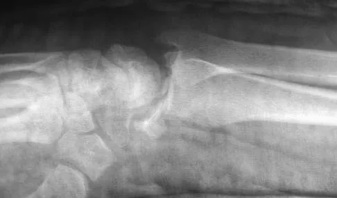
Figures 44a through 44c show the radiographs of an 18-year-old female soccer player who fell on her outstretched hand 1 day ago. She denies any history of wrist pain. Examination reveals tenderness at the anatomic snuffbox. Management should consist of


Explanation

44b 44c The treatment of choice for proximal pole scaphoid fractures is open reduction and internal fixation with a differential pitch screw via a dorsal approach. Healing rates of 100% have been reported for these acute fractures. Casting results in slow healing, with recommendations including 16 weeks or more in a cast. Vascularized bone grafts are not indicated for acute fractures. Rettig ME, Raskin KB: Retrograde compression screw fixation of acute proximal pole scaphoid fractures. J Hand Surg 1999;24:1206-1210.

Question 11

An excessively large radial styloidectomy poses a risk for wrist instability. What ligament is at greatest risk for injury?


Explanation

The radioscaphocapitate ligament is the most radial of the extrinsic volar ligaments of the wrist. It has a mean attachment to the radius 4 mm from the tip of the radial styloid. Nakamura T, Cooney WP III, Lui WH, et al: Radial styloidectomy: A biomechanical study on the stability of the wrist joint. J Hand Surg Am 2001;26:85-93.

Question 12

What joint always remains uninvolved in all stages of scapholunate advanced collapse (SLAC) deformity of the wrist?


Explanation

The development of arthritis in SLAC wrist follows a consistent pattern. Beginning at the radial styloid to the scaphoid articulation, it progresses through the entire radioscaphoid joint and the midcarpal joint. In all stages, the radiolunate joint is spared, which is the basis for a scaphoid excision and four-corner fusion performed as a motion-sparing procedure for treatment of this condition. Wyrick JD: Proximal row carpectomy and intercarpal arthrodesis for the management of arthritis. J Am Acad Orthop Surg 2003;11:277-281. Watson HK, Ballett FL: The SLAC wrist: Scapholunate advanced collapse pattern of degenerative arthritis. J Hand Surg Am 1984;9:358-365.

Question 13

Free flap coverage for severe trauma to the upper extremity has the fewest complications when performed within what time period after injury?


Explanation

Flap necrosis and infection rates are lowest if free flap coverage is performed within 72 hours of injury. Delays beyond 72 hours are associated with a higher rate of complications. Godina M: Early microsurgical reconstruction of complex trauma of the extremities. Plast Reconstr Surg 1986;78:285-292.

Question 14

A 54-year-old woman with idiopathic carpal tunnel syndrome undergoes open carpal tunnel release with a flexor tenosynovectomy. The pathology from the tenosynovium is likely to show


Explanation

The tenosynovium excised at the time of a carpal tunnel release for idiopathic carpal tunnel syndrome rarely shows signs of acute or chronic inflammation. Fibrosis, edema, and vascular sclerosis are the most common histologic findings. A tenosynovectomy with a carpal tunnel release usually is not necessary in the treatment of idiopathic carpal tunnel syndrome. Shum C, Parisien M, Strauch RJ, et al: The role of flexor tenosynovectomy in the operative treatment of carpal tunnel syndrome. J Bone Joint Surg Am 2002;84:221-225. Fuchs PC, Nathan PA, Myers LD: Synovial histology in carpal tunnel syndrome. J Hand Surg Am 1991;16:753-758.

Question 15

Examination of a 10-year-old girl with a hypoplastic breast and atrophic pectoralis major may also reveal which of the following findings?


Explanation

Poland's syndrome has four main features: 1) short digits as the result of absence or shortening of the middle phalanx; 2) syndactyly of the short digits usually consisting of a simple, complete type; 3) hypoplasia of the hand and forearm; and 4) absence of the sternocostal head of the pectoralis major on the same side. Wilson MR, Louis DS, Stevenson TR: Poland's syndrome: Variable expression and associated anomalies. J Hand Surg 1988;13:880-882.

Question 16

Figures 45a and 45b show the radiographs of a 40-year-old woman with rheumatoid arthritis who is unable to straighten her ring and little fingers. Examination reveals that the fingers can be passively corrected, but she is unable to actively maintain the fingers in extension. Management should consist of


Explanation

45b The patient has extensor tendon ruptures at the level of the wrist that are the result of synovitis at the distal radioulnar joint (Vaughn-Jackson syndrome). Extensor indius proprius transfer appropriately matches strength and excursion of the ruptured extensor digiti quinti and extensor digitorum communis tendons. An extensor tenosynovectomy with distal radioulnar joint resection decreases the synovitis, which if left untreated may cause additional tendon ruptures. Radial head resection is used for posterior interosseous nerve compression secondary to radial head synovitis, and in this patient only two fingers are involved, which rules out this diagnosis. Dynamic splinting is not indicated for ruptured tendons. Metacarpophalangeal arthroplasties and imbrication of the sagittal bands are used for metacarpophalangeal arthritis and extensor tendon subluxation. If this was the problem, the patient should be able to maintain the fingers in extension after they are passively extended. Total wrist arthrodesis prevents the tenodesis effect, thus limiting effective tendon excursion and making the proposed transfer less effective. Feldon P, Terrono AL, Nalebuff EA, et al: Rheumatoid arthritis and other connective tissue diseases: Tendon ruptures, in Green DP, Hotchkiss RN, Pederson WC (eds): Green's Operative Hand Surgery, ed 4. New York, NY, Churchill Livingstone, 1999, pp 1669-1684. Moore JR, Weiland AJ, Valdata L: Tendon ruptures in the rheumatoid hand: Analysis of treatment and functional results in 60 patients. J Hand Surg Am 1987;12:9-14.

Question 17

Figures 46a through 46e show the radiographs of a 22-year-old man who injured his wrist in a motorcycle accident. He has no other injuries. What is the best course of action?


Explanation

46b 46c 46d 46e The patient has a fracture-dislocation of the radiocarpal joint. Attached to the large radial styloid fragment are the extrinsic wrist ligaments to the carpus. This injury should be treated with open reduction and internal fixation of the styloid fracture. Radiolunate fusion or extrinsic ligament repair is suggested when the extrinsic ligaments are ruptured, resulting in ulnar translocation of the carpus. Dumontier C, Meyer ZU, Reckendorf G, et al: Radiocarpal dislocations: Classification and proposal for treatment: A review of twenty-seven cases. J Bone Joint Surg Am 2001;83:212.

Question 18

A 36-year-old nurse has had redness, pain, and small vesicles on the pulp of her middle finger for the past 3 days. Management should consist of


Explanation

Small vesicles on the fingers of a health care worker suggest a herpetic infection, and the management of choice is observation. Incision and drainage may result in a bacterial infection. Marsupialization is used in the treatment of a chronic paronychia. Calcium gluconate is used for hydrofluoric acid burns, and copper sulfate is used for white phosphorus burns. Fowler JR: Viral Infections. Hand Clin 1989;5:613-627.

Question 19

A 35-year-old man has numbness and tingling in the index, middle, and ring fingers. History reveals that he also has had vague wrist pain and stiffness since being injured in a motorcycle accident 1 year ago. Radiographs are shown in Figures 47a through 47c. Management should consist of


Explanation

47b 47c The patient has a chronic unrecognized volar lunate dislocation. Median nerve compression is the result of the lunate displaced into the carpal tunnel. The diagnosis can be made by radiographs; MRI is not necessary. A volar approach allows median nerve decompression with excision of the lunate, whereas a dorsal approach facilitates excision of the scaphoid and triquetrum. Rettig ME, Raskin KB: Long-term assessment of proximal row carpectomy for chronic perilunate dislocations. J Hand Surg Am 1999;24:1231-1236.

Question 20

A 42-year-old woman has persistent thumb pain that she notes is worse with opening jars and turning her car key. Opponens splinting provides some relief, but she is poorly tolerant of the splint. Finkelstein's test is negative, and a carpometacarpal grind test is positive. The radiographs shown in Figures 48a and 48b reveal minimal degenerative changes at the first carpometacarpal joint. What is the best course of action?


Explanation

48b The woman has early basilar thumb arthritis. An extension osteotomy will redirect the force to the dorsal, more uninvolved portion of the first carpometacarpal joint and has been reported to alleviate pain in these patients. Arthrodesis is usually reserved for young, typically male laborers. Thermal shrinkage and denervation are considered experimental at this time. Interposition arthroplasty is typically used for more advanced stages of arthritis. Tomaino MM: Treatment of Eaton stage I trapeziometacarpal disease with thumb metacarpal extension osteotomy. J Hand Surg Am 2000;25:1100-1106. Pellegrini VD Jr, Parentis M, Judkins A, et al: Extension metacarpal osteotomy in the treatment of trapeziometacarpal osteoarthritis: A biomechanical study. J Hand Surg Am 1996;21:16-23.

Question 21

A 45-year-old man sustains a low-velocity gunshot wound to the base of the right thumb. The open wound is allowed to heal by secondary intention, resulting in a contracture of the first web space. Clinical photographs are shown in Figures 49a through 49c. Treatment should now consist of


Explanation

49b 49c The contracture is too large for a Z-plasty, which allows a 75% increase in length. Excision of the scar with placement of a skin graft is prone to contracture. A posterior interosseous fasciocutaneous flap will provide enough well-vascularized tissue and is well suited to reach the first dorsal web space. Buchler U, Frey HP: Retrograde posterior interosseous flap. J Hand Surg Am 1991;16:283-292.

Question 22

The vessel seen in the clinical photographs shown in Figures 50a and 50b (1,2 intercompartmental supraretinacular artery) is being dissected to be used as a source of vascularized bone graft for a patient who is scheduled to undergo internal fixation of a scaphoid nonunion. This vessel is a branch of what artery?


Explanation

50b The 1,2 intercompartmental supraretinacular artery is a branch of the radial artery. The vessel provides a reliable source of vascularized bone graft with an adequate pedicle length for use in scaphoid nonunions. Sheetz KK, Bishop AT, Berger RA: The arterial blood supply of the distal radius and ulna and its potential use in vascularized pedicled bone grafts. J Hand Surg 1995;20:902-914.

Question 23

The flap shown in the clinical photograph seen in Figure 51 is based on what arterial supply?


Explanation

The groin flap is based on the superficial circumflex iliac artery, an axial flap that has been a mainstay of providing soft-tissue coverage of the upper extremity. Flaps as large as 35 cm in length and 15 cm in width have been reported. An advantage of the flap is that when used as a pedicle flap, the donor site can be closed directly. A disadvantage of the flap is that it can be quite bulky and can have a thick layer of subcutaneous fat. The superficial circumflex iliac artery travels lateral and superficial to the fascia and below and parallel to the inguinal ligament. It is helpful to elevate the fascia at the medial border of the sartorius muscle to include the deep and superficial branches of the artery for improved flap survival. McGregor IA, Jackson IT: The groin flap. Br J Plast Surg 1972;25:3-9.

Question 24

A 63-year-old woman who sustained a distal radial fracture 2 months ago now reports that she is unable to achieve active extension of the thumb at the interphalangeal joint. What type of trauma may lead to this clinical finding?


Explanation

Nondisplaced distal radial fractures have a higher rate of spontaneous rupture of the extensor pollicis longus tendon. The extensor mechanism is felt to impinge on the tendon following a nondisplaced fracture and causes either a mechanical attrition of the tendon or a local area of ischemia in the tendon. Helal B, Chen SC, Iwegbu G: Rupture of the extensor pollicis longus tendon in undisplaced Colles' type of fracture. Hand 1982;14:41-47.

Question 25

What radiographic view will best reveal degeneration of the pisotriquetral joint in a patient who is being evaluated for pisotriquetral arthrosis?


Explanation

The pisotriquetral joint is best seen on a lateral view in 30 degrees of supination. The carpal tunnel view provides visualization of the joint but to a lesser extent. The other views do not provide clear and accurate visualization. Paley D, McMurty RY, Cruickshank B: Pathologic conditions of the pisiform and pisotriquetral joint. J Hand Surg Am 1987;12:110-119.

Question 26

A surgeon is performing an open reduction and internal fixation of a complex capitellar fracture involving the trochlea. The surgical plan requires exposing the radiocapitellar joint. If the surgeon utilizes the Kaplan approach, which of the following describes the correct internervous/intermuscular interval?


Explanation

The Kaplan approach to the lateral elbow utilizes the interval between the extensor carpi radialis brevis (ECRB, innervated by the radial nerve) and the extensor digitorum communis (EDC, innervated by the posterior interosseous nerve). In contrast, the Kocher approach utilizes the interval between the anconeus (radial nerve) and the extensor carpi ulnaris (posterior interosseous nerve).

Question 27

A 68-year-old man presents with chronic right shoulder pain and an inability to actively elevate his arm above 40 degrees. Passive elevation is preserved to 160 degrees. MRI demonstrates a massive, retracted tear of the supraspinatus and infraspinatus with advanced fatty infiltration (Goutallier stage 4), while the subscapularis and teres minor are intact. What is the most reliable surgical option to restore active forward elevation in this patient?


Explanation

The patient presents with pseudoparalysis of elevation secondary to a massive, irreparable posterosuperior rotator cuff tear. In elderly patients with pseudoparalysis and advanced fatty infiltration, reverse total shoulder arthroplasty (rTSA) provides the most reliable restoration of active elevation by medializing the center of rotation and maximizing the deltoid moment arm. Tendon transfers and SCR are generally less reliable for reversing true pseudoparalysis.

Question 28

A 30-year-old man sustains a midshaft humerus fracture resulting in a complete, high radial nerve palsy. After 14 months of observation and serial EMGs showing no signs of reinnervation, tendon transfers are planned. To optimally restore active wrist extension, which of the following is the most standard and reliable donor muscle?


Explanation

The pronator teres (PT) to extensor carpi radialis brevis (ECRB) transfer is the workhorse procedure for restoring wrist extension in patients with an irreparable radial nerve palsy. The PT is chosen due to its favorable excursion, strength, and synergistic function with wrist extension. The ECRB is preferred over the ECRL as the recipient because its more central insertion at the base of the third metacarpal minimizes radial deviation during wrist extension.

Question 29

A 42-year-old woman reports sudden onset of severe, unprovoked left forearm and shoulder pain that lasted for two weeks and has now resolved. However, she currently complains of difficulty writing and an inability to form an 'OK' sign with her thumb and index finger. She has no numbness or tingling. What is the most likely diagnosis and recommended initial management?


Explanation

The patient's presentation of intense pain followed by purely motor weakness is classic for Parsonage-Turner syndrome (brachial neuritis). Her inability to make the 'OK' sign indicates involvement of the anterior interosseous nerve (AIN), which supplies the flexor pollicis longus, the radial half of the flexor digitorum profundus, and the pronator quadratus. Brachial neuritis frequently mimics compressive neuropathies but is primarily managed non-operatively with observation and supportive care.

Question 30

A 45-year-old man complains of chronic right wrist pain 10 years after a fall.

Radiographs reveal a scaphoid nonunion with arthritic changes at the radioscaphoid joint and the capitolunate joint. The radiolunate joint is completely spared. What is the most appropriate surgical treatment?


Explanation

This patient has Stage III Scaphoid Nonunion Advanced Collapse (SNAC). SNAC Stage I involves the radial styloid; Stage II involves the entire radioscaphoid joint; Stage III involves the capitolunate joint; and Stage IV involves pancarpal arthritis. Because the capitolunate joint is arthritic, a proximal row carpectomy (PRC) is contraindicated (as it requires a pristine capitate head to articulate with the lunate fossa). Therefore, scaphoid excision and four-corner fusion is the best motion-preserving option.

Question 31

A 32-year-old patient presents with a sensation of their elbow 'giving way' and clicking when pushing up from a chair with the forearm supinated. Clinical examination reveals a positive lateral pivot-shift test of the elbow. Deficiency of which of the following structures is the primary cause of this condition?


Explanation

The patient describes symptoms and exam findings pathognomonic for posterolateral rotatory instability (PLRI) of the elbow. The primary restraint to PLRI is the lateral ulnar collateral ligament (LUCL). Insufficiency of the LUCL allows the radial head to subluxate posterolaterally away from the capitellum, especially during axial loading, valgus stress, and supination.

Question 32

A 55-year-old woman is seen 6 months after undergoing volar locking plate fixation for a displaced distal radius fracture.

She reports a recent, sudden inability to flex the interphalangeal joint of her thumb. She experienced some volar wrist crepitus in the weeks prior. What is the most likely etiology of her new deficit?


Explanation

The patient has suffered an iatrogenic rupture of the flexor pollicis longus (FPL) tendon, which is the most common flexor tendon to rupture following volar plating of the distal radius. This complication is classically associated with placing the plate distal to the 'watershed line'—the bony ridge at the distal margin of the pronator fossa. Prominence of hardware here causes frictional wear and subsequent tendon attrition. Dorsal screw prominence typically endangers the extensor tendons, such as the extensor pollicis longus (EPL).

Question 33

A 35-year-old carpenter presents with worsening dorsal wrist pain. Radiographs show sclerosis and fragmentation of the lunate, with preserved carpal height (no collapse). The patient has a negative ulnar variance of 3 mm. According to the Lichtman classification, the patient has Stage IIIa Kienbock's disease. Which of the following is the most widely accepted surgical intervention for this specific presentation?


Explanation

In the setting of Kienböck's disease (avascular necrosis of the lunate) prior to carpal collapse (Lichtman Stages I, II, or IIIa) in a patient with negative ulnar variance, joint-leveling procedures are indicated to mechanically offload the lunate. A radial shortening osteotomy is the most common and reliable method, preferred over ulnar lengthening due to lower rates of nonunion and hardware complications.

Question 34

A 24-year-old motorcyclist sustains a severe traction injury to his left brachial plexus. He has a flail, completely insensate left arm. Physical examination also reveals ipsilateral ptosis, miosis, and anhidrosis. The presence of these specific facial and ocular findings most strongly indicates which of the following injury patterns?


Explanation

The patient is exhibiting Horner's syndrome (ptosis, miosis, anhidrosis), which in the context of a brachial plexus injury signifies damage to the sympathetic chain. The sympathetic fibers to the head and neck exit the spinal cord at T1 and travel near the C8 and T1 nerve roots before joining the sympathetic chain. Horner's syndrome indicates a pre-ganglionic injury (root avulsion) of the lower plexus roots (C8, T1), suggesting a poor prognosis for spontaneous recovery and precluding the use of those roots as donors for nerve transfers.

Question 35

A 22-year-old male sustains a completely displaced midshaft clavicle fracture following a cycling fall. In evaluating whether to proceed with non-operative management versus open reduction and internal fixation (ORIF), which of the following physical examination or radiographic findings is considered an absolute or strong relative indication for immediate ORIF?


Explanation

Absolute or strong relative indications for acute operative fixation of clavicle fractures include open fractures, impending skin breakdown (characterized by skin tenting with blanching or necrosis), associated neurovascular injury, and 'floating shoulder' (ipsilateral displaced scapular neck fracture). While shortening is a relative indication, modern literature typically uses a threshold of >2 cm (not 1 cm) of shortening to justify the benefits of surgery versus non-operative treatment.

Question 36

A 40-year-old man falls from a height and sustains a posterolateral elbow dislocation, radial head fracture, and coronoid fracture. Following closed reduction, the joint remains unstable in extension. During operative management, what is the generally recommended sequence of reconstruction to restore elbow stability?


Explanation

The standard surgical protocol for addressing a 'terrible triad' of the elbow (elbow dislocation, radial head fracture, coronoid fracture) typically proceeds from deep to superficial and anterior to posterior: 1. Fixation of the coronoid fracture to restore the anterior buttress, 2. Fixation or replacement of the radial head to restore the lateral column, and 3. Repair of the lateral ulnar collateral ligament (LUCL) complex to the lateral epicondyle. If the elbow remains unstable after these steps (usually assessed in extension), the medial collateral ligament (MCL) may be repaired or a hinged external fixator applied.

Question 37

A 65-year-old woman undergoes open reduction and internal fixation of a distal radius fracture with a volar locking plate. Six months postoperatively, she presents with inability to actively flex the interphalangeal joint of her thumb. Radiographs show the fracture is fully healed. What was the most likely surgical error that led to this complication?


Explanation

The patient is experiencing a flexor pollicis longus (FPL) tendon rupture. The most common cause of FPL rupture following volar plating of the distal radius is prominent hardware. Placing the plate distal to the 'watershed line' of the distal radius positions the hardware directly against the flexor tendons. The watershed line marks the distal margin of the pronator fossa; plates placed over or distal to this line cause frictional attrition of the FPL tendon during wrist and finger motion, eventually leading to rupture.

Question 38

A 50-year-old manual laborer presents with chronic right wrist pain. Radiographs reveal advanced osteoarthritis involving the radioscaphoid and capitolunate joints, with preservation of the radiolunate joint. Which of the following is the most appropriate motion-preserving surgical treatment?


Explanation

The patient has Stage III Scapholunate Advanced Collapse (SLAC), characterized by arthritis involving the capitolunate joint in addition to the radioscaphoid joint. The radiolunate joint is characteristically spared because of the congruent spherical articulation and intact short radiolunate ligament. Proximal row carpectomy (PRC) is contraindicated in Stage III SLAC because it relies on a healthy articulation between the capitate head and the lunate fossa of the radius; placing an arthritic capitate head into the lunate fossa would lead to continued pain and failure. Therefore, scaphoid excision and four-corner fusion (capitate, lunate, triquetrum, hamate) is the preferred motion-preserving procedure for Stage III SLAC.

Question 39

A 22-year-old athlete sustains a proximal pole scaphoid fracture. Due to the high risk of nonunion and avascular necrosis, operative fixation is planned. Which surgical approach and fixation strategy is most appropriate for this specific fracture pattern?


Explanation

The scaphoid receives its blood supply predominantly from the dorsal carpal branch of the radial artery, which enters at the distal waist and flows in a retrograde fashion to the proximal pole. Proximal pole fractures are highly prone to avascular necrosis. Due to the intra-articular position of the proximal pole and the natural flexion of the scaphoid, a dorsal approach is strongly preferred. It allows for direct visualization of the proximal pole, preservation of the remaining blood supply, and placement of a central axis screw from proximal to distal, which biomechanically provides superior compression and stability for this specific region.

Question 40

A 28-year-old overhead athlete complains of poorly localized posterior shoulder pain and paresthesias over the lateral deltoid. MRI of the shoulder reveals isolated atrophy and fatty infiltration of the teres minor muscle. Which of the following anatomic structures form the borders of the space where the affected nerve is likely compressed?


Explanation

The clinical presentation and MRI findings (isolated teres minor atrophy) are classic for Quadrilateral Space Syndrome. This syndrome involves compression of the axillary nerve and posterior humeral circumflex artery. The anatomical boundaries of the quadrilateral space are the teres minor (superiorly), teres major (inferiorly), long head of the triceps (medially), and the surgical neck of the humerus (laterally). Compression in this space typically leads to lateral deltoid paresthesias and selective denervation of the teres minor and/or deltoid.

Question 41

A 45-year-old competitive weightlifter suffers an acute distal biceps tendon rupture. The surgeon utilizes a two-incision technique (modified Boyd-Anderson) to reattach the tendon to the radial tuberosity. Compared to a single anterior incision technique, the two-incision approach is associated with a higher risk of which of the following postoperative complications?


Explanation

Operative repair of a distal biceps tendon rupture can be performed via a single anterior incision or a two-incision technique. The single-incision technique carries a higher risk of injury to anterior structures, specifically the lateral antebrachial cutaneous (LABC) nerve and the posterior interosseous nerve (PIN). The two-incision technique was developed to protect these nerves but carries a historically higher risk of proximal radioulnar synostosis (heterotopic ossification bridging the radius and ulna) due to subperiosteal dissection and the potential creation of bone debris in the highly reactive interosseous space.

Question 42

A 35-year-old man presents with a sense of clicking and instability in his elbow when pushing himself up from a chair. He underwent a lateral epicondylar release for recalcitrant 'tennis elbow' one year ago. On physical examination, with the patient supine and the shoulder flexed, applying an axial load, valgus stress, and supination to the elbow as it is moved from extension to flexion produces a palpable clunk. Deficiency of which of the following structures is most likely responsible for his symptoms?


Explanation

The patient's history of prior lateral epicondylar surgery and physical examination findings (positive lateral pivot-shift test of the elbow) indicate Posterolateral Rotatory Instability (PLRI). PLRI is caused by insufficiency of the lateral ulnar collateral ligament (LUCL). The LUCL can be iatrogenically injured during a lateral epicondylar release (e.g., Nirschl procedure) if the surgical dissection is carried too far posterior and distal to the extensor carpi radialis brevis (ECRB) origin.

Question 43

A 32-year-old carpenter presents with a 6-month history of central dorsal wrist pain and decreased grip strength. Radiographs reveal sclerosis and early fragmentation of the lunate, but no carpal collapse is noted. Radiographic measurements demonstrate a negative ulnar variance of 3 mm. Which of the following surgical interventions is most appropriate to halt disease progression?


Explanation

The patient has Kienbock's disease (avascular necrosis of the lunate) Lichtman Stage II or early IIIa (fragmentation but no carpal collapse) associated with negative ulnar variance. Negative ulnar variance increases compressive load transmission across the radiolunate joint. For patients with negative ulnar variance and no advanced carpal collapse, joint-leveling procedures such as a radial shortening osteotomy are indicated. This decreases the mechanical stress on the lunate, potentially allowing for revascularization and halting collapse. While ulnar lengthening achieves the same biomechanical goal, radial shortening osteotomy is technically preferred due to higher union rates and fewer complications.

Question 44

A 72-year-old man with severe pseudoparalysis and glenohumeral osteoarthritis secondary to a massive, irreparable rotator cuff tear is scheduled for a reverse total shoulder arthroplasty (RTSA). Preoperative evaluation demonstrates absent active external rotation with the arm at the side (positive Hornblower's sign and dropped arm sign). In addition to RTSA, what adjunctive procedure is most appropriate to optimize this patient's postoperative ability to perform activities of daily living?


Explanation

The patient has a massive rotator cuff tear involving the posterior superior cuff (infraspinatus and teres minor), as evidenced by the lack of active external rotation (positive Hornblower's and drop sign). A reverse total shoulder arthroplasty (RTSA) will restore anterior elevation by utilizing the intact deltoid, but it does not adequately restore active external rotation if the teres minor is nonfunctional. To restore active external rotation and allow the patient to perform essential activities of daily living (like eating, grooming, and reaching the face), an adjunctive latissimus dorsi and/or teres major transfer (L'Episcopo procedure) is frequently performed concurrently with the RTSA.

Question 45

A 25-year-old man sustains a closed, distal-third spiral fracture of the humeral shaft (Holstein-Lewis fracture). Upon initial presentation in the emergency department, he exhibits a complete inability to extend his wrist, thumb, and metacarpophalangeal joints, along with numbness in the first dorsal web space. According to current orthopedic guidelines, what is the most appropriate initial management of this injury?


Explanation

The patient has a primary radial nerve palsy associated with a closed distal-third spiral humeral shaft fracture (Holstein-Lewis fracture). Despite the high historical association of radial nerve entrapment or laceration with this specific fracture pattern, the vast majority (>85%) of these injuries represent a neuropraxia (axonotmesis) that will spontaneously recover. Current AAOS and orthopedic trauma guidelines stipulate that the presence of a primary radial nerve palsy in the setting of a closed humeral shaft fracture is not an absolute indication for immediate surgical exploration. The standard of care is nonoperative fracture management (e.g., coaptation splint followed by a functional Sarmiento brace) and observation of the nerve palsy for 3 to 4 months. Surgical exploration is reserved for open fractures, failure to achieve closed reduction, vascular injury, or failure of the nerve to show clinical or EMG recovery by 3-4 months.

Question 46

A 65-year-old female undergoes a reverse total shoulder arthroplasty (RTSA) for a comminuted 4-part proximal humerus fracture. Six months postoperatively, she presents with severe shoulder aching and stiffness, though she lacks systemic symptoms. Laboratory results show a normal ESR and CRP. Joint aspiration cultures grow Cutibacterium acnes after 10 days. Which of the following is true regarding this infection in the setting of shoulder arthroplasty?


Explanation

Cutibacterium acnes (formerly Propionibacterium acnes) is a gram-positive anaerobic rod that is part of the normal skin flora, particularly in the shoulder region. It is a slow-growing, fastidious organism, and cultures should be held for at least 14 days in anaerobic environments to avoid false-negative results. Preoperative preparation with chlorhexidine/alcohol or hydrogen peroxide is more effective than povidone-iodine in reducing the burden of C. acnes. These infections often present indolently without systemic signs such as fever or significantly elevated inflammatory markers.

Question 47

A 42-year-old male construction worker presents with chronic numbness in his small and ring fingers and weakness in grip strength. Exam reveals a positive Froment's sign and intrinsic muscle atrophy. Intraoperatively, during decompression, the ulnar nerve is found to subluxate anteriorly over the medial epicondyle upon elbow flexion. What is the most appropriate surgical management?


Explanation

In patients with ulnar nerve subluxation or instability at the elbow during flexion, an in situ decompression alone is contraindicated. Decompressing without stabilizing the nerve can lead to continued or worsened subluxation, causing a severe friction neuritis over the medial epicondyle. An anterior transposition (subcutaneous, intramuscular, or submuscular) or a medial epicondylectomy is recommended to address both the compression and the dynamic instability.

Question 48

A 30-year-old male presents with chronic radial-sided wrist pain following a remote injury. Radiographs demonstrate a scaphoid nonunion with radioscaphoid and capitolunate arthritis. The radiolunate joint is radiographically preserved. What is the most appropriate surgical intervention for this stage of Scaphoid Nonunion Advanced Collapse (SNAC)?


Explanation

The scenario describes SNAC Stage III, characterized by capitolunate arthritis with a spared radiolunate joint. A proximal row carpectomy (PRC) requires a pristine capitate head and lunate fossa to function as the new articulation; therefore, capitolunate arthritis is a contraindication to PRC. Four-corner arthrodesis (capitate, hamate, lunate, triquetrum) with scaphoid excision is the most appropriate motion-preserving salvage procedure for SNAC Stage III, as it relies on the preserved radiolunate joint.

Question 49

A 48-year-old weightlifter feels a 'pop' in his anterior elbow during a heavy deadlift, followed by ecchymosis and weakness in supination. MRI confirms a complete distal biceps tendon avulsion. He opts for surgical repair using a single-incision anterior approach. Which nerve is at greatest risk of iatrogenic injury during the superficial dissection of this specific surgical approach?


Explanation

The single-incision anterior approach for distal biceps tendon repair primarily places the lateral antebrachial cutaneous nerve (LABCN) at risk during the superficial exposure. The LABCN exits the deep fascia lateral to the biceps tendon and must be identified and protected. While the posterior interosseous nerve (PIN) is at risk during deep retractor placement or drilling of the posterior radius cortex, the LABCN is statistically the most commonly injured nerve overall in the single-incision anterior approach.

Question 50

A 6-year-old boy sustains an extension-type Gartland type III supracondylar humerus fracture. Upon initial evaluation, he has an absent radial pulse, but the hand is pink, warm, and has a brisk capillary refill. Following closed reduction and percutaneous pinning, the radial pulse remains absent, but the hand's perfusion status is unchanged (pink and warm). What is the next most appropriate step in management?


Explanation

In a pediatric supracondylar humerus fracture with a 'pink, pulseless hand' after acceptable closed reduction and pinning, the standard of care is close observation. The collateral circulation in children is generally sufficient to maintain hand viability even if the brachial artery is in spasm or occluded. Immediate vascular exploration is indicated if the hand is ischemic ('white, pulseless hand') that does not improve after fracture reduction. Serial neurovascular checks are crucial to monitor for compartment syndrome or secondary loss of perfusion.

Question 51

A 24-year-old female gymnast presents with progressive dorsal wrist pain. Radiographs reveal sclerosis of the lunate with no carpal collapse (Lichtman Stage II). Ulnar variance is determined to be negative 3 mm. MRI confirms Kienböck's disease. Which of the following is the most appropriate surgical treatment?


Explanation

Kienböck's disease is avascular necrosis of the lunate. In the early stages (Lichtman Stage II or IIIa - sclerosis or mild collapse without carpal instability) in a patient with negative ulnar variance, a joint-leveling procedure such as a radial shortening osteotomy is indicated. This unloads the radiolunate joint, decreasing the forces transmitted through the lunate, and has a high success rate in halting disease progression. Salvage procedures like PRC or STT arthrodesis are reserved for more advanced stages with carpal collapse.

Question 52

A 72-year-old male presents with pseudoparalysis of the right shoulder and severe glenohumeral osteoarthritis secondary to massive rotator cuff arthropathy. He is planned for a Grammont-style reverse total shoulder arthroplasty (RTSA). How is the center of rotation biomechanically altered in this implant design compared to the native shoulder?


Explanation

The classic Grammont-style reverse total shoulder arthroplasty (RTSA) alters the biomechanics by medializing and inferiorizing the center of rotation. Medialization decreases the torque and shear forces on the glenoid component, reducing the risk of loosening, and recruits more deltoid muscle fibers. Inferiorization tensions the deltoid, increasing its lever arm and restoring active elevation in the setting of a deficient rotator cuff.

Question 53

A 35-year-old male sustains a 'terrible triad' injury of the elbow (elbow dislocation, radial head fracture, and coronoid fracture). When operating to restore elbow stability, which of the following is the generally accepted sequence of surgical repair?


Explanation

The standard surgical protocol for a 'terrible triad' of the elbow typically proceeds from deep to superficial, or 'inside-out' through a lateral approach. The sequence is: 1) Fixation of the coronoid fracture (or anterior capsule), 2) Fixation or replacement of the radial head, 3) Repair of the lateral collateral ligament (LCL) complex. After these steps, the elbow is examined for stability. The medial collateral ligament (MCL) is repaired only if the elbow remains grossly unstable in extension after the lateral structures are secured.

Question 54

A 28-year-old tennis player complains of chronic ulnar-sided wrist pain. MRI confirms a central articular disc tear of the triangular fibrocartilage complex (TFCC) (Palmer Type 1A). Radiographs reveal a positive ulnar variance of 4 mm. Conservative management has failed. What is the most appropriate surgical management?


Explanation

The patient has a Palmer Type 1A TFCC tear (central, avascular portion of the articular disc) associated with significant positive ulnar variance, indicative of ulnocarpal impaction syndrome. Central tears lack blood supply and are generally treated with arthroscopic debridement rather than repair. However, addressing the underlying ulnocarpal impaction with an ulnar shortening osteotomy (USO) is crucial to relieve the excessive mechanical load on the ulnar carpus. Debridement alone without joint leveling would lead to persistent symptoms.

Question 55

A 19-year-old male presents to the trauma bay after a high-speed motor vehicle collision complaining of severe pain at the base of his neck, dysphagia, and a choking sensation. The medial end of the right clavicle is not palpable anteriorly. A CT scan of the chest confirms a posterior sternoclavicular dislocation. As the surgical team prepares for closed reduction in the operating room, which of the following specialist teams must ideally be immediately available?


Explanation

Posterior sternoclavicular dislocations are high-energy injuries associated with potentially life-threatening complications due to the proximity of mediastinal structures. Symptoms such as dysphagia, dyspnea, choking, or venous congestion indicate mediastinal compression. Closed reduction should be attempted in the operating room under general anesthesia. Because of the high risk of catastrophic hemorrhage from the great vessels (e.g., brachiocephalic vein, superior vena cava, aorta) lying directly posterior to the joint, a cardiothoracic or vascular surgeon must be immediately available to perform an emergency sternotomy if needed.

Question 56

A 42-year-old male falls on an outstretched hand and sustains a terrible triad injury of the elbow. Standard surgical protocol dictates repairing structures from deep to superficial to restore elbow stability. Which of the following describes the most appropriate sequence of surgical repair?


Explanation

The standard surgical algorithm for a terrible triad injury of the elbow involves repairing structures from deep to superficial. The classic sequence is coronoid fixation, followed by radial head repair or arthroplasty, and finally lateral collateral ligament (LCL) repair. If the elbow remains unstable after these steps, the medial collateral ligament (MCL) is repaired, or a hinged external fixator is placed.

Question 57

A 65-year-old woman is 6 months status-post open reduction and internal fixation of a distal radius fracture with a volar locking plate. She presents with a sudden inability to actively flex the interphalangeal joint of her thumb. Which of the following surgical errors is most commonly associated with this complication?


Explanation

Flexor pollicis longus (FPL) tendon rupture is a well-known complication of volar plating of distal radius fractures. It is most commonly caused by placement of the plate distal to the watershed line, resulting in prominence of the distal edge of the plate and subsequent attrition and rupture of the FPL tendon. Prominent dorsal screws typically cause extensor tendon irritation or rupture (e.g., extensor pollicis longus).

Question 58

A 24-year-old elite volleyball player complains of vague posterior shoulder pain and weakness with overhead serving. Physical examination reveals atrophy of the infraspinatus but normal bulk of the supraspinatus. There is notable weakness in external rotation but normal abduction. An MRI shows a paralabral cyst. In which of the following anatomic locations is the cyst most likely compressing the involved nerve?


Explanation

The suprascapular nerve innervates the supraspinatus and infraspinatus. It passes through the suprascapular notch (where compression affects both muscles) and then winds around the spinoglenoid notch to innervate the infraspinatus. Compression at the spinoglenoid notch, often by a paralabral cyst associated with a posterior labral tear, results in isolated infraspinatus atrophy and weakness in external rotation, while the supraspinatus remains spared.

Question 59

A 72-year-old female sustains a severe 4-part proximal humerus fracture. She undergoes a reverse total shoulder arthroplasty (RTSA). Which of the following represents the most significant biomechanical advantage of RTSA over an anatomic total shoulder arthroplasty in this specific clinical setting?


Explanation

Reverse total shoulder arthroplasty (RTSA) is highly effective for 4-part proximal humerus fractures in the elderly, especially when rotator cuff function is compromised or tuberosity healing is unreliable. Biomechanically, RTSA medializes and distalizes the center of rotation of the glenohumeral joint. This significantly increases the moment arm and tension of the deltoid muscle, allowing it to initiate and maintain shoulder elevation without a functioning rotator cuff.

Question 60

A 21-year-old male fell on an outstretched hand 3 months ago and was treated conservatively for a 'sprained wrist.' He now presents with persistent radial-sided wrist pain. Radiographs reveal a scaphoid waist fracture with cystic changes and 2 mm of displacement. What is the primary blood supply to the proximal pole of the scaphoid, which places it at high risk for avascular necrosis in this fracture pattern?


Explanation

The primary blood supply to the scaphoid is retrograde. The dorsal carpal branch of the radial artery enters the scaphoid at the dorsal ridge (distal to the waist) and supplies the proximal 80% of the bone via intraosseous retrograde flow. A fracture at the scaphoid waist disrupts this blood supply to the proximal pole, placing it at high risk for avascular necrosis (AVN) and nonunion. The palmar carpal branch supplies only the distal 20%.

Question 61

A 45-year-old male undergoes surgical repair of an acute distal biceps tendon rupture using a standard 2-incision technique. Postoperatively, he exhibits a specific nerve palsy. Which of the following nerves is at greatest risk during the posterior approach of the 2-incision technique if the forearm is not fully pronated during surgical exposure?


Explanation

The 2-incision technique for distal biceps tendon repair aims to reduce the risk of lateral antebrachial cutaneous nerve and radial nerve injuries associated with a single anterior incision. However, it places the posterior interosseous nerve (PIN) at risk during the posterior dissection. To protect the PIN, the forearm must be maximally pronated during the posterior approach and retractor placement, as this displaces the PIN anteriorly and medially, away from the surgical field.

Question 62

A 32-year-old male sustains a closed, distal-third spiral fracture of the humeral shaft (Holstein-Lewis fracture). On initial presentation, he has a complete radial nerve palsy. He is treated with functional bracing. Twelve weeks later, there is radiographic evidence of early bridging callus, but the patient still has no clinical or electromyographic (EMG) evidence of radial nerve recovery. What is the most appropriate next step in management?


Explanation

The initial management of a closed humeral shaft fracture with a primary radial nerve palsy is observation, as the vast majority are neuropraxias that will spontaneously recover. However, if there is no clinical or EMG evidence of nerve recovery by 12 weeks (3 months), the standard recommendation is surgical exploration of the radial nerve. This allows for neurolysis, direct repair, or grafting if the nerve is transected or entrapped within the fracture callus.

Question 63

A 55-year-old manual laborer presents with chronic, intractable posterior shoulder pain and profound weakness in external rotation. He has a positive Hornblower's sign and a positive dropping sign. MRI demonstrates a massive, retracted, and irreparably atrophic tear of the infraspinatus and teres minor, with an intact subscapularis. Which of the following tendon transfers is most appropriate to restore external rotation in this patient?


Explanation

This patient has an isolated loss of active external rotation due to irreparable tears of the infraspinatus and teres minor. The lower trapezius transfer is highly effective for restoring external rotation in this setting because the line of pull of the lower trapezius closely replicates the vector of the infraspinatus. While the latissimus dorsi transfer is used for massive posterosuperior tears, its vector is less ideal for isolated external rotation compared to the lower trapezius. Pectoralis major transfers are used for irreparable subscapularis tears.

Question 64

A 28-year-old male sustains a diaphyseal fracture of the middle third of the radius with an associated disruption of the distal radioulnar joint (DRUJ) after a fall. Intraoperatively, after rigid open reduction and internal fixation of the radius, the DRUJ is found to reduce anatomically and is stable in supination, but it readily subluxates when the forearm is placed in pronation. What is the most appropriate management of the DRUJ?


Explanation

This describes a Galeazzi fracture-dislocation. Following rigid internal fixation of the radial shaft, the stability of the DRUJ must be assessed. If the DRUJ is reducible and stable in supination (the position that tightens the palmar radioulnar ligaments and stabilizes dorsal dislocations), the recommended management is immobilization in a long arm splint or cast in supination for 4 to 6 weeks. If the DRUJ remains unstable in all positions or cannot be reduced, open reduction and TFCC repair or percutaneous pinning of the DRUJ is indicated.

Question 65

A 34-year-old carpenter presents with an 8-month history of insidious onset, progressive dorsal wrist pain and limited extension. Radiographs show sclerosis and partial fragmentation of the lunate, with no evidence of carpal collapse or secondary osteoarthritis (Lichtman Stage IIIA). His ulnar variance is negative (-3 mm). Which of the following is the most appropriate surgical intervention?


Explanation

The patient has Kienböck's disease (avascular necrosis of the lunate) stage IIIA (lunate sclerosis and fragmentation, but normal carpal height) with negative ulnar variance. The goal of surgery in early stages with negative ulnar variance is to unload the lunate by leveling the distal radioulnar joint. A radial shortening osteotomy (or ulnar lengthening osteotomy) decreases the compressive forces on the lunate and is the procedure of choice. Proximal row carpectomy or intercarpal fusions are typically reserved for advanced stages (IIIB or IV) where carpal collapse or arthritis has occurred.

Question 66

A 70-year-old woman with advanced rotator cuff tear arthropathy is scheduled for a reverse total shoulder arthroplasty (RTSA). During preoperative templating and intraoperative execution, which of the following glenosphere positioning strategies is most effective in minimizing the risk of scapular notching?


Explanation

Scapular notching is a common and highly recognized complication of reverse total shoulder arthroplasty (RTSA). It occurs due to mechanical impingement of the humeral polyethylene cup against the inferior scapular neck during arm adduction. Inferior placement of the baseplate along with inferior tilt of the glenosphere, and lateralization of the center of rotation, have been proven to significantly decrease the incidence of scapular notching by increasing the impingement-free range of motion.

Question 67

A 34-year-old man presents with recurrent clicking, apprehension, and a sensation of 'giving way' in his right elbow, particularly when attempting to push himself out of a chair. Physical examination reveals a positive lateral pivot-shift test of the elbow. Which of the following ligamentous structures is primarily deficient in this specific instability pattern?


Explanation

The patient describes symptoms and demonstrates physical exam signs consistent with posterolateral rotatory instability (PLRI) of the elbow. PLRI is primarily caused by insufficiency of the lateral ulnar collateral ligament (LUCL), which acts as the main restraint to varus and posterolateral rotatory stress. The pathognomonic mechanism that reproduces symptoms involves a combination of axial load, valgus stress, and external rotation (supination) of the forearm.

Question 68

A 55-year-old woman sustained a non-displaced distal radius fracture treated non-operatively in a short arm cast. Eight weeks post-injury, she reports a sudden, painless inability to extend her thumb interphalangeal joint. On examination, she is unable to lift her thumb off the table when the palm is laid flat. What is the most appropriate surgical management to restore thumb kinematics?


Explanation

The patient has experienced an extensor pollicis longus (EPL) tendon rupture, a known complication of distal radius fractures, particularly non-displaced fractures due to localized ischemia and attrition at Lister's tubercle. Because the ruptured tendon ends are typically severely frayed, degenerated, and retracted, primary repair is usually impossible. An EIP to EPL tendon transfer is the standard of care, providing appropriately matched excursion and vector pull without significant donor site morbidity.

Question 69

During an open carpal tunnel release, the surgeon carefully dissects the transverse carpal ligament and identifies the recurrent motor branch of the median nerve piercing directly through the substance of the ligament. According to the Lanz classification of median nerve variations, which subtype does this represent?


Explanation

The Lanz classification describes anatomical variations of the recurrent motor branch of the median nerve. The normal and most common anatomy is extraligamentous (the nerve branches distally and turns radially to the transverse carpal ligament). A transligamentous branch (Lanz group 1 variation) arises within the tunnel and pierces directly through the transverse carpal ligament to reach the thenar musculature. Recognizing this variation is critical to avoid iatrogenic nerve transection during standard ligament release.

Question 70

A 24-year-old male presents with a displaced fracture involving the proximal pole of the scaphoid. Regarding the surgical management and relevant vascular anatomy, which of the following statements is true?


Explanation

The scaphoid receives its primary blood supply from dorsal branches of the radial artery that enter the distal ridge and flow retrograde to the proximal pole. This tenuous retrograde blood supply puts proximal pole fractures at exceedingly high risk for avascular necrosis and nonunion. For proximal pole fractures, a dorsal surgical approach is heavily favored as it provides direct visualization, easier anatomic reduction, and a more biomechanically sound central axis for screw placement without disrupting the volar ligaments or the remaining critical dorsal blood supply.

Question 71

A 68-year-old female sustains a 3-part anterior fracture-dislocation of her right proximal humerus after a mechanical fall. Upon presentation in the emergency department, she exhibits decreased sensation over the lateral aspect of her shoulder. Which of the following physical examination findings would most likely accompany this isolated neurological deficit once the fracture is stabilized?


Explanation

Decreased sensation over the lateral aspect of the shoulder (the territory of the superior lateral brachial cutaneous nerve) strongly indicates an axillary nerve injury. The axillary nerve is the most commonly injured nerve in anterior shoulder dislocations and proximal humerus fracture-dislocations. It provides motor innervation to the deltoid and teres minor. Because the supraspinatus (suprascapular nerve) initiates the first 15 degrees of shoulder abduction, an isolated axillary nerve injury will present clinically as profound weakness in active shoulder abduction beyond those initial 15 degrees.

Question 72

A 42-year-old male weightlifter feels a sudden 'pop' in his anterior elbow while performing heavy eccentric bicep curls. He presents with ecchymosis in the antecubital fossa and a positive 'hook test.' To restore maximum functional strength, surgical repair should anatomically reattach the tendon to which of the following structures?


Explanation

The patient has sustained a rupture of the distal biceps tendon, indicated by the mechanism of injury, ecchymosis, and a positive hook test (inability to hook the finger under the intact biceps tendon from the lateral side). The distal biceps tendon anatomically inserts onto the radial tuberosity. The primary function of the biceps brachii at the elbow is robust forearm supination, followed by elbow flexion. Anatomic repair to the radial tuberosity is mandatory to restore full supination torque, which is otherwise permanently decreased by roughly 40% if left unrepaired or repaired non-anatomically (e.g., to the brachialis).

Question 73

A 55-year-old carpenter presents with a 6-month history of paresthesias in the right small and ulnar half of the ring finger, along with subjective weakness in hand grip. Electromyography confirms a compressive ulnar neuropathy at the elbow. During surgical decompression, the surgeon must systematically release several potential sites of compression. Which of the following anatomic structures is NOT a recognized site of ulnar nerve compression in this region?


Explanation

The Ligament of Struthers is a potential site of MEDIAN nerve compression in the distal arm (associated with an anomalous supracondylar process of the humerus), not the ulnar nerve. Recognized sites of ulnar nerve compression around the elbow include the Arcade of Struthers (a fascial band extending from the medial head of the triceps to the medial intermuscular septum), the medial intermuscular septum (especially susceptible after anterior transposition of the nerve), the medial epicondyle, Osborne's ligament (the retinaculum bridging the two heads of the FCU), and the deep flexor-pronator aponeurosis.

Question 74

A 32-year-old manual laborer presents with progressive dorsal wrist pain and decreased grip strength. Radiographs demonstrate increased sclerosis of the lunate without carpal collapse or fragmentation, and a negative ulnar variance of 3 mm. MRI confirms avascular necrosis of the lunate. According to the Lichtman classification, this represents Stage II disease. Which of the following surgical interventions is most appropriate for this patient?


Explanation

The patient has Kienböck's disease (avascular necrosis of the lunate) Lichtman stage II (sclerosis without collapse) associated with ulnar negative variance. In patients with early-stage Kienböck's (Lichtman I, II, or IIIa) and ulnar negative variance, joint-leveling procedures such as a radial shortening osteotomy are the treatment of choice. This biomechanically unloads the lunate by shifting compressive forces to the ulnocarpal joint, thereby promoting revascularization and preventing further collapse. Salvage procedures like proximal row carpectomy (PRC) or intercarpal fusions are reserved for advanced stages with carpal collapse and secondary osteoarthritis (Lichtman IIIb and IV).

Question 75

While managing a 28-year-old cyclist who sustained a midshaft clavicle fracture, the orthopedic surgeon reviews the indications for operative intervention. Which of the following is considered an ABSOLUTE indication for open reduction and internal fixation of an acute clavicle fracture?


Explanation

Absolute indications for the operative fixation of an acute clavicle fracture include open fractures, fractures with associated progressive neurovascular compromise, and arguably the true 'floating shoulder' (ipsilateral displaced clavicle and scapular neck fractures, though some debate exists, open fracture remains universally absolute). Relative indications include shortening greater than 2 cm, >100% displacement, severe skin tenting (specifically with impending skin compromise/blanching), polytrauma, and symptomatic nonunions. Skin tenting without blanching or ischemia is considered a relative, not absolute, indication.

Question 76

A 72-year-old woman is 3 years post Reverse Total Shoulder Arthroplasty (RTSA). Radiographs show Grade 3 scapular notching. Which of the following surgical techniques or implant designs would most effectively minimize the risk of this complication?


Explanation

Scapular notching is a well-known complication of RTSA caused by mechanical impingement of the humeral component against the inferior scapular neck during adduction. Factors that decrease the risk of scapular notching include inferior placement of the glenoid baseplate, inferior tilt of the baseplate, lateralization of the glenosphere, use of a larger glenosphere, and a lower humeral neck-shaft angle (e.g., 135 degrees vs 155 degrees).

Question 77

A 22-year-old collegiate baseball pitcher undergoes ulnar collateral ligament (UCL) reconstruction using a palmaris longus autograft via the docking technique. What is the most common complication following this procedure?


Explanation

Ulnar neuropathy is the most common complication following UCL reconstruction, occurring in up to 10-15% of patients depending on the surgical technique (muscle-splitting vs. detachment) and whether routine ulnar nerve transposition is performed. Symptoms are often transient but can be persistent and may require later neurolysis.

Question 78

A 45-year-old male falls on an outstretched hand, sustaining a 'terrible triad' injury of the elbow. Intraoperatively, after secure fixation of the coronoid process and stable radial head arthroplasty, the elbow remains unstable and tends to subluxate posteriorly in extension. What is the next most appropriate step in management?


Explanation

The standard surgical algorithm for a terrible triad injury involves: 1) Coronoid fixation or anterior capsule repair, 2) Radial head fixation or replacement, and 3) LCL (specifically LUCL) repair. If the elbow remains unstable after LUCL repair, the MCL may then be repaired, or an external fixator applied. Since the LUCL has not yet been addressed in this scenario, repairing it is the critical next step to restore lateral column stability.

Question 79

A 38-year-old weightlifter undergoes a single-incision anterior approach for distal biceps tendon repair using suture anchors. Postoperatively, he notes a new onset of numbness along the radial aspect of his volar forearm. Which of the following nerves is most likely injured, and what is its motor innervation?


Explanation

The lateral antebrachial cutaneous nerve (LABCN), the terminal sensory branch of the musculocutaneous nerve, is the most commonly injured nerve during a single-incision anterior distal biceps repair due to vigorous lateral retraction. It provides sensation to the radial/lateral aspect of the forearm and has strictly sensory function, thus providing no motor innervation.

Question 80

A 28-year-old professional volleyball player presents with insidious onset of right shoulder pain and weakness. Examination reveals isolated atrophy of the infraspinatus muscle with normal bulk of the supraspinatus. MRI demonstrates a paralabral cyst. At which anatomic location is the nerve compression most likely occurring?


Explanation

The suprascapular nerve innervates both the supraspinatus and infraspinatus. Compression at the suprascapular notch affects both muscles. Compression at the spinoglenoid notch, often due to a paralabral cyst associated with a posterior SLAP tear in overhead athletes, affects only the distal infraspinatus branch, leading to isolated infraspinatus atrophy and weakness in external rotation.

Question 81

A 55-year-old man presents with a chronic, massive, irreparable posterosuperior rotator cuff tear. He has preserved forward elevation but a severe lack of active external rotation with a positive Hornblower's sign. He undergoes a lower trapezius tendon transfer prolonged with an Achilles tendon allograft. Which of the following nerves must be carefully protected during the harvest and mobilization of the lower trapezius?


Explanation

The lower trapezius is innervated by the spinal accessory nerve (CN XI). During its harvest and mobilization for a tendon transfer to restore external rotation, meticulous care must be taken to identify and protect the spinal accessory nerve and the transverse cervical artery, which course on the deep surface of the muscle.

Question 82

A 62-year-old woman sustains a displaced 3-part proximal humerus fracture. The orthopaedic surgeon plans open reduction and internal fixation via a deltopectoral approach. To avoid iatrogenic injury, the surgeon must be mindful of the axillary nerve. Which of the following accurately describes the normal anatomic course of the axillary nerve?


Explanation

The axillary nerve originates from the posterior cord of the brachial plexus. It courses anterior to the subscapularis muscle, then passes inferior to the shoulder capsule to exit posteriorly through the quadrilateral space, accompanied by the posterior humeral circumflex artery. The quadrilateral space is bounded by the teres minor (superior), teres major (inferior), long head of the triceps (medial), and humerus (lateral).

Question 83

A 42-year-old man presents with a history of sudden, severe, unremitting right shoulder pain lasting for 2 weeks that woke him from sleep. The pain has now largely resolved, but he has noticed profound weakness in shoulder abduction and external rotation. There is no history of trauma. EMG at 4 weeks reveals acute denervation changes in the supraspinatus and deltoid. What is the most likely diagnosis and appropriate initial management?


Explanation

Parsonage-Turner syndrome (idiopathic brachial neuritis) typically presents with acute, severe shoulder pain that lasts for 1-3 weeks. As the pain subsides, profound weakness and muscle atrophy (commonly affecting the deltoid, supraspinatus, and infraspinatus) become evident. It is typically a self-limiting condition, and the initial management consists of pain control, physical therapy, and observation.

Question 84

During an in situ ulnar nerve decompression for cubital tunnel syndrome, a surgeon sequentially releases the structures of the cubital tunnel. Which of the following structures constitutes the primary roof of the cubital tunnel?


Explanation

The roof of the cubital tunnel is primarily formed by Osborne's ligament (the cubital tunnel retinaculum spanning from the medial epicondyle to the olecranon) and the aponeurosis of the two heads of the flexor carpi ulnaris (FCU). The Arcade of Struthers is a fascial band located proximal to the medial epicondyle.

Question 85

A 33-year-old carpenter falls from a ladder, sustaining a comminuted radial head fracture. Following radial head excision at an outside facility, he develops chronic wrist pain and proximal migration of the radius. This complication is a result of the undiagnosed disruption of which of the following structures?


Explanation

The patient is presenting with a longitudinal radioulnar dissociation (Essex-Lopresti injury), characterized by a radial head fracture, tear of the forearm interosseous membrane (IOM), and disruption of the distal radioulnar joint (DRUJ). If the radial head is excised without recognizing the IOM injury, the radius will migrate proximally, leading to chronic wrist pain and ulnar-sided abutment. Management requires restoring radial length and stability, typically with radial head arthroplasty.

Question 86

A 65-year-old woman sustained a non-displaced distal radius fracture treated in a short arm cast for 4 weeks. Six weeks post-injury, she reports a sudden, painless loss of the ability to extend her thumb at the interphalangeal joint. Tenodesis effect of the thumb is absent. What is the most likely diagnosis?


Explanation

Extensor pollicis longus (EPL) tendon rupture is a known complication following non-displaced or minimally displaced distal radius fractures. It occurs secondary to mechanical attrition or relative ischemia within the intact third dorsal compartment as a fracture hematoma expands. A painless drop of the thumb IP joint and loss of thumb retropulsion are classic findings.

Question 87

A 38-year-old man presents with chronic, progressive wrist pain and stiffness. Radiographs demonstrate a scaphoid nonunion advanced collapse (SNAC) pattern. There is established arthritis of the radioscaphoid joint and the capitolunate joint, but the radiolunate joint is completely spared. Which of the following is the most appropriate surgical intervention?


Explanation

The patient has Stage III SNAC wrist, characterized by arthritis of the radioscaphoid and capitolunate joints with a preserved radiolunate joint. The standard motion-preserving salvage procedures for this stage are proximal row carpectomy (PRC) or scaphoid excision with four-corner fusion. Radial styloidectomy is reserved for Stage I SNAC. Total wrist fusion is typically indicated for pan-carpal arthritis (Stage IV).

Question 88

A 6-year-old boy is brought to the emergency department after falling from monkey bars. He has a widely displaced, extension-type supracondylar humerus fracture. His hand is pink and warm, but the radial pulse is absent to palpation. He undergoes closed reduction and percutaneous pinning. Post-operatively, the hand remains pink, warm, and well-perfused with brisk capillary refill, but the radial pulse remains non-palpable and is faintly audible on Doppler. What is the most appropriate next step in management?


Explanation

In a pediatric patient with a supracondylar humerus fracture presenting with a "pulseless, pink" hand, the most appropriate management after successful closed reduction and pinning—provided the hand remains well-perfused (warm, pink, brisk capillary refill)—is admission for close observation and serial neurovascular checks. Routine vascular exploration is not indicated if distal perfusion is clinically adequate.

Question 89

A 72-year-old woman sustains a 3-part proximal humerus fracture after a fall from standing height. Which of the following physical examination findings is the most reliable acute indicator of injury to the nerve most commonly affected by this fracture pattern?


Explanation

The axillary nerve is the most commonly injured nerve in proximal humerus fractures. It provides motor innervation to the deltoid and teres minor, and sensory innervation to the lateral shoulder via the superior lateral cutaneous nerve of the arm. Testing for loss of sensation over the lateral deltoid is the most reliable acute assessment, as pain often precludes accurate motor testing of the shoulder.

Question 90

A 28-year-old skier sustains an acute abduction injury to his right thumb. Examination reveals significant laxity to valgus stress at the metacarpophalangeal (MCP) joint with no firm endpoint. An MRI demonstrates the adductor aponeurosis interposed between the ruptured ulnar collateral ligament (UCL) and its insertion site on the proximal phalanx. What is the name of this pathoanatomic lesion and the recommended treatment?


Explanation

A Stener lesion occurs when the torn ulnar collateral ligament of the thumb MCP joint displaces superficial to the adductor aponeurosis. Because the aponeurosis blocks the ligament from returning to its anatomic insertion on the proximal phalanx, anatomic healing cannot occur conservatively, making surgical repair the standard of care.

Question 91

A 45-year-old male weightlifter felt a sudden pop in his anterior elbow while performing heavy bicep curls. He presents with local ecchymosis and weakness in forearm supination. Which of the following clinical tests has the highest sensitivity and specificity for diagnosing a complete rupture of the distal biceps tendon?


Explanation

The Hook test involves having the patient flex the elbow to 90 degrees and actively supinate the forearm. The examiner attempts to hook an index finger under the lateral edge of the intact distal biceps tendon. It has a reported sensitivity and specificity approaching 100% for diagnosing complete distal biceps tendon ruptures.

Question 92

Which of the following scenarios is considered an absolute indication for operative fixation of an acute midshaft clavicle fracture?


Explanation

Absolute indications for operative fixation of a clavicle fracture include open fractures, neurovascular compromise, and impending skin breakdown (severe skin tenting that does not resolve with reduction). Shortening, comminution, and athletic status are considered relative indications.

Question 93

During an open carpal tunnel release, the surgeon must completely divide the transverse carpal ligament to decompress the median nerve. Which specific carpal bones serve as the radial and ulnar osseous attachment sites for this ligament?


Explanation

The transverse carpal ligament (flexor retinaculum) forms the roof of the carpal tunnel. It attaches radially to the tuberosity of the scaphoid and the crest of the trapezium, and ulnarly to the pisiform and the hook of the hamate.

Question 94

A 50-year-old mechanic complains of numbness in his small and ring fingers, accompanied by intrinsic muscle weakness. Electromyography (EMG) confirms compressive neuropathy of the ulnar nerve at the elbow. Which of the following anatomic structures represents the most common site of ulnar nerve compression in this syndrome?


Explanation

Cubital tunnel syndrome is the second most common compressive neuropathy of the upper extremity. The most frequent site of ulnar nerve compression at the elbow is between the humeral and ulnar heads of the flexor carpi ulnaris (FCU), deep to Osborne's ligament (the fascial band connecting the two heads). The Arcade of Struthers and the medial intermuscular septum are less common sites.

Question 95

A 34-year-old man sustains a "terrible triad" injury of the elbow after falling from a ladder. What three anatomic injuries characterize this condition, and what is the standard recommended surgical repair sequence?


Explanation

The 'terrible triad' of the elbow consists of an elbow dislocation, a radial head fracture, and a coronoid fracture. The classic surgical protocol described by Pugh et al. involves a deep-to-superficial repair approach: first fix or reconstruct the coronoid (often accessed through the radial head defect), then fix or replace the radial head, and finally repair the lateral collateral ligament (LCL) complex to restore posterolateral rotatory stability.

Question 96

A 35-year-old man sustains a closed transverse fracture of the middle third of the humerus. On initial evaluation in the emergency department, he has an isolated, complete radial nerve palsy. Radiographs show acceptable fracture alignment. What is the most appropriate initial management?


Explanation

Radial nerve palsy occurs in up to 18% of closed humeral shaft fractures. The vast majority of these injuries are neuropraxias that will spontaneously recover. The gold standard for initial management of a closed humeral shaft fracture with an associated primary radial nerve palsy is functional bracing and observation. Immediate exploration is indicated for open fractures, penetrating trauma, associated vascular injuries requiring repair, or in some cases if the palsy occurs secondarily after a closed reduction maneuver. EMG is not useful in the acute setting because Wallerian degeneration takes approximately 3 weeks to demonstrate signs of denervation.

Question 97

A 42-year-old male undergoes a two-incision technique for repair of a ruptured distal biceps tendon. Postoperatively, he is noted to have a new-onset nerve deficit characterized by the inability to actively extend his thumb and fingers at the metacarpophalangeal joints. When he attempts to extend his wrist, it deviates radially. Injury to which of the following nerves is the most likely cause?


Explanation

The posterior interosseous nerve (PIN) is at high risk during the two-incision technique for distal biceps repair, particularly if the forearm is not fully pronated during the posterior dissection or through aggressive retraction. The PIN supplies the ECU, EDC, EDM, APL, EPB, EPL, and EIP. Injury results in the inability to actively extend the fingers and thumb. Because the extensor carpi radialis longus and brevis (ECRL, ECRB) are innervated by the radial nerve proper prior to its bifurcation, wrist extension is preserved but occurs with strong radial deviation due to the loss of the counterbalancing ECU.

Question 98

A 28-year-old carpenter presents with a swollen, painful index finger 3 days after sustaining a puncture wound from a wood splinter. Examination reveals a finger held in slight flexion, fusiform swelling, and tenderness along the entire flexor tendon sheath. Which of the following signs is considered the most reliable and specific for diagnosing suppurative flexor tenosynovitis in its early stages?


Explanation

Kanavel's four cardinal signs of flexor tenosynovitis are 1) fusiform swelling of the digit, 2) resting posture of the digit in slight flexion, 3) tenderness along the flexor tendon sheath, and 4) excruciating pain with passive extension of the digit. Pain on passive extension is widely considered the most specific and earliest reliable clinical sign of suppurative flexor tenosynovitis.

Question 99

During an anterior submuscular transposition of the ulnar nerve for refractory cubital tunnel syndrome, the surgeon must mobilize the nerve proximally to prevent tethering. Which of the following structures is located approximately 8 cm proximal to the medial epicondyle and must be carefully released to prevent a new site of nerve compression?


Explanation

The arcade of Struthers is a fascial band extending from the medial head of the triceps to the medial intermuscular septum, located approximately 8 cm proximal to the medial epicondyle. During anterior transposition of the ulnar nerve, failure to release the arcade of Struthers can lead to proximal tethering and iatrogenic compression of the nerve. The ligament of Struthers is an anomalous structure in the distal humerus associated with median nerve and brachial artery compression. Osborne's ligament forms the roof of the cubital tunnel. The arcade of Frohse is associated with PIN compression.

Question 100

A 52-year-old man presents with chronic wrist pain and decreased grip strength. Radiographs reveal a chronic scaphoid nonunion with advanced osteoarthritic changes at the radioscaphoid and capitolunate joints. The radiolunate joint shows no evidence of arthritis. Which of the following is the most appropriate surgical treatment?


Explanation

The patient's clinical and radiographic presentation is consistent with Scaphoid Nonunion Advanced Collapse (SNAC) stage III, which is characterized by arthrosis of the radioscaphoid and capitolunate joints with preservation of the radiolunate joint. Because the capitate head is arthritic, a proximal row carpectomy (PRC) is generally contraindicated, as the arthritic capitate would articulate directly with the lunate fossa, leading to persistent pain and failure. Therefore, a scaphoid excision and four-corner arthrodesis (capitate, hamate, lunate, triquetrum) is the motion-preserving procedure of choice. Total wrist arthrodesis is reserved for SNAC stage IV (pancarpal arthritis).

None

Clinic OS
Medically Verified Content by
Prof. Clinic OS
Consultant Orthopedic & Spine Surgeon
Chapter Index