العربية
Part of the Master Guide

General Orthopedics 2026 Practice Questions: Set 13 (Solved)

Orthopedic Sports Medicine 2026 MCQs: Board Review Questions & Answers (Part 2)

27 Apr 2026 85 min read 80 Views
Figure for Sports Medicine 2007 MCQs - Part 2 - Question 26

Key Takeaway

Here are the crucial details you must know about Orthopedic Sports Medicine 2026 MCQs: Board Review Questions & Answers (Part 2). Top-rated Orthopedic Sports Medicine 2026 MCQs bank. Practice with clinical case questions, orthopedic surgery board review, and evidence-based answers updated for 2026.

Orthopedic Sports Medicine 2026 MCQs: Board Review Questions & Answers (Part 2)

Comprehensive 100-Question Exam


00:00

Start Quiz

Question 1

What type of exercise is used early in the rehabilitation process to safely stimulate co-contraction of the scapular and rotator cuff muscles?


Explanation

Closed kinetic chain exercises are used early in the rehabilitation process. The distal segment is fixed, and an axial load is applied which provides glenohumeral compression and reduces the demand on the rotator cuff. These exercises stimulate co-contractions of the scapular and rotator cuff muscles, load scapular stabilizers, and facilitate active motion. Facilitated active motion exercises use proximal segment motion to stimulate and facilitate motion in the target tissue. These exercises are often performed in diagonal movements. Resistive active motion exercises are used later in the rehabilitation process. These are typically open kinetic chain exercises that involve active glenohumeral motion with extrinsic loads such as weights or exercise tubing. During the later stages of upper extremity rehabilitation, plyometrics are added. These exercises help to prepare the athlete for return to sport. When performed at slower speeds, these exercises emphasize stabilization and control. As the speeds increase, muscles begin to work in the stretch-shortening sequence associated with sports participation. Garrick JG (ed): Orthopaedic Knowledge Update: Sports Medicine 3. Rosemont, IL, American Academy of Orthopaedic Surgeons, 2004, pp 129-136.

Question 2

Which of the following cardiac conditions is considered an absolute contraindication to vigorous exercise?


Explanation

Hypertrophic cardiomyopathy (HCM) accounts for up to 50% of cases of sudden death in young athletes. HCM phenotype becomes evident by age 13 to 14 years. Those at higher risk include individuals with cardiac symptoms, a family history of inherited cardiac disease, and those with a family history of premature sudden death. Echocardiography is useful for detecting structural heart disease, including the cardiomyopathies and valvular abnormalities. Trained adolescent athletes demonstrated greater absolute left ventricular wall thickness (LVWT) compared to controls. HCM should be considered in any trained adolescent male athlete with a LVWT of more than 12 mm (female of more than 11 mm) and a nondilated ventricle. Adolescent and adult athletes differ with respect to the range of LVWT measurements, as a manifestation of left ventricular hypertrophy (LVH). Differentiating LVH ("athlete's heart") from HCM involves looking at additional echocardiographic features. Sharma and associates reported that adolescents with HCM had a small or normal-sized left ventricle (less than 48 mm) chamber size, while those with LVH had a chamber size at the upper limits of normal (52 mm to 60 mm). Sharma S, Maron BJ, Whyte G, et al: Physiologic limits of left ventricular hypertrophy in elite junior athletes: Relevance to differential diagnosis of athlete's heart and hypertrophic cardiomyopathy. J Am College Cardiol 2002;40:1431-1436. Maron BJ, Spirito P, Wesley Y, et al: Development and progression of left ventricular hypertrophy in children with hypertrophic cardiomyopathy. N Engl J Med 1986;315:610-614.

Question 3

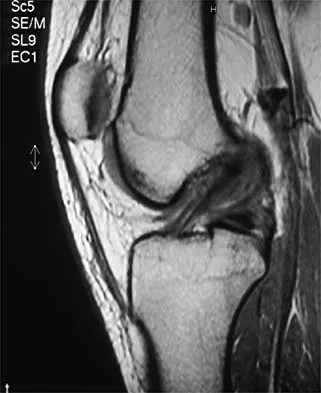
A 36-year-old professional baseball player reports the acute onset of severe right groin pain while attempting to avoid being hit by a baseball while at bat. Examination reveals tenderness, soft-tissue swelling, and ecchymosis in the right groin extending over the medial thigh. MRI scans are shown in Figures 8a and 8b. Management should consist of


Explanation

8b The MRI scans reveal a severe avulsion injury of the adductor muscle from the pubic ramus with muscle edema and hemorrhage. Injury to the adductor muscle group, a "pulled groin," is caused by forceful external rotation of an abducted leg. Pain is immediate and severe in the groin region. Tenderness is at the site of injury along the subcutaneous border of the pubic ramus. Treatment is dictated by the severity of the symptoms but generally consists of rest, ice, and protected weight bearing, followed by a rehabilitation program that begins with gentle stretching and progresses to resistance exercise with a gradual return to sports. Immobilization should be avoided because this promotes muscle tightness and scarring. No data exist to suggest that open repair yields a better outcome than nonsurgical management. Tenotomy has been performed in high-level athletes with chronic groin pain following injury. Gilmore J: Groin pain in the soccer athlete: Fact, fiction, and treatment. Clin Sports Med 1998;17:787-793.

Question 4

An 18-year-old football player is injured after making a tackle with his left shoulder. He has decreased sensation over the lateral aspect of the left shoulder and radial aspect of the forearm. Motor examination reveals weakness to shoulder abduction and external rotation as well as elbow flexion. He has decreased reflexes of the biceps tendon on the left side but full, nontender range of motion of the cervical spine. What anatomic site has been injured?


Explanation

The athlete has symptoms referable to the axillary, musculocutaneous, and suprascapular nerves resulting from an injury to the upper trunk of the brachial plexus. This portion of the plexus is formed by contributions of the fourth through sixth cervical nerve roots. This area is often contused or stretched following a tackling maneuver that results in either depression of the shoulder from contact at Erb's point or traction of the upper plexus from forced stretching of the neck to the contralateral side. Schenck CD: Anatomy of the innervation of the upper extremity, in Torg JS (ed): Athletic Injuries to the Head, Neck, and Face, ed 2. St Louis, MO, Mosby-Year Book, 1991.

Question 5

Which of the following is considered the most common long-term effect on the spine of a professional race horse jockey?


Explanation

Horseback riding is a sport that directly affects the jockey's spine. Tsirikos and associates reported the results of a study of 32 jockeys. They found that equestrian sports, especially professional horseback riding, apart from the increased risk of direct spinal injury caused by a fall from the horse, can lead to progressive spinal degeneration as a result of repetitive trauma and increased physical stress on the spine. It is associated with spondylosis of the cervical spine and lumbar spine. Tsirikos A, Papagelopoulos PJ, Giannakopoulos PN, et al: Degenerative spondyloarthropathy of the cervical and lumbar spine in jockeys. Orthop 2001;24:561-564.

Question 6

An 18-year-old lacrosse player is diagnosed with infectious mononucleosis. What is the recommendation for return to play?


Explanation

Infectious mononucleosis commonly affects adolescents and young adults. It is a febrile illness accompanied by acute pharyngitis. Splenomegaly may occur and predispose the athlete to splenic rupture. Splenic rupture has been reported in nonathletes as well as in patients with normal-sized spleens. Clinical evidence supports a return to all sports 4 weeks after the onset of symptoms provided that the spleen has returned to normal size. Auwaerter PG: Infectious mononucleosis: Return to play. Clin Sports Med 2004;23:485-497.

Question 7

A 30-year-old patient reports chronic medial knee pain and swelling. Figure 9a shows an articular cartilage lesion observed during arthroscopy. The surgeon decides to treat the lesion with the microfracture technique seen in Figure 9b. A biopsy of the repaired tissue 1 year after treatment is likely to show which of the following findings?


Explanation

9b Microfracture is a marrow stimulation technique where stem cells from the underlying subchondral bone marrow can form at the base of the lesion. The rationale for this technique is based on these cells differentiating into cells that will produce an articular cartilage repair. Biopsy findings in animals and humans have demonstrated primarily a fibrocartilagenous repair tissue and not articular cartilage. The collagen type found in hyaline or articular cartilage is of the type II variety. Fibrocartilage possesses mostly type I and III cartilage. Buckwalter JA, Mankin HJ: Articular cartilage: Degeneration and osteoarthritis, repair, regeneration, and transplantation. Instr Course Lect 1998;47:487-504.

Question 8

A 24-year-old dancer reports posterior ankle pain when in the "en pointe" position. Examination reveals posteromedial tenderness, no pain reproduction with passive forced planter flexion, and pain with motion of the hallux. What is the most likely diagnosis?


Explanation

Flexor hallucis longus tendinitis is a common cause of posterior ankle pain in dancers. It tends to be more posteromedial and is characterized by a clicking or catching sensation posteromedially with motion of the great toe. A painful os trigonum typically causes more posterolateral ankle pain and may occur after an ankle sprain or plantar flexion injury where there may be a fracture of the os trigonum. Garrick JG (ed): Orthopaedic Knowledge Update: Sports Medicine 3. Rosemont, IL, American Academy of Orthopaedic Surgeons, 2004, pp 249-261.

Question 9

Kinematic analysis of the medial and lateral menisci has demonstrated that the lateral meniscus has which of the following characteristics compared with the medial meniscus?


Explanation

Kinematic analysis of both menisci demonstrates anterior movement with extension and posterior movement with flexion. The lateral meniscus has more mobility than the medial meniscus because of less soft-tissue attachments. Insall JN, Scott WN (eds): Surgery of the Knee, ed 3. New York, NY, Churchill Livingstone, 2001, vol 1, p 474. Thompson WO, Thaete FL, Fu FH, et al: Tibial meniscal dynamics using 3D reconstructions of MR images, in Proceedings of the Orthopaedic Research Society 1990;389.

Question 10

A 24-year-old professional basketball player reports the gradual onset of pain that is poorly localized to the left midfoot for the past 2 months. Examination reveals diffuse tenderness to palpation, full range of motion of the ankle and subtalar joint, and a normal neurovascular examination to the foot. An AP radiograph is shown in Figure 10. Definitive treatment should include


Explanation

The imaging studies reveal a navicular stress fracture. This condition is secondary to chronic overuse (often running on hard surfaces) and results in vague, ill-defined pain in the midfoot. These fractures can be missed on radiographs but are well-defined on CT or MRI. Tarsal navicular fractures are typically oriented in the sagittal plane. Surgery is typically indicated for the high-level athlete because of the high risk for nonunion and persistent symptoms following nonsurgical management. Internal fixation is the treatment of choice. Torg JS, Pavlov H, Cooley JH, et al: Stress fractures of the tarsal navicular. J Bone Joint Surg Am 1982;64:700-712.

Question 11

A 19-year-old college football player reports persistent weakness, tingling, and numbness of both upper extremities at half time. He states that these symptoms initially occurred after tackling an opposing player with his head early in the game. History reveals that he has had "burners" in the past that typically resolved within 15 to 30 minutes. Examination reveals pain-free cervical motion, weakness to shoulder abduction testing bilaterally, normal upper extremity reflexes, and decreased sensation over both shoulders and the upper arms. Appropriate initial management should consist of


Explanation

The player's symptoms represent more than the mere "burner syndrome," which leads to unilateral symptoms that typically last less than 1 minute. Return to play following a burner is allowed following nonsurgical management and once the symptoms have subsided and the player exhibits normal strength and motion of the neck and upper extremities. This player has the history, symptoms, and examination findings that are consistent with cervical neurapraxia. Return to play in contact sports is contraindicated with bilateral symptoms prior to MRI evaluation of the cervical spine. CT of the brain is indicated with a history of loss of consciousness or other symptoms suggestive of a concussion. Torg JS, Sennett B, Pavlov H, et al: Spear tackler's spine: An entity precluding participation in tackle football and collision activities that expose the cervical spine to axial energy inputs. Am J Sports Med 1993;21:640-649. Torg JS: Cervical spinal stenosis with cord neurapraxia and transient quadriplegia. Sports Med 1995;20:429-434.

Question 12

Which of the following is the most relevant clinical factor in the maturation assessment of an adolescent female athlete contemplating anterior cruciate ligament (ACL) reconstruction?


Explanation

Age of menarche is the most accurate clinical factor to assess the degree of skeletal maturity in the female athlete. Such an assessment is necessary prior to ACL reconstruction in a skeletally immature female because of the risk of damage to the distal femoral and proximal tibial physes. Height of an older male sibling is not relevant to the female athlete. Parental height and recent change in shoe size are only moderately useful in predicting final growth, and hence, skeletal maturity. The presence of breast buds occurs early in adolescent development; therefore, its presence suggests a high likelihood of future growth. Micheli LJ, Foster TE: Acute knee injuries in the immature athlete. Instr Course Lect 1993;42:473-481. Stanitski CL: Anterior cruciate ligament injury in the skeletally immature patient: Diagnosis and treatment. J Am Acad Orthop Surg 1995;3:146-158.

Question 13

A 28-year-old woman fell on her right wrist while rollerblading 2 days ago. She was seen in the emergency department at the time of injury and was told she had a sprain. Examination now reveals dorsal tenderness in the proximal wrist but no snuffbox or ulnar tenderness. Standard wrist radiographs are normal. What is the next most appropriate step in management?


Explanation

When considering the diagnosis of scapholunate ligament injury, standard radiographic views of the hand will not always reveal widening of the scapholunate gap. Although MRI may reveal injury to the ligaments, the PA clenched fist view can be obtained in the office during the initial patient visit. Arthroscopy is not a first-line diagnostic tool. Walsh JJ, Berger RA, Cooney WP: Current status of scapholunate interosseous ligament injuries. J Am Acad Orthop Surg 2002;10:32-42.

Question 14

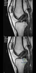
A 17-year-old basketball player and pole vaulter who has had anterior knee pain for the past 18 months now reports a recent inability to jump. Based on the MRI scan shown in Figure 11, management should consist of


Explanation

The MRI scan reveals a partial patellar tendon rupture in conjunction with chronic patellar tendinitis. Mild and moderate patellar tendinitis may be treated nonsurgically with rest, stretching, strengthening, and anti-inflammatory drugs. Severe tendinopathy or extensor mechanism disruption is best treated surgically with tendon debridement and repair. Al-Duri ZA, Aichroth PM: Surgical aspects of patella tendonitis: Techniques and results. Am J Knee Surg 2001;14:43-50.

Question 15

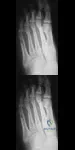
Figures 12a through 12c show the radiographs of a 28-year-old professional baseball player who has ulnar-sided wrist pain and numbness and tingling in the fourth and fifth digits for the past 6 weeks. Management should consist of


Explanation

12b 12c Hook of the hamate fractures typically occur as a result of direct force from swinging a bat, golf club, or racket. Pain is localized to the hypothenar eminence. The injury is best seen on a carpal tunnel view. CT will confirm the diagnosis. Chronic cases can be associated with neuropathy of the ulnar nerve. Excision of the hook through the fracture site usually yields satisfactory results, allowing the athlete to return to competition. Parker RD, Berkowitz MS, Brahms MA, et al: Hook of the hamate fractures in athletes. Am J Sports Med 1986;14:517-523.

Question 16

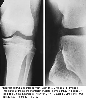
Figure 13 shows the radiographs of a 20-year-old intercollegiate basketball player who was injured 6 weeks prior to the start of the season. What is the most appropriate treatment?


Explanation

A Jones fracture occurs at the metaphyseal-diaphyseal junction of the fifth metatarsal. It is often an acute fracture in conjunction with a chronic stress-related injury. It requires either a short leg cast with strict non-weight-bearing or surgical fixation. In the high performance athlete, the need for rapid return to sport activity usually requires surgical intervention, most commonly with an intramedullary screw. Brodsky JW, Krause JO: Stress fractures of the foot and ankle, in Delee JC, Drez D (eds): Orthopaedic Sports Medicine, ed 2. Philadelphia, PA, Saunders, 2003, vol 2, pp 2391-2409.

Question 17

A 12-year-old boy reports the acute onset of pain and a pop over the right side of his pelvis while swinging a baseball bat during a Little League game. Radiographs reveal an avulsion of the anterior superior iliac spine with 2 cm of displacement. Management should consist of


Explanation

Anterior superior iliac spine avulsion fractures are caused by sudden, forceful contractions of the sartorius and tensor fascia lata. These injuries occur in young athletes through the growth plate with the hip extended and the knee flexed, such as while sprinting or swinging a baseball bat. The athlete will often report a pop or snap at the time of injury. Displaced fractures usually can be seen on radiographs. CT or MRI can be obtained to confirm the diagnosis. In most patients, nonsurgical management consisting of rest and protected weight bearing yields satisfactory outcomes. Surgery is usually reserved for fractures with displacement of more than 3 cm and painful nonunions. Garrick JG (ed): Orthopaedic Knowledge Update: Sports Medicine 3. Rosemont, IL, American Academy of Orthopaedic Surgeons, 2004, pp 139-153.

Question 18

Which of the following best describes heat stroke?


Explanation

Heat stroke consists of hyperthermia (greater than 105.8 degrees F [41 degrees C]), central nervous system dysfunction, and cessation of sweating with hot, dry skin. It is a medical emergency that results from failure of the thermoregulatory mechanisms of the body. It has a high death rate and requires rapid reduction in body core temperature. Heat syncope is characterized by a transient loss of consciousness with peripheral vasodilation and decreased cardiac output with normal body temperature. Heat cramps involve painful contractions of large muscle groups because of decreased hydration and a decrease of serum sodium and chloride. Heat exhaustion is distinguished by a core temperature of less than 102.2 degrees F (39 degrees C) and an absence of central nervous system dysfunction. Hypernatremic heat exhaustion results from inadequate water replacement. Knochel JP: Environmental heat illness: An eclectic review. Arch Intern Med 1974;133:841-864. Hubbard RW, Gaffin SL, Squire DL: Heat related illness, in Wilderness Medicine, ed 3. St Louis, MO, Mosby, 1995, p 167.

Question 19

Which of the following factors is most critical to the success of a meniscal allograft transplantation?


Explanation

Success of a meniscal allograft transplantation is strongly dependent on accurate graft sizing, typically within 5% of the native meniscus. Previous studies have established that donor cell viability is not mandatory for the survival of these grafts since they are replaced by the recipient's cells (at least peripherally) within several weeks. Thus, cryopreservation of the graft to ensure cell viability is not necessary. There is a limited immune response to musculoskeletal allografts; therefore, immunosuppression, as is required for visceral organ transplantation, is not indicated. Wirth CA, Kohn D: Meniscal transplantation and replacement, in Fu FH, Harner CD, Vince JG (eds): Knee Surgery. Baltimore, MD, Williams & Wilkins, 1994, vol 1, pp 631-641. Brautigan BE, Johnson DL, Caborn DM, et al: Allograft tissues, in Drez D, DeLee JD, Miller MD (eds): Orthopaedic Sports Medicine: Principles and Practice, ed 2. Philadelphia, PA, WB Saunders, 2003, pp 205-213.

Question 20

What is the most common behavioral effect of anabolic steroid use in athletes?


Explanation

Users of anabolic steroids often display increased feelings of hostility and aggression. Although reports of psychotic, depressive, and manic behavior have been reported with the use of steroids, they are rare. Drug dependence, such as seen with narcotics, is not a feature of steroid use. Hartgens F, Kuipers H: Effects of androgenic-anabolic steroids in athletes. Sports Med 2004;34:513-554.

Question 21

What is the effect on knee kinematics following placement of an anterior cruciate ligament (ACL) graft at the 12 o'clock position?


Explanation

Endoscopic ACL reconstructive techniques may result in a vertical graft placement. The reconstructed ligament will resist anterior translation of the tibia but the graft will not restore rotatory stability. Decreased flexion and extension are caused by placement of the femoral tunnel too anterior and posterior, respectively. Impingement of the graft on the femoral notch is caused by anterior placement of the tibial tunnel or inadequate notchplasty. Scopp JM, Jasper LE, Belkoff SM, et al: The effect of oblique femoral tunnel placement on rotational constraint of the knee reconstructed using patellar tendon autografts. Arthroscopy 2004;20:294-299.

Question 22

The superior glenohumeral ligament primarily restrains


Explanation

Several cutting studies have evaluated the primary static restraints and the role of the glenohumeral ligaments in providing static stability. With the arm at the side in adduction, the superior glenohumeral ligament and coracohumeral ligament are the primary restraints to inferior translation. The middle glenohumeral ligament functions with the arm in 45 degrees of abduction and resists anterior translation. The inferior glenohumeral ligament is the primary restraint to anterior translation at 90 degrees of abduction. Warner JJ, Deng XH, Warren RF, et al: Static capsuloligamentous restraints to superior-inferior translation of the glenohumeral joint. Am J Sports Med 1992;20:675-685.

Question 23

Which of the following best describes carbohydrate loading?


Explanation

Carbohydrate loading is the practice of maximizing glycogen stores by decreasing training and increasing carbohydrate intake the week before an endurance event. Nonendurance athletes do not benefit from this because glycogen depletion is not the limiting factor during a normal competition. Potential side effects of carbohydrate loading are water retention, muscle stiffness, and weight gain. Coyle EF, Hagberg JM, Hurley BF, et al: Carbohydrate feeding during prolonged strenuous exercise can delay fatigue. J Appl Physiol 1983;55:230-235. Costill DL, Sherman WM, Fink WJ, et al: The role of dietary carbohydrates in muscle glycogen resynthesis after strenuous running. Am J Clin Nutr 1981;34:1831-1836.

Question 24

A 29-year-old quarterback falls onto his dominant shoulder and sustains the injury shown in Figures 14a and 14b. Management should consist of


Explanation

14b Type V acromioclavicular dislocations are characterized by elevation of the clavicle of 100% to 300% and involve extensive soft-tissue stripping. The treatment of choice is surgical reduction of the acromioclavicular joint and some type of stabilization. Treatment of type III injuries is controversial. Lemos MJ: The evaluation and treatment of the injured acromioclavicular joint in athletes. Am J Sports Med 1998;26:137-144.

Question 25

A 27-year-old professional baseball pitcher who underwent arthroscopic olecranon debridement continues to have medial-sided elbow pain during late cocking. Physical examination reveals laxity and pain with valgus stress testing. What is the most likely cause of his pain?


Explanation

Both the medial collateral ligament and the olecranon contribute to valgus stability of the elbow. Excessive olecranon resection increases the demand placed on the medial collateral ligament in resisting valgus forces during throwing. Bone removal from the olecranon should be limited to osteophytes. Garrick JG (ed): Orthopaedic Knowledge Update: Sports Medicine 3. Rosemont, IL, American Academy of Orthopaedic Surgeons, 2004, pp 101-111.

Question 26

During the 'ligamentization' process of a free tendon autograft used for an anterior cruciate ligament (ACL) reconstruction, what is the correct temporal sequence of the biologic phases?


Explanation

The ligamentization process of a free tendon autograft in ACL reconstruction typically occurs in four overlapping phases: necrosis, revascularization, cellular proliferation, and remodeling. Initially, the graft undergoes ischemic necrosis and cellular death. This is followed by a period of revascularization originating from the synovial tissue and infrapatellar fat pad, which supports cellular proliferation (fibroblast infiltration). The final phase is remodeling (maturation), where the collagen cross-linking and architecture adapt to resemble a native ligament, although it never fully regains the identical histological or mechanical properties of an intact ACL.

Question 27

When performing an anatomic reconstruction of the posterolateral corner (PLC) of the knee, identifying the exact femoral attachments is critical to restore native biomechanics. Which of the following describes the correct anatomic location of the fibular collateral ligament (FCL) origin relative to the popliteus tendon origin on the lateral femoral condyle?


Explanation

Based on quantitative anatomic studies by LaPrade et al., the femoral attachment of the fibular collateral ligament (FCL) is consistently located 18.5 mm proximal and 4.3 mm posterior to the popliteus tendon attachment on the lateral femoral condyle. Recognizing this relationship is crucial during anatomic PLC reconstructions to avoid graft anisometry and subsequent failure.

Question 28

A 22-year-old rugby player undergoes an open Latarjet procedure for recurrent anterior shoulder instability with significant anterior glenoid bone loss. Postoperatively, he has profound weakness in shoulder external rotation, but abduction initiation and elbow flexion are intact. Sensation over the lateral shoulder is normal. Which nerve was most likely injured, and what is the typical mechanism in this setting?


Explanation

The patient exhibits isolated weakness in external rotation (infraspinatus) with intact abduction initiation (supraspinatus) and intact sensation. This points to a distal injury of the suprascapular nerve at the spinoglenoid notch. During a Latarjet procedure, if the coracoid graft screws are too long and directed too posteriorly or inferiorly, they can penetrate the posterior cortex of the glenoid neck and directly injure the suprascapular nerve as it courses toward the infraspinatus fossa. Musculocutaneous nerve injury is a common complication but would present with elbow flexion weakness. Axillary nerve injury would result in deltoid weakness and lateral sensory loss.

Question 29

A 26-year-old male hockey player undergoes hip arthroscopy for symptomatic femoroacetabular impingement (FAI). A prominent anterolateral cam lesion is identified and resected. To minimize the risk of a catastrophic post-operative femoral neck fracture, the maximum recommended depth of the osteochondroplasty relative to the native femoral neck diameter should not exceed:


Explanation

Biomechanical studies have demonstrated that resecting more than 30% of the anterolateral femoral neck diameter significantly alters the load-bearing capacity of the proximal femur. Exceeding this 30% threshold exponentially increases peak stresses, putting the patient at a highly elevated risk for an iatrogenic femoral neck fracture, especially in athletes returning to high-impact activities.

Question 30

Based on recent Level I randomized controlled trials comparing operative and non-operative management of acute Achilles tendon ruptures using early functional rehabilitation protocols, which of the following outcomes remains significantly higher in the non-operative group?


Explanation

Recent high-level evidence, including the landmark RCT by Willits et al., demonstrated that when aggressive early functional rehabilitation protocols are utilized, there is no clinically significant difference in functional outcomes, range of motion, or time to return to work/sport between operative and non-operative management of acute Achilles tendon ruptures. However, the re-rupture rate, while lower than historical cast-immobilization cohorts, still remains slightly higher in the non-operative group. Operative management inherently carries risks of surgical site infection and sural nerve injury.

Question 31

During the baseball pitching motion, at which phase does the ulnar collateral ligament (UCL) of the elbow experience the highest valgus stress, placing it at the greatest risk for injury?


Explanation

The late cocking phase of the pitching motion places the highest valgus stress on the elbow. At maximum external rotation during late cocking, the valgus torque peaks. The magnitude of this stress frequently approaches or exceeds the ultimate tensile strength of the native UCL, requiring dynamic stabilization from the flexor-pronator mass and often leading to cumulative microtrauma or acute rupture over time.

Question 32

A 45-year-old female sustains an acute posterior root tear of the medial meniscus. Biomechanically, what is the consequence on contact pressures in the medial compartment if this tear is left untreated, compared to a totally meniscectomized knee?


Explanation

A posterior root tear of the medial meniscus disrupts the circumferential hoop fibers, resulting in catastrophic failure of the meniscus to distribute axial loads. This leads to meniscal extrusion. Biomechanical studies have conclusively shown that a posterior root tear is functionally and biomechanically equivalent to a total medial meniscectomy, significantly decreasing the contact area and increasing peak contact pressures by over 200%.

Question 33

A 45-year-old recreational tennis player presents with persistent deep shoulder pain. MRI arthrogram demonstrates an isolated Type II superior labrum anterior and posterior (SLAP) tear. He has failed 6 months of conservative management. According to current evidence-based guidelines, which surgical intervention provides the most reliable clinical outcomes and lowest revision rate for this patient demographic?


Explanation

In patients older than 35-40 years with a symptomatic Type II SLAP tear, primary biceps tenodesis has been shown to have superior clinical outcomes, more reliable pain relief, and significantly lower revision and complication rates compared to arthroscopic SLAP repair. SLAP repair in this older demographic has a notably high risk of postoperative stiffness, persistent pain, and subsequent need for revision surgery.

Question 34

During reconstruction of the medial patellofemoral ligament (MPFL) for recurrent patellar instability, identifying the anatomic femoral attachment is critical to avoid graft anisometry. Radiographically, Schöttle's point is best described on a true lateral radiograph of the knee as:


Explanation

Schöttle's point is the established radiographic landmark for the anatomic femoral origin of the MPFL. On a strict lateral radiograph, it is defined as: 1 mm anterior to a line extending the posterior femoral cortex, 2.5 mm distal to the posterior origin (articular border) of the medial femoral condyle, and proximal to the posterior point of Blumensaat's line (the posterior notch).

Question 35


A 12-year-old boy sustains a knee injury while skiing. Radiographs reveal a completely displaced, non-comminuted fracture of the tibial eminence without hinging (Meyers and McKeever Type III injury). What is the most appropriate definitive management to restore joint kinematics and prevent long-term morbidity?


Explanation

Meyers and McKeever Type III tibial eminence (tibial spine) fractures are completely displaced. The standard of care for displaced Type III and Type IV (comminuted) injuries is anatomic reduction and internal fixation (using sutures, screws, or K-wires, often performed arthroscopically) to restore ACL tension and avoid a mechanical block to extension. Nonoperative management is reserved for non-displaced (Type I) or minimally displaced, reducible (Type II) fractures. Primary ACL reconstruction is not indicated for acute, fixable bony avulsions.

Question 36

A 25-year-old professional football player undergoes an isolated posterior cruciate ligament (PCL) reconstruction using an Achilles tendon allograft following a direct blow to the proximal tibia. Which of the following accurately describes the biomechanical properties of the native PCL bundles and the primary goal of a single-bundle reconstruction?


Explanation

The native PCL consists of two distinct functional bundles: the larger anterolateral (AL) bundle and the smaller posteromedial (PM) bundle. Biomechanically, the AL bundle is tightest in knee flexion and provides the primary restraint to posterior tibial translation at 90 degrees of flexion. The PM bundle is tightest in extension and deep flexion. Standard single-bundle PCL reconstruction techniques aim to restore the larger and biomechanically dominant AL bundle to re-establish posterior stability in flexion, which is critical for athletic function.

Question 37

A 21-year-old collegiate rugby player presents with recurrent anterior shoulder instability, reporting four dislocation events this season. A representative imaging study demonstrates an 'off-track' engaging Hill-Sachs lesion and 22% anterior glenoid bone loss.

What is the most appropriate definitive management to minimize the risk of recurrence?


Explanation

The patient has significant anterior glenoid bone loss (>20%) and an engaging, off-track Hill-Sachs lesion. Isolated soft-tissue procedures (arthroscopic or open Bankart repairs) have an unacceptably high failure rate in the setting of critical bone loss (>15-20%). The Latarjet procedure (transfer of the coracoid process with the attached conjoined tendon to the anterior glenoid) provides a triple blocking effect (bone block, sling effect of the conjoined tendon, and capsular repair) and is the standard of care for collision athletes with significant bipolar bone loss.

Question 38

A 45-year-old marathon runner feels a sharp 'pop' in the posterior aspect of his knee while performing a deep squat. MRI reveals a posterior root tear of the medial meniscus. Radiographs show no significant osteoarthritis (Kellgren-Lawrence grade 1). Which of the following best describes the biomechanical consequence of leaving this tear untreated?


Explanation

A posterior root tear of the medial meniscus disrupts the circumferential hoop stresses that normally distribute axial loads across the joint. Biomechanical studies have demonstrated that a medial meniscus posterior root tear is functionally and biomechanically equivalent to a total medial meniscectomy. This results in significantly decreased contact area and increased peak contact pressures, leading to rapid chondral wear and extrusion of the meniscus if left unmanaged. Surgical repair is indicated in active patients without advanced osteoarthritis.

Question 39

A 9-year-old gymnast sustains an acute anterior cruciate ligament (ACL) rupture confirmed by MRI. She is Tanner stage 1 with wide-open physes and substantial remaining growth. She experiences recurrent giving-way episodes during daily activities despite a rigorous conservative management trial. Which of the following surgical techniques is most appropriate to minimize the risk of iatrogenic growth arrest?


Explanation

In prepubescent children with wide-open physes (Tanner stage 1 or 2), standard transphyseal ACL reconstructions carry a significant risk of physeal injury, potentially leading to limb length discrepancy or angular deformity. Physeal-sparing techniques, such as the iliotibial band (ITB) extra-articular reconstruction (e.g., MacIntosh or modified MacIntosh procedure), are highly recommended. These techniques avoid drilling across the distal femoral and proximal tibial physes while restoring knee stability.

Question 40

A 22-year-old collegiate baseball pitcher reports insidious onset of medial elbow pain and decreased pitching velocity. Physical examination reveals pain with the moving valgus stress test. An MRI confirms a high-grade partial tear of the ulnar collateral ligament (UCL). Which specific anatomic structure is the primary restraint to valgus stress at the elbow during the late cocking and early acceleration phases of throwing?


Explanation

The ulnar collateral ligament (UCL) complex of the elbow consists of the anterior bundle, posterior bundle, and transverse ligament. The anterior bundle is the primary restraint to valgus stress at the elbow from 30 to 120 degrees of flexion, which perfectly correlates with the elbow position during the late cocking and early acceleration phases of the throwing motion. The posterior bundle provides restraint in deep flexion, and the transverse ligament contributes little to no valgus stability.

Question 41

A 26-year-old professional hockey player presents with chronic groin pain exacerbated by hip flexion, adduction, and internal rotation. Radiographs demonstrate an alpha angle of 65 degrees on the Dunn lateral view. Which of the following best describes the pathophysiology and typical location of the primary osseous deformity?


Explanation

An alpha angle greater than 50-55 degrees is diagnostic of Cam-type femoroacetabular impingement (FAI). Cam morphology is characterized by an aspherical femoral head with an osseous bump or decreased offset, most commonly located at the anterosuperior aspect of the femoral head-neck junction. During hip flexion and internal rotation, this prominence is driven into the acetabulum, causing shear forces on the anterosuperior labrum and adjacent articular cartilage. Pincer impingement refers to acetabular overcoverage, not femoral-sided deformities.

Question 42

A 17-year-old female dancer suffers her third lateral patellar dislocation. Evaluation reveals normal lower extremity alignment and a tibial tubercle-trochlear groove (TT-TG) distance of 14 mm. An isolated medial patellofemoral ligament (MPFL) reconstruction is planned.

Which of the following statements is true regarding the biomechanics of the MPFL?


Explanation

The medial patellofemoral ligament (MPFL) is the primary passive soft-tissue restraint against lateral patellar translation, providing 50% to 60% of the total restraining force from 0 to 30 degrees of knee flexion. Beyond 30 degrees of flexion, the patella engages the trochlear groove, and bony architecture becomes the primary stabilizer. The anatomic femoral origin of the MPFL (Schottle's point) is located between the medial epicondyle and the adductor tubercle. Over-tensioning an MPFL graft typically restricts flexion and increases medial compartment contact pressures.

Question 43

A 20-year-old collegiate cross-country runner presents with recurrent, bilateral anterolateral leg pain that reliably begins 15 minutes into a run and resolves within 30 minutes of rest. Suspecting chronic exertional compartment syndrome, the physician performs intracompartmental pressure testing. According to the Pedowitz criteria, which of the following measurements confirms the diagnosis?


Explanation

The diagnosis of chronic exertional compartment syndrome (CECS) is typically confirmed using the Pedowitz criteria for intracompartmental pressure measurements. The criteria require at least one of the following findings: a resting (pre-exercise) pressure >= 15 mm Hg, a 1-minute post-exercise pressure >= 30 mm Hg, or a 5-minute post-exercise pressure >= 20 mm Hg. An option of 22 mm Hg at 5 minutes post-exercise meets the diagnostic criteria.

Question 44

A 32-year-old male weightlifter presents with vague posterior shoulder pain and selective weakness in external rotation. An MRI reveals a large paralabral cyst located strictly in the spinoglenoid notch, extending from a posterior superior labral tear. Based on the anatomic location of this cyst, which examination finding is most expected?


Explanation

Paralabral cysts associated with superior or posterior labral tears can compress the suprascapular nerve. The location of the compression dictates the clinical deficit. Compression at the suprascapular notch affects both the supraspinatus and infraspinatus muscles. However, compression strictly at the spinoglenoid notch affects the nerve after it has already given off its motor branches to the supraspinatus. Therefore, it results in isolated denervation and atrophy of the infraspinatus, leading to weakness in external rotation with preserved abduction.

Question 45

A 28-year-old downhill skier sustains a high-energy multi-ligamentous knee dislocation (KD-III L) involving the anterior cruciate ligament, posterior cruciate ligament, and posterolateral corner. In the emergency department, the patient exhibits a complete foot drop and cannot actively extend the toes. Given the expected nerve injury, which of the following sensory deficits is most likely to accompany this motor finding?


Explanation

Knee dislocations involving the posterolateral corner have a high association (up to 40%) with common peroneal nerve injury due to traction or direct trauma as the nerve winds around the fibular neck. The common peroneal nerve bifurcates into the deep and superficial peroneal nerves. Injury results in loss of ankle dorsiflexion and toe extension (foot drop) and a sensory deficit over the lateral aspect of the lower leg (superficial peroneal nerve) and the dorsum of the foot, specifically including the first web space (deep peroneal nerve). Medial numbness indicates saphenous nerve involvement, while plantar numbness suggests tibial nerve injury.

Question 46

A 25-year-old male sustains a severe twisting injury to his right knee while playing soccer. On physical examination, the dial test reveals 15 degrees of increased external rotation at 30 degrees of knee flexion compared to the contralateral side. At 90 degrees of knee flexion, the dial test shows 20 degrees of increased external rotation compared to the normal knee. Based on these examination findings, which of the following injury patterns is most likely present?


Explanation

The dial test is utilized to evaluate for posterolateral instability. Increased external rotation (>10 degrees compared to the contralateral side) isolated at 30 degrees of flexion indicates an isolated posterolateral corner (PLC) injury. However, if there is increased external rotation at both 30 degrees and 90 degrees of flexion, it signifies a combined injury to both the PLC and the posterior cruciate ligament (PCL). Isolated PCL injuries may show slight asymmetry at 90 degrees but are not the primary driver of the marked external rotation seen in combined injuries.

Question 47

A 22-year-old collegiate baseball pitcher presents with medial elbow pain that is worse during the late cocking and early acceleration phases of throwing. The moving valgus stress test is positive. An MRI confirms a full-thickness tear of the anterior bundle of the ulnar collateral ligament (UCL). He elects to undergo UCL reconstruction utilizing the docking technique. During the surgical approach to expose the sublime tubercle, which of the following muscle-fascia intervals or techniques is typically utilized?


Explanation

The typical surgical exposure for an ulnar collateral ligament (UCL) reconstruction involves a muscle-splitting approach through the flexor carpi ulnaris (FCU) or utilizing the interval between the FCU and the palmaris longus. This approach safely exposes the sublime tubercle (the distal insertion of the anterior bundle of the UCL on the ulna) while minimizing the risk to the ulnar nerve and surrounding musculature.

Question 48

A 28-year-old male undergoes right hip arthroscopy for femoroacetabular impingement (cam lesion and labral tear). The procedure requires 90 minutes of traction. In the recovery room, he complains of numbness over the dorsum of his right foot and demonstrates weakness in ankle dorsiflexion. What is the most likely pathophysiologic mechanism for this specific complication?


Explanation

Sciatic nerve palsy, specifically affecting the peroneal division, is a known complication of hip arthroscopy related to excessive or prolonged longitudinal traction. The peroneal division is more susceptible to stretch injury because it is tethered at both the sciatic notch and the fibular head. Pudendal nerve injury is also a well-known complication but presents with perineal numbness or sexual dysfunction, driven by compression against the perineal post rather than traction.

Question 49

A 50-year-old active female experiences a 'pop' in the back of her knee while descending stairs. An MRI demonstrates a complete radial tear at the posterior root of the medial meniscus with no significant osteoarthritis (Outerbridge grade II). If left untreated, what is the primary biomechanical consequence of this specific injury pattern?


Explanation

A complete meniscal root tear disrupts the circumferential collagen fibers of the meniscus. Biomechanically, this results in meniscal extrusion under axial load and a complete loss of meniscal hoop stresses. The resulting peak tibiofemoral contact pressures in the affected compartment are equivalent to those seen after a total meniscectomy, leading to rapid progression of articular cartilage degeneration.

Question 50

A 35-year-old recreational athlete sustains an acute, closed Achilles tendon rupture. He is evaluating treatment options with his orthopedic surgeon. Based on current high-level evidence and AAOS guidelines regarding the comparison between operative and nonoperative management utilizing modern early functional rehabilitation protocols, which of the following statements is most accurate?


Explanation

Recent high-level randomized controlled trials and meta-analyses have demonstrated that when a strict, early functional rehabilitation protocol is employed, the rerupture rates between operative and nonoperative management of acute Achilles tendon ruptures are statistically similar. However, operative management carries a higher risk of surgical complications, including wound infections, delayed healing, and sural nerve injury.

Question 51

A 42-year-old recreational tennis player presents with vague, deep anterior shoulder pain exacerbated by overhead serving. Examination reveals a positive O'Brien's test and dynamic labral shear test. MRI arthrogram confirms an isolated type II SLAP tear. After 6 months of failed conservative management, surgical intervention is planned. Based on recent literature for patients in this age demographic (>40 years), which procedure is recommended to minimize postoperative stiffness and maximize the rate of return to sport?


Explanation

In patients older than 35 to 40 years of age with symptomatic Type II SLAP tears that fail conservative treatment, biceps tenodesis is highly favored over SLAP repair. Studies have shown that SLAP repair in this older demographic is associated with higher rates of postoperative stiffness, lower patient satisfaction, and a higher rate of revision surgery compared to primary biceps tenodesis.

Question 52

A 17-year-old female presents with recurrent lateral patellar instability.

Radiographs demonstrate a Caton-Deschamps ratio of 1.1 and normal trochlear depth. A CT scan measures the tibial tubercle-trochlear groove (TT-TG) distance at 14 mm. MRI reveals an incompetent medial patellofemoral ligament (MPFL) with no loose bodies. What is the most appropriate surgical management for this patient?


Explanation

This patient has recurrent patellar instability with an incompetent MPFL, which is the primary restraint to lateral patellar translation at early flexion. Her anatomic risk factors are within normal limits: a normal TT-TG distance (<20 mm indicates no need for medialization), normal patellar height (Caton-Deschamps ratio 0.8-1.2, no need for distalization), and no significant trochlear dysplasia. Therefore, isolated MPFL reconstruction is the most appropriate and biomechanically sound surgical treatment.

Question 53

A 26-year-old mountain biker falls directly onto his shoulder. Clinical examination reveals a prominent distal clavicle. Radiographs demonstrate a 150% superior displacement of the clavicle relative to the acromion, and the coracoclavicular (CC) distance is markedly increased compared to the contralateral side. The deltotrapezial fascia is clinically disrupted. According to the Rockwood classification, what type of injury is this, and what is the standard management approach?


Explanation

According to the Rockwood classification of acromioclavicular (AC) joint injuries, a Type V injury is characterized by 100% to 300% superior displacement of the clavicle relative to the acromion, along with severe disruption of the CC ligaments, AC ligaments, and the deltotrapezial fascia. Because of the severe displacement and fascial stripping, Type V injuries are generally treated operatively with surgical reduction and CC ligament reconstruction/fixation.

Question 54

A 23-year-old male presents with a re-rupture of his hamstring autograft anterior cruciate ligament (ACL) reconstruction, sustained during a non-contact pivoting event 2 years postoperatively.

Standing lateral knee radiographs demonstrate a posterior tibial slope (PTS) of 16 degrees. He is planned for a revision ACL reconstruction. To minimize the risk of a second graft failure, which of the following concomitant procedures is most strongly indicated based on his radiographic findings?


Explanation

An abnormally increased posterior tibial slope (PTS), typically defined as greater than 12 to 14 degrees, is a significant biomechanical risk factor for ACL graft failure because it dramatically increases the anterior shear forces on the tibia during axial loading. In the setting of a revision ACL reconstruction with a high PTS (>12-14 degrees), an anterior closing wedge osteotomy of the proximal tibia is indicated to flatten the slope, thereby protecting the revision graft.

Question 55

A 19-year-old competitive swimmer presents with bilateral, vague shoulder pain and a sensation of her shoulders 'sliding out of joint.' She has no history of distinct trauma. Physical examination demonstrates a positive sulcus sign, a positive load and shift test both anteriorly and posteriorly, and generalized ligamentous laxity (Beighton score of 7/9). She is diagnosed with multidirectional instability (MDI). If nonoperative management is chosen, which of the following should be the primary focus of her rehabilitation program?


Explanation

Multidirectional instability (MDI) of the shoulder is typically atraumatic, bilateral, and associated with generalized ligamentous laxity. The cornerstone of treatment for MDI is a prolonged, dedicated physical therapy program focused on strengthening the dynamic stabilizers of the glenohumeral joint. This primarily involves neuromuscular re-education, dynamic rotator cuff strengthening, and scapular stabilization exercises. Aggressive stretching is contraindicated as it exacerbates the underlying capsular laxity.

Question 56

A 52-year-old female presents with the sudden onset of posteromedial knee pain and a "pop" that occurred while deep squatting to lift a box. She has no significant history of knee pain. An MRI scan reveals a medial meniscus extrusion of 4 mm and a radial defect at the posterior root attachment. What is the most appropriate management to prevent the rapid progression of osteoarthritis in this patient?


Explanation

Medial meniscus posterior root tears result in the loss of hoop stresses, rendering the knee biomechanically similar to a total meniscectomy. This leads to increased peak contact pressures and rapid progression of osteoarthritis. In a patient without severe pre-existing osteoarthritis, the recommended treatment is surgical repair, commonly utilizing a transtibial pull-out technique or suture anchors, to restore meniscal hoop stresses.

Question 57

A 28-year-old competitive powerlifter felt a sudden tearing sensation in his anterior chest wall while performing a heavy bench press. Examination reveals loss of the normal anterior axillary fold and weakness in internal rotation.

Which portion of the affected tendon is most commonly ruptured during this activity, and what is its normal anatomic insertion relative to the other heads?


Explanation

Pectoralis major ruptures most commonly occur during eccentric loading (e.g., bench pressing) and typically involve the sternocostal head. Due to the 180-degree twist of the pectoralis major tendon as it travels to its insertion on the lateral lip of the bicipital groove, the inferiorly originating sternal head fibers insert proximal and deep to the superiorly originating clavicular head fibers.

Question 58

A 22-year-old collegiate rugby player presents with recurrent anterior shoulder instability. A 3D reconstructed CT scan reveals a 26% anterior glenoid bone loss with an engaging Hill-Sachs lesion. What is the most appropriate surgical management to minimize his risk of recurrent instability?


Explanation

Critical anterior glenoid bone loss (>20-25%) in a contact athlete is an indication for a bony augmentation procedure. Soft-tissue repairs alone (like a Bankart repair) have an unacceptably high failure rate in the setting of critical bone loss. The Latarjet procedure transfers the coracoid process and the attached conjoint tendon to the anterior glenoid, providing both a bony block and a dynamic sling effect.

Question 59

A 31-year-old male sustains a multiligamentous knee injury (MLKI) following a tackle in soccer. The knee is grossly deformed but is reduced in the emergency department. Post-reduction, the pedal pulses are palpable and symmetric. However, the ankle-brachial index (ABI) is measured at 0.8. What is the most appropriate next step in management?


Explanation

A knee dislocation is associated with a high risk of popliteal artery injury. Even in the presence of palpable pedal pulses, an ABI < 0.9 is highly suspicious for a vascular injury (e.g., an intimal tear). The gold standard for evaluating this finding is a CT angiogram, which guides further vascular intervention. Serial examinations alone are inadequate given the low ABI.

Question 60

A 24-year-old minor league baseball pitcher presents with chronic posterior shoulder pain during the late cocking phase of throwing. Physical examination reveals a 25-degree loss of internal rotation (GIRD) compared to the contralateral shoulder, with normal total arc of motion.

What is the most appropriate initial management for this condition?


Explanation

Glenohumeral internal rotation deficit (GIRD) is common in overhead throwing athletes and is caused by a contracture of the posterior band of the inferior glenohumeral ligament (posterior capsule). This leads to a posterosuperior shift of the humeral head during the cocking phase, causing internal impingement. Initial management is always nonoperative, primarily focusing on stretching the posterior capsule using the "sleeper stretch."

Question 61

A 20-year-old collegiate baseball pitcher is undergoing an ulnar collateral ligament (UCL) reconstruction using a palmaris longus autograft and a muscle-splitting approach (modified Jobe technique). During the surgical approach to the medial elbow, which nerve is most commonly at risk and must be meticulously identified and protected?


Explanation

During the medial approach for UCL reconstruction, the medial antebrachial cutaneous nerve (MABC) and its branches cross the operative field and are highly at risk of injury (reportedly injured in up to 10-20% of cases if not careful). Neuroma formation or numbness over the medial forearm can cause significant postoperative morbidity. While the ulnar nerve is also protected, MABC neuropraxia/injury is the most common iatrogenic nerve complication during the approach.

Question 62

A 42-year-old competitive water skier fell forward with his knee extended and hip flexed. He presents with severe posterior thigh pain, profound ecchymosis, and a palpable defect at the ischial tuberosity. MRI reveals a complete avulsion of the proximal hamstring conjoined tendon with 6 cm of distal retraction. What is the most appropriate treatment?


Explanation

Acute, complete, multi-tendon proximal hamstring avulsions with significant retraction (>2 cm) in active patients are best treated with early open surgical repair. Surgery restores the length-tension relationship for strength, minimizes the risk of chronic sciatic nerve tethering/neuralgia, and provides the best chance of returning to high-level athletic function.

Question 63

A 12-year-old skeletally immature male presents with vague anterior knee pain. Radiographs demonstrate an osteochondritis dissecans (OCD) lesion on the lateral aspect of the medial femoral condyle. MRI confirms an intact cartilage cap with no T2 fluid signal behind the bony fragment.

What is the most appropriate initial management?


Explanation

In skeletally immature patients, a stable OCD lesion (intact cartilage cap, absence of fluid behind the fragment on MRI) has a high propensity for spontaneous healing. The standard initial management is a 3- to 6-month trial of nonoperative treatment consisting of activity restriction, immobilization, and/or weight-bearing modifications before considering surgical intervention.

Question 64

A 25-year-old professional hockey player sustains an external rotation injury to his ankle. He exhibits localized tenderness over the anterior inferior tibiofibular ligament (AITFL) and a positive squeeze test. Stress radiographs show a normal medial clear space and no tibiofibular diastasis. MRI confirms an isolated tear of the AITFL with an intact deltoid ligament. What is the most appropriate treatment?


Explanation

Isolated syndesmotic injuries without radiographic diastasis and without deltoid ligament compromise are classified as stable (Grade I or II). The standard of care for stable syndesmosis sprains is nonoperative management with a brief period of immobilization in a walking boot, followed by early weight-bearing as tolerated and progressive functional rehabilitation.

Question 65

A 19-year-old female gymnast undergoes an acute lateral patellar dislocation which is reduced in the emergency department. This is her first dislocation. MRI reveals a tear of the medial patellofemoral ligament (MPFL) at its femoral origin, with no osteochondral fractures. There is no evidence of severe trochlear dysplasia. What is the primary patellar restraint provided by the MPFL, and what is the recommended initial management?


Explanation

The MPFL is the primary soft-tissue restraint against lateral patellar translation, providing over 50% of the restraining force, and it functions maximally in early flexion (0 to 30 degrees) before the patella fully engages the trochlear groove. For a first-time dislocator without osteochondral loose bodies or severe anatomic variants, nonoperative treatment with brief immobilization, physical therapy, and bracing is the recommended initial standard of care.

Question 66

A 19-year-old female collegiate soccer player undergoes anterior cruciate ligament (ACL) reconstruction using a bone-patellar tendon-bone (BPTB) autograft. Compared to a hamstring autograft, which of the following is the most commonly reported long-term complication associated specifically with this graft choice?


Explanation

Bone-patellar tendon-bone (BPTB) autografts are considered a gold standard for ACL reconstruction but are historically associated with a significantly higher incidence of anterior knee pain and kneeling pain (donor site morbidity) compared to hamstring autografts. Both grafts provide excellent functional stability, and recent studies show similar re-rupture rates when properly sized and positioned.

Question 67

A 24-year-old male is brought to the emergency department after a high-velocity motorcycle accident. Examination reveals a multiligamentous knee injury (Schenck KD III). The foot is warm, but the Ankle-Brachial Index (ABI) on the injured extremity is 0.8. Which of the following is the most appropriate next step in management?


Explanation

In the setting of a knee dislocation, vascular status must be carefully assessed. An ABI < 0.9 is highly suspicious for an occult vascular injury and warrants advanced imaging, primarily CT angiography, to evaluate the popliteal artery. Immediate surgical exploration is indicated for 'hard signs' of arterial ischemia, such as absent pulses, an expanding or pulsatile hematoma, or active pulsatile bleeding.

Question 68

A 22-year-old professional rugby player undergoes a Latarjet procedure for recurrent anterior shoulder instability with 25% glenoid bone loss. Postoperatively, he exhibits weakness in initiating shoulder abduction and decreased sensation over the lateral aspect of the proximal arm. Which nerve was most likely injured during the procedure?


Explanation

The axillary nerve is at high risk during the Latarjet procedure, particularly during the exposure of the inferior glenoid and capsular release. It innervates the deltoid (shoulder abduction) and provides sensation to the lateral arm via the superior lateral cutaneous nerve of the arm. The musculocutaneous nerve is also at risk during coracoid preparation and transfer, but injury would present with biceps weakness and lateral forearm sensory deficits.

Question 69

A 28-year-old competitive weightlifter feels a sudden "pop" in his anterior chest wall while performing a heavy bench press. Examination demonstrates loss of the anterior axillary fold and significant weakness with internal rotation and adduction. MRI confirms a complete tear of the sternocostal head of the pectoralis major at its humeral insertion. What is the optimal management?


Explanation

Complete ruptures of the pectoralis major, which most commonly involve the sternocostal head avulsing from the humeral insertion during eccentric loading (e.g., bench press), are best treated with surgical repair in young, active patients to restore strength and cosmesis. Suture anchors, cortical buttons, or transosseous sutures are typically used to reattach the tendon to the humerus.

Question 70

A 14-year-old elite female gymnast presents with lateral elbow pain and catching. Radiographs show a radiolucency in the capitellum. MRI reveals an osteochondritis dissecans (OCD) lesion of the capitellum with intact articular cartilage, but there is a rim of T2 hyperintense fluid behind the lesion. What is the most appropriate management?


Explanation

The presence of fluid behind the OCD lesion on MRI indicates instability. However, because the articular cartilage is still intact and the patient is young, the lesion is salvageable. The gold standard for an unstable but intact/salvageable OCD lesion is in situ internal fixation (e.g., using bioabsorbable pins or headless compression screws) to promote healing and preserve the native joint surface.

Question 71

A 45-year-old recreational tennis player complains of deep, vague anterior shoulder pain for 6 months. He has failed a comprehensive physical therapy program. MRI arthrogram reveals a Type II SLAP (Superior Labrum Anterior to Posterior) tear. Based on current evidence, what is the best surgical option for this patient?


Explanation

In patients older than 40-45 years with a symptomatic Type II SLAP tear who have failed conservative management, primary biceps tenodesis is widely recommended over SLAP repair. Studies have demonstrated better clinical outcomes, a lower complication rate, and a lower incidence of postoperative stiffness and revision surgery with tenodesis compared to SLAP repair in this age demographic.

Question 72

A 26-year-old ice hockey player presents with chronic groin pain exacerbated by hip flexion and internal rotation. Radiographs demonstrate a pistol grip deformity and an alpha angle of 70 degrees. This specific morphologic abnormality primarily leads to articular cartilage damage in which region of the acetabulum?


Explanation

The scenario describes a CAM-type femoroacetabular impingement (FAI), characterized by a nonspherical femoral head-neck junction (pistol grip deformity, elevated alpha angle >50-55 degrees). During deep hip flexion and internal rotation, the aspherical CAM lesion engages the acetabulum, causing shear stress and delamination of the cartilage and labrum primarily in the anterosuperior quadrant of the acetabulum.

Question 73

A 21-year-old cross-country runner complains of bilateral anterolateral leg pain that reliably begins 15 minutes into her run and resolves 30 minutes after resting. Which of the following intracompartmental pressure measurements confirms the diagnosis of chronic exertional compartment syndrome (CECS) according to the Pedowitz criteria?


Explanation

The modified Pedowitz criteria for diagnosing chronic exertional compartment syndrome (CECS) require at least one of the following intracompartmental pressure measurements: a pre-exercise resting pressure ≥ 15 mmHg, a 1-minute post-exercise pressure ≥ 30 mmHg, or a 5-minute post-exercise pressure ≥ 20 mmHg.

Question 74

A 16-year-old female presents to the clinic after suffering a first-time lateral patellar dislocation while dancing. The patella was reduced in the emergency department. Which of the following is considered an absolute indication for acute surgical stabilization in this patient?


Explanation

First-time patellar dislocations are generally managed nonoperatively with bracing and physical therapy. However, the presence of a displaced osteochondral loose body (often from the medial patellar facet or lateral femoral condyle) is an absolute indication for acute surgical intervention to remove or fix the fragment and address the medial patellofemoral ligament (MPFL). Patella alta and trochlear dysplasia are risk factors for recurrence but do not dictate acute surgery.

Question 75

A 30-year-old male sustains a severe varus and hyperextension injury to his knee. Examination shows a positive dial test at 30 degrees of flexion, with a 15-degree increase in external rotation compared to the contralateral knee. However, the dial test is symmetric at 90 degrees of flexion. Which structure is most likely injured in isolation?


Explanation

The dial test evaluates for external rotation asymmetry. An increase in external rotation of >10 degrees compared to the uninjured side at 30 degrees of knee flexion, but symmetric rotation at 90 degrees, is classic for an isolated posterolateral corner (PLC) injury. If the external rotation asymmetry is present at both 30 and 90 degrees of flexion, it indicates a combined PLC and posterior cruciate ligament (PCL) injury.

Question 76

A 19-year-old female collegiate soccer player undergoes anterior cruciate ligament (ACL) reconstruction using a bone-patellar tendon-bone (BTB) autograft. When counseling her preoperatively, which of the following is considered the most common long-term complication associated with this specific graft choice compared to a hamstring autograft?


Explanation

Bone-patellar tendon-bone (BTB) autografts are considered a gold standard for ACL reconstruction but are notably associated with a higher incidence of donor-site morbidity. The most common long-term complications compared to hamstring autografts are anterior knee pain, pain with kneeling, and a slight extension deficit. Graft rupture rates are generally equivalent or slightly lower for BTB grafts. Patellar tendon rupture is a known but extremely rare complication.

Question 77

A 25-year-old football player sustains a knee dislocation after a violent tackle. The knee is reduced on the field. In the emergency department, his Ankle-Brachial Index (ABI) is calculated to be 0.8. He has palpable distal pulses, no expanding hematoma, and no active bleeding. What is the most appropriate next step in management?


Explanation

In the setting of a knee dislocation, vascular status must be carefully assessed. An ABI of less than 0.9 strongly indicates abnormal arterial flow and mandates advanced vascular imaging; CT angiography is the current gold standard. Immediate surgical exploration is reserved for patients presenting with 'hard signs' of ischemia (absent pulses, expanding/pulsatile hematoma, active hemorrhage, or overt distal ischemia). Observation alone is inappropriate given the abnormal ABI.

Question 78

A 22-year-old rugby player presents with recurrent anterior shoulder instability following an initial dislocation sustained two years ago. A 3D CT scan demonstrates 28% anterior glenoid bone loss. Which of the following is the most appropriate surgical management to minimize his risk of recurrent instability?


Explanation

Critical glenoid bone loss (>20-25%) in a collision athlete with recurrent anterior shoulder instability is a strong contraindication to an isolated arthroscopic or open Bankart repair, as the recurrence rate is unacceptably high. The Latarjet procedure (coracoid transfer to the anterior glenoid) is the most appropriate treatment. It restores the glenoid articular arc and provides a dynamic 'sling' effect via the conjoint tendon to prevent anterior translation.

Question 79

A 32-year-old male powerlifter feels a sudden 'pop' in his chest while performing a heavy bench press. Examination reveals an asymmetric loss of the anterior axillary fold and weakness in internal rotation and adduction of the shoulder. MRI confirms a complete rupture of the pectoralis major. Where is the most common anatomical site of rupture for this injury?


Explanation

Pectoralis major ruptures most commonly occur during forceful eccentric contraction, such as the lowering phase of a bench press. The most common location of the tear is an avulsion of the distal tendon from its insertion site on the lateral lip of the bicipital groove of the proximal humerus. Surgical repair is generally recommended for complete ruptures at the insertion in active individuals.

Question 80

A 21-year-old collegiate baseball pitcher presents with medial elbow pain and a significant decrease in throwing velocity. A moving valgus stress test is positive. MRI arthrography demonstrates a high-grade partial tear of the ulnar collateral ligament (UCL). During the late cocking and early acceleration phases of throwing, which structure serves as the primary restraint to valgus stress at the elbow?


Explanation

The anterior bundle of the ulnar collateral ligament (UCL) is the primary restraint to valgus stress at the elbow, particularly between 30 and 120 degrees of flexion. This arc of motion corresponds to the late cocking and early acceleration phases of the throwing motion, which place the highest valgus torque on the elbow. The posterior bundle acts as a secondary restraint at higher flexion angles.

Question 81

A 45-year-old recreational weightlifter feels a pop in the posterior aspect of his right knee while deep squatting. An MRI reveals a complete radial tear of the posterior root of the medial meniscus. If left untreated, what is the primary biomechanical consequence of this specific injury?


Explanation

The meniscal roots anchor the meniscus to the tibial plateau, converting axial loads into circumferential hoop stresses. A complete tear of the posterior root of the medial meniscus disrupts its ability to generate these hoop stresses, leading to functional meniscal incompetence. This results in meniscal extrusion and significantly increased peak contact pressures in the medial compartment, biomechanically equivalent to a total meniscectomy, predisposing the joint to rapid osteoarthritis.

Question 82

A 14-year-old female gymnast complains of lateral elbow pain, mechanical clicking, and a 15-degree extension deficit. Radiographs and an MRI demonstrate an osteochondritis dissecans (OCD) lesion of the capitellum with an unstable 10 mm osteochondral fragment and fluid tracking behind the lesion. What is the most appropriate next step in management?


Explanation

In adolescent overhead athletes or gymnasts, OCD of the capitellum can cause significant morbidity. Nonoperative management is indicated for stable lesions with an open capitellar physis. However, the presence of mechanical symptoms (clicking), an extension deficit, and MRI findings of instability (fluid tracking behind the fragment) are indications for surgical intervention. Treatment involves either fixation of the fragment (if viable and adequately sized) or excision with microfracture/marrow stimulation for smaller, non-viable fragments.

Question 83

A 24-year-old professional hockey player presents with an insidious onset of groin pain that is exacerbated by hip flexion and internal rotation. Radiographs demonstrate an elevated alpha angle of 68 degrees and a prominent osseous bump at the anterolateral femoral head-neck junction. During dynamic motion, what is the primary pathomechanism of acetabular cartilage damage in this condition?


Explanation

The patient has Cam-type femoroacetabular impingement (FAI), characterized by an aspherical femoral head-neck junction (elevated alpha angle). During hip flexion and internal rotation, this non-spherical portion forcefully enters the acetabulum, creating outside-in shear forces. This specific mechanism leads to chondral delamination at the anterosuperior acetabulum and subsequent separation of the labrum from the transitional zone cartilage. Pincer FAI, in contrast, typically causes direct, linear compression/contusion of the labrum.

Question 84

A 28-year-old overhead athlete presents with deep shoulder pain and clicking. A Type II SLAP (Superior Labrum Anterior to Posterior) lesion is identified on MR arthrography. According to Snyder's classification, which of the following describes the pathologic anatomy of a Type II SLAP lesion?


Explanation

Snyder's classification of SLAP lesions: Type I is degenerative fraying of the superior labrum with an intact biceps anchor. Type II, the most common type requiring surgical intervention in athletes, involves detachment of the superior labrum and the biceps anchor from the superior glenoid tubercle. Type III is a bucket-handle tear of the labrum with an intact biceps anchor. Type IV is a bucket-handle tear extending into the long head of the biceps tendon.

Question 85

A 20-year-old basketball player lands awkwardly after a jump and sustains a twisting knee injury.

A sagittal T2-weighted MRI demonstrates a complete disruption of the anterior cruciate ligament (ACL) and a characteristic 'bone bruise' pattern. In an acute, non-contact ACL tear, where are these bone bruises most typically located on MRI?


Explanation

In a non-contact ACL injury, the typical mechanism involves valgus stress and internal rotation of the femur on a fixed tibia, leading to a pivot-shift phenomenon. As the tibia subluxates anteriorly, the posterolateral aspect of the tibial plateau impacts the lateral femoral condyle (often near the terminal sulcus). This impact results in the classic 'kissing' bone bruise pattern seen on T2-weighted fat-suppressed MRI in the posterolateral tibial plateau and the lateral femoral condyle.

Question 86

A 22-year-old female collegiate soccer player undergoes primary anterior cruciate ligament (ACL) reconstruction using a quadrupled hamstring autograft. Compared to a bone-patellar tendon-bone (BTB) autograft, which of the following is the most likely long-term functional deficit?


Explanation

Hamstring autograft ACL reconstruction is associated with decreased peak isokinetic knee flexion strength, particularly at deep flexion angles (>70 degrees), compared to BTB autograft. BTB autografts are typically associated with a higher incidence of anterior knee pain and kneeling pain. Both grafts have comparable long-term outcomes regarding clinical stability (Lachman) and functional hop testing.

Question 87

A 23-year-old male competitive rugby player presents with recurrent anterior shoulder instability. He has experienced 4 dislocations this season. Advanced imaging demonstrates an anterior glenoid bone loss of 28% and an engaging 'off-track' Hill-Sachs lesion. Which of the following is the most appropriate surgical management?


Explanation

In young, collision athletes with significant anterior glenoid bone loss (>20-25%) and an 'off-track' or engaging Hill-Sachs lesion, an isolated soft tissue stabilization (Bankart repair) has an unacceptably high failure rate. The Latarjet procedure (coracoid transfer) addresses the glenoid bone loss and provides a sling effect via the conjoint tendon, making it the procedure of choice in this scenario. Remplissage is typically indicated for off-track Hill-Sachs lesions in the setting of subcritical glenoid bone loss (<15-20%).

Question 88

A 50-year-old woman reports feeling a 'pop' in her posterior knee while squatting to garden, followed by medial-sided knee pain and a mild effusion. MRI confirms a medial meniscus posterior root tear with no significant osteoarthritis. What biomechanical consequence is most likely if this injury is treated nonoperatively?


Explanation

A posterior root tear of the medial meniscus functionally acts as a total meniscectomy. It disrupts the circumferential hoop fibers of the meniscus, leading to radial displacement (extrusion) of the meniscus under axial load. This significantly increases peak contact pressures in the medial compartment and rapidly accelerates the development of osteoarthritis. Repair of the root restores the hoop stresses and normalizes contact pressures.

Question 89

A 20-year-old collegiate baseball pitcher complains of medial elbow pain that is most severe during the late cocking and early acceleration phases of throwing. On physical examination, what is the most sensitive test for diagnosing ulnar collateral ligament (UCL) insufficiency?


Explanation

The moving valgus stress test is considered the most sensitive physical examination maneuver for evaluating UCL insufficiency in throwing athletes. It is performed by applying a constant valgus stress to the elbow while flexing and extending it. A positive test is the reproduction of medial elbow pain between 70 and 120 degrees of flexion (the 'shear zone'). Valgus stress at 0 degrees primarily tests the bony articulation and joint capsule, whereas the anterior bundle of the UCL is the primary restraint to valgus stress between 30 and 120 degrees of flexion.

Question 90

A 26-year-old professional hockey player presents with chronic, deep anterior groin pain exacerbated by hip flexion and internal rotation. Radiographs demonstrate a pistol-grip deformity and an alpha angle of 65 degrees on the Dunn lateral view. Which of the following pathophysiological mechanisms is most responsible for the articular cartilage damage in this condition?


Explanation

The patient has Cam-type femoroacetabular impingement (FAI), characterized by an aspherical femoral head (pistol-grip deformity, alpha angle > 55 degrees). During hip flexion and internal rotation, the non-spherical cam lesion engages the acetabular rim, causing shear forces that delaminate the articular cartilage from the underlying subchondral bone, typically at the anterosuperior chondrolabral junction. Pincer impingement is characterized by acetabular overcoverage, leading to linear impact at the rim and 'contre-coup' cartilage lesions posteroinferiorly.

Question 91

A 28-year-old mountain biker falls directly onto the point of his shoulder. Clinical examination reveals a prominent distal clavicle with a reducible step-off. Bilateral Zanca view radiographs show that the coracoclavicular (CC) distance on the injured side is 150% greater than the contralateral uninjured side. The acromioclavicular (AC) joint is completely displaced superiorly. What is the Rockwood classification of this injury?


Explanation

The Rockwood classification of AC joint separations is based on the direction and degree of clavicular displacement. Type V injuries are characterized by severe superior displacement of the clavicle, with the CC distance increased by 100% to 300% compared to the normal contralateral side. The deltotrapezial fascia is severely stripped from the acromion and clavicle. Type III injuries have a 25% to 100% increase in CC distance.

Question 92

A 32-year-old male sustains a high-energy knee dislocation in a motor vehicle collision. The knee is reduced in the emergency department. The pedal pulses are palpable, but the ankle-brachial index (ABI) is measured at 0.85. What is the most appropriate next step in management?


Explanation

In the setting of a knee dislocation, vascular injury (especially the popliteal artery) must be meticulously excluded. An ABI less than 0.9 is a highly sensitive indicator of an arterial injury, even in the presence of palpable pulses, because collateral circulation can preserve distal pulses. The appropriate next step is advanced vascular imaging, most commonly a CT angiogram, to definitively diagnose and localize the injury. Immediate surgical exploration is indicated only for 'hard signs' of ischemia (absent pulses, expanding hematoma, pulsatile bleeding).

Question 93

A 62-year-old heavy laborer presents with right shoulder pain and profound weakness in external rotation with the arm at the side. MRI reveals a massive, retracted, and fatty-infiltrated tear of the supraspinatus and infraspinatus tendons (Goutallier stage 4), with an intact subscapularis and teres minor. Radiographs show a normal acromiohumeral distance and no osteoarthritis (Hamada grade 1). After failing 6 months of conservative management, which of the following surgical interventions is most appropriate?


Explanation

The patient has a massive, irreparable posterosuperior rotator cuff tear without glenohumeral arthritis. Primary repair is contraindicated due to severe fatty infiltration (Goutallier 4). Reverse total shoulder arthroplasty is an option but is generally reserved for older, lower-demand patients or those with cuff tear arthropathy (Hamada > 2). For a posterosuperior defect (loss of external rotation and elevation) in a higher-demand patient without arthritis, a lower trapezius transfer is highly effective and provides a more synergistic vector for restoring external rotation than a latissimus dorsi transfer.

Question 94

A 21-year-old collegiate cross-country runner presents with bilateral anterolateral leg pain that reliably begins after 1.5 miles of running and resolves completely within 30 minutes of rest. He describes the pain as a tight, burning sensation accompanied by transient numbness over the dorsum of his feet. Pre-exercise compartment pressures are 18 mm Hg. At 1 minute post-exercise, pressures in the anterior compartment are 40 mm Hg. What is the most appropriate definitive management?


Explanation

The patient's history and compartment pressure measurements are diagnostic of chronic exertional compartment syndrome (CECS). Pedowitz criteria for CECS include one or more of the following: resting pressure > 15 mm Hg, 1-minute post-exercise pressure > 30 mm Hg, or 5-minute post-exercise pressure > 20 mm Hg. The symptoms and pressure readings point to the anterior (and often concomitant lateral) compartment. Definitive treatment in a competitive athlete who wishes to continue running is a surgical fasciotomy of the affected compartments (anterior and lateral).

Question 95

A 15-year-old female gymnast presents with an acute lateral patellar dislocation after an awkward landing. The patella is spontaneously reduced. On MRI, there is a full-thickness rupture of the primary soft-tissue restraint to lateral patellar translation. At what degree of knee flexion does this specific ligament provide the maximum proportional contribution to restraining lateral patellar displacement?


Explanation

The medial patellofemoral ligament (MPFL) is the primary soft-tissue restraint to lateral patellar translation, contributing up to 50-60% of the restraining force. It is most crucial in early knee flexion, specifically between 0 and 30 degrees. Beyond 30 degrees of flexion, the patella engages deeply within the trochlear groove, and the bony architecture provides the primary stability. MPFL injuries are almost universally present following acute lateral patellar dislocations.

Question 96

A 25-year-old football player sustains a direct blow to the anteromedial aspect of his proximal tibia while his foot is planted and the knee is in extension. He complains of lateral knee pain and instability. On physical examination, there is an asymmetric increase in external rotation on the dial test at 30 degrees of knee flexion, but the side-to-side difference resolves at 90 degrees of knee flexion.

Based on these findings, which of the following structures is most likely injured?


Explanation

A positive dial test (an increase in external rotation of more than 10 degrees compared to the contralateral side) at 30 degrees of flexion that normalizes at 90 degrees of flexion indicates an isolated posterolateral corner (PLC) injury. The primary stabilizers of the PLC include the lateral collateral ligament (LCL), popliteus tendon, and popliteofibular ligament. If the dial test is positive at both 30 and 90 degrees, it indicates a combined PLC and posterior cruciate ligament (PCL) injury.

Question 97

A 20-year-old collegiate rugby player with a history of recurrent anterior shoulder instability presents after another dislocation. Imaging reveals a bipolar bone loss condition with 25% glenoid bone loss and an off-track Hill-Sachs lesion. A Latarjet procedure is planned. Which of the following describes the most significant primary stabilizing mechanism of the Latarjet procedure?


Explanation

The Latarjet procedure provides stability through a 'triple effect'. The most significant stabilizing factor is the dynamic 'sling effect' of the conjoint tendon (short head of the biceps and coracobrachialis) which tension across the anterior-inferior capsule when the arm is positioned in abduction and external rotation. The other two effects are the bone block effect (restoring the anteroposterior diameter of the glenoid) and the capsule repair to the stump of the coracoacromial (CA) ligament.

Question 98

A 45-year-old active female reports feeling a 'pop' in the back of her knee while squatting to pick up a box, followed by posterior knee pain and mild effusion. MRI demonstrates a complete radial tear of the posterior root of the medial meniscus with 4 mm of medial meniscal extrusion. Biomechanical studies have shown that if this injury is left untreated, it alters knee joint contact mechanics to most closely resemble which of the following conditions?


Explanation

A complete medial meniscus posterior root tear severely disrupts the hoop stresses of the meniscus, rendering it biomechanically incompetent. In cadaveric studies, a complete root tear has been shown to lead to peak contact pressures and decreased contact area in the medial compartment that are equivalent to those seen in a total medial meniscectomy. This results in rapid progression of medial compartment osteoarthritis if not surgically repaired in suitable candidates.

Question 99

A 32-year-old competitive weightlifter feels a sudden tear in his chest while performing a heavy bench press. Examination reveals extensive ecchymosis over the anterior arm and chest, a palpable defect in the anterior axillary fold, and profound weakness with resisted shoulder internal rotation and adduction. Which of the following accurately describes the most common location of a pectoralis major rupture and the optimal timing for surgical repair in an athlete?


Explanation

The most common mechanism for a pectoralis major rupture is an eccentric load during a bench press. The vast majority of these tears involve an avulsion of the sternal head tendon from its insertion on the proximal humerus, while the clavicular head remains intact. Early surgical repair (typically within the first 6 weeks before significant retraction and scarring occur) yields significantly better functional outcomes, peak torque recovery, and cosmetic satisfaction compared to delayed repair or nonoperative management in athletic populations.

Question 100

A 28-year-old male sustains a high-energy traumatic knee dislocation (KD-III) in a motorcycle collision. The knee is grossly reduced in the emergency department. Upon initial assessment, pedal pulses are palpable but slightly asymmetric compared to the uninjured limb. The ankle-brachial index (ABI) is measured at 0.85.

What is the most appropriate next step in management?


Explanation

In the setting of a knee dislocation, an ABI of less than 0.90 is highly concerning for a vascular injury to the popliteal artery. According to modern trauma algorithms, an ABI < 0.90 or asymmetric pulses mandates further advanced vascular imaging, most commonly a CT angiogram, to identify intimal tears or partial occlusions. Immediate OR exploration is reserved for 'hard' signs of ischemia (e.g., absent pulses, active pulsatile hemorrhage, expanding hematoma). Serial checks alone are inadequate for an abnormal ABI < 0.90.

None

Clinic OS
Medically Verified Content by
Prof. Clinic OS
Consultant Orthopedic & Spine Surgeon
Chapter Index