العربية
Part of the Master Guide

General Orthopedics 2026 Practice Questions: Set 13 (Solved)

Orthopedic Sports Medicine 2026 MCQs: Board Review Questions & Answers (Part 1)

23 Apr 2026 82 min read 67 Views
Figure for Sports Medicine 2007 MCQs - Part 1 - Question 1

Key Takeaway

Discover the latest medical recommendations for Orthopedic Sports Medicine 2026 MCQs: Board Review Questions & Answers (Part 1). Top-rated Orthopedic Sports Medicine 2026 MCQs bank. Practice with clinical case questions, orthopedic surgery board review, and evidence-based answers updated for 2026.

Orthopedic Sports Medicine 2026 MCQs: Board Review Questions & Answers (Part 1)

Comprehensive 100-Question Exam


00:00

Start Quiz

Question 1

A 22-year-old college baseball pitcher reports the recent onset of anterior and posterosuperior shoulder pain in his throwing shoulder. Examination shows a 15-degree loss of internal rotation, tenderness over the coracoid, and a positive relocation test. Radiographs are normal, and an MRI scan without contrast shows no definitive lesions. A rehabilitation program is prescribed. Which of the following regimens should be initially employed?


Explanation

Throwing athletes, particularly pitchers, have a high incidence of shoulder pain. Recent evidence suggests that posteroinferior capsular tightness and scapular dyskinesis may play a substantial role in the pathologic cascade, culminating in the development of articular surface rotator cuff tears and tearing of the posterosuperior labrum. These patients have posterosuperior shoulder pain primarily. Furthermore, these athletes are susceptible to a muscular fatigue syndrome, the SICK (Scapular malposition, Inferior medial border prominence, Coracoid pain and malposition, and dysKinesis of scapular movement) scapula syndrome. This patient has an internal rotation deficit and tenderness over the coracoid. The internal rotation deficit is addressed by stretching the posterior capsule. The tenderness over the coracoid has been attributed to a contracture of the pectoralis minor tendon secondary to scapular malposition. The initial phase of the rehabilitation regimen is directed at stretching the posterior capsule and pectoralis minor tendon. Burkhart SS, Morgan CD, Kibler WB: The disabled throwing shoulder: Spectrum of pathology. Part III. Arthroscopy 2003;19:641-661.

Question 2

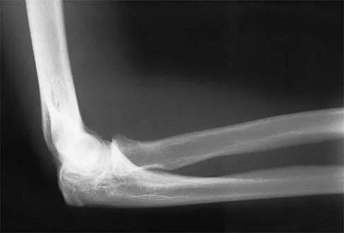
A 28-year-old professional football player reports painless loss of ankle motion after sustaining a "severe" ankle sprain 12 months ago. A mortise radiograph is shown in Figure 1. Surgical treatment should be reserved for which of the following conditions?


Explanation

The radiograph shows posttraumatic tibiofibular synostosis. This condition typically follows an eversion (high) ankle sprain that results in disruption of the interosseous membrane. Ossification usually develops within 6 to 12 months after the injury. Return to sports is possible despite the lack of normal ankle dorsiflexion and mobility between the tibia and fibula. Surgical excision is reserved for persistent pain that fails to respond to nonsurgical management once the ossification is "cold" on bone scintigraphy. Whiteside LA, Reynolds FC, Ellsasser JC: Tibiofibular synostosis and recurrent ankle sprains in high performance athletes. Am J Sports Med 1978;6:204-208. Henry JH, Andersen AJ, Cothren CC: Tibiofibular synostosis in professional basketball players. Am J Sports Med 1993;21:619-622.

Question 3

The most common mechanism of injury to the triangular fibrocartilage complex (TFCC) involves


Explanation

TFCC tears are common in athletes. As the athlete braces for a fall, the wrist is most commonly in an extended position and the forearm is pronated. Cohen MS: Ligamentous injuries of the wrist in the athlete. Clin Sports Med 1998;17:533-552.

Question 4

The force generated by a muscle is most highly dependent on its


Explanation

The cross-sectional area of a muscle determines to a great extent the force generated by the muscle. The force of a muscle contraction is controlled by the amount of myofibrils that contract; the greater the amount of contracting myofibrils, the greater the force of contraction. Fiber types have less to do with the force of contraction and more to do with the duration and speed of contraction. Muscle length affects contraction force through the Blix curve. The morphology of a muscle can affect the cross-sectional area by varying the angle of the fibers in relation to the force vector. Conditioning mostly affects duration and fatigability. Buckwalter JA, Mow VC, Ratcliffe A: Restoration of injured or degenerated articular cartilage. J Am Acad Orthop Surg 1994;2:192-201.

Question 5

A 31-year-old woman has increasing pain and tightness in her right knee, with occasional stiffness and recurrent hemorrhagic effusions. MRI scans are shown in Figures 2a and 2b. What is the most likely diagnosis?


Explanation

2b PVNS is a rare inflammatory granulomatous condition of unknown etiology, and causes proliferation of the synovium of joints, tendon sheaths, or bursa. The disorder occurs most commonly in the third and fourth decades but can occur at any age. MRI provides excellent delineation of the synovial disease. Characteristic features of PVNS on MRI include the presence of intra-articular nodular masses of low signal intensity on T1- and T2-weighted images and proton density-weighted images. Synovial biopsy should be performed if there is any doubt of the diagnosis. Total synovectomy (open or arthroscopic) is required for the diffuse form, although recurrence is common. Rheumatoid arthritis and synovial chondromatosis are not typically associated with hemorrhagic effusions. De Ponti A, Sansone V, Malchere M: Result of arthroscopic treatment of pigmented villonodular synovitis of the knee. Arthroscopy 2003;19:602-607. Chin KR, Barr SJ, Winalski C, et al: Treatment of advanced primary and recurrent diffuse pigmented villonodular synovitis of the knee. J Bone Joint Surg Am 2002;84:2192-2202.

Question 6

A 30-year-old elite marathon runner reports chronic pain over the lateral aspect of the distal right leg and dysesthesia over the dorsum of the foot with active plantar flexion and inversion of the foot. Examination reveals a tender soft-tissue fullness approximately 10 cm proximal to the lateral malleolus. The pain is exacerbated by passive plantar flexion and inversion of the ankle. There is also a positive Tinel's sign over the site of maximal tenderness. There is no motor weakness, and deep tendon reflexes are normal. Radiographs and MRI of the leg are normal. What is the next most appropriate step in management?


Explanation

The patient has entrapment of the superficial peroneal nerve against its fascial opening in the distal leg. It is typically exacerbated by passive or active plantar flexion and inversion of the foot, which leads to traction of the nerve as it exits this opening. Treatment involves release of the fascial opening to reduce this traction phenomenon. Closure of the defect will only aggravate the condition and potentially result in an exertional compartment syndrome. A four-compartment fasciotomy is only indicated for an established compartment syndrome of the leg. Styf J: Diagnosis of exercise-induced pain in the anterior aspect of the lower leg. Am J Sports Med 1988;16:165-169. Sridhara CR, Izzo KL: Terminal sensory branches of the superficial peroneal nerve: An entrapment syndrome. Arch Phys Med Rehabil 1985;66:789-791.

Question 7

A 21-year-old soccer player reports pain and is unable to straighten his knee following an acute injury during a game. He is unable to continue to play. An MRI scan is shown in Figure 3. What is the next most appropriate step in management?


Explanation

The patient has a locked knee that cannot be fully extended. This is most likely the result of the mechanical block of a bucket-handle tear that has flipped into the notch. Also, the pain may be so severe that the muscle spasm prevents the knee from straightening out. When the patient is anesthetized, the muscle spasm relaxes and the meniscus can be reduced out of the notch. Arthroscopy is the treatment of choice. A meniscal repair is usually possible in large bucket-handle tears because the meniscus is torn in the red-red zone where most of the vascular supply is located. If the handle portion is badly frayed or damaged, a partial meniscectomy should be performed. The classic finding on MRI is a "double PCL sign." This is due to the flipped portion of the meniscus in the notch. Critchley IJ, Bracey DJ: The acutely locked knee: Is manipulation worthwhile? Injury 1985;16:281-283.

Question 8

When performing an inside-out lateral meniscal repair, capsule exposure is provided by developing the


Explanation

Capsular exposure for an inside-out lateral meniscal repair is performed by developing the interval between the iliotibial band and biceps tendon. Posterior retraction of the biceps tendon exposes the lateral head of the gastrocnemius. Posterior retraction of the gastrocnemius provides access to the posterolateral capsule. Miller DB Jr: Arthroscopic meniscus repair. Am J Sports Med 1988;16:315-320.

Question 9

A 50-year-old man reports left shoulder pain and weakness after undergoing a lymph node biopsy in his neck 2 years ago. Examination reveals winging of the left scapula. Electromyography shows denervation of the trapezius. Surgical treatment for this condition involves


Explanation

The muscle transfer procedure most commonly performed for trapezius paralysis is the Eden-Lange procedure. Trapezius paralysis in this patient is secondary to iatrogenic injury to the spinal accessory nerve during lymph node biopsy. In this procedure, the levator scapulae and rhomboid minor and major muscles are transferred laterally. Pectoralis transfer to the inferior border of the scapula is used as a dynamic transfer for serratus anterior winging. Kuhn JE, Plancher KD, Hawkins RJ: Scapular winging. J Am Acad Orthop Surg 1995;3:319-325. Langenskiold A, Ryoppy S: Treatment of paralysis of the trapezius muscle by Eden-Lange operation. Acta Orthop Scand 1973;44:383-388.

Question 10

A 15-year-old female field hockey player sustains a blow to the mouth from a hockey stick. Three front teeth are knocked out and shown in Figure 4. In addition to calling a dentist immediately, what is the next best step in management?


Explanation

Tooth avulsions can occur in contact or collision sports. An avulsed tooth is a medical emergency. The likelihood of survival of the tooth depends on the length of time that the tooth is out of the socket and the degree to which the periodontal ligament is damaged. The tooth should be handled only by the crown end and not the root end. It can be rinsed of debris with water or normal saline solution. The tooth should not be brushed or cleaned otherwise. During transport, the tooth must be kept moist. An avulsed tooth can be transported in whole milk, saliva, sterile saline solution, or commercially available kits with physiologic buffer solutions. The tooth and the athlete should be transported to the dentist for reinsertion as soon as possible and preferably within an hour. Krasner P: Management of sports-related tooth displacements and avulsions. Dent Clin North Am 2000;44:111-135. Sullivan JA, Anderson SJ (eds): Care of the Young Athlete. Rosemont IL, American Academy of Orthopaedic Surgeons, Elk Grove Village, IL, American Academy of Pediatrics, 2000, p 190.

Question 11

Commotio cordis is best treated with


Explanation

Commotio cordis is a rare but catastrophic condition that is caused by blunt chest trauma. It results in cardiac fibrillation and is universally fatal unless immediate defibrillation is performed. Although case reports of successful use of the chest thump maneuver exist, the best method of treatment is cardiac defibrillation. IV fluids, epinephrine, and albuterol inhalers are used to treat dehydration, anaphylactic shock, and bronchospasm respectively, and are not effective in the treatment of commotio cordis. McCrory P: Commotio cordis. Br J Sports Med 2002;36:236-237.

Question 12

Which of the following is considered an advantage of arthroscopic distal clavicle excision compared with open distal clavicle excision?


Explanation

Arthroscopic versus open distal clavicle excision has the advantage of allowing evaluation of the glenohumeral joint arthroscopically prior to moving into the subclavicular and subacromial space to perform the distal clavicle excision. This can be of value in both confirming the diagnosis as well as avoiding diagnostic errors. Berg and Ciullo showed that in 20 patients who underwent open distal clavicle excision that resulted in failure, 15 of those patients had a superior labral anterior posterior (SLAP) lesion. Of these 15 patients who had the lesion treated surgically, 9 went on to a good to excellent result after the surgery was performed arthroscopically. Fewer complications, lower infection rate, and decreased surgical time have not been documented in the literature. Arthroscopic technique sacrifices the inferior acromioclavicular ligament and preserves the superior acromioclavicular ligament. Berg EE, Ciullo JV: The SLAP lesion: A cause of failure after distal clavicle resection. Arthroscopy 1997;13:85-89.

Question 13

A 40-year-old woman reports the atraumatic onset of severe knee pain and swelling after undergoing an uncomplicated elective cholecystectomy 1 week ago. She denies any history of diabetes mellitus or HIV but has had occasional episodes of mild knee pain and swelling that have always responded to nonsteroidal anti-inflammatory drugs. Radiographs are shown in Figures 5a and 5b. A knee aspiration yields a WBC count of 35,000/mm3. The aspirate should also yield which of the following findings?


Explanation

5b The radiographs reveal chondrocalcinosis of the menisci. This is caused by calcium pyrophosphate crystals, which are weakly positive birefringent rhomboid-shaped crystals. Frequently, this condition is asymptomatic; however, routine abdominal surgery may cause precipitation of these crystals and pain. Gout, which is caused by strongly negative birefringent needle-shaped sodium urate crystals, is not associated with chondrocalcinosis and is rare in younger women. Gross blood is uncommon without trauma. Infection is not likely in a healthy patient who underwent uncomplicated surgery. Fisseler-Eckhoff A, Muller KM: Arthroscopy and chondrocalcinosis. Arthroscopy 1992;8:98-104.

Question 14

What is the maximum acceptable amount of divergence of the interference screw in the femoral tunnel from the bone plug of a bone-patellar tendon-bone graft in anterior cruciate ligament (ACL) reconstruction before pull-out strength is statistically decreased?


Explanation

In the early 1990s, a transition was made from a two-incision ACL reconstruction to a single-incision ACL reconstruction, and there was concern over divergence of the femoral screws. It was shown radiographically that approximately 5% of the time, divergence of the screw was greater than 15 degrees from the bone plug. In a bovine model, there was significant loss of pull-out strength with an increase in divergence from 15 degrees to 30 degrees. Therefore, attempts should be made to minimize divergence to 15 degrees or less. Lemos MJ, Jackson DW, Lee TO, et al: Assessment of initial fixation of endoscopic interference femoral screws with divergent and parallel placement. Arthroscopy 1995;11:37-41.

Question 15

A 21-year-old professional ballet dancer reports a painful popping sensation over her right hip joint. Examination reveals that symptoms are reproduced with hip flexion and external rotation. Which of the following studies will best confirm the diagnosis?


Explanation

The patient has snapping hip syndrome of the internal type, which is more common in ballet dancers. It is caused by the iliopsoas tendon gliding over the iliopectineal line or the femoral head. The diagnosis usually can be made by the history and physical examination. Snapping is reproduced by hip flexion and extension or flexion with external rotation and abduction. Conventional and dynamic ultrasonography will confirm the snapping structure. Radiographs occasionally show calcifications near the lesser trochanter. MRI can be used to rule out other diagnoses that can simulate snapping hip. Gruen GS, Scioscia TN, Lowenstein JE: The surgical treatment of internal snapping hip. Am J Sports Med 2002;30:607-613.

Question 16

The posterior circumflex artery provides blood supply to what portion of the proximal humerus?


Explanation

The posterior circumflex artery provides blood supply only to the posterior portion of the greater tuberosity and a small posteroinferior portion of the humeral head. The humeral head is supplied primarily by the anterolateral ascending branch of the anterior circumflex artery; the terminal branch of this artery is termed the arcuate artery. Norris TR (ed): Orthopaedic Knowledge Update: Shoulder and Elbow 2. Rosemont, IL, American Academy of Orthopaedic Surgeons, 2002, pp 267-274.

Question 17

Use of prophylactic knee bracing in contact sports participants results in which of the following?


Explanation

Several studies have looked at the effects of knee bracing, and it appears to be effective in prophylactically decreasing the incidence of medial collateral ligament sprains. Najibi and Albright reported that although evidence is not conclusive, bracing appears to help decrease the incidence of medial collateral ligament injuries. Albright and associates showed similar findings. Prophylactic knee braces have been associated with an increased incidence of ankle injuries. Albright JP, Powell JW, Smith W, et al: Medial collateral ligament knee sprains in college football: Effectiveness of preventive braces. Am J Sports Med 1994;22:12-18.

Question 18

A 22-year-old college football player reports shortness of breath and dyspnea after a tackle. Examination reveals tachypnea, tachycardia, the trachea is shifted to the right, and there are decreased breath sounds on the left lung fields. The first line of treatment on the field should be


Explanation

The patient has a tension pneumothorax. This is a life-threatening emergency where air is trapped between the pleura and the lung, which prevents expansion of the lung. This causes hypoxia and cardiopulmonary compromise. The first line of treatment is to place a needle into the second intercostal space in the midclavicular line. The athlete should then be transported to the emergency department for chest tube placement. The athlete cannot return to play, and resuscitation is not necessary because he has not gone into cardiopulmonary arrest. Amaral JF: Thoracoabdominal injuries in the athlete. Clin Sports Med 1997;16:739-753.

Question 19

Anabolic steroid use has which of the following effects on serum lipoprotein levels?


Explanation

The use of anabolic steroids causes a decrease in high-density lipoprotein levels but has no effect on low-density lipoprotein levels. An abnormally low high-density lipoprotein level should alert the physician to the possibility of steroid use in an athlete. Hartgens F, Rietjens G, Keizer HA, et al: Effects of androgenic-anabolic steroids on apolipoproteins and lipoprotein (a). Br J Sports Med 2004;38:253-259.

Question 20

A 20-year-old professional female jockey who is wearing a helmet is thrown from her horse. What is the most likely location of her injury?


Explanation

The incidence of injury associated with horseback rising is estimated to be one per 350 riding hours to one per 1,000 riding hours. Of these injuries, approximately 15% to 27% are severe enough to warrant hospital admission. Significant and serious injuries in equestrian activities are associated with recreational riders and those not wearing a helmet. Head and spine injuries are more common in recreational and nonhelmeted riders. Extremity injuries are more common in professional and helmeted riders. Professional riders are less likely to be admitted to the hospital than recreational riders, and are about half as likely to be disabled at 6 months after injury as recreational riders. Lim J, Puttaswamy V, Gizzi M, et al: Pattern of equestrian injuries presenting to a Sydney teaching hospital. ANZ J Surg 2003;73:567-571.

Question 21

A 62-year-old man with a long history of right shoulder pain and weakness is scheduled to undergo hemiarthroplasty. Based on the radiographs shown in Figures 6a through 6c, what preoperative factor will most affect postoperative functional outcome?


Explanation

6b 6c The radiographs reveal osteoarthritis and proximal humeral head migration. Integrity of the rotator cuff must be questioned based on these radiographic changes. The status of the rotator cuff is the most influential factor affecting postoperative function in shoulder hemiarthroplasty. The coracoacromial ligament provides a barrier to humeral head proximal migration in the face of a rotator cuff tear. The radiographs do not indicate significant humeral head or glenoid erosion. Acromioclavicular arthritis is often asymptomatic. Iannotti JP, Norris TR: Influence of preoperative factors on outcome of shoulder arthroplasty for glenohumeral osteoarthritis. J Bone Joint Surg Am 2003;85:251-258.

Question 22

Which of the following complications is more likely with an inside-out repair technique compared to an all-inside techniques for a medial meniscus tear?


Explanation

All of the answers are possible complications of meniscal repair. There are large volumes of literature evaluating the results of meniscal repair, both for the all-inside technique, as well as the inside-out technique. Failure rates are similar. Intra-articular synovitis occurs with absorbable sutures and absorbable implants. Peroneal nerve injuries are more common with the lateral-sided repairs. Saphenous nerve injuries are more common with medial-sided tears. Because of the incision required and the technique of tying over soft tissue, the risk of a saphenous nerve injury is greater with an inside-out technique than with an all-inside technique. Farng E, Sherman O: Meniscal repair devices: A clinical and biomechanical literature review. Arthroscopy 2004;20:273-286.

Question 23

Figure 7 shows the CT scan of a 22-year-old professional baseball pitcher who has had elbow pain for the past 6 months despite rest from throwing. Management should consist of


Explanation

The CT scan shows a stress fracture of the olecranon. This injury is the result of repetitive abutment of the olecranon into the olecranon fossa, traction from triceps activity during the deceleration phase of the throwing motion, and impaction of the medial olecranon onto the olecranon fossa from valgus forces. Fractures may be either transverse or oblique in orientation. Initial treatment consists of rest and temporary splinting. Electrical bone stimulation may also be considered. Open fixation with a large compression screw is recommended when nonsurgical management has failed to provide relief. Ahmad CS, ElAttrache NS: Valgus extension overload syndrome and stress injury of the olecranon. Clin Sports Med 2004;23:665-676.

Question 24

A 17-year-old football player is injured during a play and reports abdominal pain that is soon followed by nausea and vomiting. What organ has most likely been injured?


Explanation

The spleen is the most common organ injured in the abdomen as the result of blunt trauma. It is also the most common cause of death because of an abdominal injury. The liver is the second most commonly injured organ. Injury to the other organs is rare. The diagnosis can be made with CT. Treatment ranges from observation to splenectomy, depending on the severity of injury. Green GA: Gastrointestinal disorders in the athlete. Clin Sports Med 1992;11:453-470.

Question 25

A 15-year-old high school soccer player collides with an opponent and is unconscious when the trainer arrives on the field. He is conscious within 15 seconds, breathing appropriately, and denies any headache, neck pain, or nausea. It is his first head injury. Provided that the athlete is free of symptoms, when should he be allowed to return to athletic activity?


Explanation

The loss of consciousness indicates a grade 2 concussion, which necessitates a 4-week period out of sport. The last week prior to return must be symptom-free and the athlete should not have symptoms in practice. Cantu RC: Return to play guidelines after a head injury. Clin Sports Med 1998;17:45-60.

Question 26

A 24-year-old male presents with knee stiffness 6 months after an anterior cruciate ligament (ACL) reconstruction using a bone-patellar tendon-bone autograft. On physical examination, he has full extension but lacks 30 degrees of terminal flexion compared to the contralateral knee. Which of the following technical errors during graft placement most likely accounts for this clinical presentation?


Explanation

An anteriorly placed femoral tunnel is a classic cause of flexion loss following ACL reconstruction. During knee flexion, an anteriorly placed femoral origin moves further away from the tibial insertion, causing the graft to abnormally tighten and restrict deep flexion. Conversely, a femoral tunnel placed too posteriorly causes the graft to tighten in extension, resulting in an extension deficit. An anteriorly placed tibial tunnel results in roof impingement and loss of extension.

Question 27

A 55-year-old physically active woman experiences a sudden 'pop' in her posterior knee while squatting. An MRI demonstrates a complete radial tear at the posterior root of the medial meniscus. If left untreated, biomechanical studies suggest this injury creates a knee environment most equivalent to which of the following?


Explanation

A complete tear of the medial meniscus posterior root inherently disrupts the circumferential collagen fibers, completely eliminating the meniscus's ability to convert axial loads into hoop stresses. Biomechanical studies have demonstrated that a medial meniscus posterior root tear results in contact pressures and kinematics that are virtually indistinguishable from a total medial meniscectomy, leading to rapid progression of osteoarthritis if not repaired.

Question 28

A 19-year-old collegiate gymnast presents with bilateral shoulder pain and a sensation of the shoulders 'slipping out' during her routines. She denies any specific traumatic event. On examination, she has 3+ sulcus signs bilaterally and positive apprehension tests that spontaneously reduce when she relaxes. Radiographs and MRI are unremarkable. What is the most appropriate initial management?


Explanation

This patient presents with classic signs of multidirectional instability (MDI) of the shoulder, characterized by generalized laxity, atraumatic onset, and bilateral involvement. The cornerstone and first-line treatment for MDI is an extensive, supervised physical therapy program focusing on strengthening the dynamic stabilizers of the shoulder (the rotator cuff and periscapular musculature). Operative management is reserved only for patients who fail a prolonged trial (usually >6 months) of nonoperative management.

Question 29

A surgeon is planning a medial patellofemoral ligament (MPFL) reconstruction for a 17-year-old female with recurrent lateral patellar dislocations. To achieve anatomic reconstruction and isometric graft behavior, the femoral tunnel must be placed at Schöttle's point. Where is this landmark located radiographically on a true lateral view of the distal femur?


Explanation

Schöttle's point defines the anatomic femoral origin of the MPFL. On a true lateral radiograph, it is found 1 mm anterior to the posterior cortex line, 2.5 mm distal to the posterior origin of the medial femoral condyle, and proximal to the level of the posterior aspect of Blumensaat's line. Anatomically, this point is located in the saddle between the medial epicondyle and the adductor tubercle.

Question 30

A 28-year-old male hockey player presents with chronic, deep groin pain exacerbated by deep hip flexion and internal rotation. An AP pelvis radiograph demonstrates a 'crossover sign.' This radiographic finding is most indicative of which of the following pathomorphologies?


Explanation

A crossover sign is present on an AP pelvis radiograph when the anterior rim of the acetabulum crosses the line of the posterior rim. This is indicative of focal or global acetabular retroversion, which leads to over-coverage of the femoral head and Pincer-type femoroacetabular impingement (FAI). Cam impingement is associated with an aspherical femoral head (decreased head-neck offset) and a high alpha angle.

Question 31

A 21-year-old collegiate baseball pitcher presents with medial elbow pain that occurs during the late cocking and early acceleration phases of throwing. On examination, the moving valgus stress test is performed, producing pain that is maximal between 70 and 120 degrees of elbow flexion. Which anatomical structure is most likely compromised?


Explanation

The anterior bundle of the ulnar collateral ligament (UCL) is the primary restraint to valgus stress at the elbow between 30 and 120 degrees of flexion. The moving valgus stress test places dynamic valgus tension on the elbow while it is rapidly extended from full flexion. Pain reproduced in the 'shear zone' (typically between 70 and 120 degrees of flexion) is highly sensitive and specific for an insufficiency or tear of the anterior bundle of the UCL.

Question 32

A 12-year-old boy complains of intermittent left knee pain over the past 3 months. Radiographs reveal an osteochondritis dissecans (OCD) lesion on the lateral aspect of the medial femoral condyle. MRI confirms the presence of the lesion but demonstrates no high T2 signal (fluid) between the fragment and the underlying bone. The physes are widely open. What is the most appropriate initial management?


Explanation

This patient has a stable osteochondritis dissecans (OCD) lesion (no fluid behind the lesion on MRI) and is skeletally immature (open physes). The most appropriate initial management for stable OCD lesions in children and younger adolescents is a trial of nonoperative treatment, consisting of activity modification, avoidance of impact sports, and potentially a period of non-weight-bearing or protected weight-bearing. Surgical intervention (e.g., drilling, fixation) is indicated if the lesion is unstable or fails prolonged conservative management.

Question 33

A 45-year-old male presents to the emergency department after feeling a 'pop' in his knee while landing from a jump playing basketball. He has a large knee effusion and is unable to perform a straight leg raise. A lateral radiograph of the knee is obtained, revealing patella baja (a low-riding patella) with an Insall-Salvati ratio of 0.6. What is the most likely diagnosis?


Explanation

A quadriceps tendon rupture classically results in patella baja (low-riding patella) because the intact patellar tendon tethers the patella to the tibial tubercle, while the superior pull of the quadriceps muscle is lost. An Insall-Salvati ratio < 0.8 confirms patella baja. In contrast, a patellar tendon rupture results in patella alta (high-riding patella, Insall-Salvati ratio > 1.2) due to the unopposed proximal pull of the intact quadriceps.

Question 34

A 28-year-old competitive weightlifter feels a sudden tearing sensation and severe pain in his anterior axillary fold while attempting a one-rep max bench press. Examination reveals extensive ecchymosis over the anterior arm and chest, with a visible loss of the normal anterior axillary contour. MRI confirms a complete rupture of the sternal head of the pectoralis major tendon at its humeral insertion. What is the recommended treatment for this patient?


Explanation

Pectoralis major ruptures most commonly occur at the insertion of the sternal head onto the humerus during eccentric loading (e.g., bench press). In young, active individuals and athletes, early operative repair (within the first few weeks) is the gold standard. Operative repair yields significantly superior functional outcomes, better return to sport strength, and corrects the cosmetic deformity compared to nonoperative management. Delayed repair is technically more demanding due to tendon retraction and scarring.

Question 35

A 35-year-old recreational basketball player sustains an acute, complete, mid-substance rupture of his Achilles tendon. He opts for nonoperative management. Based on recent Level I evidence, which of the following rehabilitation protocols provides re-rupture rates most comparable to operative treatment?


Explanation

Recent high-quality Level I evidence (including randomized controlled trials) has demonstrated that when an acute Achilles tendon rupture is treated nonoperatively using an early functional rehabilitation protocol (which includes early protected weight-bearing and active plantarflexion in a functional orthosis), the re-rupture rates are statistically similar to operative repair. Traditional strict casting (prolonged immobilization) has historically higher re-rupture rates and greater functional deficits.

Question 36

A 45-year-old active female reports a 'pop' in the posterior aspect of her knee while squatting, followed by acute posterior knee pain and mild effusion. Weight-bearing radiographs show no significant osteoarthritis. MRI reveals a radial tear at the posterior horn of the medial meniscus near its tibial attachment, accompanied by a 'ghost sign' on sagittal sequences and 4 mm of medial meniscal extrusion. What is the primary biomechanical consequence if this injury is left untreated?


Explanation

A posterior medial meniscal root tear results in a complete loss of meniscal hoop stresses. Biomechanically, this failure of the root attachment is equivalent to a total meniscectomy. It leads to decreased contact area and significantly increased peak contact pressures in the medial compartment, which rapidly progresses to early-onset osteoarthritis and subchondral insufficiency fractures if not surgically repaired.

Question 37

A 42-year-old recreational weightlifter complains of persistent deep anterior shoulder pain, particularly during bench press and biceps curls. Physical examination reveals a positive O'Brien test that is relieved when the test is repeated with the forearm in supination, and distinct tenderness in the bicipital groove. MRI reveals a Type II SLAP lesion with concomitant severe tenosynovitis and partial tearing of the long head of the biceps tendon. What is the most appropriate definitive surgical management for this patient?


Explanation

In patients older than 35-40 years, especially those with concomitant pathology of the long head of the biceps (LHB) tendon, biceps tenodesis combined with SLAP debridement provides more reliable pain relief and functional improvement. SLAP repair in this older demographic is associated with higher rates of postoperative stiffness, persistent pain, and subsequent revision surgery.

Question 38

A 20-year-old collegiate baseball pitcher presents with medial elbow pain occurring during the late cocking and early acceleration phases of throwing, accompanied by a decline in pitching velocity. Examination reveals tenderness just distal to the medial epicondyle and a positive moving valgus stress test. An MRI arthrogram confirms a high-grade partial tear of the anterior bundle of the ulnar collateral ligament (UCL). After 3 months of failed conservative management, he opts for surgical reconstruction using a palmaris longus autograft. During the surgical approach for the UCL reconstruction, which neural structure is at greatest risk of iatrogenic injury?


Explanation

The medial antebrachial cutaneous nerve (MABC) is at the highest risk of iatrogenic injury during ulnar collateral ligament (UCL) reconstruction due to its course traversing the medial epicondyle and its highly variable branching pattern. Neuroma formation or numbness in the MABC distribution is a well-documented complication of the medial approach to the elbow.

Question 39

A 28-year-old male bodybuilder feels a sudden, painful pop in his right anterior chest wall while performing a heavy bench press. Examination reveals an asymmetric chest wall with a palpable defect medial to the axillary fold, and profound weakness with adduction and internal rotation of the arm. Which of the following best describes the typical anatomic site and mechanism of the majority of these injuries?


Explanation

The pectoralis major is most commonly injured during maximal eccentric contraction, such as the eccentric lowering phase of a bench press. The sternocostal head is typically the most frequently injured portion due to its mechanical disadvantage at the inferior aspect of the tendon footprint. Ruptures most commonly occur as tendon avulsions from the humeral insertion or at the musculotendinous junction. Early surgical repair is indicated for complete tears in active individuals.

Question 40

A 31-year-old male is evaluated in the emergency department after a motorcycle accident. He has a grossly unstable knee diagnosed as a KD-III-M injury (ACL, PCL, and MCL tears). His pedal pulses are palpable, symmetric, and an ABI is 1.0. However, he demonstrates a complete foot drop and sensory loss over the dorsum of his foot. Assuming vascular stability, if the patient's neurologic deficit persists without signs of recovery, what is the most appropriate management regarding the injured nerve?


Explanation

Peroneal nerve palsy associated with knee dislocations is most often a stretch injury in continuity (neuropraxia or axonotmesis). The standard of care is observation for approximately 3 months, often accompanied by serial clinical exams and EMG/NCS. If there is no evidence of reinnervation at 3-6 months, surgical options such as nerve exploration/decompression, nerve grafting, or tendon transfer (e.g., posterior tibial tendon transfer) should be considered.

Question 41

A 35-year-old recreational basketball player sustains an acute Achilles tendon rupture. After an extensive discussion of the risks and benefits of all treatment options, he elects for nonoperative management. What rehabilitation protocol has been shown in recent literature to reduce the re-rupture rate in nonoperatively managed Achilles tendon ruptures to a level comparable to surgical repair?


Explanation

Recent high-quality, randomized controlled trials demonstrate that early functional rehabilitation protocols—involving early protected range of motion and controlled weight-bearing in a functional brace—for nonoperatively treated Achilles tendon ruptures yield functional outcomes and re-rupture rates that are comparable to operative treatment. Traditional prolonged rigid immobilization is associated with higher re-rupture rates and poorer functional recovery.

Question 42

A 19-year-old elite hockey player presents with gradual onset of deep groin pain exacerbated by hip flexion and internal rotation. Radiographs demonstrate a crossover sign and a prominent ischial spine sign. The alpha angle is measured at 45 degrees. Which of the following best describes the underlying morphologic abnormality and its typical associated labral and chondral pathology?


Explanation

A crossover sign and prominent ischial spine sign are radiographic markers of acetabular retroversion, which constitutes pincer-type femoroacetabular impingement. An alpha angle of 45 degrees is normal, ruling out cam morphology. Pincer impingement typically causes a linear compression (crushing) mechanism of the labrum and can lead to contrecoup (posteroinferior) chondral lesions due to a levering mechanism of the femoral head against the acetabular rim.

Question 43

A 25-year-old professional soccer player presents after an external rotation injury to his right ankle. On examination, he is tender over the anterior inferior tibiofibular ligament (AITFL) and proximally along the interosseous membrane. The external rotation stress test is markedly positive. Weight-bearing radiographs show a tibiofibular clear space of 7 mm, and an MRI confirms a complete rupture of the AITFL and the interosseous ligament. What is the most appropriate management?


Explanation

The clinical scenario and findings describe an unstable syndesmotic (high ankle) sprain. Radiographic evidence of widening (clear space > 5 mm is abnormal) along with MRI confirmation of complete ligamentous disruption indicates mechanical instability. The appropriate treatment for an unstable syndesmosis is surgical stabilization (using syndesmotic screws or dynamic suture button fixation) to restore the normal anatomic relationship of the ankle mortise and prevent early post-traumatic arthritis.

Question 44

A 16-year-old female high school soccer player with generalized ligamentous laxity (Beighton score 7/9) undergoes primary anterior cruciate ligament (ACL) reconstruction. Considering her age, sex, and hyperlaxity profile, which of the following graft choices is associated with the lowest risk of graft failure?


Explanation

Young, active female athletes with generalized ligamentous laxity are at a significantly higher risk for ACL graft failure. The literature consistently demonstrates that allografts have an unacceptably high failure rate in this demographic. Furthermore, hamstring autografts have been shown to have a higher failure rate compared to bone-patellar tendon-bone (BTB) autografts in young, highly active patients, particularly those with underlying hyperlaxity. BTB autograft provides rigid bone-to-bone healing and is favored to minimize re-rupture risk in this high-risk population.

Question 45

A 23-year-old rock climber presents with recurrent anterior shoulder instability. An MRI arthrogram shows an anteroinferior labral tear and a large posterolateral humeral head defect (Hill-Sachs lesion). A 3D CT scan reveals 12% anterior glenoid bone loss. On dynamic arthroscopic evaluation, the Hill-Sachs lesion 'engages' the anterior glenoid rim when the arm is placed in abduction and external rotation. Which of the following is the most appropriate surgical intervention?


Explanation

The patient has recurrent anterior instability with an 'engaging' Hill-Sachs lesion and subcritical glenoid bone loss (12%, defined typically as < 15-20%). While an isolated Bankart repair has a high failure rate for engaging lesions, adding a Remplissage (infraspinatus tenodesis and posterior capsulodesis into the Hill-Sachs defect) converts the intra-articular defect to an extra-articular one, thereby preventing engagement. Since glenoid bone loss is subcritical, a Latarjet procedure is not strictly indicated, making Bankart with Remplissage the most appropriate treatment.

Question 46

A 24-year-old male presents 6 months after an anterior cruciate ligament (ACL) reconstruction using a bone-patellar tendon-bone autograft. He complains of pain at the anterior aspect of the knee during terminal extension. Physical examination reveals a 15-degree extension lag and a palpable, audible clunk when the knee is passively brought into full extension. Sagittal MRI shows a nodular soft-tissue mass anterior to the ACL graft. What is the most likely diagnosis?


Explanation

A cyclops lesion is a form of localized anterior arthrofibrosis that typically presents weeks to months following ACL reconstruction. It is characterized by loss of terminal extension, an audible or palpable clunk near full extension, and pain. MRI typically demonstrates a localized nodular fibrovascular mass anterior to the ACL graft in the intercondylar notch. Arthroscopic excision is the treatment of choice and usually restores full extension.

Question 47

A 25-year-old professional hockey player presents with chronic groin pain that worsens with prolonged sitting and deep hip flexion. Physical examination reveals a positive FADIR test. Radiographs demonstrate a pistol grip deformity of the proximal femur and an alpha angle of 65 degrees. Which of the following is the primary pathophysiologic mechanism for his intra-articular pathology?


Explanation

The patient has Cam-type femoroacetabular impingement (FAI), characterized by an aspherical femoral head-neck junction (pistol grip deformity, alpha angle >55 degrees). During hip flexion, the aspherical portion enters the acetabulum, creating massive shear forces that lead to separation of the cartilage from the subchondral bone (chondral delamination) and subsequent labral tears, typically at the anterosuperior chondrolabral junction. Linear impaction is the mechanism for Pincer-type FAI.

Question 48

A 22-year-old collegiate football player is struck on the anteromedial aspect of his knee. He presents with lateral knee pain and a feeling of instability. Physical examination demonstrates excessive external rotation of the tibia compared to the contralateral knee when evaluated at 30 degrees of knee flexion. However, external rotation is symmetric when tested at 90 degrees of knee flexion. Which of the following structures is primarily injured?


Explanation

A positive dial test (excessive external rotation >10 degrees compared to the uninjured side) isolated to 30 degrees of flexion indicates an isolated posterolateral corner (PLC) injury. The primary structures of the PLC are the fibular collateral ligament (FCL), popliteus tendon, and popliteofibular ligament. If the dial test is positive at both 30 degrees and 90 degrees of flexion, it indicates a combined injury of the PLC and the posterior cruciate ligament (PCL).

Question 49

A 19-year-old collegiate baseball pitcher experiences an acute 'pop' and medial elbow pain while throwing. Physical examination reveals tenderness just distal to the medial epicondyle and a positive moving valgus stress test. After failing non-operative management, he is scheduled for an ulnar collateral ligament (UCL) reconstruction. During the surgical approach, which of the following cutaneous nerves is at greatest risk of iatrogenic injury, particularly if an ulnar nerve transposition is performed?


Explanation

The medial antebrachial cutaneous (MABC) nerve provides sensation to the medial forearm. Its posterior branch consistently crosses the surgical field during medial elbow approaches, including UCL reconstruction and ulnar nerve transpositions. Iatrogenic injury to this nerve can lead to painful neuromas or troublesome numbness for the throwing athlete.

Question 50

A 45-year-old female sustains sudden knee pain while deep squatting. An MRI is obtained, revealing a radial tear at the posterior horn of the medial meniscus, located 5 mm from its tibial attachment, accompanied by a 4 mm medial meniscal extrusion. Biomechanically, this specific injury profile is most equivalent to which of the following conditions?


Explanation

A meniscal root tear disrupts the circumferential hoop stresses of the meniscus, causing the meniscus to extrude radially under axial load. Biomechanical studies have demonstrated that a posterior root tear of the medial meniscus is functionally equivalent to a total medial meniscectomy in terms of decreased contact area and increased peak contact pressures in the medial compartment.

Question 51

A 30-year-old competitive weightlifter feels a sudden tear in his anterior shoulder while performing a heavy bench press. He presents with extensive ecchymosis over the medial arm and loss of the normal anterior axillary fold contour. Surgery is planned. In a complete rupture of the sternocostal head of the pectoralis major, what is the most appropriate anatomical location for surgical footprint repair?


Explanation

The pectoralis major tendon inserts onto the lateral lip of the bicipital groove of the humerus. The sternocostal head is the most commonly injured component during bench press exercises. Surgical repair is indicated for complete tears in young, active patients, and anatomic reattachment to the lateral lip of the bicipital groove yields the best functional outcomes.

Question 52

A 21-year-old cross-country runner complains of bilateral anterolateral leg pain that reliably begins 15 minutes into a run and resolves within 30 minutes of rest. She occasionally experiences mild weakness in ankle dorsiflexion immediately following a run. Which of the following post-exercise intracompartmental pressure readings is definitively diagnostic for chronic exertional compartment syndrome (CECS) according to the modified Pedowitz criteria?


Explanation

The Pedowitz criteria for the diagnosis of chronic exertional compartment syndrome (CECS) include one or more of the following intracompartmental pressure measurements: a pre-exercise (resting) pressure ≥ 15 mmHg, a 1-minute post-exercise pressure ≥ 30 mmHg, or a 5-minute post-exercise pressure ≥ 20 mmHg.

Question 53

A 13-year-old male presents with right knee pain and mechanical catching symptoms. Radiographs reveal an osteochondral lesion on the lateral aspect of the medial femoral condyle. MRI confirms the 1.5 cm lesion and demonstrates high T2 signal (fluid) interposing behind the lesion and the native subchondral bone, though the articular cartilage cap appears intact. His physes are wide open. What is the most appropriate initial surgical management?


Explanation

The patient has juvenile osteochondritis dissecans (JOCD). While stable JOCD lesions in patients with open physes are initially treated non-operatively, this patient has an unstable lesion as evidenced by mechanical symptoms and synovial fluid behind the lesion on MRI. Because the cartilage cap is intact (salvageable), the treatment of choice is arthroscopic fixation. Drilling alone is reserved for stable lesions failing non-operative care.

Question 54

A 28-year-old cyclist falls directly onto his right shoulder. Clinical examination reveals a prominent distal clavicle. Radiographs demonstrate an acromioclavicular (AC) joint separation with 150% superior displacement of the distal clavicle relative to the acromion. Which of the following ligamentous structures are completely disrupted in this injury?


Explanation

Displacement of the distal clavicle superiorly by more than 100% relative to the acromion classifies the injury as at least a Rockwood Type III AC joint separation. Type III (and above) injuries involve the complete rupture of both the acromioclavicular (AC) ligaments and the coracoclavicular (CC) ligaments (conoid and trapezoid).

Question 55

A 16-year-old female experiences an acute lateral patellar dislocation while dancing. After reduction, she is evaluated for risk factors for recurrent instability. Which of the following radiographic parameters is considered a primary major anatomic risk factor for recurrent lateral patellar dislocation?


Explanation

An Insall-Salvati ratio of >1.2 indicates patella alta, which is one of the most significant primary anatomic risk factors for recurrent lateral patellar instability. A normal TT-TG distance is generally < 15 mm (abnormal >20 mm). A normal sulcus angle is <145 degrees. A Caton-Deschamps ratio of 1.0 is normal.

Question 56

A 50-year-old active female feels a pop in the posterior aspect of her knee while squatting. MRI reveals a medial meniscus posterior root tear with 3 mm of meniscal extrusion. Which of the following biomechanical consequences is most directly associated with this specific injury if left untreated?


Explanation

A medial meniscus posterior root tear disrupts the hoop stresses of the meniscus, rendering it biomechanically equivalent to a total meniscectomy. This leads to an inability to convert axial loads into circumferential tension, resulting in significantly increased peak contact pressures in the medial compartment, medial joint space narrowing, and rapid progression to osteoarthritis.

Question 57

A 25-year-old male presents with recurrent knee instability 18 months after an anatomic single-bundle anterior cruciate ligament (ACL) reconstruction using a bone-patellar tendon-bone autograft. On physical examination, he has a positive pivot shift test but full range of motion. Radiographs demonstrate the femoral tunnel positioned too anteriorly (shallow) on the lateral femoral condyle. What is the most likely clinical consequence of an anteriorly placed femoral tunnel in ACL reconstruction?


Explanation

Femoral tunnel malposition is the most common technical error leading to ACL reconstruction failure. An anteriorly (shallow) placed femoral tunnel results in a graft that becomes unphysiologically tight in flexion (often restricting flexion) and loose (lax) in extension, leading to a positive Lachman and pivot shift test. Conversely, a posteriorly placed tunnel results in a graft that is tight in extension and loose in flexion.

Question 58

A 28-year-old hockey player undergoes hip arthroscopy for a symptomatic cam-type femoroacetabular impingement and labral tear. During the establishment of the anterior portal, the surgeon uses fluoroscopy and anatomic landmarks to ensure safe trajectory. Which of the following nerves is at greatest iatrogenic risk during the placement of the anterior portal?


Explanation

The anterior portal in hip arthroscopy is typically established at the intersection of a sagittal line drawn distally from the ASIS and a transverse line at the level of the greater trochanter. The lateral femoral cutaneous nerve (LFCN) and its branches are highly variable and are at the greatest risk of injury during the establishment of this portal. The anterolateral portal is generally considered the safest but risks the superior gluteal nerve if extended too proximally.

Question 59

A 62-year-old male laborer presents with chronic right shoulder pain and profound pseudoparalysis. Radiographs reveal superior migration of the humeral head with an acromiohumeral interval of 3 mm and severe glenohumeral osteoarthritis (Hamada Grade 4). MRI confirms a massive, retracted, irreparable tear of the supraspinatus and infraspinatus with grade 4 fatty infiltration. What is the most appropriate surgical management?


Explanation

In a patient with an irreparable massive rotator cuff tear, pseudoparalysis, and concurrent advanced glenohumeral arthritis (cuff tear arthropathy), reverse total shoulder arthroplasty (RTSA) is the gold standard treatment. RTSA relies on the deltoid muscle to elevate the arm, bypassing the deficient rotator cuff. Superior capsular reconstruction (SCR), anatomic TSA, and tendon transfers are explicitly contraindicated in the presence of severe glenohumeral osteoarthritis.

Question 60

A 19-year-old female presents with recurrent lateral patellar dislocations after failing 6 months of targeted physical therapy. Imaging demonstrates a normal trochlea, but a CT scan reveals a tibial tubercle-trochlear groove (TT-TG) distance of 22 mm and patella alta (Caton-Deschamps index 1.4). Which of the following surgical strategies is most appropriate to normalize her patellofemoral biomechanics?


Explanation

A normal TT-TG distance is generally < 15 mm. A TT-TG distance > 20 mm in the setting of recurrent instability is an indication for a medializing tibial tubercle osteotomy (TTO) to correct the lateralized extensor mechanism. Because the patient also has significant patella alta (Caton-Deschamps index > 1.2 is abnormal), a distalizing component should be added to the osteotomy. Concurrent MPFL reconstruction is required to address the essential soft-tissue lesion of lateral patellar dislocations.

Question 61

A 24-year-old professional soccer player has a symptomatic 1.5 cm² focal grade IV osteochondral defect on the weight-bearing surface of the medial femoral condyle. He wishes to return to high-level play as rapidly as possible. Which of the following surgical interventions provides the highest rate of rapid return to sport for this specific lesion size and patient profile?


Explanation

Osteochondral autograft transfer (OATS) is highly indicated for small to medium-sized focal osteochondral defects (< 2 cm²) in high-demand athletes. OATS replaces the defect with mature, intact hyaline cartilage and subchondral bone, allowing for immediate graft integration and a significantly faster return to sport compared to staged cell-based therapies like MACI, or marrow-stimulation techniques (microfracture) which yield less durable fibrocartilage.

Question 62

A 20-year-old collegiate baseball pitcher presents with medial elbow pain, decreased pitching velocity, and ulnar nerve paresthesias. MRI confirms a full-thickness tear of the anterior bundle of the ulnar collateral ligament (UCL). During UCL reconstruction, the ulnar bone tunnel is typically created at the sublime tubercle. Which nerve is at greatest risk of iatrogenic injury during the exposure and drilling of this ulnar tunnel?


Explanation

The anterior bundle of the UCL inserts distally on the sublime tubercle of the ulna. The ulnar nerve runs immediately posterior to the medial epicondyle and the sublime tubercle in the cubital tunnel. During the dissection and drilling of the ulnar bone tunnels for UCL reconstruction, the ulnar nerve is at high risk of iatrogenic injury. While the medial antebrachial cutaneous nerve (MACN) is at risk during the superficial skin incision, the ulnar nerve is most at risk during the deep tunnel preparation.

Question 63

A 30-year-old male sustains a direct blow to the anteromedial aspect of his proximal tibia while his knee is fully extended. He presents with posterolateral knee pain and a varus thrust during gait. Physical examination reveals a positive Dial test, demonstrating 15 degrees of increased external rotation of the tibia at 30 degrees of knee flexion compared to the contralateral side, but symmetric rotation at 90 degrees of flexion. Which of the following structures is most likely injured?


Explanation

The finding of increased external rotation at 30 degrees of knee flexion that normalizes at 90 degrees of flexion on the Dial test is pathognomonic for an isolated posterolateral corner (PLC) injury. The primary stabilizers of the PLC include the fibular collateral ligament (FCL), popliteus tendon, and popliteofibular ligament. If the Dial test showed increased external rotation at both 30 degrees and 90 degrees, it would indicate a combined PLC and PCL injury.

Question 64

A 28-year-old male falls directly onto the point of his shoulder. Radiographs demonstrate a Rockwood Type V acromioclavicular (AC) joint separation, characterized by >100% superior displacement of the clavicle relative to the acromion. Operative stabilization is planned. Which of the following ligaments are primarily targeted for reconstruction to restore vertical stability to the distal clavicle?


Explanation

Vertical stability of the distal clavicle is primarily provided by the coracoclavicular (CC) ligaments, which consist of the conoid (medial) and trapezoid (lateral) ligaments. The acromioclavicular (AC) ligaments primarily provide anteroposterior (horizontal) stability. Surgical reconstruction of high-grade AC joint separations (Types IV, V, VI) focuses on repairing or reconstructing the CC ligaments to reduce the distal clavicle back to the coracoid process.

Question 65

A 45-year-old competitive water skier sustains a forced hyperflexion injury of the hip with the knee fully extended. She experiences a loud pop and profound weakness in knee flexion and hip extension. MRI confirms a complete 3-tendon avulsion of the proximal hamstring origin with 4 cm of distal retraction. During surgical repair, what anatomic landmark relationship is most critical for locating and protecting the sciatic nerve?


Explanation

The sciatic nerve exits the pelvis through the greater sciatic foramen and descends lateral to the ischial tuberosity. As it courses distally into the posterior thigh, it lies deep (anterior) and slightly lateral to the conjoint tendon (the long head of the biceps femoris and semitendinosus). During a proximal hamstring repair, specifically when mobilizing retracted tendons, identifying and protecting the sciatic nerve in this lateral and deep position is the most critical step to prevent catastrophic iatrogenic nerve injury.

Question 66

A 24-year-old female soccer player undergoes an ACL reconstruction with a quadrupled hamstring autograft. During the rehabilitation phase, she struggles with deep knee flexion strength. Which of the following best describes the expected persistent muscle strength deficit following this specific graft choice compared to patellar tendon autograft?


Explanation

Hamstring autograft ACL reconstruction is commonly associated with a postoperative deficit in deep knee flexion strength (flexion >90 degrees) when compared to bone-patellar tendon-bone (BPTB) autografts. While both grafts have excellent clinical outcomes, the harvest of the semitendinosus and gracilis permanently alters the morphology and strength profile of the hamstrings, particularly in deep flexion and internal tibial rotation.

Question 67

A 28-year-old male is brought to the emergency department after a high-speed motorcycle accident. He has a grossly deformed left knee that spontaneously reduces. Examination reveals a 3+ posterior drawer, 3+ Lachman, and significant varus laxity. Ankle-brachial index (ABI) is 0.85. What is the most appropriate next step in management regarding his vascular status?


Explanation

An ankle-brachial index (ABI) less than 0.9 in the setting of a knee dislocation is highly suspicious for a vascular injury (e.g., intimal tear or occlusion of the popliteal artery). This is a hard indication for advanced vascular imaging, most commonly a CT angiography, to accurately diagnose and localize the lesion. Immediate exploration is reserved for 'hard signs' of ischemia (e.g., absent pulses, expanding hematoma, active pulsatile bleeding, cold/pale limb).

Question 68

A 21-year-old collegiate rugby player sustains recurrent anterior shoulder dislocations. An en face 3D CT reconstruction of the glenoid demonstrates 22% anterior glenoid bone loss. Which of the following surgical procedures is most appropriate to restore stability?


Explanation

Critical glenoid bone loss, typically defined as greater than 15-20% in collision athletes, leads to unacceptably high failure rates following isolated arthroscopic Bankart repair. The Latarjet procedure (coracoid transfer to the anterior glenoid) is the treatment of choice as it restores the bony arc and provides a dynamic sling effect from the conjoint tendon, effectively stabilizing the shoulder.

Question 69

A 45-year-old female presents with acute onset of medial joint line pain after a deep squat. MRI reveals a medial meniscus posterior root tear. Which of the following best describes the biomechanical consequence if this tear is left untreated?


Explanation

A meniscal root tear completely disrupts the hoop stresses of the meniscus, rendering it functionally incompetent. Biomechanical studies have demonstrated that a posterior root tear leads to peak contact pressures and joint kinematics that are essentially equivalent to a total meniscectomy. This drastically accelerates the progression of medial compartment osteoarthritis if left untreated.

Question 70

A 32-year-old male bodybuilder feels a pop in his anterior chest while bench pressing. Examination reveals loss of the anterior axillary fold and weakness in internal rotation. Surgery is planned for a pectoralis major tendon rupture. To restore the native footprint anatomy, the sternal head of the pectoralis major should be reattached in which anatomical position relative to the clavicular head on the humerus?


Explanation

The pectoralis major tendon undergoes a 180-degree twist before inserting onto the lateral lip of the bicipital groove of the humerus. Due to this twist, the inferiorly originating fibers (sternal head) insert proximal and posterior (deep) to the superiorly originating fibers (clavicular head). Accurate restoration of this footprint is crucial during surgical repair.

Question 71

A 40-year-old recreational basketball player sustains an acute Achilles tendon rupture. He opts for functional rehabilitation (nonoperative management) with an early weight-bearing protocol. Compared to surgical repair, which of the following outcomes is most likely expected?


Explanation

Recent high-quality meta-analyses show that functional rehabilitation with early weight-bearing for acute Achilles tendon ruptures yields similar re-rupture rates and functional outcomes compared to operative repair. However, nonoperative management avoids surgical complications, most notably infection and sural nerve injury.

Question 72

A 24-year-old professional hockey player sustains a rotational injury to his right ankle. Radiographs show no fracture and a normal tibiofibular clear space. However, MRI reveals disruption of the anterior inferior tibiofibular ligament (AITFL) and interosseous membrane. Intraoperative fluoroscopy with a Cotton test shows 4 mm of diastasis. Which of the following is the most appropriate management?


Explanation

A purely ligamentous syndesmotic injury (high ankle sprain) with dynamic instability demonstrated intraoperatively (positive Cotton test >2mm of diastasis) is an indication for surgical stabilization. Treatment involves reduction of the syndesmosis and fixation, commonly achieved with syndesmotic screws (across 3 or 4 cortices) or suture button constructs. Isolated ligament repair or nonoperative management is inadequate for an unstable syndesmosis.

Question 73

A 28-year-old male undergoes hip arthroscopy for cam-type femoroacetabular impingement. Postoperatively, he complains of numbness over the dorsal aspect of his foot and weakness in ankle dorsiflexion and great toe extension. Which of the following intraoperative factors most likely contributed to this complication?


Explanation

The patient is presenting with a sciatic nerve neuropraxia, specifically affecting the common peroneal division, which causes foot drop and dorsal foot numbness. This is a known complication of hip arthroscopy due to excessive or prolonged traction. To minimize the risk of neurapraxia (pudendal or sciatic), traction times should ideally be minimized and strictly kept under 2 hours.

Question 74

A 22-year-old collegiate baseball pitcher presents with vague posterior shoulder pain and a decline in pitching velocity. Physical exam reveals a positive O'Brien test and positive posterior impingement sign. A peel-back mechanism of the superior labrum is visualized on MRI arthrogram. Which of the following physical exam findings is most commonly associated with this pathology?


Explanation

Throwing athletes commonly develop Glenohumeral Internal Rotation Deficit (GIRD), characterized by a loss of internal rotation and a compensatory increase in external rotation when measured in 90 degrees of abduction. This altered kinematics shifts the humeral head posterosuperiorly during the late cocking phase, leading to internal impingement and the 'peel-back' mechanism that causes Type II SLAP lesions.

Question 75

An 18-year-old football player sustains a contact injury to his knee resulting in a posterolateral corner (PLC) injury. During anatomical reconstruction of the PLC, the surgeon aims to reconstruct the three major static stabilizing structures. Which of the following structures must be reconstructed to restore normal biomechanics?


Explanation

The posterolateral corner (PLC) of the knee contains numerous structures, but biomechanical studies have identified three primary static stabilizers: the fibular collateral ligament (FCL/LCL), the popliteus tendon (PT), and the popliteofibular ligament (PFL). Anatomic reconstructions of the PLC are designed to specifically recreate these three main components to adequately restore varus and external rotational stability.

Question 76

A 45-year-old recreational runner feels a "pop" in the posterior aspect of her knee while performing a deep squat. An MRI confirms a complete radial tear of the posterior horn of the medial meniscus at its root attachment. If left untreated, which of the following biomechanical alterations is most likely to occur in the affected knee compartment?


Explanation

A meniscal root tear structurally defunctions the meniscus, preventing it from converting axial loads into hoop stresses. Biomechanically, an unrepaired medial meniscus posterior root tear is equivalent to a total medial meniscectomy. This results in a significantly decreased tibiofemoral contact area and a consequent profound increase in peak contact pressure, predisposing the patient to rapid articular cartilage degeneration and subchondral insufficiency fractures.

Question 77

A 21-year-old collegiate rugby player presents with recurrent anterior shoulder instability. A 3D CT scan reveals 25% anterior glenoid bone loss and an engaging Hill-Sachs lesion. The surgeon plans an open Latarjet procedure. During the transfer of the coracoid process through the split in the subscapularis tendon, which of the following neurologic structures is at greatest risk of iatrogenic injury?


Explanation

The musculocutaneous nerve typically enters the coracobrachialis muscle 3 to 8 cm distal to the coracoid tip. During a Latarjet procedure, aggressive retraction, mobilization, or inferior placement of the coracoid graft places the musculocutaneous nerve at significant risk. Although the axillary nerve is also at risk inferiorly, the musculocutaneous nerve is the classic nerve at risk during the coracoid mobilization and transfer phase.

Question 78

A 9-year-old male (Tanner stage 1) sustains a complete midsubstance ACL rupture. The surgeon elects to perform a transphyseal ACL reconstruction using soft tissue autograft.

If the tibial tunnel is inadvertently placed too anteriorly, violating the tibial apophysis, which of the following growth disturbances is most likely to manifest?


Explanation

In skeletally immature patients, the anterior tibial tubercle apophysis contributes to the anterior growth of the proximal tibia. Iatrogenic injury to this anterior structure—such as placing the tibial tunnel too anteriorly or fixing hardware across it—can cause a premature growth arrest of the anterior physis. As the posterior physis continues to grow, it results in a genu recurvatum deformity.

Question 79

A 25-year-old professional hockey player undergoes hip arthroscopy for femoroacetabular impingement (FAI). Preoperative imaging demonstrated a prominent cam lesion with an alpha angle of 72 degrees. The surgeon performs an arthroscopic osteochondroplasty of the femoral head-neck junction. Resection of more than what percentage of the femoral neck diameter substantially increases the risk of a postoperative femoral neck fracture?


Explanation

During osteochondroplasty for a cam deformity in FAI, careful attention must be paid to the depth of resection. Biomechanical studies have demonstrated that resecting more than 30% of the anterolateral femoral neck diameter significantly decreases the load to failure, substantially increasing the risk of an iatrogenic femoral neck fracture.

Question 80

A 20-year-old collegiate baseball pitcher presents with medial elbow pain and decreased velocity. MRI reveals a high-grade partial tear of the ulnar collateral ligament (UCL). Biomechanical testing of the elbow indicates that the primary restraint to valgus stress at 90 degrees of elbow flexion is the:


Explanation

The anterior bundle of the ulnar collateral ligament (UCL) is the primary restraint to valgus stress of the elbow between 30 and 120 degrees of flexion. The posterior bundle acts as a secondary restraint, specifically at higher degrees of flexion, while the radiocapitellar articulation provides secondary stability at 0 to 30 degrees of flexion.

Question 81

A 16-year-old female presents with recurrent lateral patellar instability. MRI demonstrates a torn medial patellofemoral ligament (MPFL). She has failed nonoperative management and is scheduled for an MPFL reconstruction.

To achieve isometric graft function, the femoral origin of the MPFL graft should be placed anatomically. Relative to the osseous landmarks on the medial distal femur, the anatomic origin of the MPFL is located:


Explanation

The anatomic femoral origin of the medial patellofemoral ligament (MPFL) is located in the saddle-shaped depression between the medial epicondyle and the adductor tubercle. Placement in this exact location (often approximated fluoroscopically by Schöttle's point) is crucial; non-anatomic placement can lead to graft laxity in flexion or excessive tightness causing medial patellofemoral cartilage overload.

Question 82

A 28-year-old elite male volleyball player presents with painless weakness of his dominant shoulder, noting a marked decrease in spiking power. Physical examination reveals normal forward elevation and abduction strength, but isolated profound weakness in external rotation. MRI reveals cystic fluid at the spinoglenoid notch. Which of the following muscles is expected to show denervation atrophy on electromyography (EMG)?


Explanation

A paralabral cyst or entrapment at the spinoglenoid notch compresses the suprascapular nerve after it has already given off its motor branch to the supraspinatus. This results in isolated denervation and atrophy of the infraspinatus muscle, leading to isolated weakness in external rotation. Entrapment at the suprascapular notch (more proximal) would affect both the supraspinatus and infraspinatus.

Question 83

A 21-year-old Division I basketball player sustains a fracture of the fifth metatarsal during a game. Radiographs reveal a transverse fracture located at the metaphyseal-diaphyseal junction (Zone 2). Given his athletic status and desire to return to play safely, what is the most appropriate management?


Explanation

A fracture at the metaphyseal-diaphyseal junction of the fifth metatarsal is a Jones fracture (Zone 2). In high-level athletes, early intramedullary screw fixation is the treatment of choice. Nonoperative management in this demographic carries an unacceptably high rate of nonunion and a prolonged time to return to play compared to surgical fixation.

Question 84

A 19-year-old male soccer player is evaluated for chronic medial knee pain and swelling. MRI and subsequent diagnostic arthroscopy reveal a symptomatic, isolated 3.5 cm squared full-thickness chondral defect on the weight-bearing surface of the medial femoral condyle. He has no malalignment or ligamentous instability. Which of the following surgical interventions is most likely to provide hyaline-like cartilage repair and is specifically indicated for a defect of this size?


Explanation

For large full-thickness chondral defects (>2 to 3 cm squared) in young, active patients, cell-based therapies like MACI (or osteochondral allograft) are indicated. They provide a durable, hyaline-like cartilage repair. Microfracture results in structurally inferior fibrocartilage and is best reserved for small lesions (<2 cm squared). OATS is also typically reserved for lesions <2 cm squared due to donor site morbidity. HTO is unwarranted here as the patient has normal alignment.

Question 85

A 26-year-old male presents with lateral knee pain and instability after being tackled directly on the anteromedial aspect of his tibia. Physical examination reveals increased external rotation of the tibia compared to the contralateral side at 30 degrees of knee flexion, but this asymmetry resolves at 90 degrees of knee flexion. Which of the following structures constitute the primary static stabilizers of the anatomically defined "posterolateral corner" (PLC) that is injured in this scenario?


Explanation

A positive dial test at 30 degrees that does not persist at 90 degrees indicates an isolated posterolateral corner (PLC) injury. (If positive at both 30 and 90, it suggests combined PLC and PCL injuries). The three primary static stabilizers of the PLC are the lateral (fibular) collateral ligament, the popliteus tendon, and the popliteofibular ligament. Anatomic reconstruction of the PLC aims to recreate these three specific structures.

Question 86

A 19-year-old female collegiate soccer player sustains a noncontact twisting injury to her left knee. MRI demonstrates a complete anterior cruciate ligament (ACL) rupture and a displaced bucket-handle tear of the medial meniscus. During arthroscopy, the medial meniscus is repaired using an all-inside technique. Which of the following factors most significantly increases the healing rate of the repaired medial meniscus?


Explanation

Concomitant ACL reconstruction is a well-established biological factor that significantly improves the healing rates of meniscal repairs. The hemarthrosis created by drilling the femoral and tibial bone tunnels introduces marrow elements, mesenchymal stem cells, and growth factors into the joint environment, which robustly promotes meniscal healing.

Question 87

A 24-year-old professional rugby player presents with a history of recurrent anterior shoulder instability, having sustained 4 dislocations in the past year. Radiographic and CT imaging reveals a 25% anterior glenoid bone loss and an engaging Hill-Sachs lesion. What is the most appropriate surgical management to minimize his risk of recurrence?


Explanation

In a young, contact athlete with critical glenoid bone loss (typically defined as >20-25%) and recurrent instability, the Latarjet procedure (coracoid transfer) is the gold standard. It provides a 'triple block' effect (bone block, sling effect of the conjoint tendon, and capsular repair). Arthroscopic Bankart repair, even with remplissage, has an unacceptably high failure rate in the setting of critical glenoid bone loss.

Question 88

A 55-year-old female presents with acute medial knee pain and a feeling of 'giving way' after descending stairs. Physical examination reveals focal joint line tenderness. MRI shows an extrusion of the medial meniscus of 4 mm and a radial tear strictly adjacent to the posterior root attachment. What is the primary biomechanical consequence of this injury if left untreated?


Explanation

A complete posterior root tear of the medial meniscus effectively detaches the meniscus from its anchor, resulting in a complete loss of circumferential hoop stresses. Biomechanically, this failure leads to decreased contact area and exponentially increased peak contact pressures in the medial compartment, which are virtually equivalent to the knee having undergone a total meniscectomy. This often accelerates joint space narrowing and osteoarthritis.

Question 89

A 28-year-old male is evaluated for knee pain and instability after a motorcycle accident. Examination reveals a normal posterior drawer test but increased varus laxity at 30 degrees of flexion. The dial test shows 15 degrees of increased external rotation at 30 degrees of knee flexion compared to the contralateral side, but symmetric external rotation at 90 degrees. Which of the following structures is most likely injured?


Explanation

The dial test is used to evaluate the posterolateral corner (PLC) and posterior cruciate ligament (PCL). Increased external rotation (>10 degrees compared to the normal knee) strictly at 30 degrees of flexion with symmetric rotation at 90 degrees indicates an isolated PLC injury. If the dial test is positive at both 30 and 90 degrees, it strongly suggests a combined PCL and PLC injury.

Question 90

A 21-year-old male hockey player presents with deep anterior groin pain that worsens with prolonged sitting and deep flexion activities. Physical exam is remarkable for a positive flexion, adduction, internal rotation (FADIR) test. Radiographs reveal an alpha angle of 65 degrees and a positive crossover sign. What is the most accurate description of his pathology?


Explanation

Femoroacetabular impingement (FAI) is typically evaluated radiographically. An elevated alpha angle (>50-55 degrees) is indicative of a Cam lesion, which is an aspherical deformity of the femoral head-neck junction. The crossover sign on an anteroposterior pelvis radiograph indicates focal cranial retroversion of the acetabulum, typical of Pincer impingement. The presence of both findings indicates combined (mixed) FAI, which is the most common clinical presentation.

Question 91

A 19-year-old collegiate baseball pitcher presents with medial elbow pain that is worst during the late cocking and early acceleration phases of throwing. He reports feeling a 'pop' followed by inability to continue pitching. The moving valgus stress test is positive. MRI confirms a full-thickness tear of the ulnar collateral ligament (UCL). Which band of the UCL is the primary restraint to valgus stress during these critical throwing phases?


Explanation

The anterior bundle of the ulnar collateral ligament (UCL) is the primary restraint to valgus stress at the elbow between 30 and 120 degrees of flexion. This arc of motion encompasses the late cocking and early acceleration phases of the overhead throwing motion, making the anterior bundle the most critical structure injured in overhead throwing athletes.

Question 92

A 16-year-old female dancer experiences her third lateral patellar dislocation. Conservative management and physical therapy have failed. Radiographs show a Caton-Deschamps index of 1.1 and a sulcus angle of 135 degrees. A CT scan reveals a tibial tubercle-trochlear groove (TT-TG) distance of 22 mm. Which of the following surgical interventions is most appropriate to restore stability?


Explanation

The patient has recurrent patellar instability with a significantly elevated TT-TG distance (normal is <15 mm; >20 mm is widely considered a biomechanical indication for a medializing tibial tubercle osteotomy). MPFL reconstruction performed in isolation in the presence of an elevated TT-TG distance places excessive tension on the graft, leading to a high risk of graft failure and recurrent instability. Combined MPFL reconstruction and medializing TTO is the most appropriate management.

Question 93

A 65-year-old male presents with chronic right shoulder pain and weakness. Physical examination reveals an inability to actively elevate the arm past 60 degrees (pseudoparalysis), a positive drop arm test, and significant external rotation weakness. MRI demonstrates a massive, retracted, irreparable tear of the supraspinatus and infraspinatus with Grade 4 fatty infiltration (Goutallier). The subscapularis and teres minor are intact. He does not have advanced glenohumeral osteoarthritis. What is the most appropriate surgical treatment?


Explanation

In an older patient with a massive, irreparable rotator cuff tear accompanied by pseudoparalysis (inability to actively elevate the arm >90° despite adequate pain control) and intact deltoid function, Reverse Total Shoulder Arthroplasty (RTSA) is the treatment of choice, even in the absence of severe arthritis. RTSA reliably restores active elevation by medializing and distalizing the center of rotation, significantly increasing the deltoid moment arm. Tendon transfers and SCR are not indicated when frank pseudoparalysis is present.

Question 94

A 10-year-old skeletally immature male sustains a mid-substance complete ACL tear. He has wide-open physes with an estimated 5 years of growth remaining. Nonoperative management is attempted, but he experiences recurrent instability episodes, prompting surgical intervention. Which of the following surgical techniques poses the highest risk for iatrogenic angular limb deformity or growth arrest in this patient?


Explanation

Using a bone block (such as a bone-patellar tendon-bone autograft) across an open physis significantly increases the risk of premature physeal closure, growth arrest, or angular deformity due to the formation of a rigid bony bridge across the growth plate. Transphyseal reconstruction using soft-tissue grafts (like hamstrings) carries a much lower risk, provided the tunnels are strictly vertical, appropriately sized, and fixation hardware does not cross the physis.

Question 95

A 35-year-old recreational basketball player sustains an acute Achilles tendon rupture 4 cm proximal to the calcaneal insertion. He undergoes surgical repair. During the procedure, whether open or minimally invasive, a specific nerve is at risk of iatrogenic injury. To minimize this risk, the surgeon should be most cautious when dissecting or passing sutures in which anatomic aspect of the Achilles tendon?


Explanation

The sural nerve courses distally along the posterolateral aspect of the leg and typically crosses the lateral border of the Achilles tendon roughly 10 cm proximal to its calcaneal insertion. Therefore, surgical dissection, clamping, or blind percutaneous suture passage in the proximal-lateral quadrant of the Achilles tendon approach poses the highest risk of iatrogenic injury to the sural nerve.

Question 96

A 15-year-old female high school soccer player sustains an anterior cruciate ligament (ACL) tear. Examination reveals a grade 3 Lachman test, a positive pivot shift, and generalized ligamentous laxity (Beighton score 6/9). Radiographs show closed physes. She wishes to return to competitive soccer. Which of the following graft choices is most strongly associated with the lowest risk of revision surgery in this patient profile?


Explanation

In young, highly active female athletes with generalized ligamentous laxity, hamstring autografts have been shown to have a significantly higher failure rate and risk of revision compared to bone-patellar tendon-bone (BTB) autografts. Allografts have an unacceptably high failure rate in young, active patients and are contraindicated in this demographic. BTB autograft provides rigid bone-to-bone fixation and has historically demonstrated the lowest revision rates in high-risk groups, including young females with hyperlaxity.

Question 97

A 25-year-old male undergoes a Latarjet procedure for recurrent anterior shoulder instability with 25% glenoid bone loss. Postoperatively, he presents with profound weakness in elbow flexion and decreased sensation over the lateral aspect of his forearm. Which of the following intraoperative maneuvers most likely caused this neurologic injury?


Explanation

The patient's presentation of elbow flexion weakness and lateral forearm sensory deficit is classic for a musculocutaneous nerve injury. The musculocutaneous nerve is the most frequently injured nerve during a Latarjet procedure. It typically enters the conjoint tendon 3 to 8 cm distal to the tip of the coracoid. Excessive medial and distal retraction of the conjoint tendon places significant traction on the musculocutaneous nerve, leading to neuropraxia or structural injury. Inferior capsular release endangers the axillary nerve, while posterior screw penetration puts the suprascapular nerve at risk.

Question 98

A 52-year-old female presents with sudden onset medial-sided knee pain and a feeling of a 'pop' while ascending stairs. Physical examination reveals a mild effusion and joint line tenderness. MRI demonstrates an extruded medial meniscus and a high signal defect at the posterior horn attachment of the medial meniscus on the coronal sequences. What is the most likely biomechanical consequence if this injury is treated nonoperatively?


Explanation

The clinical scenario and MRI findings are consistent with a medial meniscus posterior root tear. The posterior root attachments are critical for converting axial loads into hoop stresses within the meniscus. A complete root tear leads to loss of these hoop stresses, resulting in meniscal extrusion and altered peak contact pressures that are biomechanically equivalent to a total meniscectomy. This significantly accelerates the progression of medial compartment osteoarthritis. Nonoperative treatment typically results in rapid joint degeneration rather than healing.

Question 99

A 26-year-old male ice hockey player presents with insidious onset right groin pain, worsened by deep flexion and internal rotation. Examination demonstrates a positive FADIR test. Radiographs reveal a prominent bony bump at the anterolateral femoral head-neck junction with an alpha angle of 65 degrees. He undergoes arthroscopic osteochondroplasty for a cam deformity. During the resection of the femoral neck deformity, over-resection of the head-neck junction poses the greatest risk for which of the following complications?


Explanation

Arthroscopic osteochondroplasty is indicated for symptomatic cam femoroacetabular impingement (FAI). Over-resection of the cam deformity significantly increases the risk of a postoperative iatrogenic femoral neck fracture. Biomechanical studies recommend resecting no more than 30% of the anterolateral femoral neck diameter to maintain structural integrity. Avascular necrosis is primarily a risk if the retinacular vessels (branches of the medial femoral circumflex artery) are damaged, which are located more posterosuperiorly, not typically at the primary site of anterolateral cam resection.

Question 100

A 20-year-old collegiate baseball pitcher presents with medial elbow pain during the late cocking and early acceleration phases of throwing. An MRI arthrogram reveals a high-grade partial tear of the ulnar collateral ligament (UCL). After failing 3 months of conservative management, surgical reconstruction is planned. Which of the following surgical approaches and techniques best minimizes the risk of postoperative ulnar neuropathy by allowing the ulnar nerve to remain in its native anatomic position?


Explanation

The muscle-splitting approach, often used in conjunction with the docking technique for UCL reconstruction, involves longitudinally splitting the flexor carpi ulnaris (FCU) muscle belly to access the sublime tubercle without detaching the flexor-pronator mass. This approach avoids obligatory handling or transposition of the ulnar nerve, allowing it to safely remain in its native cubital tunnel. In contrast, the classic Jobe technique involved detachment of the flexor-pronator mass and routine ulnar nerve transposition, which was historically associated with a higher rate of postoperative ulnar neuropathy.

None

Clinic OS
Medically Verified Content by
Prof. Clinic OS
Consultant Orthopedic & Spine Surgeon
Chapter Index