العربية
Part of the Master Guide

General Orthopedics 2026 Practice Questions: Set 13 (Solved)

Orthopedic Sports Medicine 2026 MCQs: Board Review Questions & Answers (Part 4)

23 Apr 2026 82 min read 80 Views
Figure for Sports Medicine 2007 MCQs - Part 4 - Question 78

Key Takeaway

Learn more about Orthopedic Sports Medicine 2026 MCQs: Board Review Questions & Answers (Part 4) and how to manage it. Top-rated Orthopedic Sports Medicine 2026 MCQs bank. Practice with clinical case questions, orthopedic surgery board review, and evidence-based answers updated for 2026.

Orthopedic Sports Medicine 2026 MCQs: Board Review Questions & Answers (Part 4)

Comprehensive 100-Question Exam


00:00

Start Quiz

Question 1

Closure of the rotator cuff interval results in elimination of which direction of shoulder instability?


Explanation

The rotator cuff interval consists of the superior glenohumeral and coracohumeral ligaments. Injury to this ligament complex leads to posteroinferior shoulder instability. Tightening of these tissues through surgical means has been shown to result in a significant reduction in posteroinferior translation of the humerus in relation to the glenoid. Harryman DT II, Sidles JA, Harris SL, et al: The role of the rotator interval capsule in passive motion and stability of the shoulder. J Bone Joint Surg Am 1992;74:53-66. O'Brien SJ, Schwartz RS, Warren RF, et al: Capsular restraints to anterior-posterior motion of the abducted shoulder: A biomechanical study. J Shoulder Elbow Surg 1995;4:298-308.

Question 2

In overhead athletic activities, the kinetic chain generates what percentage of force from the leg and trunk segments of the chain?


Explanation

The leg and trunk provide a stable base for arm motion, supply rotational momentum for force generation, and generate 50% to 55% of the total force and kinetic energy in the tennis serve. Garrick JG (ed): Orthopaedic Knowledge Update: Sports Medicine 3. Rosemont, IL, American Academy of Orthopaedic Surgeons, 2004, p 47. McClure PW, Michener LA, Sennett BJ, et al: Direct 3-dimensional measurement of scapular kinematics during dynamic movements in vivo. J Shoulder Elbow Surg 2001;10:269-277.

Question 3

A 40-year-old right-handed professional football player reports persistent right wrist pain after falling during a game 5 days ago. A radiograph is shown in Figure 21. Management should consist of


Explanation

The radiograph reveals an increased distance between the scaphoid and the lunate, which is indicative of scapholunate disassociation. A ring sign is also present, which represents the distal pole of the scaphoid viewed end on in a palmarly flexed position. In the acute setting, the scapholunate can be repaired. Open repair and percutaneous pinning is the treatment of choice. Dorsal capsulodesis is performed in the chronic setting if such an injury is initially missed. Cohen MS: Ligamentous injuries of the wrist in the athlete. Clin Sports Med 1998;17:533-552.

Question 4

A 29-year-old ultramarathoner, who is halfway into a 50-mile race, is sweating profusely. He suddenly collapses, is unresponsive, and has violent muscle contractions. Prior to these symptoms, he had been drinking water at every water stop (every 1 mile). What is the most likely diagnosis?


Explanation

Hyponatremia ("water intoxication") can occur in endurance athletes such as ultramarathoners who are sweating profusely and drinking only water as fluid replacement. Sports drinks which contain electrolytes are a better replacement in this group of athletes. Sodium is the mineral most commonly affected by physical exercise. Sodium concentration in sweat depends on diet, hydration, and heat acclimation. In most cases, sodium lost in sweat can be replaced by regular diet. Potassium plays an important role in nerve conduction and muscle contraction but is not lost in excessive amounts in sweat during exercise. The most frequent loss of potassium is through gastrointestinal disorders or excessive loss from the kidneys. Rehrer reported that overhydrating during very long-lasting exercise in the heat with low or negligible sodium intake can result in reduced performance and hyponatremia. With hyponatremia, the serum sodium is abnormally low, resulting in brain swelling, seizures, coma, and potentially death. Interestingly, hyponatremia is rarely seen in adolescent athletes and young children. Griffin LY: Emergency preparedness: Things to consider before the game starts. J Bone Joint Surg Am 2005;87:894-902.

Question 5

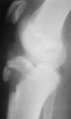
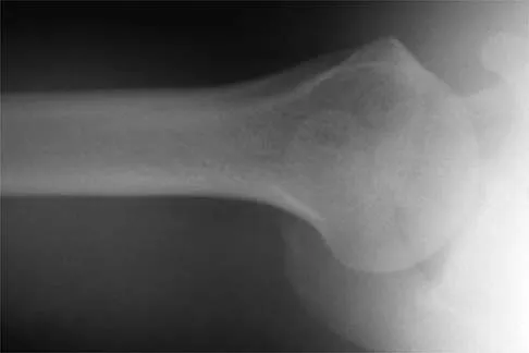
A 12-year-old Little League pitcher reports lateral elbow pain and "catching." Examination reveals painful pronation and supination and tenderness over the lateral elbow. Radiographs are shown in Figures 22a and 22b. Initial management should consist of


Explanation

22b Osteochondritis of the capitellum is a common problem in young throwing athletes and gymnasts. The mechanism of injury involves lateral compression and axial loading of the capitellum. Repetitive trauma causes ischemia with resultant osteochondral necrosis and sometimes eventual separation. Initial management includes rest for a minimum of 6 weeks; occasionally bracing is used. At long-term follow-up, there is typically an observed radiographic abnormality indicating incomplete healing even in asymptomatic patients. Arthroscopy with in situ drilling is reserved for symptomatic lesions that have an intact articular surface. Lesions with partial separation often require fixation. Lateral column osteotomy is a new investigational procedure designed to relieve lateral compression forces and may be used in salvage cases. Kobayashi K, Burton KJ, Rodner C, et al: Lateral compression injuries in the pediatric elbow: Panner's disease and osteochondritis dissecans of the capitellum. J Am Acad Orthop Surg 2004;12:246-254.

Question 6

Medial dislocation of the long head of the biceps tendon in the shoulder is most commonly caused by a


Explanation

Medial dislocation of the biceps tendon in the shoulder is commonly associated with subscapularis tendon tears. Although type II SLAP tears can result in bicipital instability, type I SLAP lesions do not. Congenitally shallow grooves and tears of the transverse ligaments usually do not lead to dislocation of the biceps tendon. Supraspinatus tendon tears are associated with long head of the biceps tendon ruptures but do not cause biceps tendon dislocations. Werner A, Mueller T, Boehm D, et al: The stabilizing sling for the long head of the biceps tendon in the rotator cuff interval: A histoanatomic study. Am J Sports Med 2000;28:28-31.

Question 7

Tension force in the anterior cruciate ligament during passive range of motion is highest at


Explanation

Tension forces in the healthy, as well as the reconstructed, anterior cruciate ligament were measured and found to be highest with the knee in full extension and decreased as the flexion increased. Markolf KL, Burchfield DM, Shapiro MM, et al: Biomechanical consequences of replacement of the anterior cruciate ligament with a patellar ligament allograft. Part II: Forces in the graft compared with forces in the intact ligament. J Bone Joint Surg Am 1996;78:1728-1734.

Question 8

Compared to eumenorrheic athletes, amenorrheic athletes have more frequent occurrences of


Explanation

In secondary amenorrhea, women do not receive the estrogen needed to maintain adequate bone mineralization. This hypoestrogenic state affects bone density, and there is evidence that stress fractures are more frequent in amenorrheic than eumenorrheic athletes. The other conditions are not seen with increased frequency in amenorrheic athletes. Warren MP: Health issues for women athletes: Exercise-induced amenorrhea. J Clin Endocrinol Metab 1999;84:1892-1896.

Question 9

Figure 23 shows the postoperative radiograph of a patient who underwent an anterior cruciate ligament (ACL) reconstruction (with bone-patella tendon-bone autograft) that failed. He initially had loss of flexion postoperatively. What is the most likely cause of this failure?


Explanation

The key to this question is the fact that the patient initially lost flexion postoperatively and this relates to anterior placement of the femoral tunnel, thus capturing the knee. The bone plug seen on the radiograph is actually from the tibial tunnel, but this occurred as the patient forced flexion until failure of the ACL graft and pullout of the plug from the tunnel. Although it could be argued that better tibial fixation would have prevented this failure, poor placement of the femoral tunnel led to the failure of this ACL reconstruction. Fu FH, Bennett CH, Latterman C, et al: Current trends in anterior cruciate ligament reconstruction: Part 1. Biology and biomechanics of reconstruction. Am J Sports Med 1999;27:821-830.

Question 10

A 22-year-old man reports anterior knee pain, swelling, and is unable to perform a straight leg raise after undergoing endoscopic anterior cruciate ligament (ACL) reconstruction with a bone-patellar tendon-bone autograft 1 week ago. He is afebrile. Examination reveals a clean incision, moderate effusion, a weak isometric quadriceps contraction, active knee range of motion of 5 degrees to 45 degrees, and the patella is ballottable. Knee radiographs show postoperative changes with good femoral and tibial tunnel placements, and normal patellar height. What is the next most appropriate step in management?


Explanation

Knee pain and swelling in the first week after ACL reconstruction is usually related to a postoperative hemarthrosis. A large hemarthrosis creates capsular distension, which inhibits active quadriceps contraction by a neurologic reflex, the H. reflex. Kennedy and associates reported that an experimentally induced knee effusion at 60 mL was found to result in profound inhibition of reflexly evoked quadriceps contraction. Removal of the hemarthrosis by aspiration will improve strength and often instantaneously restore the ability to contract the quadriceps muscle. A large effusion will also limit knee flexion. EMG and NCVS are not necessary unless there is a high index of suspicion of a femoral neuropathy. Diagnostic ultrasonography is not necessary in this patient but can be useful in the assessment of patellar tendon integrity. MRI is not indicated and would most likely be limited by artifact and postoperative changes. Continuous passive motion is not indicated and would most likely worsen the patient's symptoms. Kennedy JC, Alexander IJ, Hayes KC: Nerve supply of the human knee and its functional importance. Am J Sports Med 1982;10:329-335.

Question 11

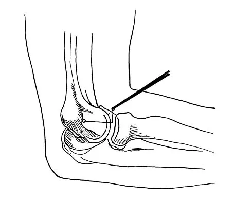
A 46-year-old woman fell from her bicycle and sustained the injury shown in Figure 24. Which of the following ligaments has been disrupted?


Explanation

The radiograph shows a type V acromioclavicular joint injury. Type V injuries involve disruption of the acromioclavicular and coracoclavicular ligaments. Type I injuries involve a sprain of the acromioclavicular joint ligaments. Type II injuries involve disruption of the acromioclavicular joint ligaments; the coracoclavicular ligaments are partially injured. Sternoclavicular ligaments stabilize the medial clavicle and the sternum; they are not damaged with acromioclavicular joint dislocations. Fukuda K, Craig EV, An KN, et al: Biomechanical study of the ligamentous system of the acromioclavicular joint. J Bone Joint Surg Am 1986;68:434-439.

Question 12

The use of knee arthroscopy following total knee arthroplasty is most effective in treating which of the following conditions?


Explanation

Patellar clunk syndrome is associated with certain types of posterior stabilized knee arthroplasties. Arthroscopic resection of the band of inflammatory tissue inferior to the patellar component is effective in treating this condition. Arthroscopic lavage of infected knee arthroplasties is not associated with an acceptable success rate. Diagnostic arthroscopy for nonspecific pain following arthroplasty is not uniformly successful. Patellar component maltracking is frequently associated with component malposition and is not alleviated by an arthroscopic lateral release. Synovitis secondary to polyethylene wear is best treated by exchange of the polyethylene spacer and not arthroscopic synovectomy. Lucas TS, DeLuca PF, Nazarian DG, et al: Arthroscopic treatment of patellar clunk. Clin Orthop 1999;367:226-229.

Question 13

Significant anterior tibial translation occurs during which of the following rehabilitation exercises?


Explanation

Terminal non-weight-bearing knee extension exercises from 60 degrees to 0 degrees of flexion increase anterior tibial translation. It is for this reason that this type of exercise should be avoided in the early phase of rehabilitation following anterior cruciate ligament reconstruction so as not to place a tensile strain on the graft. The other rehabilitation exercises either lead to posterior tibial translation in relation to the femur or have no significant effect on tibial translation. Grood ES, Suntay WJ, Noyes FR, et al: Biomechanics of the knee extension exercise: Effect of cutting the anterior cruciate ligament. J Bone Joint Surg Am 1984;66:725-734. Lutz GE, Palmitier RA, An KN: Comparison of tibiofemoral joint forces during open-kinetic-chain and closed-kinetic-chain exercises. J Bone Joint Surg Am 1993;75:732-739.

Question 14

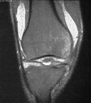
A 22-year-old volleyball player reports the insidious onset of superior and posterior shoulder pain. Radiographs are normal. An MRI scan is shown in Figure 25. What is the most specific physical examination finding?


Explanation

Overhead athletes are prone to a number of problems involving the shoulder. Pitchers and volleyball players are susceptible to posterior superior labral tears and internal impingement. These patients will have posterior superior shoulder pain, a positive relocation sign, and a positive active compression test. Occasionally, these posterior superior labral tears are associated with a spinoglenoid cyst as seen in the MRI scan. These cysts cause compression of the suprascapular nerve which manifests primarily as weakness of the infraspinatus, resulting in weakness of external rotation. Romeo AA, Rotenberg DD, Bach BR Jr: Suprascapular neuropathy. J Am Acad Orthop Surg 1999;7:358-367.

Question 15

A 20-year-old male lacrosse player sustains an anterior dislocation of the shoulder. He is extremely concerned about recurrent dislocations. Which of the following treatments has been shown to reduce the risk of recurrent dislocation?


Explanation

Recent evidence has shown that the position of immobilization of the shoulder after a dislocation influences the reduction of the Bankart lesion. In an MRI study in patients who sustained an anterior dislocation, the Bankart lesion was reduced to the glenoid anatomically with the arm in 30 degrees of external rotation. Subsequently, a clinical follow-up study has shown a reduction in recurrence rates when the arm is immobilized in external rotation compared to internal rotation. Itoi E, Hatakeyama Y, Kido T, et al: A new method of immobilization after traumatic anterior dislocation of the shoulder: A preliminary study. J Shoulder Elbow Surg 2003;12:413-415.

Question 16

A 43-year-old soccer player who had knee pain following a twisting injury underwent an arthroscopic meniscectomy 6 months ago. He continues to report posterior knee pain. Examination reveals soft-tissue fullness and tenderness just above the popliteal fossa, trace knee effusion, full range of knee motion, no instability, and negative meniscal signs. Radiographs show some mild medial joint space narrowing but no other bony changes. What is the next most appropriate step in management?


Explanation

The phenomenon of tumors misdiagnosed as athletic injuries has been termed "sports tumors." Lewis and Reilly presented a series of 36 patients who initially were thought to have a sports-related injury but ultimately were diagnosed with a primary bone tumor, soft-tissue tumor, or tumor-like condition. Muscolo and associates presented a series of 25 tumors that had been previously treated with an intra-articular procedure as a result of a misdiagnosis of an athletic injury. Initial diagnoses included 21 meniscal lesions, one traumatic synovial cyst, one patellofemoral subluxation, one anterior cruciate ligament tear, and one case of nonspecific synovitis. The final diagnoses were a malignant tumor in 14 patients and a benign tumor in 11 patients. The authors noted that oncologic surgical treatment was affected in 15 of the 25 patients. The most frequent causes of erroneous diagnosis were initial poor quality radiographs and an unquestioned original diagnosis despite persistent symptoms. Persistent symptoms warrant further diagnostic studies, not additional treatment such as physical therapy, corticosteroid injection, or an unloader brace. Although a bone scan may be helpful in this case and confirm arthrosis of the medial compartment, the suspicion of a soft-tissue mass makes MRI the imaging modality of choice. Muscolo DL, Ayerza MA, Makino A, et al: Tumors about the knee misdiagnosed as athletic injuries. J Bone Joint Surg Am 2003;85:1209-1214.

Question 17

Figures 26a through 26c show the MRI scans of a 47-year-old man who underwent arthroscopic shoulder surgery 6 months ago and continues to have pain despite a prolonged course of rehabilitation. Management should now consist of


Explanation

26b 26c The MRI scans show an os acromiale of the mesoacromion type. This represents an unfused acromial apophysis. Pain is thought to be caused by either motion at the site or downward displacement of the anterior aspect of the acromion onto the rotator cuff, causing impingement. Most patients can be treated nonsurgically as they are usually asymptomatic. In those patients with persistent symptoms of pain and tenderness over the acromion, surgery consisting of rigid internal fixation and bone grafting has yielded satisfactory results. Excision may be a viable treatment option for the preacromion type. Herzog RJ: Magnetic resonance imaging of the shoulder. Instr Course Lect 1998;47:3-20. Warner JP, Beim GM, Higgins L: The treatment of symptomatic os acromiale. J Bone Joint Surg Am 1998;80:1320-1326.

Question 18

An 18-year-old rugby player has had pain in his ring finger after missing a tackle 1 week ago. Examination reveals tenderness in the distal palm, and he is unable to actively flex the distal interphalangeal (DIP) joint. Radiographs are normal. What is the most appropriate management?


Explanation

Flexor digitorum profundus rupture or "rugger jersey finger" often occurs in the ring finger after the player misses a tackle and catches the digit on the shirt of the opposing player. Surgical repair is required for zone I-type injuries. Moiemen NS, Elliot D: Primary flexor tendon repair in zone I. J Hand Surg Br 2000;25:78-84.

Question 19

Storage of musculoskeletal allografts by cryopreservation is achieved by


Explanation

Cryopreservation uses chemicals to remove cellular water and controlled rate freezing to prevent ice crystal formation. The tissue is procured, cooled to wet ice temperature for quarantine, and then stored in a container with cryoprotectant solution of dimethyl sulfoxide or glycerol which displaces the cellular water. The controlled rate freezing is then done to prevent ice crystal formation. Fresh allografts are not frozen in order to maintain maximum cellular viability, and this process limits the shelf life of osteochondral allografts. Freeze-drying involves replacement of water in the tissue with alcohol to a moisture level of 5% and then uses a vacuum process to remove the alcohol from the tissue. Preparation of fresh frozen grafts involves freezing the graft twice and packaging the tissue without solution at minus 80 degrees C. American Association of Tissue Banks: Standards for Tissue Banking. MacLean, VA, American Association of Tissue Banks, 1999. Vangsness CT Jr, Triffon MJ, Joyce MJ, et al: Soft tissue allograft reconstruction of the human knee: A survey of the American Association of Tissue Banks. Am J Sports Med 1996;24:230-234.

Question 20

A 15-year-old wrestler sustains an abduction, hyperextension, and external rotation injury to his right shoulder. The MRI scan findings shown in Figures 27a and 27b are most consistent with


Explanation

27b An isolated avulsion of the lesser tuberosity occurs very rarely and usually is found in 12- and 13-year-old adolescents. The MRI scans reveal a tear of the humeral attachment of the inferior glenohumeral ligament, a so-called HAGL lesion. This injury to the inferior glenohumeral ligament occurs much less commonly than the classic Bankart lesion (anterior inferior labral tear). A tear of the subscapularis occurs with a similar mechanism of injury but generally occurs in older individuals. Bokor DJ, Conboy VB, Olson C: Anterior instability of the glenohumeral joint with humeral avulsion of the glenohumeral ligament: A review of 41 cases. J Bone Joint Surg Br 1999;81:93-96.

Question 21

Closed-chain exercise differs from open-chain exercise in which of the following ways?


Explanation

Closed-chain exercise requires the distal portion of the extremity to be fixed. It is more commonly used in lower extremity exercise, and movement is produced by co-contraction of muscles. Joint compression is increased, and multiple joints are involved with closed-chain exercise. In open-chain exercise, the distal portion of the extremity is free. Braddom RL (ed): Physical Medicine and Rehabilitation, ed 2. Philadelphia, PA, Saunders, 2000, pp 975-976.

Question 22

What procedure can eliminate a sulcus sign?


Explanation

A sulcus sign represents inferior subluxation of the shoulder. The elimination of this sign and correction of the inferior subluxation is best achieved through either an open or arthroscopic rotator interval closure. A SLAP repair stabilizes the biceps anchor but does not affect the sulcus sign. A Bankart repair, which corrects anterior-inferior laxity, is not sufficient to eliminate a sulcus sign. Subacromial decompression and supraspinatus repairs have no effect on inferior subluxation. Field LD, Warren RF, O'Brien SJ, et al: Isolated closure of rotator interval defects for shoulder instability. Am J Sports Med 1995;23:557-563.

Question 23

An eversion mechanism of injury is associated with which of the following ankle conditions?


Explanation

An inversion ankle injury typically involves ligamentous damage to the lateral ligaments of the ankle to include the anterior talofibular ligament and calcaneofibular ligament. Acute and particularly chronic ankle sprains also can have associated injuries. The inversion mechanism has been implicated in osteochondral and transchondral talar dome lesions, producing splits in the peroneus tendons, and in the development of meniscoid and soft-tissue impingement lesions in the anterolateral ankle. An inversion mechanism can also stretch the superficial peroneal nerve, leading to pain and paresthesias along its distribution. A fracture of the anterior colliculus is typically the result of an eversion mechanism resulting in a bony avulsion of the deltoid ligament from the anterior colliculus of the medial malleolus. Casillas MM: Ligament injuries of the foot and ankle in the athlete, in DeLee JC, Drez D, Miller MD (eds): Orthopaedic Sports Medicine: Principles and Practice, ed 2. Philadelphia, PA, WB Saunders, 2003, pp 2323-2357.

Question 24

Which of the following anatomic structures are in contact with internal impingement in the throwing athlete?


Explanation

Internal impingement occurs in the late cocking phase of throwing with humeral head abduction and maximal external rotation. It is a physiologic phenomonon occurring in 85% of patients undergoing arthroscopy for various indications in one study. Internal impingement is defined as impingement of the posterior-superior rotator cuff between the humerus and posterior-superior glenoid rim. Symptomatic internal impingement is felt to be due to the frequency and magnitude of the impingement in throwers. Koval KJ (ed): Orthopaedic Knowledge Update 7. Rosemont, IL, American Academy of Orthopaedic Surgeons, 2002, p 252.

Question 25

Second impact syndrome (SIS) after head injury is characterized by which of the following?


Explanation

SIS is a devastating but preventable complication of head injury. It occurs when return to activities is allowed prior to complete resolution of the symptoms of the first head injury. A second, sometimes trivial, head injury can lead to a devastating series of events that can result in sudden death. The symptoms tend to progress rapidly and often involve the brain stem. The prognosis is poor. Cantu RC: Second-impact syndrome. Clin Sports Med 1998;17:37-44. Saunders RL, Harbaugh RE: Second impact in catastrophic contact-sports head trauma. JAMA 1984;252:538-539.

Question 26

Comparing bone-patellar tendon-bone (BPTB) autograft to hamstring autograft for anterior cruciate ligament (ACL) reconstruction, what is a well-established difference in long-term outcomes based on current evidence?


Explanation

Long-term follow-up studies consistently demonstrate that BPTB autografts are associated with a higher incidence of donor-site morbidity, particularly anterior knee pain and pain with kneeling, compared to hamstring autografts. Rates of graft rupture are either similar or slightly lower for BPTB, while hamstring grafts tend to have a slightly higher rate of minor laxity but less donor site pain.

Question 27

In a throwing athlete with a type II superior labrum anterior and posterior (SLAP) lesion, the 'peel-back' mechanism exerts maximal torsional force on the biceps anchor. This mechanism is primarily observed during which specific phase of throwing?


Explanation

The 'peel-back' mechanism occurs during the late cocking phase of throwing. As the shoulder is placed into maximal abduction and external rotation, the biceps vector shifts posteriorly, transmitting a torsional force to the superior labrum and causing it to peel back or detach from the superior glenoid rim.

Question 28

During medial patellofemoral ligament (MPFL) reconstruction, accurate femoral tunnel positioning is crucial to avoid non-physiologic graft tension and altered patellofemoral kinematics. According to Schöttle's method, what is the correct radiographic location of the MPFL femoral insertion on a strict lateral radiograph?


Explanation

Schöttle et al. described highly reliable radiographic landmarks for the femoral insertion of the MPFL on a true lateral radiograph: 1 mm anterior to a line extending the posterior femoral cortex, 2.5 mm distal to the posterior origin of the medial femoral condyle, and proximal to the level of the posterior aspect of Blumensaat's line.

Question 29

A 22-year-old rugby player undergoes an open Latarjet procedure for recurrent anterior shoulder instability with 25% glenoid bone loss. Postoperatively, he presents with profound weakness with shoulder abduction and absent sensation over the lateral aspect of the shoulder. Which nerve was most likely injured, and what is its pertinent anatomic relationship during the procedure?


Explanation

The patient's presentation of deltoid weakness and lateral shoulder numbness indicates an axillary nerve injury. During a Latarjet procedure, the axillary nerve is at risk during the inferior capsular release and subscapularis split/tenotomy, as it courses closely inferior to the subscapularis and capsule to enter the quadrangular space.

Question 30

A 25-year-old male hockey player presents with chronic groin pain exacerbated by flexion and internal rotation. Imaging reveals a significant Cam-type morphology. In femoroacetabular impingement (FAI), where does the primary cartilage damage classically initiate in the setting of an isolated Cam lesion?


Explanation

Cam impingement is caused by an aspherical femoral head-neck junction that is forced into the acetabulum during flexion and internal rotation. This causes excessive shear forces that typically lead to characteristic anterosuperior labral tears and adjacent articular cartilage delamination on the acetabular side.

Question 31

Following a posterolateral corner (PLC) and ACL injury, a patient is undergoing surgical reconstruction. When drilling the fibular collateral ligament (FCL) femoral tunnel, understanding the anatomy is essential. What is the anatomic relationship of the FCL femoral footprint to the popliteus sulcus/attachment?


Explanation

On the lateral femoral epicondyle, the fibular collateral ligament (FCL) originates an average of 1.4 mm proximal and 3.1 mm posterior to the lateral epicondyle, effectively positioning it proximal and posterior to the popliteus femoral attachment.

Question 32

A 30-year-old cyclist sustains a direct blow to his superior shoulder during a fall. Radiographs demonstrate a type III acromioclavicular (AC) joint separation. While deciding between surgical and non-operative management, which of the following is considered an absolute indication for acute surgical intervention?


Explanation

While type III AC joint separations are controversial and generally treated non-operatively in the acute setting, absolute indications for acute surgical stabilization include open injuries, skin tenting causing impending necrosis, and concomitant neurovascular compromise.

Question 33

During the late cocking and early acceleration phases of throwing, which specific component of the ulnar collateral ligament (UCL) complex of the elbow serves as the primary restraint to valgus stress?


Explanation

The anterior bundle of the UCL is the primary restraint to valgus stress at the elbow. It is further divided into the anterior and posterior bands. The anterior band is tight in extension and serves as the primary restraint to valgus stress throughout the critical degrees of flexion seen during the acceleration phase of throwing.

Question 34

A 45-year-old woman experiences a painful pop in the posterior aspect of her knee while descending stairs. MRI demonstrates a complete radial tear of the posterior root of the medial meniscus with 3 mm of meniscal extrusion. If this injury is left untreated, the knee joint contact mechanics will most closely resemble which of the following conditions?


Explanation

A complete radial tear at the posterior root of the medial meniscus completely disrupts the circumferential hoop stresses of the meniscus. Biomechanical studies have demonstrated that this effectively renders the meniscus non-functional, resulting in joint contact pressures and areas equivalent to those seen in a knee that has undergone a total medial meniscectomy.

Question 35

When evaluating the non-operative management of acute Achilles tendon ruptures utilizing an early functional rehabilitation protocol compared to surgical repair, current high-level evidence demonstrates:


Explanation

Recent high-quality randomized controlled trials and meta-analyses comparing functional bracing/rehabilitation to surgical repair for acute Achilles tendon ruptures have shown similar re-rupture rates between the two groups. However, non-operative management avoids surgical complications such as wound breakdown, nerve injury, and deep infection.

Question 36

A 25-year-old athlete undergoes an isolated posterior cruciate ligament (PCL) reconstruction. The surgeon debates between a transtibial and a tibial inlay technique. Which of the following is the primary biomechanical advantage of the tibial inlay technique compared to the transtibial technique?


Explanation

The tibial inlay technique avoids the acute angle ('killer turn') at the posterior aspect of the tibia seen in transtibial PCL reconstruction. Biomechanical studies have shown that this sharp angle in the transtibial technique can cause graft abrasion and attenuation over time, whereas the inlay technique provides a more direct pull and reduces this risk.

Question 37

A 45-year-old overhead worker presents with shoulder pain. MRI reveals a type II SLAP tear. After failed conservative management, surgical intervention is planned. Compared to SLAP repair, primary biceps tenodesis in this age group is associated with:


Explanation

In patients over 40 years old, isolated SLAP repair is associated with higher rates of postoperative stiffness, continued pain, and higher reoperation rates compared to biceps tenodesis. Primary biceps tenodesis yields more predictable pain relief, functional outcomes, and lower reoperation rates in this older demographic.

Question 38

A 21-year-old collegiate baseball pitcher undergoes ulnar collateral ligament (UCL) reconstruction using the docking technique. He is concerned about potential postoperative complications. Which of the following is the most common complication following this procedure?


Explanation

Ulnar neuropathy is the most common complication following UCL reconstruction, occurring in 5% to 10% of cases depending on the technique used. While modern techniques like the docking approach have helped decrease this incidence compared to historical techniques requiring routine ulnar nerve transposition, transient ulnar neuropraxia remains the most frequently encountered adverse event.

Question 39

During a medial patellofemoral ligament (MPFL) reconstruction for recurrent patellar instability, identifying the correct femoral footprint is critical for proper graft isometry. Radiographically, Schöttle's point representing the femoral origin of the MPFL is located:


Explanation

Schöttle's point is a radiographic landmark used to identify the anatomic femoral origin of the MPFL on a perfect lateral radiograph. It is located 1 mm anterior to the posterior femoral cortical extension line, 2.5 mm distal to the posterior origin of the medial femoral condyle, and proximal to Blumensaat's line.

Question 40

A 19-year-old female soccer player sustains a twisting injury to her knee. Radiographs reveal an avulsion fracture of the lateral tibial plateau.

Which of the following intra-articular structures is most likely to be injured concurrently?


Explanation

A Segond fracture is an avulsion fracture of the anterolateral proximal tibia at the attachment of the anterolateral ligament (ALL) and the anterolateral capsule. It is considered a pathognomonic radiographic sign for an anterior cruciate ligament (ACL) tear.

Question 41

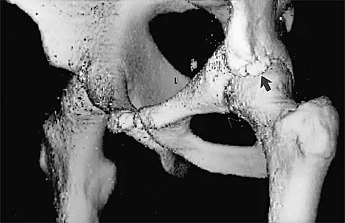
A 22-year-old rugby player presents with recurrent anterior shoulder instability. A 3D CT scan is obtained to evaluate anterior glenoid bone loss.

The 'glenoid track' concept is utilized to determine the risk of an engaging Hill-Sachs lesion. The glenoid track width is calculated as:


Explanation

The glenoid track is defined as 83% of the intact glenoid width minus the width of the anterior glenoid bone defect. If the Hill-Sachs interval (the width of the Hill-Sachs lesion plus the bone bridge to the rotator cuff footprint) is greater than the glenoid track, the lesion is 'off-track' and at risk of engaging the anterior glenoid rim during abduction and external rotation.

Question 42

A 50-year-old female experiences a sudden pop in her posterior knee while squatting. MRI demonstrates a radial tear at the posterior root of the medial meniscus.

Biomechanically, leaving this medial meniscus posterior root tear untreated is most comparable to which of the following?


Explanation

A posterior root tear of the medial meniscus completely disrupts the circumferential hoop stresses of the meniscus, allowing the meniscus to extrude radially under axial load. Biomechanical studies demonstrate that an untreated posterior root tear alters knee kinematics, decreases contact area, and increases peak contact pressures to levels equivalent to those seen after a total medial meniscectomy, accelerating osteoarthritis.

Question 43

A 28-year-old male undergoes hip arthroscopy for femoroacetabular impingement (FAI) with a symptomatic CAM lesion. During the osteochondroplasty of the anterolateral femoral head-neck junction, the surgeon must be cautious to avoid injury to the primary blood supply of the femoral head. Which vessel is at greatest risk if the resection is carried too far posterosuperiorly?


Explanation

The deep branch of the medial femoral circumflex artery (MFCA) gives rise to the superior retinacular vessels, which provide the primary blood supply to the femoral head. These vessels run along the posterosuperior aspect of the femoral neck. Osteochondroplasty for CAM lesions is typically performed anterolaterally; however, extending the resection excessively to the posterosuperior region places these vital retinacular vessels at risk, which could lead to avascular necrosis.

Question 44

A 30-year-old cyclist falls directly onto his shoulder. Clinical examination reveals significant superior displacement of the clavicle with a prominent step-off. Radiographs show superior displacement of the clavicle by 150% compared to the contralateral side.

According to the Rockwood classification, what specific anatomical disruption distinguishes this injury from a Type III AC joint separation?


Explanation

This patient has a Rockwood Type V acromioclavicular (AC) joint injury, which is defined by greater than 100% (up to 300%) superior displacement of the clavicle relative to the acromion. Both Type III and Type V injuries involve complete tears of the AC and CC ligaments. However, Type V is distinguished by the complete disruption and stripping of the deltotrapezial fascia from the distal clavicle. Type IV involves posterior displacement, and Type VI involves inferior displacement.

Question 45

A 35-year-old recreational basketball player sustains an acute Achilles tendon rupture. He elects to undergo a minimally invasive percutaneous repair to minimize wound complications. During the procedure, the surgeon places sutures through the proximal stump of the tendon. Which of the following structures is at the highest risk of iatrogenic injury during this specific step?


Explanation

The sural nerve is at significant risk of entrapment or laceration during percutaneous or minimally invasive Achilles tendon repair, particularly when placing sutures in the proximal stump. The nerve courses distally along the posterior calf, migrating laterally to run closely adjacent to the lateral border of the Achilles tendon in the middle and distal thirds of the leg.

Question 46

A 25-year-old professional rugby player sustains a contact injury to his right knee. Physical examination reveals a positive dial test with 20 degrees of increased external rotation compared to the contralateral side at both 30 degrees and 90 degrees of knee flexion. Varus stress testing demonstrates grade III laxity at both 0 degrees and 30 degrees of flexion. Which combination of ligamentous structures is most likely injured?


Explanation

A positive dial test showing >10 degrees of asymmetric external rotation at 30 degrees of knee flexion indicates a posterolateral corner (PLC) injury. When this asymmetry persists or increases at 90 degrees of flexion, it indicates a combined PLC and posterior cruciate ligament (PCL) injury. Grade III varus laxity at 30 degrees confirms LCL injury (a component of the PLC), and grade III varus laxity at 0 degrees confirms the involvement of a cruciate ligament, classically the PCL in this combined injury pattern.

Question 47

A 22-year-old collegiate baseball pitcher complains of posterior shoulder pain that is most severe during the late cocking phase of throwing. He has a loss of 25 degrees of internal rotation compared to his non-throwing shoulder. Which of the following describes the primary pathophysiologic mechanism for his pain?


Explanation

Internal impingement is a common cause of posterior shoulder pain in overhead throwing athletes, specifically during the late cocking phase (maximum abduction and external rotation). In this position, the greater tuberosity abuts the posterosuperior glenoid rim, causing the undersurface of the posterior rotator cuff (supraspinatus/infraspinatus) and the posterosuperior labrum to become pinched. The patient's glenohumeral internal rotation deficit (GIRD) further exacerbates this altered kinematic pattern.

Question 48

A 28-year-old male undergoes right hip arthroscopy for femoroacetabular impingement. Postoperatively in the recovery room, he complains of profound numbness in the perineum and scrotum, and difficulty achieving an erection over the next several days. Which mechanism is the primary cause of this complication?


Explanation

Pudendal nerve neurapraxia is a well-documented complication of hip arthroscopy due to compression against the perineal post during prolonged or excessive joint traction. Symptoms include numbness in the perineum, scrotum/labia, and potential sexual dysfunction. Proper padding, limiting traction time to less than 2 hours, and considering postless distraction techniques are utilized to prevent this complication. Lateral femoral cutaneous nerve injury presents with lateral thigh numbness and is related to anterior portal placement.

Question 49

A 16-year-old female experiences a first-time lateral patellar dislocation while playing soccer. The patella spontaneously reduces. Initial radiographs reveal no acute fractures. Which of the following is an absolute indication for surgical intervention following this primary dislocation event?


Explanation

The standard of care for a primary, uncomplicated patellar dislocation is non-operative management with a short period of immobilization followed by physical therapy focusing on vastus medialis obliquus (VMO) strengthening. However, absolute indications for early surgical intervention include a displaced osteochondral fracture/loose body, an avulsion fracture of the medial patellar border, or a massive medial soft tissue avulsion with lateral patellar subluxation that fails to spontaneously reduce. MPFL tears are expected in acute dislocations and do not mandate early surgery without other complications.

Question 50

When comparing the outcomes of acute Achilles tendon ruptures treated with operative repair versus non-operative management utilizing early functional bracing and mobilization, non-operative management is associated with which of the following?


Explanation

High-quality randomized controlled trials (such as Willits et al.) have demonstrated that when non-operative treatment of acute Achilles tendon ruptures is paired with an early functional rehabilitation protocol, the re-rupture rates are statistically similar to those of operative repair. Non-operative management eliminates the risks associated with surgery, most notably surgical site infections, wound healing complications, and iatrogenic sural nerve injury. Plantar flexion strength and return to work times are generally comparable between the two groups.

Question 51

A 20-year-old collegiate pitcher undergoes ulnar collateral ligament (UCL) reconstruction utilizing a palmaris longus autograft. Which of the following technical factors is most critical for successfully restoring the normal kinematics and stability of the elbow?


Explanation

The anterior band of the anterior bundle of the UCL is the primary restraint to valgus stress at the elbow. In UCL reconstruction (e.g., Tommy John surgery), anatomic placement of the graft tunnels at the native footprints—the sublime tubercle on the proximal ulna and the anteroinferior aspect of the medial epicondyle—is the most critical factor for restoring kinematics. Non-anatomic placement leads to graft stretching or loss of motion due to lack of isometry. Ulnar nerve transposition is not routinely required unless there are preoperative nerve symptoms.

Question 52

A 45-year-old man falls onto his outstretched arm while skiing and sustains an acute, traumatic isolated full-thickness tear of the subscapularis tendon. Which of the following physical examination findings is most specific for this injury?


Explanation

The bear hug test, along with the belly-press and lift-off tests, specifically evaluate the integrity of the subscapularis tendon. The patient places their hand on their contralateral shoulder, and the examiner attempts to externally rotate the arm to break the patient's internal rotation force. Hornblower's sign evaluates the teres minor. Jobe's test (empty can) evaluates the supraspinatus. O'Brien's test evaluates the acromioclavicular joint and superior labrum. Neer's test is a general test for subacromial impingement.

Question 53

A 24-year-old athlete presents with a failed anterior cruciate ligament (ACL) reconstruction.

Computed tomography evaluation demonstrates significant femoral and tibial tunnel widening, with both tunnels measuring >16 mm in diameter. What is the most appropriate next step in management?


Explanation

In the setting of a failed ACL reconstruction with significant tunnel widening (typically defined as >14-15 mm), a two-stage revision is indicated. The first stage consists of hardware removal and bone grafting of the enlarged tunnels to restore bone stock. Once the bone graft has consolidated (usually after 4 to 6 months), the second stage involving revision ACL reconstruction can be safely performed. Attempting a single-stage revision with >15 mm tunnels risks poor graft fixation, hardware failure, and recurrent instability.

Question 54

During the biomechanical evaluation of the acromioclavicular (AC) joint complex, which specific ligamentous structure provides the primary restraint against superior displacement of the distal clavicle?


Explanation

The coracoclavicular (CC) ligaments consist of the conoid and trapezoid ligaments. The conoid ligament is located more medially and posteriorly, and it provides the primary restraint to superior displacement of the clavicle. The trapezoid ligament is located more laterally and anteriorly, primarily resisting axial compression into the acromion. The AC capsular ligaments primarily provide restraint to anteroposterior translation.

Question 55

A 13-year-old skeletally immature male gymnast complains of ongoing knee pain.

MRI reveals a 1.5 cm x 1.5 cm osteochondritis dissecans (OCD) lesion on the lateral aspect of the medial femoral condyle. The overlying cartilage is intact, and there is no fluid behind the lesion. Non-operative management, including restricted weight-bearing and activity modification, has failed after 6 months. What is the most appropriate surgical treatment?


Explanation

For a stable osteochondritis dissecans (OCD) lesion (intact cartilage, no synovial fluid behind the lesion on MRI) in a skeletally immature patient that has failed an adequate trial of non-operative management, subchondral drilling is the gold standard surgical treatment. Drilling (either retroarticular or transarticular) violates the sclerotic border of the lesion to promote vascular ingrowth and healing of the fragment. Procedures like OATS, ACI, or allograft are reserved for unstable, detached, or unsalvageable lesions, particularly in skeletally mature patients.

Question 56

A 24-year-old female athlete undergoes an anterior cruciate ligament (ACL) reconstruction using a bone-patellar tendon-bone autograft. Postoperatively, she demonstrates a severe loss of terminal knee flexion. During physical examination under anesthesia, the reconstructed graft is noted to be excessively tight in flexion, but relatively lax in extension. What is the most likely technical error that occurred during the reconstruction?


Explanation

A femoral tunnel placed too anteriorly (high in the notch when the knee is extended) is a common technical error in ACL reconstruction. This non-anatomic placement causes the distance between the femoral and tibial attachments to increase as the knee flexes. As a result, the graft captures the joint, becoming excessively tight in flexion (limiting flexion) and relatively lax in extension. Conversely, an excessively anterior tibial tunnel leads to roof impingement and a loss of extension.

Question 57

A 28-year-old soccer player sustains a direct blow to the anteromedial aspect of the proximal tibia while the knee is flexed. On physical examination, the dial test reveals 25 degrees of external rotation of the tibia compared to 10 degrees on the contralateral side at 30 degrees of knee flexion. However, at 90 degrees of knee flexion, the external rotation is symmetric bilaterally. Which of the following structures is most likely injured?


Explanation

The dial test is utilized to evaluate for posterolateral instability. An increase in external rotation of greater than 10 degrees compared to the normal, contralateral knee is considered positive. Increased external rotation at 30 degrees of knee flexion, which then reduces to symmetric rotation at 90 degrees, is pathognomonic for an isolated posterolateral corner (PLC) injury. If the external rotation remains asymmetrical and increased at both 30 and 90 degrees of flexion, it indicates a combined injury to both the PLC and the posterior cruciate ligament (PCL).

Question 58

A 22-year-old collegiate baseball pitcher complains of vague anterior shoulder pain and a 'dead arm' sensation that exclusively occurs during the late cocking phase of throwing. An MRI arthrogram confirms an isolated Type II SLAP (Superior Labrum Anterior and Posterior) tear. What is the primary biomechanical mechanism responsible for generating this specific pathology in an overhead athlete?


Explanation

In overhead throwing athletes, a Type II SLAP tear is classically caused by the 'peel-back' mechanism. During the late cocking phase of throwing, the shoulder is placed in maximum abduction and external rotation. In this extreme position, the vector of the long head of the biceps tendon shifts posteriorly, creating a torsional force that peels the posterosuperior labrum away from the glenoid rim. This unique dynamic mechanism is a primary driver for symptomatic SLAP lesions in pitchers.

Question 59

A 50-year-old active female feels a sharp 'pop' in her posterior knee while rising from a deep squat. MRI reveals a complete radial tear immediately adjacent to the posterior horn medial meniscus attachment, with 4 mm of meniscal extrusion seen on coronal sequences. Biomechanically, in terms of tibiofemoral contact pressures, this injury is most equivalent to which of the following?


Explanation

A complete medial meniscus posterior root tear disrupts the crucial circumferential hoop stresses of the meniscus. Without intact osseous attachments, the meniscus is extruded radially under axial load. Extensive biomechanical studies have demonstrated that a posterior root tear effectively abolishes the load-sharing function of the meniscus, dramatically increasing peak contact pressures in the medial compartment to levels essentially equivalent to those observed after a total medial meniscectomy, thereby predisposing the patient to rapid articular cartilage wear and osteoarthritis.

Question 60

A 26-year-old professional hockey player presents with chronic groin pain exacerbated by deep hip flexion and internal rotation. A Dunn lateral radiograph is obtained, and the alpha angle is measured to be 68 degrees. This radiographic finding is most consistent with which of the following pathomorphologies?


Explanation

The alpha angle is a radiographic measurement used to quantify the sphericity of the anterior femoral head and the head-neck offset, typically measured on a lateral projection such as the Dunn view. An alpha angle greater than 50-55 degrees (with 68 degrees being clearly pathologic) indicates a loss of the normal concave junction between the anterior femoral head and neck. This morphologic abnormality represents a Cam deformity, which engages and damages the anterosuperior acetabular labrum and articular cartilage during hip flexion and internal rotation (Cam-type Femoroacetabular Impingement).

Question 61

During a medial patellofemoral ligament (MPFL) reconstruction for recurrent patellar instability, the surgeon utilizes intraoperative fluoroscopy to identify the anatomic femoral attachment site (Schöttle's point). Which of the following accurately describes the correct radiographic landmarks for this location on a strict lateral radiograph?


Explanation

Schöttle's point represents the radiographic femoral footprint of the MPFL on a true lateral radiograph. It is accurately located 1 mm anterior to the posterior cortex line extension, 2.5 mm distal to the posterior articular border of the medial femoral condyle, and proximal to the level of the posterior point of Blumensaat's line. Precise placement is crucial; a femoral tunnel placed too proximal or anterior leads to a graft that is non-isometric and excessively tight in flexion, causing increased patellofemoral contact pressures and potential graft failure.

Question 62

A 28-year-old marathon runner presents with persistent anterior knee pain. MRI and subsequent diagnostic arthroscopy reveal a symptomatic, 4.5 cm² full-thickness unipolar chondral defect on the weight-bearing surface of the medial femoral condyle. He has failed exhaustive nonoperative management. His mechanical alignment is neutral, and both menisci are intact. According to current treatment algorithms, what is the most appropriate primary cartilage restoration procedure?


Explanation

The treatment of chondral defects depends on the size of the lesion, patient age, and physical demands. For large symptomatic full-thickness defects (> 2-3 cm²), cell-based therapies such as Matrix-induced Autologous Chondrocyte Implantation (MACI) or fresh osteochondral allografts are the standard of care. OATS (osteochondral autograft) is typically reserved for smaller defects (< 2 cm²) due to significant donor site morbidity when harvesting multiple plugs. Microfracture is generally not recommended for large defects as the resulting fibrocartilage lacks durability. Because alignment is neutral, HTO is not indicated.

Question 63

A 23-year-old collegiate baseball pitcher requires an ulnar collateral ligament (UCL) reconstruction after suffering a complete rupture of the anterior bundle. During reconstruction using the docking technique, the surgeon aims to anatomically recreate the primary restraint to valgus stress. To which specific anatomic footprints must the graft be secured to accurately recreate the anterior bundle of the UCL?


Explanation

The anterior bundle of the ulnar collateral ligament (UCL) is the primary restraint to valgus stress at the elbow, particularly between 30 and 120 degrees of flexion. It originates on the anteroinferior surface of the medial epicondyle and inserts distally on the sublime tubercle of the proximal ulna. Anatomic UCL reconstruction techniques specifically target these footprints to restore native elbow kinematics and stability.

Question 64

A 31-year-old male sustains a knee dislocation (Schenck KD-IIIL) after a high-speed motorcycle accident. On initial presentation, he has an absent dorsalis pedis pulse with an ABI of 0.6, and a profound 'foot drop' with absent sensation in the first web space. Following a successful vascular bypass, orthopedic ligamentous reconstruction is planned. Which of the following statements regarding his neurologic deficit is most accurate?


Explanation

Common peroneal nerve palsy is a devastating complication of knee dislocations, especially those involving the posterolateral corner (KD-IIIL or KD-IV). The injury is typically a high-energy stretch/traction injury over a long segment, making primary end-to-end repair nearly impossible without grafting. The prognosis for spontaneous functional recovery (useful motor function for dorsiflexion) is historically poor, occurring in less than 30-40% of cases. Consequently, tendon transfers (such as a posterior tibial tendon transfer) or an ankle-foot orthosis (AFO) are frequently required for long-term functional management.

Question 65

A 35-year-old competitive weightlifter presents to the emergency department with acute severe pain, swelling, and ecchymosis over the right anterior axilla and medial arm. He reports feeling a sudden 'tearing' sensation at the bottom phase of a maximal bench press. Physical examination demonstrates a notable loss of the anterior axillary contour. Based on the biomechanics of this injury pattern, which portion of the pectoralis major muscle is placed under the greatest tension during this movement and most commonly ruptures first?


Explanation

Pectoralis major tendon ruptures classically occur in weightlifters performing the bench press, and the most common location of failure is at the humeral insertion (tendon avulsion). The pectoralis major tendon undergoes a complex 180-degree twist before inserting on the humerus, such that the inferior (sternocostal/sternal) fibers insert most superiorly and proximally. When the arm is in maximum extension, abduction, and external rotation (the lowest point of the bench press), these inferior sternal fibers are stretched maximally. Thus, the sternal head at its humeral insertion undergoes the highest disproportionate tension and is consistently the first portion to fail.

Question 66

A 24-year-old professional soccer player presents with recurrent knee instability 2 years after an arthroscopic anterior cruciate ligament (ACL) reconstruction using a bone-patellar tendon-bone autograft. He reports a 'giving way' episode without a new traumatic event. A sagittal MRI from his recent evaluation is shown in Figure 1.

What is the most common etiology for early clinical failure of this reconstructed ligament?


Explanation

The most common cause of recurrent instability and failure following primary ACL reconstruction is non-anatomic tunnel placement. Specifically, a femoral tunnel placed too anteriorly or vertically results in a graft that is non-isometric, leading to over-tensioning in flexion and stretching or rupture over time. While missed concomitant injuries (like posterolateral corner injuries or ramp lesions) are important secondary causes of failure, surgical technique errors regarding tunnel positioning remain the leading overall cause.

Question 67

During the arthroscopic repair of a Type II Superior Labrum Anterior to Posterior (SLAP) tear in a 21-year-old collegiate baseball pitcher, anchors are placed to secure the labrum. To avoid altering the complex biomechanics of the superior labrum and to minimize postoperative stiffness, anchor placement should strictly avoid which of the following areas?


Explanation

In repairing a Type II SLAP lesion, anchors should be placed posterior to the biceps anchor (and sometimes anteriorly if the tear extends into that region) but NEVER directly through or securing the biceps tendon root itself. Tying down the biceps anchor directly restricts its normal physiologic rolling and excursion during overhead motions, which consistently leads to severe postoperative stiffness, particularly in overhead throwing athletes.

Question 68

A 20-year-old collegiate baseball pitcher presents with medial elbow pain that occurs predominantly during the late cocking and early acceleration phases of throwing. On physical examination, the moving valgus stress test is markedly positive. Magnetic resonance imaging confirms a high-grade partial tear of the ulnar collateral ligament (UCL), and he elects to undergo UCL reconstruction. Which bundle of the UCL is the primary restraint to valgus stress, and what is its true anatomic footprint?


Explanation

The anterior bundle of the ulnar collateral ligament (UCL) is the primary restraint to valgus stress of the elbow between 30 and 120 degrees of flexion. It originates on the anterior undersurface of the medial epicondyle and inserts on the sublime tubercle of the anteromedial facet of the coronoid process. The posterior bundle forms the floor of the cubital tunnel and is only a secondary restraint to valgus stress at higher degrees of flexion.

Question 69

A 24-year-old male hockey player presents with chronic, deep groin pain exacerbated by hip flexion and internal rotation. Radiographs demonstrate a pistol-grip deformity. A coronal T2-weighted MRI of his right hip is shown in Figure 11.

During arthroscopic intervention for this condition, where is the most common location of concomitant articular cartilage damage expected to be found?


Explanation

The patient's presentation and imaging findings are classic for Cam-type femoroacetabular impingement (FAI), characterized by a reduced head-neck offset (pistol-grip deformity). During hip flexion and internal rotation, the aspherical femoral head abuts the acetabular rim. This mechanical impingement most commonly causes chondral delamination and labral tears in the anterosuperior quadrant of the acetabulum.

Question 70

A 32-year-old weightlifter presents with right shoulder pain and weakness after feeling a clunk during a heavy bench press exercise. He reports difficulty externally rotating the arm. A modified axillary radiograph is provided in Figure 5.

Imaging reveals a reverse Hill-Sachs lesion that involves approximately 25% of the articular surface. What is the most appropriate surgical management?


Explanation

The patient has suffered a posterior shoulder dislocation, classically associated with a reverse Hill-Sachs lesion (an impaction fracture of the anteromedial humeral head). For lesions involving 20% to 40% of the articular surface, the modified McLaughlin procedure is indicated. This involves the transfer of the subscapularis tendon (and sometimes the lesser tuberosity) into the anterior humeral head defect to prevent it from engaging the posterior glenoid rim. Infraspinatus transfer (Remplissage) is used for anterior dislocations with a standard Hill-Sachs lesion.

Question 71

A 26-year-old rugby player sustained a direct blow to the anteromedial aspect of his knee while it was fully extended. On physical examination, he demonstrates 15 degrees of increased external tibial rotation at 30 degrees of knee flexion compared to the contralateral side. At 90 degrees of flexion, the external rotation side-to-side difference is only 3 degrees. Which of the following structures is most likely injured?


Explanation

The patient's physical examination describes a positive Dial test at 30 degrees of knee flexion but a negative Dial test at 90 degrees. This finding is indicative of an isolated posterolateral corner (PLC) injury. The PLC (which includes the LCL, popliteus tendon, and popliteofibular ligament) is the primary restraint to external tibial rotation at 30 degrees. If the Dial test is positive at both 30 and 90 degrees, it suggests a combined PCL and PLC injury.

Question 72

A 19-year-old elite collegiate basketball player sustains a fracture at the metaphyseal-diaphyseal junction of the fifth metatarsal. He undergoes intramedullary screw fixation to expedite return to play. The high risk of nonunion in this specific fracture pattern is primarily attributed to a watershed vascular zone. From which aspect does the primary nutrient artery enter the fifth metatarsal to supply this area?


Explanation

A Jones fracture occurs at the metaphyseal-diaphyseal junction (Zone 2) of the fifth metatarsal. This area is prone to nonunion because it resides in a vascular watershed area. The primary intraosseous blood supply comes from the nutrient artery, which typically enters the medial cortex at the middle third of the diaphysis and branches proximally. The proximal base is supplied by metaphyseal vessels, leaving the junction zone relatively avascular.

Question 73

A 17-year-old gymnast undergoes a medial patellofemoral ligament (MPFL) reconstruction using a semitendinosus autograft for recurrent patellar dislocations. The surgeon uses fluoroscopy to identify Schöttle's point for the femoral anchor placement. Which of the following best describes the radiographic landmarks for the anatomic femoral attachment of the MPFL on a true lateral radiograph?


Explanation

Schöttle's point is the recognized radiographic landmark for the anatomic femoral attachment of the MPFL on a true lateral radiograph. It is located 1 mm anterior to a line extending the posterior femoral cortex, 2.5 mm distal to the posterior origin of the medial femoral condyle, and just proximal to the level of the posterior medial epicondyle. Accurate placement is critical, as non-anatomic femoral tunnel placement (especially too proximal) leads to excessive graft tension in flexion and loss of knee motion.

Question 74

A 40-year-old marathon runner feels a sudden pop in the posterior aspect of his knee while decelerating. MRI demonstrates a complete radial tear at the posterior root of the medial meniscus. If left untreated, this specific injury pattern most closely mimics the biomechanical effects of which of the following?


Explanation

A medial meniscus posterior root tear effectively completely disrupts the continuity of the meniscal ring. This leads to an inability to convert axial joint loads into hoop stresses. Biomechanically, this results in medial compartment peak contact pressures and contact areas that are functionally equivalent to those seen after a total medial meniscectomy, rapidly accelerating the progression of osteoarthritis. Thus, root repairs are strongly advocated in active, appropriately selected patients.

Question 75

A 35-year-old recreational tennis player presents with acute posterior ankle pain after lunging for a drop shot. He has a positive Thompson test. An MRI of the ankle is shown in Figure 7.

He elects to undergo minimally invasive surgical repair of the Achilles tendon. During percutaneous suture passage in the proximal stump, which nerve is at the greatest risk of iatrogenic injury?


Explanation

The sural nerve courses distally along the posterolateral aspect of the calf, crossing the lateral border of the Achilles tendon roughly 10 cm proximal to its insertion. During percutaneous or minimally invasive Achilles tendon repair, the sural nerve is at significant risk of being captured, tethered, or injured during the blind passage of sutures in the proximal tendon stump. Surgeons must carefully map or protect the nerve laterally when passing sutures.

Question 76

A 25-year-old athlete sustains a high-energy knee dislocation during a football game. Magnetic resonance imaging demonstrates complete tears of the anterior cruciate ligament (ACL), posterior cruciate ligament (PCL), and the posteromedial corner (PMC). The patient undergoes single-stage multi-ligament knee reconstruction 3 weeks post-injury. What is the most common complication following surgical treatment of a multi-ligament knee injury?


Explanation

Arthrofibrosis (joint stiffness) is the most common complication following multi-ligament knee injury and reconstruction, particularly when surgery is performed in the early acute phase or when multiple ligaments are reconstructed simultaneously. Meticulous postoperative rehabilitation is essential to restore range of motion.

Question 77

A 30-year-old competitive weightlifter feels a sudden, painful 'pop' in his anterior chest wall while performing a heavy bench press. Physical examination reveals ecchymosis, loss of the anterior axillary fold, and notable weakness in internal rotation and adduction of the humerus. MRI confirms a complete tear of the pectoralis major at its humeral insertion. What is the optimal management for this patient?


Explanation

Pectoralis major ruptures at the sternal head insertion are classic weightlifting injuries. In young, active patients or athletes, early surgical repair (within 6 to 8 weeks) yields significantly better outcomes in restoring strength and cosmesis compared to nonoperative management or delayed repair.

Question 78

A 21-year-old rugby player presents with recurrent anterior shoulder instability following an initial dislocation one year ago. A 3D reconstructed CT scan of the glenoid demonstrates an 'inverted pear' appearance with approximately 26% anterior bone loss.

What is the most appropriate surgical intervention to prevent recurrent instability?


Explanation

Anterior glenoid bone loss exceeding 20-25% alters the biomechanics of the glenohumeral joint, resulting in an 'inverted pear' shaped glenoid. Soft tissue stabilization (Bankart repair) alone has an unacceptably high failure rate in this scenario. Bony augmentation, such as the Latarjet procedure, is required to restore the articular arc and provide a 'sling' effect via the conjoined tendon.

Question 79

A 22-year-old collegiate baseball pitcher undergoes ulnar collateral ligament (UCL) reconstruction utilizing a palmaris longus autograft (Tommy John surgery). Which of the following is the most frequent postoperative complication associated with this procedure in overhead throwers?


Explanation

Ulnar neuropathy is the most common complication following UCL reconstruction. It can occur due to traction, compression, or ischemia during the procedure, especially if the nerve is handled or transposed. Most cases are transient, but a subset may require secondary neurolysis.

Question 80

A 45-year-old woman experiences a sudden 'pop' in the back of her knee while squatting. MRI demonstrates a medial meniscus posterior root tear and localized marrow edema in the medial femoral condyle. What is the primary biomechanical consequence of leaving this root tear untreated?


Explanation

A meniscal root tear disrupts the circumferential continuity of the meniscus, causing a complete loss of its ability to convert axial loads into hoop stresses. This leads to radial extrusion of the meniscus, biomechanically equating to a total meniscectomy and rapidly resulting in increased peak contact pressures and accelerated osteoarthritis.

Question 81

During hip arthroscopy for a 28-year-old hockey player with femoroacetabular impingement (FAI), an osteochondroplasty is performed for a large cam lesion at the femoral head-neck junction. Resection of more than what percentage of the femoral neck diameter substantially increases the risk of an iatrogenic postoperative femoral neck fracture?


Explanation

Biomechanical studies have demonstrated that resecting more than 30% of the anterolateral femoral neck diameter significantly decreases the load to failure of the proximal femur, thereby increasing the risk of a catastrophic iatrogenic femoral neck fracture postoperatively.

Question 82

A 14-year-old female gymnast presents with lateral elbow pain, clicking, and a 15-degree extension deficit. Radiographs demonstrate a lucency in the capitellum. MRI reveals an osteochondral defect with high T2 signal fluid surrounding the fragment, indicating instability. Her capitellar physis is open. What is the recommended treatment?


Explanation

In a juvenile patient (open physis) with an unstable but intact osteochondritis dissecans (OCD) lesion of the capitellum (indicated by fluid behind the fragment on MRI), the standard of care is surgical fixation of the fragment to preserve the native articular cartilage. Marrow stimulation or OATS are typically reserved for unsalvageable fragments or failed primary fixation.

Question 83

A 24-year-old recreational soccer player sustains an acute anterior cruciate ligament (ACL) rupture. During a subsequent gait analysis assessment, which of the following kinematic adaptations is most characteristic of a classic ACL-deficient 'quadriceps avoidance gait' during the stance phase?


Explanation

Patients with an ACL-deficient knee often adopt a 'quadriceps avoidance gait' to dynamically stabilize the knee. Because contraction of the quadriceps pulls the tibia anteriorly, the patient unconsciously decreases peak knee flexion during the stance phase, relying more on hip extensors and hamstrings to prevent the tibia from subluxating anteriorly.

Question 84

A 35-year-old woman presents with persistent anterior hip pain. Imaging shows a positive crossover sign, a center-edge angle of 45 degrees, and a labral tear. Which of the following is the defining pathomechanical feature of this specific type of femoroacetabular impingement (FAI)?


Explanation

The scenario describes Pincer-type FAI, characterized by acetabular overcoverage (e.g., retroversion shown by a crossover sign, or deep socket/coxa profunda shown by an increased CE angle). The defining feature is linear contact (abutment) between the prominent acetabular rim and the femoral head-neck junction. Cam impingement, conversely, is caused by an aspherical femoral head.

Question 85

A 28-year-old cyclist falls directly onto his shoulder. Radiographs show superior displacement of the distal clavicle.

Measurements reveal the coracoclavicular distance is increased by 150% compared to the contralateral side. According to the Rockwood classification, what type of acromioclavicular (AC) joint injury is this, and what is the typical recommended management?


Explanation

A coracoclavicular (CC) distance increased by 100% to 300% relative to the contralateral side indicates a Rockwood Type V AC joint injury. This is accompanied by severe soft-tissue stripping, including detachment of the deltoid and trapezius from the distal clavicle. Unlike Type III injuries (up to 100% displacement), which are usually treated nonoperatively, Type V injuries generally require surgical reconstruction to restore shoulder biomechanics and relieve severe symptoms.

Question 86

A 25-year-old professional hockey player sustains a high-grade acromioclavicular (AC) joint separation and is scheduled for coracoclavicular (CC) ligament reconstruction. To accurately recreate the native anatomy, the surgeon must be aware of the specific insertion footprints of the CC ligaments. Which of the following describes the anatomical insertion of the conoid ligament relative to the trapezoid ligament on the clavicle?


Explanation

The coracoclavicular (CC) ligaments consist of the conoid and trapezoid. The conoid ligament inserts medially and posteriorly on the conoid tubercle of the clavicle, approximately 4.5 cm from the distal end. It is the primary restraint to superior translation of the clavicle. The trapezoid ligament inserts laterally and anteriorly, approximately 3 cm from the distal end, and is the primary restraint to axial compression of the clavicle towards the acromion.

Question 87

A 22-year-old collegiate soccer player is evaluated for posterolateral knee pain and a feeling of instability after a twisting injury. On physical examination, the dial test demonstrates 15 degrees of increased external rotation compared to the contralateral normal knee at 30 degrees of knee flexion, but symmetric external rotation at 90 degrees of knee flexion. This finding is most indicative of an isolated injury to which of the following structures?


Explanation

The dial test is used to evaluate combined or isolated injuries of the posterolateral corner (PLC) and the posterior cruciate ligament (PCL). An increase of >10 degrees of external rotation compared to the contralateral side at 30 degrees of flexion, with symmetric rotation at 90 degrees, is indicative of an isolated PLC injury. If external rotation is increased at both 30 and 90 degrees, it suggests a combined PCL and PLC injury.

Question 88

A 24-year-old male presents with deep groin pain exacerbated by hip flexion and internal rotation. Radiographs demonstrate a 'pistol grip' deformity of the proximal femur. Which of the following radiographic parameters is most diagnostic of the Cam-type femoroacetabular impingement (FAI) suspected in this patient?


Explanation

Cam-type FAI is characterized by an aspherical femoral head-neck junction. An alpha angle greater than 55 degrees (typically measured on a cross-table lateral or Dunn view) is indicative of a Cam lesion. A center-edge angle < 20 degrees suggests developmental dysplasia of the hip (DDH), while a lateral center-edge angle > 40 degrees or a crossover sign indicates focal or global acetabular overcoverage, typical of Pincer-type FAI.

Question 89

A 28-year-old overhead throwing athlete presents with deep shoulder pain and clicking. An MRI is obtained as shown in Figure 4.

After failing conservative management, he undergoes arthroscopic repair for a Type II SLAP lesion. Which of the following portals provides the most optimal trajectory for anchor placement at the posterosuperior glenoid rim to address this pathology?


Explanation

The Port of Wilmington (posterolateral portal) is located approximately 1 cm anterior and 1 cm lateral to the posterolateral corner of the acromion. This portal provides the optimal 'deadman' angle of approach (typically 45 degrees to the articular surface) for placing suture anchors into the posterosuperior glenoid rim during SLAP lesion repairs. The Neviaser portal (suprascapular) is superior and is sometimes used but risks the suprascapular nerve and provides a steeper angle.

Question 90

A 45-year-old recreational athlete feels a 'pop' in the back of his knee while performing a deep squat. MRI reveals a complete posterior root tear of the medial meniscus. According to biomechanical studies, leaving a complete posterior root tear of the medial meniscus unrepaired is biomechanically equivalent to which of the following?


Explanation

The meniscal roots anchor the meniscus to the tibial plateau, allowing for the conversion of axial compressive forces into circumferential hoop stresses. Biomechanical studies have demonstrated that a complete disruption of the medial meniscus posterior root completely disrupts these hoop stresses, rendering the meniscus non-functional and leading to medial compartment contact pressures equivalent to a total meniscectomy.

Question 91

A 21-year-old collegiate baseball pitcher reports medial elbow pain and decreased velocity during the late cocking and early acceleration phases of throwing. An MRI is shown in Figure 15.

He undergoes an ulnar collateral ligament (UCL) reconstruction using a docking technique. During the surgical exposure, the surgeon utilizes a muscle-splitting approach through the flexor-pronator mass. Care must be taken to identify and protect which of the following cutaneous nerves to prevent painful neuroma formation?


Explanation

During the medial approach to the elbow for UCL reconstruction, branches of the medial antebrachial cutaneous (MABC) nerve frequently cross the operative field. They lie superficial to the flexor-pronator mass. Injury to the MABC can cause numbness over the medial forearm or a painful neuroma, which is a significant complication for a throwing athlete. The ulnar nerve is deeper and must be managed (either protected in situ or transposed), but the MABC is the specific superficial cutaneous nerve at high risk.

Question 92

A 55-year-old man presents with right shoulder weakness after a fall on an outstretched arm. He specifically complains of difficulty tucking in his shirt behind his back and bringing his hand to his abdomen. Physical examination reveals a positive bear-hug test and increased passive external rotation compared to the contralateral side. Which of the following special tests is also most likely to be positive in this patient?


Explanation

The patient's clinical presentation (weakness with internal rotation, increased passive external rotation, positive bear-hug test) is highly indicative of a subscapularis tendon tear. The belly-press test (Napoleon test) isolates the subscapularis and is positive when the patient cannot maintain pressure on their abdomen without extending the shoulder and flexing the wrist. Hornblower's sign evaluates the teres minor. O'Brien's test evaluates for SLAP tears or AC joint pathology. Jobe's test isolates the supraspinatus. Yergason's test evaluates the long head of the biceps.

Question 93

A 19-year-old competitive skier sustains a twisting injury to her left knee. A radiograph reveals a small avulsion fracture of the lateral aspect of the proximal tibia, known as a Segond fracture.

This specific radiographic finding is highly associated with an anterior cruciate ligament (ACL) tear and represents a bony avulsion of which of the following structures?


Explanation

A Segond fracture is an avulsion fracture of the lateral tibial plateau and is considered pathognomonic for an anterior cruciate ligament (ACL) tear. Recent anatomical and biomechanical literature has clarified that the Segond fracture represents a bony avulsion of the anterolateral ligament (ALL) of the knee, which acts as a secondary restraint to internal tibial rotation. It is distinct from the IT band (which avulses at Gerdy's tubercle) and the LCL.

Question 94

A 28-year-old physically active female presents with a 3.5 cm² symptomatic, unipolar, full-thickness chondral defect on the medial femoral condyle. She has failed 6 months of non-operative management. Diagnostic arthroscopy reveals normal mechanical alignment, an intact meniscus, and a stable knee. Which of the following surgical interventions is most appropriate to provide hyaline-like cartilage restoration for a defect of this size?


Explanation

For symptomatic, unipolar, full-thickness cartilage defects of the femoral condyle >2 cm² in a young, active patient without subchondral bone loss, Autologous Chondrocyte Implantation (ACI) or Matrix-induced ACI (MACI) is indicated and aims to produce hyaline-like cartilage. Microfracture is typically reserved for smaller defects (<2 cm²) and produces predominantly fibrocartilage. OATS is also generally used for smaller defects due to donor site morbidity. Osteochondral allograft is favored for very large defects (>3 cm²) or those with significant subchondral bone involvement.

Question 95

A 25-year-old professional baseball pitcher presents with vague posterior shoulder pain and a noted decrease in pitching velocity. Physical exam reveals a Glenohumeral Internal Rotation Deficit (GIRD) of 25 degrees compared to the non-throwing shoulder, along with a loss of total arc of motion. What is the most common pathomechanical cause of symptomatic GIRD contributing to internal impingement in the overhead throwing athlete?


Explanation

Symptomatic GIRD (defined as a loss of internal rotation >20 degrees with a corresponding loss of total arc of motion >5 degrees) in overhead throwers is most commonly caused by contracture and thickening of the posteroinferior capsule. This contracture occurs due to repetitive eccentric microtrauma during the deceleration phase of throwing. The tight posteroinferior capsule causes a posterosuperior shift of the humeral head during the cocking phase, leading to internal impingement and placing increased strain on the superior labrum. Acquired humeral retroversion causes an altered arc of motion but preserves the total arc and does not typically result in pathological GIRD.

Question 96

A 17-year-old female high school soccer player tears her ACL during a game. She and her parents are discussing graft options for surgical reconstruction. What is the most accurate information regarding the use of bone-patellar tendon-bone (BPTB) allograft compared to BPTB autograft in this specific patient population?


Explanation

Multiple large-cohort studies (including the MARS and MOON cohorts) have demonstrated that in young, active patients (especially those under 20 years old), allograft ACL reconstruction is associated with a significantly higher failure and revision rate compared to autograft. Allografts tend to have a slower biological incorporation process and undergo a prolonged remodeling phase, which increases the risk of graft failure in highly active young cohorts returning to pivot-shift sports.

Question 97

A 22-year-old male rugby player presents with recurrent anterior shoulder instability after a primary dislocation 2 years ago. He reports 5 subsequent dislocations requiring closed reduction. A 3D CT scan of the shoulder is shown in Figure 14, demonstrating an 'inverted pear' glenoid with 27% anterior bone loss. What is the most appropriate definitive management?


Explanation

Critical anterior glenoid bone loss (>20-25%) alters the biomechanics of the glenohumeral joint, rendering soft-tissue stabilization alone (arthroscopic or open Bankart) insufficient due to unacceptably high recurrence rates. The 'inverted pear' appearance indicates significant bone loss where the inferior width is narrower than the superior width. The Latarjet procedure (coracoid transfer) is the gold standard for recurrent anterior shoulder instability with critical glenoid bone loss, providing stability via a triple effect: a bone block, a sling effect from the conjoint tendon, and capsular repair.

Question 98

A 30-year-old male sustains a severe knee hyperextension injury during American football, resulting in a knee dislocation. After closed reduction, his vascular exam is normal with biphasic pulses, but he exhibits a profound foot drop and absent sensation in the first web space. An MRI shown in Figure 5 demonstrates complete disruption of the ACL, PCL, and posterolateral corner (PLC). Which of the following anatomical structures is most closely associated with the pathway of the injured nerve and serves as a critical surgical landmark?


Explanation

The clinical presentation describes a common peroneal nerve injury (foot drop, numbness in the first dorsal web space), a well-known complication of posterolateral corner (PLC) injuries and knee dislocations. The common peroneal nerve courses distally and laterally through the popliteal fossa, wrapping around the fibular neck just posterior and deep to the long and short heads of the biceps femoris tendon. The biceps femoris tendon is the key anatomical landmark for locating, protecting, and decompressing the common peroneal nerve during surgical approaches to the posterolateral knee.

Question 99

A 24-year-old male collegiate hockey player complains of insidious onset, worsening deep right groin pain that is exacerbated by prolonged sitting and deep hip flexion. A radiograph is shown in Figure 19, demonstrating an abnormal alpha angle of 65 degrees. What is the primary pathophysiologic mechanism of chondral injury in this specific morphological variant of femoroacetabular impingement (FAI)?


Explanation

An abnormal alpha angle (>50-55 degrees) signifies a Cam lesion, characterized by a lack of sphericity at the femoral head-neck junction. During hip flexion and internal rotation, this aspherical prominence is forcefully introduced into the acetabulum, causing significant shear stress at the anterosuperior chondrolabral junction. This repetitive shear typically results in 'inside-out' delamination of the adjacent acetabular articular cartilage and subsequent labral detachment. In contrast, Pincer impingement (acetabular over-coverage) primarily causes labral crushing or degeneration and contrecoup chondral lesions in the posteroinferior acetabulum.

Question 100

A 21-year-old collegiate baseball pitcher underwent a right elbow ulnar collateral ligament (UCL) reconstruction using a palmaris longus autograft 6 weeks ago. He now presents with persistent tingling in his small and ring fingers, as well as subjective weakness when gripping. Which intraoperative factor or surgical step is most commonly associated with this specific postoperative complication?


Explanation

Ulnar neuropathy is the most frequent complication following UCL reconstruction (Tommy John surgery). It is most commonly associated with excessive handling, traction, or mobilization of the ulnar nerve during the medial approach. When the nerve is aggressively retracted to expose the sublime tubercle and medial epicondyle but left in situ (or transposed with kinking/devascularization), the risk of postoperative ulnar neuritis increases significantly. Modern techniques emphasize minimal handling and in situ preservation of the nerve, or a meticulous submuscular transposition if the nerve subluxates or is heavily involved in scar tissue.

None

Clinic OS
Medically Verified Content by
Prof. Clinic OS
Consultant Orthopedic & Spine Surgeon
Chapter Index