العربية
Part of the Master Guide

General Orthopedics 2026 Practice Questions: Set 13 (Solved)

Orthopedic Basic 2026 MCQs: Board Review Questions & Answers (Part 4)

23 Apr 2026 80 min read 71 Views
Figure for Mtd 2008 MCQs - Part 4 - Question 76

Key Takeaway

Discover the latest medical recommendations for Orthopedic Basic 2026 MCQs: Board Review Questions & Answers (Part 4). Top-rated Orthopedic Basic 2026 MCQs bank. Practice with clinical case questions, orthopedic surgery board review, and evidence-based answers updated for 2026.

Orthopedic Basic 2026 MCQs: Board Review Questions & Answers (Part 4)

Comprehensive 100-Question Exam


00:00

Start Quiz

Question 1

A 23-year-old man has had right posterolateral knee pain and occasional lateral calf dysesthesias for the past 8 months. A radiograph, CT scan, MRI scans, and a biopsy specimen are shown in Figures 62a through 62e. What is the most likely diagnosis?


Explanation

62b 62c 62d 62e The radiograph shows an eccentric, cortically based lytic lesion in the proximal fibula. The CT and MRI scans confirm that it is well circumscribed and cortically based with significant surrounding edema. The radiographic differential diagnosis would be a Brodie's abscess or osteoid osteoma. An osteoblastoma would have to be greater than 2 cm in size. A chondroblastoma may also have significant edema around it, but it is an epiphyseal-based lesion, not cortically based. The well-circumscribed nature of the lesion is not consistent with osteosarcoma. The pathology shows a very cellular and vascular stroma with plump, but not atypical osteoblast cells making a matrix of immature woven bone. There are no abundant inflammatory cells or dead bone suggestive of osteomyelitis or a Brodie's abscess. Therefore, the clinical and histologic picture is most consistent with an osteoid osteoma. Percutaneous radiofrequency ablation, usually with CT guidance, has become the preferred method for treating most cases of osteoid osteoma. Rosenthal DI: Radiofrequency treatment. Orthop Clin North Am 2006;37:475-484.

Question 2

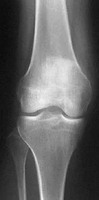
Figures 63a and 63b show the radiographs of an 11-year-old girl who sustained a twisting injury of the knee playing soccer. She is now asymptomatic. What is the appropriate treatment of the lesion?


Explanation

63b This is a nonossifying fibroma of the proximal tibia. The lesion is eccentric, cortically based, with sclerotic margins and no evidence of a soft-tissue mass. Nonossifying fibromas are benign lesions that need no biopsy or surgical treatment when classic findings appear on radiographs. A follow-up radiograph should be performed 2 to 3 months after the initial presentation to ensure that the lesion is not progressive. Surgery is reserved for large lesions with risk of pathologic fracture or for cases where a displaced pathologic fracture has occurred and internal fixation is needed for fracture treatment. Nondisplaced pathologic fractures through nonossifying fibromas are best treated by allowing the fracture to heal and observation of the lesion. Vaccaro AR (ed): Orthopaedic Knowledge Update 8. Rosemont, IL, American Academy of Orthopaedic Surgeons, 2005, pp 197-215.

Question 3

A 21-year-old man is referred for evaluation of a lesion in the left proximal femur that was discovered when he was undergoing an upper gastrointestinal series. He reports no hip or thigh pain. Radiographs are shown in Figures 64a and 64b. What is the best course of action?


Explanation

64b Melorheostosis is a rare disorder characterized by the classic radiographic appearance of flowing hyperostosis in a long bone. The hyperostosis may be on the periosteal or endosteal surface of the bone and frequently gives the appearance of wax falling down the side of a candle. The radiographs are diagnostic; therefore, no further work-up is indicated. Menendez LR (ed): Orthopaedic Knowledge Update: Musculoskeletal Tumors. Rosemont, IL, American Academy of Orthopaedic Surgeons, 2002, pp 87-102.

Question 4

A 17-year-old boy has had elbow pain for the past 6 weeks. A radiograph, MRI scans, and biopsy specimens are shown in Figures 65a through 65e. What is the most likely diagnosis?


Explanation

65b 65c 65d 65e The findings are consistent with an osteoblastoma. The radiographs show a bone-forming lesion of the distal humerus. The lesion has an osseous component extending out of the native cortex with a thin sclerotic border. The T2-weighted MRI scan shows the lesion extending anteriorly beyond the native cortex. No fluid-fluid levels are seen. Histology shows large osteoblasts producing osteoid and woven bone. The tissue between the spicules of bone and osteoid contains thin fibrous tissue and capillaries. Osteoid osteoma is a smaller lesion usually with sclerotic reactive bone around a small nidus. The histology differentiates osteoblastoma from osteosarcoma because no malignant cells are seen. There is no cartilage production or chondroblasts in the histologic specimen, eliminating chondroblastoma. Giant cell tumors of bone typically occur in a epiphyseal metaphyseal location, most commonly after skeletal maturity, and contain numerus giant cells. Menendez LR (ed): Orthopaedic Knowledge Update: Musculoskeletal Tumors. Rosemont, IL, American Academy of Orthopaedic Surgeons, 2002, pp 87-102.

Question 5

What is the most common site of metastases from a soft-tissue sarcoma?


Explanation

The most common site of metastases from a soft-tissue sarcoma is the lungs and occurs in 40% to 60% of patients. The second most common site of metastases in soft-tissue sarcomas is the lymph nodes. Nodal metastases are seen with regularity in synovial sarcoma, epithelioid sarcoma, and rhabdosarcoma. The liver, brain, bone, and muscle are occasional sites of spread, but the occurrence is very rare. Simon SR (ed): Orthopaedic Basic Science. Rosemont, IL, American Academy of Orthopaedic Surgeons, 1994, pp 219-276.

Question 6

A 45-year-old woman has had right wrist pain for the past 2 months. A radiograph, bone scan, and MRI scan are shown in Figures 66a through 66c, and a photomicrograph is shown in Figure 66d. What is the most likely diagnosis?


Explanation

66b 66c 66d The radiograph shows a purely radiolucent lesion without matrix mineralization in the epiphysis of the distal radius. The lesion is "hot" on bone scan, and the MRI scan reveals cortical destruction with a soft-tissue mass. These findings are most consistent with giant cell tumor. The distal radius is a common location for giant cell tumors. The other options would be very uncommon in this location in a 45-year-old patient. The photomicrograph demonstrates multinucleated giant cells in a sea of mononuclear cells. The nuclei of the giant cells and the nuclei of the mononuclear stromal cells are identical. This feature helps distinguish giant cell tumor from other lesions that might contain giant cells. Cheng CY, Shih HN, Hsu KY, et al: Treatment of giant cell tumor of the distal radius. Clin Orthop Relat Res 2001;383:221-228.

Question 7

An otherwise healthy 16-year-old dancer reports a 1-month history of leg pain. AP and lateral radiographs of the distal femur are shown in Figures 67a and 67b. What is the next most appropriate step in management?


Explanation

67b The radiographs show a very ill-defined, aggressive, moth-eaten bony destruction involving the distal left femoral diaphysis just above the metaphyseal junction. The differential diagnosis includes Ewing's sarcoma, osteosarcoma, lymphoma of bone, eosinophilic granuloma, osteomyelitis, and others. MRI would further define the lesion, and soft-tissue and intramedullary extension. There is aggressive periosteal reaction in the posteromedial aspect of the adjacent lesion with some multilayered components in the distal interface of the periosteum. The lateral radiograph shows cortical penetration and irregular periosteal reaction of the posterior margin, suggesting some posterior soft-tissue extension at this site as well. Gebhardt MC, Ready JE, Mankin HJ: Tumors about the knee in children. Clin Orthop Relat Res 1990;255:86-110.

Question 8

Acral metastases are most commonly seen in what type of carcinoma?


Explanation

Metastatic lesions to bone are usually located in the axial and proximal appendicular skeleton. Metastases below the elbow and knee are rare, but when they do occur they are most commonly from lung carcinoma. Hayden RJ, Sullivan LG, Jebson PJ: The hand in metastatic disease and acral manifestations of paraneoplastic syndromes. Hand Clin 2004;20:335-343.

Question 9

A 47-year-old woman has had left medial clavicle pain for the past 6 months. History is remarkable for mediastinal non-Hodgkin's lymphoma, treated with mantel radiation 22 years ago. A radiograph, CT scan, MRI scan, and a biopsy specimen are shown in Figures 68a through 68d. What is the most likely diagnosis?


Explanation

68b 68c 68d Radiation-associated sarcomas typically occur at least 5 years following radiation therapy, in the radiation therapy field, and with different histology than the original disease. The radiograph shows a lytic destructive lesion of the medial clavicle. The radiographic differential could include any of the above etiologies. The CT and MRI scans show this same reaction with extension into the adjacent soft tissue and periosteal reaction. These findings eliminate a degenerative process or radiation-induced osteonecrosis but do not distinguish between a neoplastic and infectious process. A PET scan showed marked uptake in the distal clavicle, which is more consistent with a malignant neoplastic process than a reactive process, like that of an infection. These findings, combined with the cellular atypia and bone formation on the biopsy specimen, confirm the diagnosis of radiation-associated sarcoma. In this older patient, radiation-associated sarcoma appears in an unusual location secondary to her previous radiation treatment in that region, which can occur 3 to 50 years after previous radiation therapy. Shaheen M, Deheshi BM, Riad S, et al: Prognosis of radiation-induced bone sarcoma is similar to primary osteosarcoma. Clin Orthop Relat Res 2006;450:76-81.

Question 10

In 1980, a 32-year-old woman was found to have right breast mass, and a biopsy revealed adenocarcinoma. She underwent a mastectomy at that time, with no other treatment. Five years later, she noticed a lump in the left breast and underwent a left mastectomy. Seven lymph nodes were positive. In 2006, she now reports hip and thigh pain for the past 3 months. Figures 69a and 69b show AP and lateral radiographs of the femur. A bone scan shows a solitary lesion. Following radiographic staging, what is the next most appropriate step in management?


Explanation

69b Solitary bone lesions require biopsy, for there is the possibility that the lesion may represent a primary bone sarcoma, which will necessitate a different treatment plan. This is especially true in patients with remote histories of cancer. The most likely cause of a lytic bone lesion in a patient older than age 40 years is a metastatic lesion. Rougraff BT, Kneisl JS, Simon MA: Skeletal metastases of unknown origin: A prospective study of a diagnostic strategy. J Bone Joint Surg Am 1993;75:1276-1281.

Question 11

What is the mechanism of action of bisphosphonates?


Explanation

Bisphosphonates are stable analogues of pyrophosphate that have a strong affinity for bone hydroxyapatite; these agents inhibit bone resorption by reducing the recruitment and activity of osteoclasts and increasing apoptosis. Bone formed while patients are receiving bisphosphonate treatment is histologically normal. Bisphosphonates have been shown to be effective in decreasing pathologic fractures, bone pain, and the need for radiation therapy in patients with multiple myeloma and metastatic carcinoma to bone. The most effective method of administration is via monthly intravenous infusion. Osteonecrosis of the mandible is sometimes a complication of this treatment. Gass M, Dawson-Hughes B: Preventing osteoporosis-related fractures: An overview. Am J Med 2006;119:S3-S11.

Question 12

A 16-year-old girl has had hip pain for 1 year. Approximately 2 months ago she noted the development of a hard mass in the right buttock that has steadily increased in size. She now reports severe pain in the right buttock, with radiation down the leg and numbness involving the right foot and toes. A radiograph is shown in Figure 70a and an axial postcontrast T1-weighted MRI scan is shown in Figure 70b. A biopsy specimen is shown in Figure 70c. The chest CT shows multiple lung metastases. Treatment of this lesion should consist of


Explanation

70b 70c Ewing's sarcoma is the second most common primary tumor of bone in children. Depending on the site and extent of disease, chemotherapy, radiation therapy, and surgery are all treatment options. In this patient with extensive pelvic and metastatic disease, chemotherapy and radiation therapy offer the best oncologic control while preserving functional outcome. Gibbs CP Jr, Weber K, Scarborough MT: Malignant bone tumors. Instr Course Lect 2002;51:413-428. Thacker MM, Temple HT, Scully SP: Current treatment for Ewing's sarcoma. Expert Rev Anticancer Ther 2005;5:319-331.

Question 13

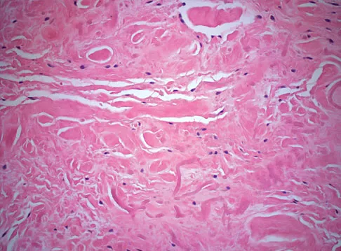
A 55-year-old woman has slowly increasing pain at the distal end of her little finger that is exacerbated by cold temperatures. She denies any history of trauma to her hands and is employed as a school teacher. The histology of the resected specimen is shown in Figure 71. What is the most likely diagnosis?


Explanation

Glomus tumors are rare vascular lesions typically occurring about the nail of the distal phalanx of the hand. The diagnostic "triad" of glomus tumors consists of local pain, sensitivity to cold, and paroxysmal pain. They tend to present with pain as the most typical symptom and this can be exacerbated by changes in temperature that is felt to cause a vascular response within the lesion. The biopsy specimen confirms a glomus tumor showing the typical vascular spaces surrounded by glomus epithelioid glomus cells. Zook EG, Brown RE: The perionychium, in Green DP, Hotchkiss RN, Pederson WC (eds): Green's Operative Hand Surgery, ed 4. Philadelphia, PA, Churchill Livingstone, 1999, vol 2, pp 1353-1380.

Question 14

A 69-year-old woman reports a painful clicking in her right shoulder. A soft-tissue mass is palpated at the lower portion of the scapula. Based on the MRI scan and biopsy specimens shown in Figures 72a through 72c, what is the most likely diagnosis?


Explanation

72b 72c Elastofibroma is a rare, benign soft-tissue pseudotumor characteristically located in the subscapular region. Most patients are between 50 and 70 years of age and have pain, decreased shoulder range of motion, or a mass. The lesion usually is best visualized with the arm elevated forward and adducted to displace the scapula laterally and allow the mass to protrude from the chest wall. They are frequently bilateral. Grossly, the mass is ill-defined, oblong or spherical, firm, and ranges in size from 5 cm to 10 cm. These masses appear infiltrative and frequently are mistaken for a sarcomatous lesion. Histologically, the mass is composed of a mixture of intertwining eosinophilic collagen and elastic fibers, and scattered fibroblasts, mucoid material, and fat. Vastamaki M: Elastofibroma scapulae. Clin Orthop Relat Res 2001;392:404-408. Nielsen T, Sneppen O, Mykre-Jensen O, et al: Subscapular elastofribroma: A reactive pseudotumor. J Shoulder Elbow Surg 1996;5:209-213.

Question 15

A 40-year-old man has a painless mass around his left ankle. He notes minimal growth over the past year. An MRI scan is shown in Figure 73a, and biopsy specimens are shown in Figures 73b and 73c. What is the most likely diagnosis?


Explanation

73b 73c The biopsy specimen is a low-power view of a soft-tissue sarcoma with a biphasic pattern of epithelial cells and fibrous spindle cells that are typical of a synovial sarcoma. A deep, painless soft-tissue mass greater than 5 cm in size is suspicious for a sarcoma. The imaging in this patient is indeterminate and the patient requires a biopsy for an accurate diagnosis. The biopsy reveals a tumor with a biphasic appearance consistent with a synovial sarcoma. These tumors are slow growing, occur primarily in the lower extremities, and are found in a younger demographic population compared to malignant fibrous histiocytoma and liposarcoma. They can occur in a biphasic pattern with clumps of epithelial cells and fibrous spindle cells or in a monophasic pattern. Synovial sarcomas stain positively for keratin. Keratin is positive in nearly all biphasic types and in many tumors of the monophasic fibrous type. Sixty percent of these tumors are found in the lower extremity. The area around the knee is the most common location, followed by the ankle and foot. Epithelioid and clear cell sarcomas are found associated with tendon sheaths of the hand and feet and are generally smaller in size. Epithelioid sarcoma may resemble granulomatous inflammation histologically; clear cell sarcoma is composed of nests of clear cells with occasional multinucleated giant cells. Pigmented villonodular synovitis is found within the synovium and is characterized by a villous histologic appearance with hemosiderin-laden macrophages. Malignant fibrous histiocytoma has a storiform histologic pattern with an abundance of pleomorphic cells. Enzinger FM, Weiss SW: Soft Tissue Tumors, ed 3. St Louis, MO, Mosby, 1995, p 757.

Question 16

A 28-year-old woman has left shoulder pain and a tender soft-tissue mass. Based on the MRI scan and biopsy specimens shown in Figures 74a through 74c, what is the most likely diagnosis?


Explanation

74b 74c Schwannomas (neurilemomas) occur at all ages but are most frequently seen in persons between the ages of 20 and 50 years. MRI features of schwannomas are fairly nonspecific, but when they are associated with a large named nerve, the identification of a mass in continuity with that nerve is highly suggestive of a schwannoma. Most have a fairly homogeneous appearance with a high water content and often fusiform shape. Classically, the histology shows alternating Antoni A (dense spindle cell region) areas and Antoni B (loose myxoid tissue) areas. They also demonstrate uniform intense immunostaining with S-100 protein. Damron TA, Sim FH: Soft-tissue tumors about the knee. J Am Acad Orthop Surg 1997;5:141-152.

Question 17

Which of the following prognostic indicators is associated with the least favorable outcome for patients newly diagnosed with osteosarcoma?


Explanation

Distant bone metastasis is associated with an extremely poor prognosis for patients with osteosarcoma (5-year survival rate of less than 10%). Most osteosarcomas are high grade and extracompartmental, and approximately half are greater than 8 cm at presentation. The 5-year survival rate for these patients is approximately 70%. Patients with a solitary pulmonary metastasis have a prognosis worse than patients without detectable metastases but not as bad as those with bone metastases. Bielack SS, Kempf-Bielack B, Delling G, et al: Prognostic factors in high-grade osteosarcoma of the extremities or trunk: An analysis of 1,702 patients treated on neoadjuvant cooperative osteosarcoma study group protocols. J Clin Oncol 2002;20:776-790. Heck RK, Stacy GS, Flaherty MJ, et al: A comparison study of staging systems for bone sarcomas. Clin Orthop Relat Res 2003;415:64-71.

Question 18

A 12-year-old girl has had increasing left knee pain for the past 3 months. A radiograph is shown in Figure 75a, and low- and high-power photomicrographs are shown in Figures 75b and 75c. What is the most appropriate treatment?


Explanation

75b 75c The radiograph reveals an aggressive purely lytic lesion of the distal femoral metaphysis. There is no apparent matrix mineralization or periosteal reaction. The photomicrographs show a malignant spindle cell neoplasm in a storiform pattern. Based on these findings, the diagnosis is malignant fibrous histiocytoma of bone. At most institutions, patients with this tumor are treated similar to patients with osteosarcoma with multi-agent chemotherapy and surgery with wide margins (resection or amputation). Menendez LR (ed): Orthopaedic Knowledge Update: Musculoskeletal Tumors. Rosemont, IL, American Academy of Orthopaedic Surgeons, 2002, pp 203-209.

Question 19

A 58-year-old woman has had a slowly progressing mass over the distal interphalangeal (DIP) joint of her dominant hand with a worsening deformity of her nail. She has no significant medical history but underwent bilateral knee arthroplasties 1 year ago. Radiographs reveal a small osteophyte at the DIP joint dorsally. A clinical photograph and a biopsy specimen are shown in Figures 76a and 76b. What is the most likely diagnosis?


Explanation

76b A mucous cyst is thought to be a ganglion arising from the DIP joint in patients with osteoarthritis. They are frequently associated with nail deformities. Treatment involves removal of the cyst with debridement of DIP joint osteophytes. Fritz GR, Stern PJ, Dickey M: Complications following mucous cyst excision. J Hand Surg Br 1997;22:222-225.

Question 20

A 13-year-old boy has had pain and swelling in his ankle for the past several months. Based on the radiograph, MRI scan, and biopsy specimen shown in Figures 77a through 77c, what is the best course of action?


Explanation

77b 77c Chondroblastomas are benign cartilage lesions frequently seen in adolescents or young adults. They are found in the epiphyseal or apophyseal regions of bones. The radiograph shows a radiolucent lesion with mineralization and a well-marginated rim of reactive bone. The lesion is composed of sheets of immature chondroblasts (polygonal cells with a clear, bluish cytoplasm and a small round central nucleus). In some regions, classic "chicken-wire" matrix calcifications and a "cobblestone" pattern of cell arrangement may be seen. Treatment consists of curettage and bone grafting. Campanacci M: Bone and Soft Tissue Tumors, ed 2. New York, NY, Springer-Verlag, 1999, pp 247-264.

Question 21

A 39-year-old man has had a foot mass for the past several months. MRI scans are shown in Figures 78a through 78c. A core biopsy specimen reveals synovial sarcoma, and a staging chest CT scan is normal. Which of the following treatments offers the best local tumor control and expedites the patient's return to normal function?


Explanation

78b 78c Certain histologic subtypes of soft-tissue sarcoma have been noted to arise preferentially in the hand and the foot, such as epithelioid sarcoma, clear cell sarcoma, and synovial sarcoma. Synovial sarcoma is the most common foot sarcoma. Frequently there is a delay in diagnosis because the lesions are rare. The lesions tend to occur in younger adults, typically between the ages of 15 and 40 years. Patients with hand and foot sarcomas have been described as having improved overall survival, but this is likely a result of the smaller size of tumors arising in these locations. In this patient, the tumor has grown to a substantial size and involves many of the bones of the midfoot. Limb salvage may be a possibility when incorporated into a multidisciplinary treatment program, but this will entail months of adjuvant treatment and significant morbidity. Amputation and early prosthetic fitting still have a role in management of some soft-tissue sarcomas, most frequently in the foot. Ferguson PC: Surgical considerations for management of distal extremity soft tissue sarcomas. Curr Opin Oncol 2005;17:366-369.

Question 22

A 10-month-old boy has multiple skeletal lesions and a skin rash that he has had since he was a newborn. Based on the radiographs and biopsy specimens shown in Figures 79a through 79d, what is the most likely diagnosis?


Explanation

79b 79c 79d Langerhans cell histiocytosis or eosinophilic granuloma is a nonneoplastic lesion that is part of a spectrum of clinical diseases featuring histiocytes. Most occur during the first two decades of life within any bone. Radiographs show a radiolucent lesion, frequently diaphyseal in location. A periosteal response is occasionally seen and can resemble more aggressive lesions such as osteomyelitis or Ewing's sarcoma. Histology demonstrates CD1a positive histiocytes with large oval-shaped nuclei with indentation, and a variable presence of eosinophils. Plasschaert F, Craig C, Bell R, et al: Eosinophilic granuloma: A different behaviour in children than in adults. J Bone Joint Surg Br 2002;84:870-872.

Question 23

Which of the following is not a characteristic of synovial sarcomas?


Explanation

Synovial sarcomas have a number of features that differentiate them from other soft-tissue sarcomas. They often have small areas of calcifications within the lesion. They occur in a younger patient population than most soft-tissue sarcomas. A subset of patients with synovial sarcoma tend to be chemosensitive. They often contain the SYT-SSX translocation. Although they can occur intra-articular, this is rare, despite their name. Campanacci M: Bone and Soft Tissue Tumors, ed 2. New York, NY, Springer-Verlag, 1999, pp 1109-1126.

Question 24

Mutations of what gene are associated with the subsequent development of osteosarcoma?


Explanation

Patients with a mutation of the retinoblastoma gene (RB) have an increased likelihood for the development of osteosarcoma. The EWS-FLI1 gene is the fusion product of a chromosomal translocation of 11:22. EGF-R and IGF-R are growth factor receptors, but mutations in these genes have not been correlated with osteosarcoma. VEGF is a proangiogenic molecule that is involved in tumor formation in multiple sites but not the development of osteosarcoma. Scholz RB, Kabisch H, Delling G, et al: Homozygous deletion within the retinoblastoma gene in a native osteosarcoma specimen of a patient cured of a retinoblastoma of both eyes. Pediatr Hematol Oncol 1990;7:265-273.

Question 25

A 27-year-old man has had pain in the right index finger for the past 9 months. The pain is completely relieved with ibuprofen. An AP radiograph and CT scan are shown in Figures 80a and 80b. What is the most likely diagnosis?


Explanation

80b Osteoid osteoma is a round or oval, well-circumscribed lesion with a radiolucent nidus. A small area of calcification may be present within the center of the nidus. The radiolucent nidus is surrounded by a thick rim of sclerotic bone. These diagnostic features are frequently better seen on CT. An increase in cyclooxygenase activity has been demonstrated within osteoid osteomas, which may explain why aspirin and other nonsteroidal anti-inflammatory drugs classically relieve the pain associated with these lesions. Kneisl JS, Simon MA: Medical management compared with operative treatment for osteoid-osteoma. J Bone Joint Surg Am 1992;74:179-185.

Question 26

Bone morphogenetic proteins (BMPs) play a crucial role in osteoinduction and fracture healing. They exert their intracellular effects primarily by binding to which of the following types of cell-surface receptors?


Explanation

BMPs are members of the transforming growth factor-beta (TGF-β) superfamily. They bind to cell surface receptors that have intrinsic serine/threonine kinase activity. Upon activation, these receptors phosphorylate intracellular Smad proteins (e.g., Smad 1, 5, 8), which then complex with Smad 4 and translocate to the nucleus to regulate target gene transcription.

Question 27

A 30-year-old woman presents with progressive knee pain. Radiographs reveal an eccentric, purely lytic epiphyseal lesion extending to the subchondral bone in the distal femur. A biopsy is performed, and the diagnosis of a Giant Cell Tumor (GCT) of bone is confirmed. Which of the following best describes the primary neoplastic cell population in this tumor?


Explanation

In a Giant Cell Tumor of bone, the actual neoplastic cells are the mononuclear spindle cells (stromal cells) of osteoblastic lineage. These neoplastic cells overexpress RANKL, which recruits and stimulates circulating monocyte-macrophage precursors to fuse and form the reactive, non-neoplastic multinucleated giant cells that are responsible for the aggressive bone resorption characteristic of the lesion.

Question 28

A 14-year-old boy presents with thigh pain and a low-grade fever. Radiographs show a permeative lytic lesion in the diaphysis of the femur with an 'onion-skin' periosteal reaction. A biopsy confirms Ewing sarcoma. Which of the following chromosomal translocations is most commonly associated with this diagnosis?


Explanation

The classic t(11;22)(q24;q12) translocation is identified in approximately 85% of Ewing sarcomas. This translocation results in the fusion of the EWSR1 gene on chromosome 22 with the FLI1 gene on chromosome 11, creating a fusion protein that acts as an aberrant transcription factor. t(X;18) is seen in synovial sarcoma; t(12;16) is seen in myxoid liposarcoma; t(2;13) is seen in alveolar rhabdomyosarcoma; and t(9;22) is associated with extraskeletal myxoid chondrosarcoma.

Question 29

Articular cartilage relies on a highly organized extracellular matrix for its biomechanical properties. In which structural zone of articular cartilage are the type II collagen fibers oriented parallel to the joint surface and the highest concentration of water found?


Explanation

The superficial (tangential) zone of articular cartilage makes up 10-20% of its thickness. In this zone, collagen fibers are oriented parallel to the joint surface to resist shear forces. It contains the highest concentration of water and the lowest concentration of proteoglycans compared to the deeper zones.

Question 30

A 4-year-old boy is evaluated for frequent fractures and progressive hearing loss. Radiographs demonstrate a diffuse, symmetric skeletal sclerosis with a 'bone within bone' appearance. The underlying pathogenesis of his disease most likely involves a defect in the function of which of the following?


Explanation

The patient's clinical and radiographic presentation is classic for malignant infantile osteopetrosis, a condition characterized by severely impaired osteoclast function. The autosomal recessive form is most often caused by mutations in genes necessary for acidifying the Howship's lacuna, such as TCIRG1, ClCN7, and Carbonic Anhydrase II (CAII). Defective Type I collagen is seen in osteogenesis imperfecta; FGFR3 mutations cause achondroplasia; Cbfa1 mutations lead to cleidocranial dysplasia; and COMP mutations cause pseudoachondroplasia.

Question 31

Denosumab is an effective pharmacological agent used in the management of osteoporosis, bone metastases, and unresectable giant cell tumors of bone. It decreases bone resorption by directly targeting and inhibiting which of the following?


Explanation

Denosumab is a fully human monoclonal antibody that binds directly to RANKL (Receptor Activator of Nuclear factor Kappa-B Ligand) with high affinity. By neutralizing RANKL, it prevents RANKL from binding to the RANK receptor on the surface of osteoclast precursors and mature osteoclasts, thereby inhibiting osteoclast formation, function, and survival.

Question 32

A 55-year-old man presents with an acute, painful, swollen left knee. Joint aspiration yields synovial fluid with a white blood cell count of 45,000 cells/mm³, consisting of 80% polymorphonuclear leukocytes. Under polarized light microscopy, rhomboid-shaped crystals are visualized within the neutrophils. Which of the following is the most likely composition of these crystals?


Explanation

The presence of rhomboid-shaped crystals in the synovial fluid that are typically weakly positively birefringent under polarized light is characteristic of calcium pyrophosphate dihydrate (CPPD) crystal deposition disease (pseudogout). Monosodium urate crystals, seen in gout, are needle-shaped and strongly negatively birefringent.

Question 33

Vitamin D homeostasis is critical for normal bone mineralization. Which enzyme is responsible for the conversion of 25-hydroxyvitamin D to its most physiologically active form, 1,25-dihydroxyvitamin D, and where does this primarily occur?


Explanation

The most biologically active form of vitamin D is 1,25-dihydroxyvitamin D. The final, tightly regulated step of its synthesis is the hydroxylation of 25-hydroxyvitamin D by the enzyme 1-alpha-hydroxylase. This conversion occurs primarily in the proximal convoluted tubules of the kidney, and the enzyme's activity is upregulated by parathyroid hormone (PTH) and hypophosphatemia.

Question 34

A 16-year-old girl is treated for a high-grade, intramedullary conventional osteosarcoma of the distal femur. After undergoing a standard regimen of neoadjuvant chemotherapy, she undergoes a wide surgical resection of the tumor. Which of the following parameters determined from the resected specimen is the most important independent prognostic indicator for her long-term survival?


Explanation

The degree of tumor necrosis following neoadjuvant chemotherapy (evaluated histologically on the definitive surgical resection specimen) is widely considered the most significant prognostic factor for survival in osteosarcoma. A good histologic response, typically defined as greater than 90% tumor necrosis, is strongly correlated with improved disease-free and overall survival.

Question 35

Vitamin C deficiency (scurvy) profoundly affects bone and connective tissue formation, leading to poor wound healing, capillary fragility, and defective osteoid matrix. This occurs because ascorbic acid is an essential cofactor in which specific step of collagen synthesis?


Explanation

Vitamin C (ascorbic acid) acts as an essential reducing agent for the enzymes prolyl hydroxylase and lysyl hydroxylase. These enzymes are responsible for the hydroxylation of proline and lysine residues on the nascent procollagen alpha chains within the rough endoplasmic reticulum. Without this hydroxylation, the collagen chains cannot form a stable triple helix at body temperature, leading to rapid degradation and weak connective tissue.

Question 36

A 45-year-old male smoker is undergoing a single-level anterior lumbar interbody fusion (ALIF). The surgeon decides to use a commercially available recombinant human bone morphogenetic protein (rhBMP) to enhance fusion. Which of the following best describes the intracellular signaling mechanism of the FDA-approved rhBMP used for this indication?


Explanation

rhBMP-2 is FDA-approved for single-level ALIF. BMPs belong to the TGF-beta superfamily. They bind to transmembrane serine/threonine kinase receptors, which then phosphorylate intracellular SMAD proteins (specifically SMAD 1, 5, and 8). These form a complex with SMAD 4, translocate to the nucleus, and regulate the transcription of osteogenic genes (e.g., Runx2). They do not act via tyrosine kinases, Wnt/beta-catenin directly as their primary receptor, or cAMP.

Question 37

Highly cross-linked polyethylene (HXLPE) has become the standard bearing surface in total hip arthroplasty to minimize wear and osteolysis. Which of the following is a direct mechanical consequence of the irradiation process used to manufacture HXLPE compared to conventional ultra-high-molecular-weight polyethylene (UHMWPE)?


Explanation

Irradiation of UHMWPE creates cross-links between polymer chains, which dramatically improves wear resistance. However, this cross-linking process adversely affects the bulk mechanical properties of the material. Specifically, it decreases ultimate tensile strength, ductility, fatigue strength, and fracture toughness. To eliminate the free radicals generated during irradiation (which cause oxidation), the polyethylene is typically melted or annealed, or doped with Vitamin E.

Question 38

A 32-year-old woman presents with a recurrence of an unresectable giant cell tumor (GCT) of the sacrum. The multidisciplinary tumor board recommends treatment with denosumab. What is the specific mechanism of action of this medication in the context of a giant cell tumor?


Explanation

Giant cell tumor of bone consists of neoplastic mononuclear cells (which express RANK ligand) and reactive, non-neoplastic, osteoclast-like giant cells (which express RANK). Denosumab is a fully human monoclonal antibody that binds directly to RANKL (secreted by the neoplastic stromal cells), preventing it from activating the RANK receptor on the reactive giant cells. This halts their recruitment, formation, and bone-resorbing activity.

Question 39

A 14-year-old boy presents with a history of multiple fractures and anemia. Radiographs demonstrate diffusely dense bones with a 'bone-in-bone' appearance. Genetic analysis reveals a mutation in the TCIRG1 gene. What is the primary cellular defect responsible for this patient's condition?


Explanation

The patient has osteopetrosis, a genetic disorder characterized by dense but brittle bones due to defective osteoclast function. The TCIRG1 gene encodes the a3 subunit of the vacuolar H+-ATPase (proton pump), which is essential for osteoclasts to pump protons and acidify the resorption pit (Howship lacuna). Without this acidic environment, hydroxyapatite cannot be dissolved, halting bone resorption.

Question 40

During a single-stage revision total knee arthroplasty for a periprosthetic joint infection, thorough debridement is performed to remove the bacterial biofilm. Which of the following stages of biofilm formation is characterized by the initial secretion of a protective extracellular polymeric substance (EPS) and the beginning of irreversible bacterial adherence?


Explanation

Biofilm formation occurs in stages. Stage 1 is Reversible Attachment, where planktonic bacteria adhere weakly to the surface via Van der Waals forces. Stage 2 is Irreversible Attachment, marked by the initial secretion of the extracellular polymeric substance (EPS), making the attachment permanent. Stages 3 and 4 (Maturation) involve thickening of the EPS, quorum sensing, and metabolic slowing. Stage 5 is Dispersion.

Question 41

In a biomechanical study, an intact anterior cruciate ligament (ACL) specimen is subjected to a constant tensile load over 24 hours. The investigators observe a progressive increase in ligament deformation (length) over time despite the load remaining unchanged. This viscoelastic phenomenon is known as:


Explanation

Creep is the continuous deformation of a viscoelastic material when subjected to a constant, sustained load. Stress relaxation (load relaxation) is the decline in stress (force) observed when a material is stretched to and held at a constant length. Hysteresis represents the energy lost (as heat) between the loading and unloading curves of a viscoelastic material.

Question 42

Tranexamic acid (TXA) is widely utilized to minimize blood loss during primary and revision total joint arthroplasty. Which of the following best describes the pharmacological mechanism of TXA?


Explanation

Tranexamic acid (TXA) is a synthetic derivative of the amino acid lysine. It competitively binds to the lysine receptor sites on plasminogen, preventing plasminogen from binding to fibrin and being activated to plasmin. By inhibiting plasmin formation, TXA prevents the degradation of fibrin clots (fibrinolysis), thereby reducing bleeding.

Question 43

A 28-year-old man sustains a closed spiral fracture of the middle third of the humerus. On examination, he is unable to extend his wrist or digits. Based on the Seddon classification, if this injury is an axonotmesis, what is the expected microscopic state of the nerve at the zone of injury?


Explanation

In the Seddon classification, Axonotmesis refers to a nerve injury where the axon and its myelin sheath are disrupted, leading to Wallerian degeneration distally. However, the supporting connective tissue framework—specifically the endoneurium, perineurium, and epineurium—remains intact. This intact framework provides a clear pathway for axonal regeneration. Neuropraxia involves focal demyelination with intact axons. Sunderland classification further divides these, but axonotmesis corresponds primarily to Sunderland 2nd degree (intact endoneurium).

Question 44

During normal human gait on a level surface, sufficient knee flexion during the swing phase is critical for foot clearance. What is the approximate maximum degree of knee flexion achieved during normal level walking?


Explanation

During normal gait on level ground, the knee reaches its maximum flexion of approximately 60 to 65 degrees during the initial to mid-swing phase, which is required for adequate foot clearance. During the stance phase, maximum knee flexion is much smaller, roughly 15 to 20 degrees (loading response). Ascending and descending stairs require greater flexion (up to 90-105 degrees).

Question 45

A 20-year-old college student complains of deep, aching anterior thigh pain that is notably worse at night. The pain is consistently and completely relieved within 30 minutes of taking ibuprofen.

A radiograph and subsequent CT scan reveal a 0.8 cm radiolucent nidus surrounded by dense reactive sclerosis in the femoral diaphysis. What is the most widely accepted definitive, minimally invasive treatment for this condition if medical management is intolerable?


Explanation

The clinical presentation (nocturnal pain relieved by NSAIDs) and imaging findings (small radiolucent nidus <1.5 cm with surrounding sclerosis) are classic for an osteoid osteoma. Prostaglandin E2 levels are highly elevated in the nidus. If non-operative management (NSAIDs) fails or the patient desires definitive treatment, percutaneous Radiofrequency Ablation (RFA) under CT guidance is the standard of care with excellent success rates and minimal morbidity.

Question 46

A 45-year-old man presents with an increasingly painful shoulder over the past 6 months. Radiographs demonstrate a lytic lesion with rings and arcs of calcification in the proximal humerus, exhibiting endosteal scalloping that exceeds two-thirds of the cortical thickness. Biopsy reveals hypercellular hyaline cartilage with distinct permeation into the surrounding marrow spaces. What is the most appropriate management for this patient?


Explanation

The clinical presentation, radiographic findings (endosteal scalloping > 2/3 cortical thickness), and histologic hallmark of permeation into surrounding bone marrow spaces are diagnostic of a conventional chondrosarcoma. Unlike enchondromas, which can be observed or treated with curettage if symptomatic, conventional chondrosarcoma (even low-grade) typically requires surgical resection. Grade 1 lesions in the appendicular skeleton can sometimes be managed with aggressive extended intralesional curettage depending on exact location, but permeation often indicates at least a grade 1 or 2 lesion where wide surgical excision is the standard of care. Chondrosarcomas are notably chemo-resistant and radio-resistant.

Question 47

A 19-year-old man reports persistent thigh pain that is significantly worse at night and reliably relieved within 30 minutes of taking ibuprofen. Computed tomography (CT) of the femur demonstrates a 1.2 cm radiolucent nidus surrounded by dense reactive sclerotic bone in the femoral diaphysis. What is the primary molecular mechanism responsible for the severe pain and its relief by nonsteroidal anti-inflammatory drugs (NSAIDs) in this condition?


Explanation

The patient's clinical and radiographic presentation is classic for an osteoid osteoma. The central nidus of an osteoid osteoma produces extraordinarily high levels of prostaglandins, particularly prostaglandin E2 (PGE2), due to up-regulation of the cyclooxygenase-2 (COX-2) enzyme. This local overproduction of prostaglandins stimulates adjacent nerve endings causing severe, characteristic nocturnal pain. NSAIDs provide dramatic relief by inhibiting COX-2, thereby halting the production of PGE2.

Question 48

A 14-year-old girl presents with a rapidly expanding, painful mass in her distal femur. MRI reveals an expansile, multiloculated lytic lesion with prominent fluid-fluid levels. A biopsy confirms blood-filled spaces lacking a true endothelial lining, with giant cells present in the fibrous septa. What genetic translocation is most specific and diagnostic for the primary form of this neoplastic lesion?


Explanation

The lesion described is an aneurysmal bone cyst (ABC). While historically considered purely reactive, it is now known that primary ABCs are true neoplasms characterized by translocations involving the USP6 gene on chromosome 17. The most common translocation is t(16;17)(q22;p13), creating a CDH11-USP6 fusion gene. EWSR1-FLI1 is characteristic of Ewing sarcoma; SYT-SSX is found in synovial sarcoma; PAX3-FOXO1 in alveolar rhabdomyosarcoma; and FUS-DDIT3 in myxoid liposarcoma.

Question 49

A 16-year-old boy has recently completed a standard course of neoadjuvant chemotherapy for a conventional high-grade osteosarcoma of the distal femur, followed by wide surgical resection. Pathological examination of the resected tumor specimen reveals 95% tumor necrosis. Which of the following statements is true regarding this specific finding?


Explanation

The degree of tumor necrosis following neoadjuvant chemotherapy is one of the most important prognostic factors for conventional osteosarcoma. A histologic response of 90% or greater tumor necrosis (Huvos grade III or IV) is considered a 'good' response and correlates strongly with improved event-free and overall survival. Despite a good response, patients must still complete the full post-operative (adjuvant) course of chemotherapy; it cannot be omitted. Osteosarcoma is generally radio-resistant, so radiotherapy is not routinely used for local control unless margins are positive or the tumor is unresectable.

Question 50

A 12-year-old boy presents with intermittent low-grade fevers, mild weight loss, and a painful, swollen mid-thigh. Radiographs demonstrate a permeative, ill-defined destructive lesion in the femoral diaphysis with an 'onion-skin' periosteal reaction. Biopsy reveals sheets of uniform, small, round, blue cells. Which immunohistochemical marker is most consistently and strongly positive in this tumor?


Explanation

The clinical and radiographic presentation, along with the small round blue cell histology, is highly characteristic of Ewing sarcoma. Ewing sarcoma cells demonstrate strong, diffuse, membranous staining for CD99 (the product of the MIC2 gene). MyoD1 is a marker for rhabdomyosarcoma; Cytokeratin is positive in epithelial tumors (carcinomas) and synovial sarcoma; S-100 is positive in neural crest tumors (e.g., melanoma, schwannoma) and chondroid tumors; LCA (CD45) is positive in lymphomas.

Question 51

A 14-year-old boy presents with multiple painless, hard bony protuberances around his knees, ankles, and shoulders that have been present since early childhood. Radiographs demonstrate multiple pedunculated and sessile bone lesions pointing away from the joints, showing continuity of the cortex and medullary cavity with the host bone. Mutations in which of the following genes are most commonly responsible for this condition, and what is their normal cellular function?


Explanation

The patient has Multiple Hereditary Exostoses (MHE), also known as multiple osteochondromatosis. This autosomal dominant condition is caused by loss-of-function mutations in the tumor suppressor genes EXT1 or EXT2. These genes encode glycosyltransferases that are essential for the synthesis of heparan sulfate. A deficiency in heparan sulfate disrupts the regulation of Indian hedgehog (Ihh) signaling at the growth plate, leading to the formation of osteochondromas. GNAS mutations cause McCune-Albright syndrome/fibrous dysplasia; FGFR3 mutations cause achondroplasia; COMP mutations cause multiple epiphyseal dysplasia / pseudoachondroplasia; RUNX2 mutations cause cleidocranial dysplasia.

Question 52

An 8-year-old boy sustains a minor fall and presents with a pathologic fracture of the proximal humerus. Radiographs demonstrate a centrally located, completely radiolucent metaphyseal lesion extending to the physis, with a small cortical fragment resting at the dependent portion of the cyst ('fallen leaf' sign). Once the fracture heals, the cyst remains persistent and active. What is the most appropriate initial management for the cyst?


Explanation

The lesion is a Unicameral Bone Cyst (UBC), which classically presents as a central, radiolucent lesion in the metaphysis of long bones (most commonly proximal humerus and proximal femur) in children. The 'fallen leaf' sign is pathognomonic. Initial management of an active, persistent UBC with a risk of refracture after fracture healing is typically minimally invasive, most commonly aspiration and injection of a corticosteroid (methylprednisolone) and/or bone marrow aspirate. Open curettage and bone grafting is reserved for refractory cases or specific locations (like the proximal femur where fracture risk is critical). Resection, radiation, and chemotherapy are entirely inappropriate for this benign fluid-filled cyst.

Question 53

A 32-year-old woman presents with persistent knee pain. Radiographs demonstrate an eccentric, lytic, expansile lesion in the distal femur epiphysis extending to the subchondral bone, with no sclerotic margin. Histological examination reveals numerous multinucleated giant cells distributed evenly throughout a background of mononuclear stromal cells. Which cell type in this lesion is the true neoplastic population, and what is the key molecular target for specific medical therapy?


Explanation

In Giant Cell Tumor of Bone (GCTB), the true neoplastic cells are the mononuclear spindle-shaped stromal cells, not the multinucleated giant cells. These neoplastic stromal cells aberrantly express high levels of Receptor Activator of Nuclear factor Kappa-B Ligand (RANKL), which recruits and stimulates normal circulating monocytes to fuse into the reactive osteoclast-like multinucleated giant cells that cause the aggressive bone resorption. Denosumab, a monoclonal antibody, specifically targets and binds to RANKL, preventing the activation of the giant cells and leading to rapid ossification of the lesion.

Question 54

A 25-year-old woman presents with hip pain and an impending pathological fracture of the proximal femur. Radiographs reveal a classic 'shepherd's crook' deformity with a diaphyseal 'ground-glass' appearance. The patient's medical history includes precocious puberty and hyperthyroidism. Which of the following best describes the underlying cellular pathophysiology of her musculoskeletal and endocrine abnormalities?


Explanation

The clinical picture describes McCune-Albright syndrome, a triad of polyostotic fibrous dysplasia, cafe-au-lait macules, and autonomous endocrine hyperfunction (such as precocious puberty). This condition is caused by a post-zygotic, somatic activating mutation in the GNAS gene, which encodes the alpha subunit of the Gs stimulating protein. This leads to constitutive activation of adenylate cyclase and uninhibited production of intracellular cyclic AMP (cAMP), driving the abnormal proliferation and differentiation in bone (fibrous dysplasia) and endocrine tissues.

Question 55

A 28-year-old man presents with a slow-growing, deep, painful mass in his popliteal fossa. MRI shows a well-circumscribed soft tissue mass adjacent to the knee joint but not originating from the joint space itself. Core needle biopsy reveals a biphasic tumor consisting of both spindle cells and epithelial-like gland formations. Which cytogenetic abnormality is diagnostic for this tumor?


Explanation

The clinical and histopathological findings (biphasic spindle and epithelial cells near a joint) describe Synovial Sarcoma. Despite its name, it does not arise from synovium. The pathognomonic chromosomal translocation for synovial sarcoma is t(X;18)(p11;q11), which fuses the SYT gene on chromosome 18 with one of the SSX genes (SSX1, SSX2, or SSX4) on the X chromosome. t(11;22) is Ewing sarcoma; t(12;16) is myxoid liposarcoma; t(2;13) is alveolar rhabdomyosarcoma; and t(9;22) is extraskeletal myxoid chondrosarcoma (or CML via Philadelphia chromosome).

Question 56

A 14-year-old boy presents with progressive night pain in his proximal tibia that is reliably relieved by oral ibuprofen. Radiographs demonstrate a 1-cm radiolucent nidus surrounded by dense reactive sclerosis. What is the primary mechanism by which the nonsteroidal anti-inflammatory drug (NSAID) relieves his symptoms in this specific condition?


Explanation

Osteoid osteoma is a benign bone-forming tumor characterized by a small radiolucent nidus surrounded by reactive sclerosis. The nidus cells secrete high levels of prostaglandins, particularly PGE2, which mediate the characteristic severe, nocturnal pain. NSAIDs provide dramatic pain relief by inhibiting cyclooxygenase (COX), thereby decreasing PGE2 production.

Question 57

According to standard orthopedic biomechanical principles, which of the following geometric modifications to a cortical screw will most effectively increase its pull-out strength in bone?


Explanation

The pull-out strength of a screw is primarily determined by the outer (major) diameter of the thread, the length of thread engagement, and the shear strength of the bone. The equation for screw pullout strength highlights that the outer diameter is the most critical geometric factor. Increasing the inner (core) diameter without proportionally increasing the outer diameter actually decreases the thread depth, which may reduce pull-out strength, though it increases the screw's overall bending strength.

Question 58

A 65-year-old woman is scheduled to undergo a total hip arthroplasty. The surgeon plans to use a highly cross-linked polyethylene (HXLPE) liner. Which of the following describes the primary tribological and mechanical trade-off associated with highly cross-linked polyethylene compared to conventional ultra-high-molecular-weight polyethylene (UHMWPE)?


Explanation

Highly cross-linked polyethylene (HXLPE) was developed to reduce volumetric wear and the subsequent risk of particle-induced osteolysis, which is its primary clinical advantage. However, the cross-linking process (typically via gamma or electron beam irradiation) followed by thermal treatment (melting or annealing) alters the material properties. The primary trade-off is a reduction in mechanical properties, specifically decreased fracture toughness, reduced fatigue resistance, and decreased ultimate tensile strength compared to conventional UHMWPE.

Question 59

A 45-year-old man presents with chronic, dull lower back and perineal pain. Advanced imaging reveals a destructive, midline expansile lytic lesion in the sacrum. Histological examination of a biopsy specimen demonstrates large, vacuolated cells with a bubbly cytoplasm arranged in cords within a myxoid background. Which of the following immunohistochemical markers is most likely to be positive in this lesion?


Explanation

The clinical presentation (midline sacral lesion) and histology (physaliferous cells with bubbly cytoplasm in a myxoid background) are highly characteristic of a chordoma. Chordomas are tumors of notochordal remnant origin. They characteristically express Brachyury, a transcription factor essential for notochord development, which is highly specific and sensitive for chordoma. They also typically express cytokeratins and EMA. CD99 is a marker for Ewing sarcoma; MUC4 for low-grade fibromyxoid sarcoma; MDM2 for well-differentiated liposarcoma and parosteal osteosarcoma.

Question 60

During a direct anterior approach to the hip for a total hip arthroplasty, the surgeon develops the internervous plane between the sartorius and the tensor fasciae latae. Which of the following nerves is at greatest risk of iatrogenic injury during the superficial dissection of this approach?


Explanation

The direct anterior approach to the hip utilizes the internervous plane between the sartorius (innervated by the femoral nerve) and the tensor fasciae latae (innervated by the superior gluteal nerve). The lateral femoral cutaneous nerve (LFCN) typically crosses the sartorius muscle distally and laterally from the anterior superior iliac spine (ASIS). It is at high risk of stretch or transection injury during the superficial dissection, which can lead to meralgia paresthetica (numbness, dysesthesia, or pain in the anterolateral thigh).

Question 61

A 62-year-old man presents with progressive anterolateral bowing of his left tibia. He reports his hat size has increased over the last few years. Laboratory evaluation reveals a markedly elevated serum alkaline phosphatase, but normal serum calcium, phosphorus, and parathyroid hormone levels. What is the fundamental cellular abnormality initiating this disease process?


Explanation

The patient's clinical presentation (tibial bowing, increasing head size) and laboratory profile (isolated elevation of alkaline phosphatase) are classic for Paget's disease of bone (osteitis deformans). The primary cellular abnormality initiating Paget's disease is intense, focal osteoclastic overactivity. The osteoclasts are markedly enlarged and hypernucleated (sometimes containing up to 100 nuclei). This is followed by a compensatory but disorganized osteoblastic response, leading to structurally weak, woven bone.

Question 62

In the ultrastructure of normal adult articular cartilage, which zone is characterized by the highest concentration of proteoglycans, the lowest water content, and collagen fibrils arranged perpendicular to the articular surface?


Explanation

Articular cartilage is divided into several zones. The deep (radial) zone is characterized by the highest proteoglycan content, the lowest water content, and large-diameter collagen fibrils (primarily Type II) that are arranged strictly perpendicular to the articular surface. This specific structural arrangement provides the deep zone with the highest resistance to compressive loads. The superficial zone has the highest water and collagen content, with fibrils arranged parallel to the joint surface to resist shear and tensile stresses.

Question 63

A 68-year-old man is prescribed rivaroxaban for venous thromboembolism prophylaxis following an elective total knee arthroplasty. What is the precise mechanism of action of this pharmacological agent?


Explanation

Rivaroxaban is an oral anticoagulant that functions as a direct, reversible, and highly selective inhibitor of Factor Xa (both free and clot-bound forms). By inhibiting Factor Xa, it interrupts the intrinsic and extrinsic pathways of the blood coagulation cascade, inhibiting both thrombin generation and thrombus development. Indirect inhibition of Factor Xa via antithrombin III is the mechanism of fondaparinux and low-molecular-weight heparins. Direct inhibition of thrombin is the mechanism of dabigatran.

Question 64

Which of the following biological descriptions accurately represents primary (strain-free) bone healing?


Explanation

Primary bone healing (also known as direct bone healing) occurs only when there is absolute fracture stability (strain < 2%) and intimate cortical contact, typically achieved via rigid internal fixation (e.g., compression plating). It bypasses the formation of a cartilaginous intermediate and fracture callus. Instead, it proceeds via direct remodeling of lamellar bone by osteoclastic 'cutting cones' that cross the fracture line, followed immediately by osteoblasts laying down new bone. Secondary bone healing involves callus formation and endochondral ossification, which occurs with relative stability (e.g., intramedullary nailing or casting).

Question 65

A 16-year-old boy is evaluated for persistent left thigh pain and swelling. Radiographs show a permeative, aggressive diaphyseal lesion in the femur with a prominent 'onion-skin' periosteal reaction. A core needle biopsy is performed. Molecular cytogenetic testing of the biopsy specimen is most likely to reveal which of the following chromosomal translocations?


Explanation

The clinical presentation of a permeative diaphyseal lesion with an 'onion-skin' (lamellated) periosteal reaction in an adolescent is highly characteristic of Ewing sarcoma. Ewing sarcoma is a small round blue cell tumor driven by specific chromosomal translocations. The most common translocation, found in approximately 85-90% of cases, is t(11;22)(q24;q12), which results in the fusion of the EWS gene on chromosome 22 with the FLI1 gene on chromosome 11. t(X;18) is associated with synovial sarcoma; t(12;16) with myxoid liposarcoma; and t(2;13) with alveolar rhabdomyosarcoma.

Question 66

A 28-year-old man presents with a slow-growing, painless mass in his foot. MRI shows a well-circumscribed soft tissue mass adjacent to the plantar fascia. Biopsy reveals a biphasic tumor with both epithelial and spindle cell components. What is the most characteristic chromosomal translocation associated with this diagnosis?


Explanation

Synovial sarcoma is characterized by the t(X;18)(p11;q11) translocation, which results in the SYT-SSX fusion gene. It commonly presents in young adults, frequently in the extremities (especially foot/ankle and knee). Biphasic histology shows both spindle and epithelial cells. t(11;22) is Ewing sarcoma, t(12;16) is myxoid liposarcoma, t(2;13) is alveolar rhabdomyosarcoma, and t(9;22) is extraskeletal myxoid chondrosarcoma.

Question 67

A 35-year-old woman presents with knee pain. Radiographs reveal an eccentric, lytic lesion in the distal femur extending to the subchondral bone without a sclerotic rim. Biopsy confirms a giant cell tumor of bone. She is treated with denosumab preoperatively. What is the mechanism of action of denosumab in this clinical setting?


Explanation

Denosumab is a human monoclonal antibody that binds to the Receptor Activator of Nuclear factor Kappa-B Ligand (RANKL). In Giant Cell Tumor of bone (GCT), the neoplastic stromal cells express RANKL, which recruits and activates the reactive multinucleated giant cells (osteoclast-like cells) expressing RANK. By binding RANKL, denosumab prevents the interaction with the RANK receptor, thereby decreasing bone resorption and causing tumor necrosis and ossification.

Question 68

A 12-year-old boy presents with thigh pain and low-grade fevers. Radiographs show a permeative diaphyseal lesion in the femur with a classic 'onion-skin' periosteal reaction. Biopsy shows small round blue cells. Immunohistochemistry is strongly positive for CD99. Which of the following gene fusions is most commonly responsible for this tumor?


Explanation

The scenario describes Ewing sarcoma, characterized by the t(11;22)(q24;q12) translocation, resulting in the EWS-FLI1 fusion protein in approximately 85-90% of cases. SYT-SSX is associated with synovial sarcoma. PAX3-FOXO1 is associated with alveolar rhabdomyosarcoma. TLS-CHOP (FUS-DDIT3) is seen in myxoid liposarcoma. COL1A1-PDGFB is seen in dermatofibrosarcoma protuberans (DFSP).

Question 69

A 16-year-old girl is diagnosed with high-grade conventional osteosarcoma of the proximal tibia. She undergoes neoadjuvant chemotherapy followed by wide surgical resection. Which of the following factors has been shown to be the most important prognostic indicator for overall survival in this patient?


Explanation

In high-grade conventional osteosarcoma, the percentage of tumor necrosis following neoadjuvant chemotherapy is the most significant and reliable prognostic factor for overall and disease-free survival. Good responders (typically >90% necrosis) have significantly better outcomes than poor responders. While baseline alkaline phosphatase and tumor size have some prognostic value, histologic response to chemotherapy is paramount.

Question 70

A 9-year-old girl presents with a limp. Radiographs of her proximal femur show a characteristic 'shepherd's crook' deformity with a ground-glass appearance of the medullary canal. She also has irregular café-au-lait spots on her trunk and a history of precocious puberty. This condition is primarily caused by a mutation leading to which of the following cellular abnormalities?


Explanation

The clinical presentation is classic for McCune-Albright syndrome, a triad of polyostotic fibrous dysplasia, café-au-lait macules ('Coast of Maine' borders), and endocrine abnormalities (such as precocious puberty). The underlying cause is a post-zygotic somatic activating mutation in the GNAS gene, which encodes the alpha subunit of the Gs G-protein. This leads to constitutive activation of adenylate cyclase and overproduction of intracellular cAMP.

Question 71

A 55-year-old man presents with progressive deep thigh pain, worse at night. Radiographs demonstrate a large, predominantly lytic lesion in the proximal femur with endosteal scalloping greater than two-thirds of the cortical thickness and focal cortical breakthrough. MRI shows a lobulated mass with high signal intensity on T2-weighted images. Biopsy is read as a grade 2 chondrosarcoma. What is the most appropriate definitive management for this patient?


Explanation

Conventional grade 2 (intermediate-grade) chondrosarcoma of the appendicular skeleton is treated with wide surgical resection. These tumors are generally resistant to chemotherapy and radiation. Intralesional curettage is reserved for benign cartilaginous lesions (e.g., enchondroma) or some selected atypical cartilaginous tumors (Grade 1 chondrosarcoma) in the appendicular skeleton, not for grade 2 lesions with cortical breakthrough.

Question 72

A 14-year-old boy presents with rapid swelling and pain in his left humerus. Radiographs show an expansile, eccentrically located, lytic metaphyseal lesion with 'blown-out' cortices. MRI reveals multiple fluid-fluid levels. Biopsy confirms a primary aneurysmal bone cyst. Primary aneurysmal bone cysts are frequently associated with a recurrent chromosomal translocation resulting in the upregulation of which of the following genes?


Explanation

Primary aneurysmal bone cysts (ABCs) are now recognized as true neoplasms, driven by recurrent translocations most commonly involving t(16;17)(q22;p13), which leads to the fusion of the promoter region of CDH11 (or other genes) to the coding region of USP6. This results in the upregulation of USP6, a deubiquitinating enzyme.

Question 73

A 19-year-old male presents with right shin pain that is worse at night and dramatically improves with ibuprofen. Radiographs reveal cortical thickening of the anterior tibial diaphysis. CT scan shows a 7-mm radiolucent nidus surrounded by dense reactive sclerosis. What is the primary biochemical mechanism responsible for the classic pain pattern in this condition?


Explanation

The classic presentation of an osteoid osteoma is night pain that is exquisitely sensitive to NSAIDs or aspirin. This is due to the very high concentration of prostaglandins (specifically PGE2) produced by the osteoblasts within the tumor nidus. Prostaglandins sensitize local nerve endings, causing pain. NSAIDs inhibit cyclooxygenase, decreasing prostaglandin production and dramatically relieving the pain.

Question 74

A 35-year-old woman presents with chronic, progressive swelling and catching of her right knee. Aspiration yields dark, bloody synovial fluid. MRI reveals a large, lobulated intra-articular mass with 'blooming artifact' on gradient-echo sequences. The pathogenesis of this condition is primarily driven by a translocation resulting in the overexpression of which of the following factors?


Explanation

Pigmented villonodular synovitis (PVNS), now more accurately termed Tenosynovial Giant Cell Tumor (TGCT), is driven by a neoplastic clone of synovial cells. In many cases, a t(1;2) translocation causes fusion of the COL6A3 gene to the CSF1 gene, leading to overexpression and secretion of Colony-Stimulating Factor 1 (CSF1). This recruits large numbers of non-neoplastic macrophages and multinucleated giant cells expressing the CSF1 receptor, which make up the bulk of the tumor mass.

Question 75

A 28-year-old woman presents with a slow-growing, painless mass on the posterior aspect of her distal thigh. Radiographs demonstrate a heavily ossified, lobulated mass attached to the posterior cortex of the distal femur by a broad base, with a radiolucent 'string sign' visible between the tumor and the underlying bone. Which of the following molecular alterations is most characteristic of this tumor?


Explanation

The clinical and radiographic presentation is classic for a parosteal osteosarcoma, a low-grade surface osteosarcoma that typically arises on the posterior aspect of the distal femur. The 'string sign' represents a radiolucent cleft between the tumor and the cortex. Molecularly, parosteal osteosarcomas are characterized by supernumerary ring chromosomes leading to the amplification of the MDM2 and CDK4 genes on chromosome 12q13-15.

Question 76

A 14-year-old boy presents with a 2-month history of worsening thigh pain, worse at night, and intermittent fevers. Radiographs show a permeative, destructive diaphyseal lesion with an 'onion-skin' periosteal reaction in the femur. Biopsy reveals small, round blue cells that strongly express CD99. Which of the following chromosomal translocations is most commonly associated with this patient's diagnosis?


Explanation

The clinical and histological presentation is classic for Ewing sarcoma. Ewing sarcoma is a small round blue cell tumor that is CD99 positive. It is most commonly characterized by the t(11;22)(q24;q12) translocation, which results in the EWS-FLI1 fusion protein. Option B t(X;18) is associated with Synovial sarcoma. Option C t(12;16) is associated with Myxoid liposarcoma. Option D t(2;13) is associated with Alveolar rhabdomyosarcoma. Option E t(9;22) is associated with extraskeletal myxoid chondrosarcoma.

Question 77

A 32-year-old woman presents with persistent knee pain. Radiographs demonstrate an eccentric, lytic epiphyseal lesion in the distal femur without sclerotic margins. A core needle biopsy reveals mononuclear cells intermixed with multinucleated giant cells. The patient is prescribed denosumab to downstage the tumor prior to surgery. What is the mechanism of action of this medication in the context of this tumor?


Explanation

In giant cell tumor of bone (GCTB), the neoplastic cells are the mononuclear stromal cells, not the giant cells. These stromal cells highly express RANKL. The RANKL stimulates the recruitment and differentiation of non-neoplastic multinucleated giant cells (osteoclast-like cells) which cause extensive bone destruction. Denosumab is a monoclonal antibody that binds to RANKL, preventing it from interacting with the RANK receptor on the multinucleated giant cells, thereby inhibiting their differentiation and osteolytic activity.

Question 78

A 65-year-old man presents with progressive bowing of his right tibia and dull, aching leg pain. Blood tests reveal an isolated, significantly elevated alkaline phosphatase level with normal calcium and phosphorus. Radiographs show cortical thickening, trabecular coarsening, and anterior bowing of the tibia. A biopsy of the bone would most likely demonstrate which of the following histologic features?


Explanation

The clinical presentation is classic for Paget's disease of bone (osteitis deformans). The classic histologic finding in the mixed and sclerotic phases of Paget's disease is a 'mosaic' or 'jigsaw puzzle' pattern of lamellar bone with irregular, prominent cement lines. This pattern is the result of erratic and decoupled osteoclast and osteoblast activity. Option A describes fibrous dysplasia. Option C describes an aneurysmal bone cyst or hyperparathyroidism (brown tumor). Option E describes fibrosarcoma.

Question 79

A 19-year-old man presents with aching pain in his left mid-thigh that has been present for 6 months. The pain is characteristically worse at night and is dramatically relieved within 30 minutes of taking ibuprofen. CT scan demonstrates a 0.8 cm radiolucent nidus surrounded by dense reactive cortical sclerosis in the femoral diaphysis. The dramatic pain relief with NSAIDs is due to the inhibition of which of the following substances synthesized in high amounts by this lesion?


Explanation

Osteoid osteomas characteristically present with intense night pain that is rapidly and dramatically relieved by NSAIDs. The nidus of the tumor produces extraordinarily high levels of prostaglandins, specifically Prostaglandin E2 (PGE2), which mediates the pain by irritating local nerve endings. Inhibition of cyclooxygenase (COX) by NSAIDs decreases PGE2 synthesis, providing symptom relief.

Question 80

A 12-year-old girl presents with pain and swelling over her left proximal humerus after a minor fall. Radiographs show an expansile, eccentric, radiolucent lesion in the metaphysis. MRI demonstrates multiple internal fluid-fluid levels. Biopsy reveals blood-filled cystic spaces separated by fibrous septa containing osteoclast-like giant cells. Which of the following genetic alterations is most characteristic of the primary form of this lesion?


Explanation

The clinical, radiographic, and MRI findings (fluid-fluid levels) represent an aneurysmal bone cyst (ABC). Primary aneurysmal bone cysts are characterized by translocations involving the USP6 gene on chromosome 17p13 in up to 70% of cases. The most common fusion partner is CDH11. GNAS mutations are associated with fibrous dysplasia. EXT1/EXT2 mutations are seen in multiple hereditary exostoses. H3F3A mutations are highly specific for giant cell tumors of bone and chondroblastomas. IDH1/2 mutations are seen in enchondromas and chondrosarcomas.

Question 81

A 68-year-old man presents with lower back pain and fatigue. Radiographs of the lumbar spine and pelvis reveal multiple 'punched-out' lytic lesions without reactive sclerosis. Laboratory studies show mild hypercalcemia and a normocytic anemia. Which of the following diagnostic tests would be most appropriate to confirm the underlying diagnosis and evaluate the extent of skeletal involvement?


Explanation

The clinical picture of age, punched-out lytic lesions, hypercalcemia, and anemia strongly suggests multiple myeloma. Serum protein electrophoresis (SPEP) and urine protein electrophoresis (UPEP) are essential to detect monoclonal paraproteins. To evaluate skeletal involvement, a whole-body low-dose CT scan (or whole-body MRI/PET-CT) is indicated. Technetium-99m bone scans depend on osteoblastic activity; since myeloma lesions are purely osteolytic and suppress osteoblasts, bone scans are classically 'cold' or falsely negative in myeloma.

Question 82

A 55-year-old man undergoes radiographs of his shoulder for rotator cuff symptoms, which incidentally reveal a 4 cm calcified lesion in the proximal humeral metaphysis. The calcifications are described as having a 'rings and arcs' appearance. Which of the following MRI findings would most strongly suggest a diagnosis of low-grade chondrosarcoma rather than a benign enchondroma?


Explanation

Differentiating an enchondroma from a low-grade chondrosarcoma can be challenging as both are cartilaginous lesions with 'rings and arcs' calcifications and high T2 signal intensity. Radiographic and MRI features suggestive of malignancy (chondrosarcoma) include deep endosteal scalloping (greater than two-thirds of the cortical thickness), cortical breakthrough, soft tissue extension, size > 5 cm, and pain at rest that is not explained by other local pathology (like a rotator cuff tear).

Question 83

An 8-year-old boy presents with mild arm pain after throwing a baseball. Radiographs of the humerus reveal a central, lytic lesion in the proximal diaphysis with a small piece of cortical bone resting at the dependent portion of the lesion. There is mild expansile remodeling but no other cortical breach. What is the most appropriate initial management for this patient?


Explanation

The presentation is classic for a simple (unicameral) bone cyst with a pathologic fracture. The 'fallen leaf' or 'fallen fragment' sign is pathognomonic. Unicameral bone cysts are benign, fluid-filled lesions that commonly occur in the proximal humerus and femur of children. The initial treatment for a non-displaced fracture through a UBC is immobilization (e.g., a sling) to allow the fracture to heal. Subsequent treatment, if the cyst does not consolidate after the fracture heals, typically involves percutaneous injections (corticosteroids, bone marrow aspirate) before considering more invasive options like curettage and bone grafting.

Question 84

A 10-year-old girl is evaluated for a leg length discrepancy and a limp. Radiographs of the left femur reveal a large medullary lesion with a 'ground-glass' appearance and a characteristic 'shepherd's crook' deformity of the proximal femur. She also has several large, irregular hyperpigmented macules on her back and signs of precocious puberty. Which of the following best describes the pathogenesis of her underlying condition?


Explanation

The patient's presentation of polyostotic fibrous dysplasia, café-au-lait spots with irregular 'coast of Maine' borders, and endocrine abnormalities (precocious puberty) is diagnostic of McCune-Albright syndrome. Fibrous dysplasia and McCune-Albright syndrome are caused by a somatic activating mutation in the GNAS gene, which encodes the alpha subunit of the Gs protein. This leads to constitutive activation of adenylyl cyclase and high levels of intracellular cAMP, affecting bone, skin, and endocrine tissues.

Question 85

A 15-year-old boy twists his ankle while playing basketball. Radiographs are negative for an acute fracture but incidentally demonstrate an eccentric, cortically based, well-circumscribed radiolucent lesion with a sclerotic margin in the distal tibial metaphysis. The lesion measures 3 cm in length. He has no pain localized to the site of the lesion itself. What is the most appropriate management of this incidental finding?


Explanation

The radiographic description is classic for a non-ossifying fibroma (NOF), a very common benign, asymptomatic fibrogenic lesion of childhood and adolescence. They present as eccentric, multi-loculated, cortically based radiolucent lesions with sclerotic margins in the metaphysis of long bones. Lesions smaller than 50% of the bone diameter are at low risk of fracture. Because NOFs typically undergo spontaneous ossification and resolve as the skeleton matures, asymptomatic, incidentally discovered NOFs require only reassurance and observation.

Question 86

A 45-year-old man undergoes a complex revision total hip arthroplasty utilizing a massive structural cortical allograft. Which of the following best describes the expected incorporation process of this allograft compared to a cancellous autograft?


Explanation

Cortical bone grafts incorporate via a process of initial osteoclastic resorption of the Haversian canals, followed by osteoblastic bone apposition (creeping substitution). Cancellous bone, in contrast, undergoes initial osteoblastic apposition on the existing trabeculae followed by subsequent remodeling. Cortical grafts remain an admixture of necrotic graft and viable host bone for years and are initially mechanically weakened during the resorptive phase.

Question 87

A 30-year-old woman presents with multiple asymmetric cartilaginous lesions in the short tubular bones of her hands and long bones, consistent with Ollier disease. What is the approximate lifetime risk of malignant transformation of these lesions, and what is the most common gene mutation associated with this condition?


Explanation

Ollier disease is characterized by multiple enchondromas typically affecting one side of the body. The lifetime risk of malignant transformation to chondrosarcoma is approximately 25-30% (whereas Maffucci syndrome carries a higher risk). It is strongly associated with somatic mutations in the IDH1 and IDH2 genes. EXT1/2 are associated with Multiple Hereditary Exostoses, and GNAS is associated with Fibrous Dysplasia.

Question 88

A 32-year-old woman presents with a lytic, expansile lesion in the distal femur extending to the subchondral bone. Biopsy confirms a giant cell tumor of bone. She is treated preoperatively with denosumab to consolidate the lesion. What is the specific mechanism of action of this medication?


Explanation

Denosumab is a fully human monoclonal antibody that binds directly to RANKL (Receptor Activator of Nuclear factor Kappa-B Ligand). By binding RANKL, it prevents RANKL from interacting with the RANK receptor on osteoclasts and their precursors, thereby inhibiting osteoclast formation, function, and survival. Bisphosphonates inhibit farnesyl pyrophosphate synthase.

Question 89

A 16-year-old boy presents with severe, progressive right thigh pain that is worse at night and dramatically improves with ibuprofen. Radiographs show a thickened cortical diaphyseal area, and a CT scan

reveals a 7-mm radiolucent nidus with central calcification. What inflammatory mediator is found in high concentrations within the nidus of this lesion?


Explanation

The clinical and radiographic presentation is classic for an osteoid osteoma. These benign bone-forming tumors secrete abnormally high levels of prostaglandins, specifically Prostaglandin E2 (PGE2), produced by cyclooxygenase enzymes within the nidus. This causes the characteristic intense night pain and explains the dramatic clinical response to NSAIDs.

Question 90

A 28-year-old man presents with a slow-growing, painful mass in his plantar foot. MRI reveals a soft tissue mass with heterogeneous signal intensity, and a core biopsy is performed. The diagnosis of synovial sarcoma is confirmed. Which of the following cytogenetic abnormalities is most characteristic of this tumor?


Explanation

Synovial sarcoma is characterized by the t(X;18)(p11;q11) translocation, which results in the fusion of the SYT gene on chromosome 18 with one of the SSX genes on the X chromosome (most commonly SSX1 or SSX2). Ewing sarcoma is associated with t(11;22). Myxoid liposarcoma has t(12;16). Alveolar rhabdomyosarcoma has t(2;13) or t(1;13).

Question 91

An asymptomatic 9-year-old boy undergoes a radiograph of his tibia after a minor contusion. The radiograph

reveals an eccentrically located, multilobulated, lytic lesion with a sclerotic rim in the distal tibial metaphysis. It measures 2 cm in length and occupies less than 20% of the bone diameter. What is the most appropriate management?


Explanation

The lesion described is a non-ossifying fibroma (NOF) or fibrous cortical defect, given the patient's age, asymptomatic nature, and classic radiographic appearance (eccentric, metaphyseal, lytic with sclerotic rim). Since it occupies less than 50% of the bone diameter and the patient is asymptomatic, the risk of pathologic fracture is extremely low. These lesions typically ossify and resolve spontaneously as the child approaches skeletal maturity, requiring only observation.

Question 92

A 35-year-old woman presents with a painless mass on the posterior aspect of her distal thigh. Radiographs

demonstrate a dense, heavily ossified, broad-based mass attached to the posterior cortex of the distal femur with a 'string sign' radiolucency. Biopsy reveals well-differentiated bone trabeculae within a low-grade fibroblastic stroma. Which of the following is true regarding this diagnosis?


Explanation

The clinical scenario describes a parosteal osteosarcoma, a low-grade, surface-based osteosarcoma typically found on the posterior aspect of the distal femur. It characteristically exhibits amplification of the MDM2 and CDK4 genes on chromosome 12q13-15. Because it is a low-grade malignancy, the primary treatment is wide surgical resection without systemic chemotherapy (unless dedifferentiated). It carries an excellent prognosis with a low risk of metastasis compared to conventional osteosarcoma.

Question 93

Which zone of normal articular cartilage contains the largest diameter collagen fibrils oriented strictly perpendicular to the articular surface, providing the primary resistance to compressive forces?


Explanation

Articular cartilage is divided into specific structural zones. The superficial (tangential) zone has densely packed, fine collagen fibrils oriented parallel to the joint surface to resist shear forces. The deep (radial) zone has the largest diameter collagen fibrils oriented strictly perpendicular to the articular surface, crossing into the tidemark. This structural arrangement allows the deep zone to provide the highest resistance to compressive loads.

Question 94

A 68-year-old woman taking oral alendronate for 10 years presents with a 3-month history of dull right thigh pain. Radiographs reveal focal lateral cortical thickening (beaking) and a transverse radiolucent line in the subtrochanteric femur. What is the fundamental alteration in bone physiology responsible for this condition?


Explanation

Long-term use of bisphosphonates suppresses normal bone turnover. By inhibiting osteoclastic resorption, targeted remodeling of bone is arrested, which prevents the repair of physiologic microcracks. Over time, this microdamage accumulates, altering the biomechanical properties of the femur and leading to an increased risk of atypical femur fractures (AFFs). These typically begin as tension-sided cortical stress reactions.

Question 95

A 14-year-old boy presents with fever, weight loss, and progressive pain in his left mid-thigh. Radiographs

show a destructive, permeative diaphyseal lesion with a large soft tissue extension and an 'onion-skin' periosteal reaction. Histological examination reveals sheets of uniform, small, round blue cells that strongly express membranous CD99. What is the most important initial step in the systemic staging of this patient?


Explanation

The presentation and pathology are diagnostic of Ewing sarcoma. Staging for Ewing sarcoma must rule out metastases, which most commonly occur in the lungs, other bones, and the bone marrow. Therefore, a CT scan of the chest and bone marrow aspirates/biopsies (often bilateral) are the most critical components of the initial staging workup to identify systemic disease spread. CD99 is a sensitive marker for Ewing sarcoma.

Question 96

A researcher is developing a novel therapeutic agent for osteoporosis that directly inhibits the physiological processes at the ruffled border of osteoclasts. Which of the following best describes the primary mechanism by which osteoclasts create the acidic environment necessary for bone resorption at this ruffled border?


Explanation

Osteoclasts resorb bone by tightly adhering to the bone surface (creating a sealed zone) and forming a ruffled border. Within the osteoclast, carbonic anhydrase II catalyzes the conversion of H2O and CO2 into hydrogen ions (H+) and bicarbonate (HCO3-). The hydrogen ions are actively pumped across the ruffled border into the resorption pit by a vacuolar H+-ATPase, creating a highly acidic environment (pH ~4) that dissolves inorganic hydroxyapatite. Once the mineral is dissolved, enzymes like Cathepsin K degrade the remaining organic collagen matrix.

Question 97

A 65-year-old man who underwent a primary total hip arthroplasty using a titanium stem and a cobalt-chromium modular head 5 years ago presents with new-onset groin pain. Laboratory tests show elevated serum cobalt levels, while chromium levels are mildly elevated. MRI reveals a solid-cystic pseudotumor around the hip joint. The primary mechanism responsible for the release of these metal ions at the modular head-neck junction is best described as:


Explanation

The release of metal ions at the modular head-neck junction (trunnion) of a total hip arthroplasty is primarily due to mechanically assisted crevice corrosion (MACC), also known as fretting corrosion. Micromotion at the modular junction continuously disrupts the protective passivation layer of the metals, leading to subsequent corrosive attack and the release of metal debris and ions (particularly cobalt). While galvanic corrosion can technically occur when two dissimilar metals are in contact, mechanically assisted crevice corrosion is the overwhelmingly dominant mode of failure driving trunnionosis in modern modular implants.

Question 98

During the process of secondary fracture healing, the differentiation of mesenchymal stem cells is highly dependent on the local mechanical and biological environment. According to Carter's mechanobiology principles and Perren's strain theory, which of the following environments most strongly favors the differentiation of these cells into chondrocytes (cartilage formation)?


Explanation

The differentiation of tissue in a fracture gap is regulated by both mechanical strain and oxygen tension. According to Perren's strain theory, tissues can only form if they can withstand the local mechanical strain without rupturing: bone can tolerate up to 2% strain, cartilage up to 10% strain, and granulation/fibrous tissue >10-20%. Furthermore, Carter's principles dictate that low oxygen tension (hypoxia) combined with moderate mechanical strain or hydrostatic compressive stress favors chondrogenesis. Conversely, low strain and high oxygen tension favor direct osteogenesis. Extremely high strain prevents both bone and cartilage formation, leading to a fibrous nonunion.

Question 99

A 22-year-old female athlete tears her anterior cruciate ligament (ACL) and undergoes reconstruction using a bone-patellar tendon-bone (BPTB) autograft. The surgeon discusses the biological remodeling process of the graft, known as 'ligamentization'. At which postoperative time point is the structural graft mechanically at its weakest, owing primarily to the phase of revascularization and cellular repopulation?


Explanation

The biological incorporation of a tendon autograft after ACL reconstruction undergoes 'ligamentization', which consists of sequential phases: an initial avascular necrosis phase, followed by revascularization and cellular proliferation, and finally ligamentous remodeling. The graft is structurally and mechanically at its weakest point during the revascularization and cellular repopulation phase, which typically occurs between 6 and 12 weeks (most notably around 6-8 weeks) postoperatively. During this window, careful progression of rehabilitation is critical to avoid elongation or failure of the graft scaffold.

Question 100

A 14-year-old boy presents with an expanding, painful mass in the diaphysis of his left femur. Radiographs display an aggressive, permeative osteolytic lesion with a 'laminated' (onion-skin) periosteal reaction. Core needle biopsy reveals sheets of uniform, small round blue cells with scant cytoplasm. Immunohistochemical staining is strongly positive for CD99. Which of the following chromosomal translocations is most characteristic of this diagnosis?


Explanation

The clinical presentation, radiographic findings ('onion-skin' laminated periosteal reaction), and histopathology (small round blue cells, strong CD99 positivity) are highly characteristic of Ewing sarcoma. The hallmark genetic abnormality in Ewing sarcoma is a balanced chromosomal translocation, most commonly t(11;22)(q24;q12), which results in the fusion of the EWS gene on chromosome 22 with the FLI1 gene on chromosome 11. In contrast, t(X;18) is associated with synovial sarcoma; t(2;13) is seen in alveolar rhabdomyosarcoma; t(9;22) in bone/soft tissue tumors often characterizes extraskeletal myxoid chondrosarcoma; and t(12;16) is associated with myxoid liposarcoma.

None

Clinic OS
Medically Verified Content by
Prof. Clinic OS
Consultant Orthopedic & Spine Surgeon
Chapter Index