العربية
Part of the Master Guide

General Orthopedics 2026 Practice Questions: Set 13 (Solved)

Orthopedic Upper Extremity 2026 MCQs: Board Review Questions & Answers (Part 4)

23 Apr 2026 83 min read 81 Views
Figure for Upper Extremity 2008 MCQs - Part 4 - Question 78

Key Takeaway

This topic focuses on Orthopedic Upper Extremity 2026 MCQs: Board Review Questions & Answers (Part 4), Top-rated Orthopedic Upper Extremity 2026 MCQs bank. Practice with clinical case questions, orthopedic surgery board review, and evidence-based answers updated for 2026.

Orthopedic Upper Extremity 2026 MCQs: Board Review Questions & Answers (Part 4)

Comprehensive 100-Question Exam


00:00

Start Quiz

Question 1

Outcome measures should have established psychometric properties of reliability, validity, and responsiveness. Reliability refers to which of the following?


Explanation

The recent JBJS article by Kocher and associates defines the different psychometric properties that are used in outcome measures. Reliability is a measure of how reproducible a test is. This can be interobserver reliability (ie, reliability between people), or intraobserver reliability (ie, reliability for the same person doing the outcome measure at different occasions).

Question 2

With the arm abducted 90 degrees and fully externally rotated, which of the following glenohumeral ligaments resists anterior translation of the humerus?


Explanation

With the arm in the abducted, externally rotated position, the anterior band of the inferior glenohumeral ligament complex moves anteriorly, preventing anterior humeral head translation. Both the coracohumeral ligament and the superior glenohumeral ligament restrain the humeral head to inferior translation of the adducted arm, and to external rotation in the adducted position. The middle glenohumeral ligament is a primary stabilizer to anterior translation with the arm abducted to 45 degrees. The posterior band of the inferior glenohumeral ligament complex resists posterior translation of the humeral head when the arm is internally rotated. Harryman DT II, Sidles JA, Harris SL, et al: The role of the rotator interval capsule in passive motion and stability of the shoulder. J Bone Joint Surg Am 1992;74:53-66.

Question 3

Figure 37 shows a coronal T2-weighted MRI scan. What is the name of the labeled torn structure?


Explanation

The labeled structure is the LCL, and it is avulsed from the lateral humeral epicondyle. This is the most common site of injury for the LCL. The biceps and brachialis tendon insertions are not well visualized in this section. The MCL and flexor/pronator origin are intact. Potter HG, Weiland AJ, Schatz JA, et al: Posterolateral rotatory instability of the elbow: Usefulness of MR imaging in diagnosis. Radiology 1997;204:185-189.

Question 4

The best candidate for a reverse total shoulder arthroplasty is a patient with rotator cuff tear arthropathy with


Explanation

Reverse total shoulder arthroplasty is relatively contraindicated in patients with acromial stress fractures and rheumatoid arthritis. A patient with active forward elevation to 130 degrees is better treated with a hemiarthroplasty because the motion already exceeds the average forward elevation attained in most studies using the reverse prosthesis. A centered case of rotator cuff tear arthropathy is also better treated with a hemiarthroplasty, especially in patients with a large external rotation lag sign because the reverse prosthesis has been shown to decrease active external rotation. However, hemiarthroplasties have not performed well in patients with anterior superior escape and in this group of patients, the reverse prosthesis is best. Rittmeister M, Kerschbaumer M: Grammont reverse total shoulder arthroplasty in patients with rheumatoid arthritis and nonreconstructible rotator cuff lesions. J Shoulder Elbow Surg 2001;10:17-22. Visotosky JL, Basamania C, Seebauer L, et al: Cuff tear arthropathy: Pathogenesis, classification, and algorithm for treatment. J Bone Joint Surg Am 2004;86:35-40.

Question 5

Which of the following findings is a contraindication to isolated percutaneous pinning of a distal radius fracture?


Explanation

Intrafocal pinning allows the Kirschner wires to be placed through a site of comminution and then drilled through intact cortex. Generally Kapandji intrafocal pinning is done for dorsal comminuted extra-articular dorsal bending fractures, but it also may be used to elevate and buttress radial comminution. Simple intra-articular fractures can also be treated with pinning alone. Intrafocal pinning works best as a dorsal or radial buttress to prevent shortening. When there is volar comminution, the fracture is prone to shortening and supplemental external fixation or plating is recommended. Trumble TE, Wagner W, Hanel DP, et al: Intrafocal (Kapandji) pinning of distal radius fractures with and without external fixation. J Hand Surg Am 1998;23:381-394. Choi KY, Chan WS, Lam TP, et al: Percutaneous Kirschner-wire pinning for severely displaced distal radial fractures in children: A report of 157 cases. J Bone Joint Surg Br 1995;77:797-801.

Question 6

Figure 38 shows the radiograph of a 75-year-old woman who has had right shoulder pain, difficulty sleeping on the affected arm, and difficulties performing activities of daily living for the past 6 weeks. Initial nonsurgical management includes analgesics, a subacromial cortisone injection, and gentle range-of-motion exercises. However, these modalities have failed to provide relief, and the patient reports that she is unable to elevate her arm. Her pain is worse and she would like the most reliable treatment method for pain relief and functional improvement. What is the best surgical treatment?


Explanation

The authors of several studies conducted in Europe have reported promising results in the short- and medium-term with use of a reversed or inverted shoulder implant. The most recent investigation, a multicenter study in Europe in which 77 patients (80 shoulders) with glenohumeral osteoarthritis and a massive rupture of the rotator cuff were treated with the Delta III prosthesis, described an improvement in the mean constant score of 42 points, an increase of 65 degrees in forward elevation, and minimal or no pain in 96% of the patients. Hemiarthroplasty, the "nonconstrained" option, has long been the standard of care for rotator cuff tear arthropathy. However, careful examination of the literature reveals that the results have not been uniform. Favard L, Lautmann S, Sirveaux F, et al: Hemiarthroplasty versus reverse arthroplasty in the treatment of osteoarthritis with massive rotator cuff tear, in Walch G, Boileau P, Mole D (eds): 2000 Shoulder Prosthesis Two to Ten Year Follow-Up. Montpellier, France, Sauramps Medical, 2001, pp 261-268. Frankle M, Siegal S, Pupello D, et al: The reverse shoulder prosthesis for glenohumeral arthritis associated with severe rotator cuff deficiency: A minimum two-year follow-up study of sixty patients. J Bone Joint Surg Am 2005;87:1697-1705.

Question 7

An extended head hemiarthroplasty (rotator cuff tear arthropathy head) has what theoretic advantage when compared to a standard hemiarthroplasty?


Explanation

The theoretic advantage of a metal-to-bone articulation with the acromion is that there is a greater arc in which a smooth metal surface contacts the glenoid and acromion. This may improve pain and function, but no studies have evaluated this to date. One study showed results comparable to that of a standard hemiarthroplasty. There are no other biomechanic advantages. Visotsky JL, Basamania C, Seebauer L, et al: Cuff tear arthropathy: Pathogenesis, classification, and algorithm for treatment. J Bone Joint Surg Am 2004;86:35-40.

Question 8

A 21-year-old right hand-dominant male collegiate swimmer reports painful clicking in the right shoulder. He states that he can occasionally feel his shoulder "slip out" when he is working out. AP, true AP, and axillary radiographs are shown in Figures 39a through 39c. What is the next most appropriate step in management?


Explanation

39b 39c The radiographs show glenoid hypoplasia. The common radiographic findings of glenoid hypoplasia include an inferior and posterior glenoid deficiency, enlargement of the distal end of the clavicle, and sometimes an indentation in the glenoid. It is usually bilateral and rarely associated with other syndromes; therefore, an echocardiogram, abdominal ultrasound, or a skeletal survey is unnecessary unless the patient has stigmata of a syndrome such as Holt-Oram or Apert's. Although posterior instability has been reported, the results of glenoid osteotomy have been variable and should not be considered initially. Physical therapy is the mainstay of initial management, but the patient should be counseled that this may be a recurrent problem with early osteoarthritis developing in many patients. Radiographs of the contralateral side should be obtained because this is usually bilateral. Wirth MA, Lyons FR, Rockwood CA Jr: Hypoplasia of the glenoid: A review of sixteen patients. J Bone Joint Surg Am 1993;75:1175-1184.

Question 9

A 55-year-old man sustained an elbow dislocation in a fall. Postreduction radiographs are shown in Figures 40a and 40b. What is the best course of management?


Explanation

40b The radiographs show an elbow dislocation associated with a comminuted radial head fracture. In the setting of comminution and instability, factures of the radial head are best managed with an arthroplasty rather than open reduction and internal fixation. Resection of the radial head will worsen the instability and is not recommended. Silastic radial head replacements are contraindicated. Hildebrand KA, Patterson SD, King GJ: Acute elbow dislocations: Simple and complex. Orthop Clin North Am 1999;30:63-79.

Question 10

Osteochondritis dissecans of the capitellum is a source of elbow pain and most commonly occurs in what patient population?


Explanation

The etiology of osteochondritis dissecans of the capitellum is somewhat unclear. However, trauma has been implicated in this disease process. Gymnasts who load their upper extremities during tumbling and throwing athletes with repetitive trauma during the throwing motion are common patient subgroups in which osteochondritis dissecans of the elbow is seen. This often occurs in the adolescent age population. Baumgarten TE, Andrews JR, Satterwhite YE: The arthroscopic classification and treatment of osteochondritis dissecans of the capitellum. Am J Sports Med 1998;26:520-523.

Question 11

An 82-year-old woman fell on her right shoulder 2 days ago. She is alert, oriented, and in mild discomfort. Prior to falling, she lived alone and functioned independently. Examination reveals extensive ecchymosis extending to the midhumeral region. Her neurovascular examination is normal. Radiographs are shown in Figures 41a and 41b. What is the most appropriate management?


Explanation

41b The patient has a displaced four-part proximal humerus fracture. Given her age and the presence of osteopenia, a cemented hemiarthroplasty is the treatment of choice. The glenoid is uninjured so a total shoulder arthroplasty is not indicated. Percutaneous pinning in younger individuals with good bone quality may be indicated but not in an 82-year-old woman with osteopenia. Sling immobilization and immediate pendulum exercises will lead to a nonunion. Sling immobilization for 6 weeks followed by active range of motion will result in a nonunion or malunion with unacceptable functional results. Neer CS II: Displaced proximal humeral fractures: I. Classification and evaluation. J Bone Joint Surg Am 1970;52:1077-1089.

Question 12

Figures 42a and 42b show the radiographs of a 52-year-old man who sustained a fall from a motorcycle 6 months ago and now reports pain and stiffness in his left shoulder. What is the most reliable treatment to improve function and comfort of the shoulder?


Explanation

42b Appropriate treatment is based on multiple considerations, which include the chronicity of the dislocation, the amount of humeral head involvement, the medical condition, and functional limitations of the patient. It has been shown that shoulder arthroplasty for locked posterior dislocation provides pain relief and improved motion. Transfer of the lesser tuberosity with its attached subscapularis tendon into the defect is recommended for anteromedial humeral defects that are smaller than approximately 40% of the joint surface. Subscapularis transfer as described by McLaughlin and the modification thereof later described by Hawkins and associates in which the lesser tuberosity is transferred into the defect, have yielded good results if the defect is less than 40% of the humeral head. Prosthetic replacement is preferred for larger defects. If the dislocation is less than 3 weeks old and has less than 25% of humeral head involvement, closed reduction with the patient under general anesthesia should be attempted and the stability assessed by internally rotating the arm. If the arm can be safely internally rotated to the abdomen, then 6 weeks of immobilization in an orthosis that maintains the shoulder in slight extension and external rotation can yield a good result. If the dislocation has been present for more than 3 weeks, closed reduction becomes exceedingly difficult. Gerber C, Lambert SM: Allograft reconstruction of segmental defects of the humeral head for the treatment of chronic locked posterior dislocation of the shoulder. J Bone Joint Surg Am 1996;78:376-382. Spencer EE Jr, Brems JJ: A simple technique for management of locked posterior shoulder dislocations: Report of two cases. J Shoulder Elbow Surg 2005;14:650-652. Sperling JW, Pring M, Antuna SA, et al: Shoulder arthroplasty for locked posterior dislocation of the shoulder. J Shoulder Elbow Surg 2004;13:522-527. Hawkins RJ, Neer CS II, Pianta RM, et al: Locked posterior dislocation of the shoulder. J Bone Joint Surg Am 1987;69:9-18.

Question 13

In a patient with rheumatoid arthritis of the wrist, which of the following extensor tendons is most at risk of rupture?


Explanation

The tendon most prone to rupture in a patient with rheumatoid arthritis of the wrist is the extensor digiti quinti. It can be a silent injury since the extensor digitorum communis can provide extension to the fifth finger. The extensor digiti quinti is at high risk since it is overlying the ulnar head where it is prone to attritional rupture (Vaughan-Jackson syndrome). Vaughan-Jackson OJ: Rupture of extensor tendons by attrition at the inferior radioulnar joint: A report of two cases. J Bone Joint Surg Br 1948;30:528-530.

Question 14

A 40-year-old right hand-dominant construction worker has had a 6-month history of aching left shoulder pain that is worse after working a long day. Examination reveals limited range of motion and good strength when compared to his asymptomatic right arm. He has not had any orthopaedic intervention to date. Radiographs are shown in Figures 43a and 43b. What is the most appropriate treatment?


Explanation

43b The patient is a young laborer with osteoarthritis. Initial treatment should begin with nonsurgical management that may include anti-inflammatory drugs, cortisone injections, and physical therapy to diminish pain and improve motion. The other choices may eventually be necessary but should only follow a course of nonsurgical management. Norris TR (ed): Orthopaedic Knowledge Update: Shoulder and Elbow 2. Rosemont, IL, American Academy of Orthopaedic Surgeons, 2002, pp 257-266.

Question 15

What is the most appropriate surgical treatment for a stage III symptomatic scapholunate advanced collapsed (SLAC) wrist?


Explanation

SLAC is the end result of chronic scapholunate instability. The arthritis follows a predictable pattern. Stage I disease involves cartilage loss between the waist of the scaphoid and the radial styloid. In stage II, the arthritis progresses to include the proximal pole of the scaphoid and the scaphoid fossa of the radius. Finally, stage III goes on to include arthritis of the capitolunate joint. The only treatment option that addresses all of the sites of arthritis is the scaphoid excision and four corner fusion. Ashmead DT IV, Watson HK, Damon C, et al: Scapholunate advanced collapse wrist salvage. J Hand Surg Am 1994;19:741-750.

Question 16

A 25-year-old man shot himself at the base of the right index finger while cleaning his handgun. Examination reveals that the finger is cool and cyanotic. A clinical photograph and radiograph are shown in Figures 44a and 44b. What is the recommended treatment?


Explanation

44b The gunshot wound has caused injury to multiple systems: bone, vascular, skin, and tendon; therefore, the treatment of choice is amputation. An immediate ray amputation allows for a more rapid return to activities and less time off work. Peimer CA, Wheeler DR, Barrett A, et al: Hand function following single ray amputation. J Hand Surg Am 1999;24:1245-1248.

Question 17

What are the two terminal branches of the lateral cord of the brachial plexus?


Explanation

The lateral cord divides into the musculocutaneous and median nerves. The posterior cord terminates into the axillary and radial nerves. The medial cord divides into the ulnar and median nerves. Hollinshead WH: Anatomy for Surgeons, ed 3. Philadelphia, PA, Harper and Row, 1982, pp 228-236.

Question 18

A 32-year-old patient reports progressively increasing pain and stiffness after undergoing arthroscopic shoulder stabilization 1 year ago. The stabilization procedure was a Bankart repair with anchor fixation and supplemented with the heat probe. Radiographs are shown in Figures 45a and 45b. What is the most likely diagnosis?


Explanation

45b Postshoulder stabilization chondrolysis is a rare but devastating complication. It has been implicated with the use of the radiofrequency heat probe in some patients. Levine WN, Clark AM Jr, D'Alessandro DF, et al: Chondrolysis following arthroscopic thermal capsulorrhaphy to treat shoulder instability: A report of two cases. J Bone Joint Surg Am 2005;87:616-621.

Question 19

A 35-year-old man who is an avid weight lifter competing in local tournaments reports new onset pain and loss of motion in his dominant right shoulder. Examination reveals joint line tenderness, active elevation to 100 degrees, and external rotation to 10 degrees. His contralateral shoulder reveals 170 degrees forward elevation and 50 degrees external rotation. Radiographs are shown in Figures 46a and 46b. What is the next most appropriate step in management?


Explanation

46b New onset pain and stiffness in the young arthritic shoulder is a difficult problem to treat. Initial management should be aimed at reducing pain and improving motion in all planes. This patient's activities and age preclude a shoulder arthroplasty at this time. If nonsurgical management fails to provide relief, then arthroscopic debridement and capsular release may be beneficial. Norris TR (ed): Orthopaedic Knowledge Update: Shoulder and Elbow 2. Rosemont, IL, American Academy of Orthopaedic Surgeons, 2002, pp 257-266.

Question 20

A 23-year-old man who is a competitive overhead athlete has shoulder pain. Based on the pathology shown in Figure 47, what treatment option would yield the highest satisfaction and return to overhead sports?


Explanation

The patient has a classic type II SLAP tear that will respond best to arthroscopic repair. Labral debridement has been shown to lead to predictably poor results, and biceps tenodesis and tenotomy may be appropriate for an older patient who is not a competitive overhead athlete. Snyder SJ, Karzel RP, Del Pizzo W, et al: SLAP lesions of the shoulder. Arthroscopy 1990;6:274-279.

Question 21

Acute redislocation of the glenohumeral joint is a complication that occurs following a first-time dislocation. This is most often seen with


Explanation

Redislocation following acute dislocation occurs in approximately 3% of patients. This redislocation tends to occur in middle-aged and elderly patients. A higher incidence of redislocation occurs when there are accompanying fractures of the glenoid rim and the greater tuberosity. Robinson CM, Kelly M, Wakefield AE: Redislocation of the shoulder during the first six weeks after a primary anterior dislocation: Risk factors and results of treatment. J Bone Joint Surg Am 2002;84:1552-1559.

Question 22

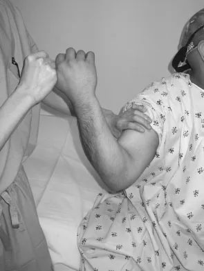
A 20-year-old college pitcher reports medial elbow pain after 3 innings of hard throwing. He recalls no injury and reports no pain with light throwing. The examination shown in the clinical photograph in Figure 48 reproduces the elbow pain. What is the most likely diagnosis?


Explanation

The milking test, as seen in the photograph, elicits pain when a tear is present in the medial collateral ligament. Complete rupture is possible but unlikely when there is no history of trauma and the patient is able to throw pain-free for several innings. Subluxation of the ulnar nerve and triceps tendon subluxation present as a painful snapping over the medial aspect of the elbow. Williams RJ III, Urquhart ER, Altchek DW: Medial collateral ligament tears in the throwing athlete. Instr Course Lect 2004;53:579-586.

Question 23

A 51-year-old woman is seen for evaluation of chronic supraspinatus and infraspinatus tendon tears. Three years ago, in an attempted repair the surgeon was unable to repair the supraspinatus and infraspinatus tendon tears. Currently she has a marked amount of pain, reduced range of motion, and weakness. Examination reveals anterosuperior escape. Radiographs show no signs of arthritic changes. You are considering a latissimus dorsi tendon transfer. During the discussion, you mention that


Explanation

Latissimus dorsi tendon transfer is considered a surgical option for treatment in patients with chronic supraspinatus and infraspinatus tendon tears. Preoperative subscapularis function is necessary for good clinical results. Additionally, men with active elevation to shoulder level and active external rotation to 20 degrees have predictably good results. Women with active shoulder elevation limited to below chest level have poor results from this procedure and should not be considered candidates. Postoperatively they lack pain control, active elevation, and active external rotation. Muscular atrophy in the latissimus dorsi does not occur, and glenohumeral arthritic changes frequently develop postoperatively. Gerber C, Maquieira G, Espinosa N: Latissimus dorsi transfer for the treatment of irreparable rotator cuff tears: Factors affecting outcome. J Bone Joint Surg Am 2006;88:113-120.

Question 24

A patient undergoes an arthroscopic debridement for lateral epicondylitis. Postoperatively she reports pain and a sense of clicking of the elbow. Examination reveals apprehension to supination, load, and extension. What structure has been injured resulting in the clinical presentation?


Explanation

The patient has an iatrogenic injury to the lateral ulnar collateral ligament following the arthroscopic procedure. Failure to adhere to known anatomic landmarks can lead to this devastating complication. The examination findings are classic for posterolateral elbow instability. Koval KJ (ed): Orthopaedic Knowledge Update 7. Rosemont, IL, American Academy of Orthopaedic Surgeons, 2002, p 318.

Question 25

A patient with refractory long head biceps pain in the shoulder undergoes biceps tenotomy. The patient is concerned about possible postoperative deformity and loss of supination strength. Which of the following techniques provides the strongest initial fixation to prevent distal migration?


Explanation

Recent articles have looked at the cyclic load failure and ultimate load failure of biceps tenodesis techniques. The interference screw has proved superior to bone tunnel, suture anchor, and soft-tissue tenodesis techniques in laboratory cadaveric testing. Whether this is clinically relevant or not is still unknown. Ozalay M, Akpinar S, Karaeminogullari O, et al: Mechanical strength of four different biceps tenodesis techniques. Arthroscopy 2005;21:992-998.

Question 26

Which of the following component design modifications or surgical techniques in reverse total shoulder arthroplasty has been shown to decrease the incidence of scapular notching?


Explanation

Scapular notching is a common mechanical complication of reverse total shoulder arthroplasty (RTSA) occurring when the medial aspect of the humeral component impinges against the inferior scapular neck during adduction. Techniques to minimize it include inferior placement of the baseplate (overhanging the inferior rim), inferior tilt of the baseplate, lateralization of the glenosphere (or center of rotation), using a larger glenosphere, and decreasing the neck-shaft angle of the humeral component (e.g., to 135 or 145 degrees).

Question 27

A 55-year-old construction worker undergoes an open subpectoral biceps tenodesis for a symptomatic SLAP tear and biceps tendinopathy. Postoperatively, he is noted to have a new-onset neurological deficit with weakness in elbow flexion and numbness over the lateral forearm. Which of the following nerves is at greatest risk of injury during the deep retractor placement for this procedure?


Explanation

The musculocutaneous nerve is at greatest risk of injury during open subpectoral biceps tenodesis, particularly with overzealous medial retraction. The nerve usually pierces the coracobrachialis 5-8 cm distal to the coracoid process and runs between the biceps and brachialis. Medial retractors (like a Hohmann retractor) placed blindly can compress or stretch this nerve.

Question 28

A 42-year-old man falls on his outstretched hand and sustains a "terrible triad" injury of the elbow. Which of the following represents the most widely accepted sequence of surgical reconstruction for this specific injury pattern?


Explanation

The standard surgical sequence for a terrible triad injury (defined as an elbow dislocation with fractures of the radial head and coronoid process) proceeds from deep to superficial. The classic algorithmic approach described by Ring and Jupiter is to first fix or reconstruct the coronoid fracture, followed by radial head fixation or arthroplasty, and finally repair of the lateral collateral ligament (LCL) complex. The medial collateral ligament (MCL) is rarely repaired unless there is gross persistent valgus instability after the lateral and anterior structures are stabilized.

Question 29

During an open carpal tunnel release, the surgeon encounters the recurrent motor branch of the median nerve piercing directly through the transverse carpal ligament. According to the Lanz classification, what type of anatomical variant does this represent?


Explanation

According to Lanz's classification of the recurrent motor branch of the median nerve, the extraligamentous type is the most common (~50-80%), where the nerve branches distal to the ligament and courses backwards. The transligamentous variant, in which the nerve pierces directly through the transverse carpal ligament, occurs in approximately 20% of individuals. This variant places the nerve at the highest risk for iatrogenic injury during a carpal tunnel release, especially in endoscopic or mini-open procedures if the ligament is divided without complete visualization.

Question 30

A 24-year-old male sustains a proximal pole scaphoid fracture. The high risk of avascular necrosis and nonunion in this fracture pattern is primarily due to the unique blood supply to the scaphoid. The predominant blood supply to the scaphoid is derived from branches of which of the following vessels?


Explanation

The scaphoid receives its primary blood supply (70-80%) from the dorsal carpal branch of the radial artery, which enters the scaphoid at the dorsal ridge (near the waist) and supplies the proximal pole in a retrograde fashion. The volar scaphoid branches (also from the radial artery) supply the distal 20-30%. Because of this retrograde flow, fractures at the waist or proximal pole disrupt the blood supply to the proximal fragment, leading to a high rate of avascular necrosis and nonunion.

Question 31

A 22-year-old rugby player presents with recurrent anterior shoulder instability. A pre-operative 3D CT scan of his shoulder reveals anterior glenoid bone loss. Historically, at which of the following percentages of inferior glenoid bone loss is an arthroscopic soft-tissue Bankart repair alone considered to have an unacceptably high failure rate, thus definitively indicating the need for a bony augmentation procedure (e.g., Latarjet)?


Explanation

Critical glenoid bone loss has traditionally been defined as >20-25% of the inferior glenoid diameter. At 25% or greater bone loss, the glenoid acts like an 'inverted pear', and a soft-tissue stabilization (Bankart repair) alone will uniformly fail due to lack of an adequate bony bumper. In these cases, a bone block procedure (such as a Latarjet coracoid transfer) is indicated. Note that recent literature has identified 'subcritical' bone loss thresholds (~13.5-15%) where outcomes may still be compromised in high-demand athletes, but 25% remains the classic absolute indication for bony augmentation.

Question 32

A 60-year-old man with a massive, retracted, chronic posterosuperior rotator cuff tear develops weakness not only in abduction and external rotation but also demonstrates electromyographic (EMG) evidence of denervation of the supraspinatus and infraspinatus. Traction on which of the following structures is most likely responsible for the suprascapular nerve injury in this specific setting?


Explanation

Massive, medially retracted tears of the supraspinatus and infraspinatus can cause a 'bowstringing' medial traction effect on the suprascapular nerve. The nerve becomes tethered at the suprascapular notch by the superior transverse scapular ligament, leading to a traction neuropathy affecting both the supraspinatus and infraspinatus. Compression at the spinoglenoid notch (under the inferior transverse scapular/spinoglenoid ligament) typically occurs secondary to paralabral cysts and isolatedly affects the infraspinatus.

Question 33

A 45-year-old woman undergoes surgical decompression of the ulnar nerve for severe cubital tunnel syndrome. During the approach, the primary structure causing compression between the olecranon and the medial epicondyle is identified and released. Which of the following anatomical structures forms the true roof of the cubital tunnel in this region?


Explanation

Osborne's ligament (also known as the cubital tunnel retinaculum) forms the roof of the cubital tunnel, spanning from the medial epicondyle to the olecranon. The floor is formed by the posterior band of the medial collateral ligament (MCL) and the joint capsule. Other potential sites of ulnar nerve compression include the Arcade of Struthers (proximal to the epicondyle), the medial intermuscular septum, and the aponeurotic heads of the FCU (distal to the tunnel).

Question 34

A 30-year-old male presents with an irreversible high radial nerve palsy following a midshaft humerus fracture sustained 18 months ago. A standard Jones tendon transfer is planned. Which of the following tendon transfers is classically utilized to restore wrist extension in this procedure?


Explanation

In a classic Jones transfer for radial nerve palsy, wrist extension is restored by transferring the pronator teres (PT) to the extensor carpi radialis brevis (ECRB). The ECRB is chosen over the ECRL because it inserts more centrally (base of the 3rd metacarpal), producing pure wrist extension and minimizing radial deviation. The rest of the classic Jones transfer includes transferring the flexor carpi ulnaris (FCU) to the extensor digitorum communis (EDC) for finger extension, and the palmaris longus (PL) to the extensor pollicis longus (EPL) for thumb extension.

Question 35

A 55-year-old female presents 6 months after a volar locking plate fixation of a distal radius fracture. She complains of suddenly losing the ability to actively flex the interphalangeal joint of her thumb. Radiographs show the fracture has healed, but the plate was placed on and slightly distal to the watershed line. Which of the following tendons is most commonly ruptured in this scenario?


Explanation

The flexor pollicis longus (FPL) tendon is at the highest risk for iatrogenic attritional rupture following volar plate fixation of distal radius fractures, particularly when the hardware is placed at or distal to the watershed line. The prominent distal edge of the plate causes friction and attritional wear of the overlying FPL tendon. Conversely, Extensor pollicis longus (EPL) ruptures are more common with non-operative management of nondisplaced distal radius fractures (due to ischemia or callus in the 3rd dorsal compartment) or from dorsally protruding screws piercing the dorsal cortex.

Question 36

In reverse total shoulder arthroplasty (RTSA), which of the following glenosphere configurations has been shown to most effectively minimize the risk of scapular notching?


Explanation

Scapular notching is a well-documented complication in RTSA resulting from mechanical impingement of the humeral component against the inferior scapular neck. Implementing an inferior tilt of the glenosphere along with lateralization increases the clearance between the humeral component and the scapular pillar during adduction, thereby significantly reducing the incidence of scapular notching.

Question 37

A 45-year-old male presents with a 'terrible triad' injury of the elbow following a fall from a height. After closed reduction, surgical management is indicated due to persistent instability. Which of the following represents the most widely accepted sequence of surgical repair for this injury pattern?


Explanation

The standard surgical protocol for a terrible triad injury involves a 'deep-to-superficial' approach working from the inside out. First, the coronoid is addressed (via fixation or anterior capsular repair), followed by the radial head (fixation or arthroplasty), and finally the lateral collateral ligament (LCL) complex is repaired. The medial collateral ligament (MCL) is typically only addressed if the elbow remains unstable after the lateral side has been stabilized.

Question 38

A 24-year-old male presents with severe radial-sided wrist pain after falling on an outstretched hand. Radiographs reveal a displaced fracture of the proximal pole of the scaphoid. The high risk of avascular necrosis in this specific fracture pattern is primarily due to the retrograde blood supply originating from branches of which of the following arteries?


Explanation

The scaphoid receives approximately 70-80% of its blood supply from the dorsal carpal branch of the radial artery, which enters the scaphoid at the dorsal ridge (distal pole) and supplies the proximal pole in a retrograde fashion. Proximal pole fractures have a notoriously high risk of nonunion and avascular necrosis because the fracture disrupts this tenuous retrograde intraosseous blood flow.

Question 39

A 38-year-old carpenter presents with an inability to flex the interphalangeal joint of the thumb and the distal interphalangeal joint of the index finger. He cannot form an 'OK' sign. Sensation in the hand and forearm is completely normal. Which of the following muscles is most likely to also demonstrate weakness on physical examination?


Explanation

The patient's presentation is classic for Anterior Interosseous Nerve (AIN) syndrome, which is characterized by paralysis of the flexor pollicis longus (FPL) and flexor digitorum profundus (FDP) to the index (and sometimes middle) finger. The AIN is a pure motor branch of the median nerve that also innervates the pronator quadratus. Weakness in resisted pronation with the elbow fully flexed (to neutralize the pronator teres) tests the pronator quadratus.

Question 40

A newborn infant is diagnosed with a brachial plexus birth palsy after a difficult forceps delivery. The child exhibits a shoulder that is internally rotated and adducted, an extended elbow, and a flexed wrist, commonly known as a 'waiter's tip' posture. Which nerve roots are predominantly injured in this classic presentation?


Explanation

Erb's palsy involves an injury to the upper trunk of the brachial plexus, specifically the C5 and C6 nerve roots. This results in the classic 'waiter's tip' deformity due to loss of shoulder abduction and external rotation (suprascapular and axillary nerves), loss of elbow flexion (musculocutaneous nerve), and weakness of wrist extensors.

Question 41

During the repair of an acute distal biceps tendon rupture using a single-incision anterior approach, the surgeon must be particularly careful to protect a specific nerve during the deep dissection and placement of retractors on the radial side of the radial tuberosity. Injury to this nerve leads to an inability to extend the digits. Which nerve is at greatest risk?


Explanation

The posterior interosseous nerve (PIN) is at significant risk during a single-incision anterior approach for distal biceps repair. The PIN wraps around the radial neck within the supinator muscle. Aggressive retraction on the radial side of the tuberosity or inadvertent plunging with a drill can injure the PIN, resulting in a loss of finger and thumb extension. The LABC nerve is at risk during the superficial approach, but its injury only causes sensory deficits.

Question 42

A 65-year-old female undergoes volar locking plate fixation for a comminuted distal radius fracture. Six weeks postoperatively, she presents with a sudden inability to flex the interphalangeal joint of her thumb. Radiographs confirm stable fracture fixation, but the plate is positioned distally, crossing the watershed line. Which of the following structures has most likely ruptured?


Explanation

A volar plate placed distal to the watershed line of the distal radius can irritate and eventually cause attrition rupture of the flexor tendons. The Flexor Pollicis Longus (FPL) tendon is the most commonly injured tendon in this scenario due to its proximity to the volar prominent hardware. Conversely, Extensor Pollicis Longus (EPL) rupture is classically associated with prominent dorsal screws or undisplaced distal radius fractures treated non-operatively.

Question 43

A 32-year-old manual laborer presents with chronic dorsal wrist pain. Radiographs demonstrate sclerosis, fragmentation, and collapse of the lunate. Additionally, there is a fixed flexed posture of the scaphoid (ring sign) and a decreased carpal height ratio. This clinical and radiographic presentation is most consistent with which Lichtman stage of Kienböck's disease?


Explanation

Lichtman Stage IIIB Kienböck's disease is characterized by lunate collapse and fragmentation accompanied by fixed scaphoid rotation (scaphoid flexion) and a decrease in carpal height ratio, indicating progressive carpal instability. Stage IIIA has lunate collapse but maintains normal carpal alignment without fixed scaphoid rotation. Stage IV includes secondary degenerative osteoarthritic changes in the radiocarpal or midcarpal joints.

Question 44

A 60-year-old male is evaluated for a massive, retracted rotator cuff tear. Preoperative MRI is obtained to assess the viability of a primary repair. According to the Goutallier classification of fatty infiltration, which stage is defined specifically by the presence of an equal amount of fat and muscle tissue within the muscle belly?


Explanation

The Goutallier classification grades fatty infiltration of the rotator cuff muscles. Stage 0 is normal muscle; Stage 1 has some fatty streaks; Stage 2 has more muscle than fat; Stage 3 has an equal amount of fat and muscle; and Stage 4 has more fat than muscle. Fatty infiltration of Stage 3 or higher generally indicates irreversible changes, carrying a poorer prognosis for successful tendon healing after repair.

Question 45

A 28-year-old skier presents with acute thumb pain and weakness of pinch after a fall with the thumb forcefully abducted. Clinical examination shows significant laxity of the thumb metacarpophalangeal (MCP) joint to valgus stress. MRI reveals a complete rupture of the ulnar collateral ligament (UCL), and the torn distal end of the ligament is displaced superficial to the adductor aponeurosis. What is the most appropriate management?


Explanation

The patient's MRI demonstrates a Stener lesion, which occurs when a completely avulsed ulnar collateral ligament (UCL) of the thumb MCP joint becomes displaced and trapped superficial to the adductor pollicis aponeurosis. This mechanical block prevents the torn ligament ends from apposing and healing. Therefore, conservative treatment is ineffective, and surgical repair is the definitive standard of care.

Question 46

A 72-year-old female undergoes an uncomplicated reverse total shoulder arthroplasty (rTSA) for massive rotator cuff tear arthropathy. Postoperatively, she demonstrates significantly improved forward elevation but exhibits an isolated, severe loss of active external rotation with the arm at the side. Preoperatively, she had a positive hornblower's sign. Deficiency in which of the following muscles is most directly responsible for this specific functional loss following an rTSA?


Explanation

In reverse total shoulder arthroplasty, the deltoid is tensioned to provide forward elevation, compensating for the absent supraspinatus. However, the teres minor remains the primary active external rotator with the arm at the side. If the teres minor is absent or non-functional (indicated preoperatively by a positive hornblower's sign or drop sign, and significant fatty infiltration on MRI), the patient will lack active external rotation postoperatively.

Question 47

A 45-year-old male laborer undergoes a two-incision approach (modified Boyd-Anderson) for the repair of an acute distal biceps tendon rupture. During the posterolateral approach (second incision) to expose the radial tuberosity, excessive retraction or improper splitting of the supinator muscle places which of the following neurovascular structures at highest risk of iatrogenic injury?


Explanation

The posterior interosseous nerve (PIN) traverses the supinator muscle and wraps around the radial neck. It is at significant risk during the posterolateral aspect of a two-incision distal biceps repair if the supinator is bluntly split or forcefully retracted. Conversely, the lateral antebrachial cutaneous nerve is most at risk during the superficial dissection of the anterior single-incision approach.

Question 48

A 28-year-old elite volleyball player presents with vague posterior shoulder pain and progressive weakness in external rotation. Examination reveals atrophy isolated to the infraspinatus fossa. MRI demonstrates a paralabral cyst in the spinoglenoid notch. Based on the site of nerve compression, which of the following findings on physical examination would also be expected?


Explanation

A cyst at the spinoglenoid notch selectively compresses the suprascapular nerve distal to its innervation of the supraspinatus muscle. Therefore, the supraspinatus remains functional, leading to normal strength in forward elevation and abduction (a negative Jobe test). The infraspinatus is denervated, resulting in isolated external rotation weakness and atrophy. There are no sensory deficits associated with isolated suprascapular nerve entrapment.

Question 49

A 32-year-old carpenter presents with progressive, activity-related dorsal wrist pain. Radiographs reveal Kienböck's disease (Lichtman Stage IIIA) with sclerosis and early fragmentation of the lunate, and a notable negative ulnar variance. There is no evidence of radioscaphoid arthritis. Which of the following surgical interventions is most appropriate to unload the lunate and halt disease progression?


Explanation

In early to moderate stages of Kienböck's disease (Stage II or IIIA) associated with negative ulnar variance (ulna minus) and preserved carpal cartilage, joint-leveling procedures are indicated. A radial shortening osteotomy unloads the radiolunate joint and redistributes forces across the wrist. Ulnar lengthening is an alternative but carries higher complication rates (e.g., nonunion). Ulnar shortening would exacerbate the negative variance.

Question 50

A 21-year-old collegiate baseball pitcher reports medial elbow pain during the late cocking phase of throwing. Magnetic resonance arthrography confirms a full-thickness tear of the anterior bundle of the ulnar collateral ligament (UCL). Which of the following biomechanical principles best describes the primary restraint provided by this specific structure?


Explanation

The anterior bundle of the ulnar collateral ligament (UCL) is the primary static restraint to valgus stress at the elbow between 30 and 120 degrees of flexion. In full extension (0 to 30 degrees), the bony articulation (olecranon engaging in the olecranon fossa) and the anterior joint capsule provide a significant portion of valgus stability.

Question 51

A 24-year-old male sustains an acute, non-displaced fracture of the proximal pole of the scaphoid after a fall. What is the primary anatomic and pathophysiological rationale for recommending surgical fixation with a headless compression screw over prolonged cast immobilization in this specific scenario?


Explanation

The scaphoid receives its primary blood supply from branches of the radial artery that enter distally and flow in a retrograde fashion. Fractures of the proximal pole profoundly disrupt this blood supply, resulting in a high risk of avascular necrosis and nonunion. Surgical fixation is highly recommended even for non-displaced proximal pole fractures to achieve rigid stability and optimize the chances of union.

Question 52

A 48-year-old female sustains a fall from a height, resulting in a complex elbow injury requiring urgent orthopedic intervention. The diagnosis of a 'terrible triad' injury of the elbow is established. Which of the following combinations of injuries strictly defines this classic clinical entity?


Explanation

The 'terrible triad' of the elbow describes a specific and highly unstable injury pattern consisting of an elbow dislocation (typically posterior or posterolateral), a radial head fracture, and a coronoid fracture. It represents a sequential failure of the primary and secondary stabilizers of the elbow, often requiring operative repair of the lateral collateral ligament complex and restoration of the radial head and coronoid to permit early range of motion.

Question 53

A 55-year-old male undergoes an in situ decompression of the ulnar nerve for severe cubital tunnel syndrome. During the release of Osborne's fascia, the surgeon identifies and meticulously protects the first motor branch of the ulnar nerve in the forearm. Which of the following muscles is innervated by this specific branch?


Explanation

The first motor branch of the ulnar nerve in the forearm typically innervates the flexor carpi ulnaris (FCU) muscle. It branches off the main ulnar nerve just distal to the medial epicondyle as the nerve passes between the humeral and ulnar heads of the FCU (Osborne's fascia). Iatrogenic injury to this branch during decompression or transposition can lead to functional deficits.

Question 54

A 30-year-old chef sustains a deep volar laceration to his left index finger at the level of the proximal phalanx (Zone II), cleanly transecting the flexor tendons. He is indicated for primary flexor tendon repair. To safely permit an early active motion rehabilitation protocol, which of the following variables is most directly correlated with the ultimate tensile strength of the repair?


Explanation

Biomechanical studies have consistently proven that the ultimate tensile strength of a flexor tendon repair is directly proportional to the number of core suture strands crossing the repair site. Modern protocols relying on early active motion generally require at least a 4-strand or 6-strand repair to prevent gap formation and subsequent rupture. While an epitendinous suture adds strength and minimizes gliding resistance, the core strand count is the primary determinant of strength.

Question 55

A 35-year-old female is diagnosed with neurogenic thoracic outlet syndrome characterized by chronic upper extremity paresthesias and weakness exacerbated by overhead activity. She has failed six months of conservative management. Surgical decompression is planned. What is the most widely accepted and definitive surgical intervention for this condition?


Explanation

Neurogenic thoracic outlet syndrome is most frequently caused by compression of the brachial plexus within the scalene triangle, which is bordered by the anterior scalene, the middle scalene, and the first rib. When surgical intervention is required, decompression classically involves resection of the first rib combined with an anterior scalenectomy (and often a middle scalenectomy) to effectively widen the thoracic outlet and relieve neurovascular compression.

Question 56

Scapular notching is a common complication following reverse total shoulder arthroplasty (RTSA). Which of the following component positioning strategies is most effective in minimizing the risk of inferior scapular notching?


Explanation

Scapular notching is a well-known complication of RTSA, caused by mechanical impingement of the humeral component against the inferior scapular neck during adduction. Placing the glenosphere more inferiorly (overhanging the inferior glenoid bone by 2-4 mm) and with an inferior tilt can help to reduce this mechanical conflict. Lateralizing the center of rotation also decreases notching. Increasing the humeral neck-shaft angle to 155 degrees (more valgus) actually increases the risk of notching compared to an angle of 135 degrees.

Question 57

The medial ulnar collateral ligament (MUCL) of the elbow consists of anterior, posterior, and transverse bundles. During the late cocking and early acceleration phases of throwing, which specific structure is the primary restraint to valgus stress?


Explanation

The anterior bundle of the MUCL is the primary restraint to valgus stress at the elbow. It is further subdivided into anterior and posterior bands. The anterior band of the anterior bundle is tight in extension and is the primary restraint to valgus stress up to 90 degrees of flexion, which covers the late cocking and early acceleration phases of throwing. The posterior band is tight in flexion (greater than 90 degrees). The radiocapitellar joint acts as a secondary restraint.

Question 58

A 28-year-old elite volleyball player presents with insidious onset of right shoulder pain and weakness, predominantly with external rotation. Examination reveals isolated atrophy of the infraspinatus muscle. The supraspinatus muscle bulk and strength are normal. Where is the most likely location of the nerve entrapment?


Explanation

The suprascapular nerve innervates the supraspinatus and infraspinatus muscles. It courses through the suprascapular notch (beneath the transverse scapular ligament), where compression affects both the supraspinatus and infraspinatus. It then passes through the spinoglenoid notch (under the spinoglenoid ligament) to innervate the infraspinatus. Compression at the spinoglenoid notch, often due to a paralabral cyst associated with a posterior SLAP or labral tear, typically results in isolated infraspinatus weakness and atrophy, while the supraspinatus is spared. This is a classic presentation in overhead athletes, particularly volleyball players.

Question 59

The primary blood supply to the articular segment of the humeral head in an adult is predominantly provided by which of the following vessels?


Explanation

Historically, the anterior humeral circumflex artery (AHCA), specifically its anterolateral ascending branch (arcuate artery), was thought to provide the main blood supply to the humeral head. However, more recent anatomic and perfusion studies (e.g., Brooks et al., Hettrich et al.) have demonstrated that the posterior humeral circumflex artery (PHCA) provides the majority (up to 64%) of the blood supply to the humeral head. This has important implications in proximal humerus fracture surgery and predicting avascular necrosis.

Question 60

In the setting of a primary linked semi-constrained total elbow arthroplasty (TEA) performed for rheumatoid arthritis, which of the following is the most common long-term complication?


Explanation

The most common long-term complication of total elbow arthroplasty, particularly in linked semi-constrained designs, is aseptic loosening. While ulnar neuropathy, infection, triceps insufficiency, and periprosthetic fractures are important complications, aseptic loosening has the highest incidence over time due to the high mechanical stresses across the implant interfaces. Bushing wear can also contribute to osteolysis and subsequent aseptic loosening.

Question 61

A 25-year-old man presents with chronic wrist pain and is diagnosed with a scaphoid waist fracture nonunion with a humpback deformity. Radiographs demonstrate a dorsal intercalated segment instability (DISI) pattern and a shortened scaphoid. There is no evidence of avascular necrosis or arthritis. Which of the following is the most appropriate surgical management?


Explanation

The patient has a scaphoid waist fracture nonunion with a humpback deformity (flexion of the scaphoid leading to a shortened volar length) and a secondary DISI deformity. Correction of the deformity is crucial to restore carpal kinematics and prevent progression to SNAC (scaphoid nonunion advanced collapse) arthritis. The standard of care for a humpback scaphoid nonunion without AVN is a volar approach, using a structural interposition wedge bone graft (typically from the iliac crest) to restore scaphoid length and correct the deformity, followed by rigid internal fixation. Percutaneous fixation without grafting will not address the deformity. Vascularized bone grafting is typically reserved for nonunions with avascular necrosis (proximal pole). Salvage procedures (four-corner fusion, PRC) are indicated if advanced arthritic changes (SNAC) are present.

Question 62

A 35-year-old male sustains a "terrible triad" injury to his left elbow following a fall from a height. The standard surgical sequence for reconstruction typically involves which of the following steps?


Explanation

The "terrible triad" of the elbow includes an elbow dislocation, a radial head fracture, and a coronoid process fracture. The classic surgical treatment algorithm, as described by Pugh and McKee, proceeds from deep to superficial and typically from medial to lateral (when approaching from the lateral side). The standard sequence is: 1) Fixation of the coronoid fracture (to restore the anterior buttress), 2) Fixation or replacement of the radial head (to restore the anterior and valgus buttress), 3) Repair of the lateral collateral ligament complex (specifically the LUCL, to restore posterolateral rotatory stability). Repair of the medial collateral ligament (MCL) or application of a hinged external fixator is reserved for cases where the elbow remains unstable after the first three steps are completed.

Question 63

A 42-year-old male bodybuilder feels a "pop" in his anterior elbow while performing heavy deadlifts. He presents with weakness in forearm supination and elbow flexion. If surgical repair is performed using a two-incision technique rather than a single anterior incision, which of the following complications is more significantly increased?


Explanation

Surgical repair of distal biceps tendon ruptures can be performed via a single anterior incision or a two-incision technique (anterior and posterolateral). The two-incision technique was historically developed to reduce the risk of radial nerve injury associated with the extensile single anterior incision. However, the two-incision technique carries a significantly higher risk of heterotopic ossification and proximal radioulnar synostosis, which can severely limit forearm rotation. Injury to the lateral antebrachial cutaneous nerve (LABCN) is the most common neurologic complication overall, but it is typically associated with the anterior approach. The radial nerve is more at risk with a single anterior incision, particularly if retractors are placed aggressively.

Question 64

According to the Snyder classification of Superior Labrum Anterior to Posterior (SLAP) lesions, a Type II tear is characterized by:


Explanation

The Snyder classification categorizes SLAP lesions into four initial types: Type I: Degenerative fraying of the superior labrum; the biceps anchor is intact. Type II: Detachment of the superior labrum and the origin of the long head of the biceps tendon from the superior glenoid rim. This is the most common type and often requires surgical repair or biceps tenodesis. Type III: A bucket-handle tear of the superior labrum with an intact biceps anchor. Type IV: A bucket-handle tear of the superior labrum that extends into the substance of the biceps tendon.

Question 65

In the evaluation of a patient with Kienböck's disease (avascular necrosis of the lunate), negative ulnar variance is often observed. Which of the following biomechanical effects does negative ulnar variance have on the wrist joint?


Explanation

Ulnar variance refers to the relative lengths of the distal articular surfaces of the radius and ulna. Negative ulnar variance means the ulna is shorter than the radius. Biomechanically, negative ulnar variance decreases the load transmitted through the ulnocarpal joint and correspondingly increases the load transmitted across the radiocarpal joint, specifically the radiolunate articulation. This increased mechanical stress on the lunate is thought to be a contributing factor to the pathogenesis of Kienböck's disease, as it may lead to microtrauma and compromise of the lunate's precarious blood supply. Joint leveling procedures (e.g., radial shortening osteotomy) aim to decrease this radiolunate load by restoring neutral variance.

Question 66

A 45-year-old male presents after a fall on an outstretched hand, sustaining a 'terrible triad' injury of the elbow. Which of the following best describes the appropriate surgical sequence and principles to restore joint stability?


Explanation

The standard surgical protocol for a terrible triad injury (coronoid fracture, radial head fracture, and elbow dislocation resulting in a LUCL tear) follows an inside-out or deep-to-superficial approach. The sequence typically involves: 1) fixing the coronoid process, 2) fixing or replacing the radial head, and 3) repairing the LUCL to the lateral epicondyle. If the elbow remains unstable after these steps, the MCL is repaired or a hinged external fixator is placed.

Question 67

A 35-year-old manual laborer presents with chronic progressive wrist pain 5 years after an untreated fall on his outstretched hand. Radiographs reveal a scaphoid nonunion with advanced radioscaphoid and capitolunate arthritis, but the radiolunate joint is spared. What is the most appropriate surgical management to provide pain relief while preserving motion?


Explanation

This patient has a Scaphoid Nonunion Advanced Collapse (SNAC) stage III wrist (characterized by radioscaphoid and capitolunate arthritis, with a preserved radiolunate joint). A proximal row carpectomy (PRC) is contraindicated in SNAC III because the capitate articular surface is degenerated, which would articulate with the lunate fossa in a PRC. Therefore, scaphoid excision and a four-corner fusion (capitate, hamate, lunate, triquetrum) is the preferred motion-sparing procedure.

Question 68

A 42-year-old male bodybuilder undergoes a single-incision anterior approach for a distal biceps tendon rupture repair using cortical button fixation. Postoperatively, he is noted to have weakness in thumb and finger extension, but normal wrist extension with radial deviation. Which of the following nerves was most likely injured during the procedure?


Explanation

The posterior interosseous nerve (PIN) is at risk during the anterior single-incision approach to the distal biceps, particularly if retractors are placed too vigorously on the lateral side or during drilling of the radius. Injury to the PIN results in weakness of finger and thumb extensors and the extensor carpi ulnaris. Wrist extension is preserved but deviates radially because the extensor carpi radialis longus and brevis are innervated by the radial nerve proper, which branches proximal to the PIN.

Question 69

During an in situ ulnar nerve decompression for cubital tunnel syndrome, the surgeon releases the nerve from the cubital tunnel. To prevent proximal entrapment, the surgeon explores the medial arm. Which fascial structure represents a potential site of ulnar nerve compression up to 8 cm proximal to the medial epicondyle?


Explanation

The Arcade of Struthers is a fascial band extending from the medial head of the triceps to the medial intermuscular septum, located approximately 8 cm proximal to the medial epicondyle. It is a known potential site of ulnar nerve compression, particularly if the nerve is transposed anteriorly and this arcade is not adequately released. In contrast, the Ligament of Struthers is associated with median nerve compression, and Osborne's ligament forms the roof of the cubital tunnel itself.

Question 70

A 65-year-old female sustains a displaced 3-part proximal humerus fracture. Based on quantitative anatomical studies, which of the following arterial structures provides the predominant blood supply to the humeral head and is most at risk in anatomic neck fractures?


Explanation

Recent anatomical studies (e.g., Hettrich et al.) have demonstrated that the posterior humeral circumflex artery provides the predominant blood supply to the humeral head (approximately 64% of the volume). This finding shifts the traditional paradigm, which historically emphasized the anterolateral branch (arcuate artery) of the anterior humeral circumflex artery. Disruption of this primary blood supply significantly increases the risk of avascular necrosis.

Question 71

A 45-year-old male construction worker presents with deep, aching shoulder pain and a positive O'Brien test. An MRI arthrogram reveals a type II SLAP tear. Nonoperative management has failed. Based on recent literature, what is the most appropriate surgical intervention to minimize postoperative stiffness and allow a predictable return to work?


Explanation

In patients older than 35-40 years with a type II SLAP tear, particularly manual laborers, primary biceps tenodesis has been shown to have lower complication rates, a lower incidence of postoperative stiffness, and higher rates of return to work compared to SLAP repair. SLAP repair in this demographic is associated with higher rates of persistent pain and stiffness.

Question 72

A 72-year-old female presents with chronic right shoulder pain and an inability to actively elevate her arm above 60 degrees. Passive range of motion is full. Radiographs show a Hamada Grade 3 arthropathy (acromiohumeral interval < 7mm with acetabularization of the acromion). MRI reveals a massive, retracted, and fatty-infiltrated tear of the supraspinatus and infraspinatus. What is the most reliable surgical option to restore active elevation?


Explanation

This patient presents with pseudoparalysis due to a massive, irreparable rotator cuff tear and concurrent rotator cuff arthropathy (Hamada grade 3). Reverse total shoulder arthroplasty (RTSA) is the most reliable treatment to restore forward elevation in this scenario. RTSA medializes and distalizes the center of rotation, recruiting the deltoid to act as the primary elevator. Superior capsular reconstruction and tendon transfers are generally contraindicated in the setting of significant glenohumeral arthritis and established pseudoparalysis.

Question 73

A 55-year-old woman undergoes volar locked plating for a displaced distal radius fracture. At her 6-week postoperative visit, she reports a sudden inability to actively flex her thumb interphalangeal joint. Radiographs show a healed fracture with the plate positioned distally, volar to the watershed line. Which of the following tendons was most likely injured?


Explanation

The flexor pollicis longus (FPL) tendon is the most commonly ruptured flexor tendon following volar plating of distal radius fractures. The mechanism is typically attrition and rupture due to the plate being placed too distally, projecting volar to the 'watershed line' where the FPL tendon is in intimate contact with the radius. Extensor pollicis longus (EPL) rupture occurs due to dorsal screw prominence, but presents with loss of thumb extension, not flexion.

Question 74

A 28-year-old female overhead athlete complains of numbness and tingling in her medial forearm and hand that worsens with overhead activity. The Adson test is positive, and EMG confirms delayed conduction across the brachial plexus. If the neurovascular compression is occurring in the primary anatomic space implicated in neurogenic thoracic outlet syndrome, what are its boundaries?


Explanation

The patient has symptoms of neurogenic thoracic outlet syndrome (nTOS). The most common site of compression in nTOS is the scalene triangle. The borders of the interscalene triangle are the anterior scalene muscle (anterior), the middle scalene muscle (posterior), and the first rib (inferiorly). The brachial plexus roots/trunks and the subclavian artery pass through this interval, whereas the subclavian vein passes anterior to the anterior scalene.

Question 75

A 22-year-old collegiate baseball pitcher presents with medial elbow pain and decreased pitching velocity. An MRI arthrogram demonstrates a full-thickness tear of the anterior bundle of the ulnar collateral ligament (UCL). During a Tommy John reconstruction, which specific anatomic footprint on the ulna must be restored to ensure normal kinematics and valgus stability?


Explanation

The anterior bundle of the ulnar collateral ligament (UCL) is the primary restraint to valgus stress at the elbow. Its anatomic distal footprint is located on the sublime tubercle, which is situated on the medial aspect of the coronoid process of the ulna. Accurately restoring this footprint during UCL reconstruction is crucial to re-establishing normal elbow kinematics and preventing recurrent valgus instability.

Question 76

A 35-year-old man presents after a high-speed motorcycle accident with a completely flail left upper extremity and a massively swollen shoulder. Radiographs show lateral displacement of the scapula and a widely displaced midshaft clavicle fracture. What vascular injury is most commonly associated with this specific pattern of injury?


Explanation

The clinical picture describes scapulothoracic dissociation, a severe, high-energy closed injury involving traumatic disruption of the scapulothoracic articulation. It is characterized by lateral displacement of the scapula, clavicle fracture or acromioclavicular/sternoclavicular joint disruption, and severe soft tissue injury. The subclavian artery and the brachial plexus are the most commonly injured neurovascular structures, often leading to a flail and ischemic limb. Amputation is frequently required due to the severity of the complete brachial plexus avulsion and vascular disruption.

Question 77

A 65-year-old female underwent an anatomic total shoulder arthroplasty (TSA) 6 weeks ago utilizing a standard deltopectoral approach with subscapularis peel and repair. She now complains of increased pain and profound weakness with internal rotation. On examination, she has a positive bear-hug test and increased passive external rotation compared to the contralateral side. What is the most appropriate next step in management?


Explanation

The patient's presentation of new-onset internal rotation weakness, a positive bear-hug test, and increased passive external rotation 6 weeks post-TSA is classic for acute subscapularis failure. Early recognition is critical. When diagnosed in the acute or subacute postoperative period, primary revision surgery to repair the subscapularis tendon is indicated to restore function and prevent secondary anterior instability of the prosthesis. If the condition is chronic and the subscapularis is irreparable or the patient has developed anterior escape, conversion to a reverse TSA may be required.

Question 78

An 81-year-old, functionally independent female with severe osteoporosis sustains a 4-part proximal humerus fracture with a 'head-splitting' component following a mechanical fall. She reports excruciating pain. What surgical option provides the most predictable restoration of forward elevation and overall functional outcome in this specific patient demographic?


Explanation

In elderly patients with severe osteopenia and complex, unreconstructible proximal humerus fractures (e.g., 4-part fractures, head-splitting components), reverse total shoulder arthroplasty (RTSA) has been shown to provide more predictable pain relief and restoration of forward elevation compared to hemiarthroplasty or ORIF. Hemiarthroplasty outcomes rely heavily on the anatomic healing of the tuberosities, which is notoriously unreliable in osteoporotic bone. RTSA bypasses the reliance on tuberosity healing to restore overhead function by utilizing the deltoid.

Question 79

A 45-year-old male presents with persistent medial elbow pain, constant numbness in the ring and small fingers, and intrinsic muscle weakness 12 months after an in situ ulnar nerve decompression at the cubital tunnel. Postoperative EMG/NCS confirms persistent, severe ulnar neuropathy localized to the elbow. Which of the following revision procedures is most appropriate?


Explanation

For failed primary in situ decompression of the ulnar nerve, revision surgery typically involves transposition of the nerve to move it out of the scarred bed. Submuscular transposition is widely favored in revision settings because it places the previously mobilized and potentially ischemic nerve into an unscarred, well-vascularized muscle bed, reducing the risk of recurrent perineural fibrosis and providing maximum protection. While subcutaneous transposition is an option, submuscular is generally preferred for revisions.

Question 80

A 28-year-old elite volleyball player presents with vague posterior shoulder pain and progressive weakness with serving. Physical examination reveals marked atrophy of both the supraspinatus and infraspinatus muscles. At what anatomical location is the neurological compression most likely occurring?


Explanation

The patient is exhibiting signs of suprascapular nerve entrapment. The suprascapular nerve innervates both the supraspinatus and infraspinatus muscles. Compression at the suprascapular notch (beneath the transverse scapular ligament) affects the nerve before it gives off branches to the supraspinatus, resulting in atrophy and weakness of BOTH the supraspinatus and infraspinatus. Conversely, compression at the spinoglenoid notch (distal to the supraspinatus innervation) typically presents with isolated infraspinatus atrophy.

Question 81

A 32-year-old male presents with profound elbow stiffness 5 months after surgical management of a terrible triad injury (radial head arthroplasty, LCL repair, coronoid fixation). Radiographs demonstrate heterotopic ossification (HO) bridging the radiocapitellar joint with mature, sharp cortical margins and distinct trabecular patterns. His clinical range of motion has plateaued despite aggressive therapy. What is the most appropriate management regarding surgical excision?


Explanation

Historically, surgical excision of heterotopic ossification (HO) was delayed until 12-18 months when bone scans showed metabolic quiescence. However, contemporary evidence demonstrates that early excision (typically between 4 to 6 months) is safe and effective once the HO is radiographically mature (showing distinct trabeculae and sharp margins) and the patient's clinical range of motion has plateaued. Early excision allows for earlier rehabilitation and does not have an increased rate of recurrence compared to delayed excision, provided proper prophylaxis (NSAIDs or radiation) is used postoperatively.

Question 82

A 42-year-old male undergoes a single-incision anterior approach for the repair of an acute distal biceps tendon rupture using a cortical button technique. Postoperatively, he is unable to actively extend his thumb and fingers. However, he is able to extend his wrist, though it deviates radially during the movement. Which nerve has been injured, and what is the most common iatrogenic mechanism?


Explanation

The clinical presentation describes a posterior interosseous nerve (PIN) palsy. The PIN is the deep motor branch of the radial nerve. Injury results in the inability to extend the digits and thumb. Wrist extension is preserved because the extensor carpi radialis longus (ECRL) is innervated by the radial nerve proper proximal to the bifurcation; however, because the extensor carpi ulnaris (ECU) is innervated by the PIN, wrist extension occurs with unopposed radial deviation. The PIN is particularly at risk during the single-incision approach to the distal biceps when retractors (such as Hohmanns) are placed blindly or aggressively around the radial neck.

Question 83

A 30-year-old heavy laborer presents with central dorsal wrist pain. Radiographs demonstrate sclerosis of the lunate with no evidence of collapse or fragmentation. He has an ulnar variance of -3 mm. MRI confirms decreased T1 signal intensity throughout the entire lunate. What is the most appropriate surgical treatment?


Explanation

This patient has Lichtman Stage II Kienböck's disease, characterized by sclerosis of the lunate on plain radiographs without lunate collapse. In the presence of ulnar negative variance, joint leveling procedures, such as a radial shortening osteotomy, are the treatment of choice. This mechanically unloads the radiolunate joint, decreasing the stress on the lunate and potentially allowing for revascularization and halting disease progression. Proximal row carpectomy or fusions are salvage procedures reserved for more advanced stages (Stage III or IV) with carpal collapse or secondary osteoarthritis.

Question 84

A 25-year-old hockey player sustains a direct blow to the superior aspect of his right shoulder. Clinical examination reveals marked prominence of the distal clavicle. Radiographs confirm a Type V acromioclavicular (AC) joint separation, with the clavicle displaced superiorly by 200% relative to the acromion. Which anatomical structure represents the primary static restraint to superior translation of the distal clavicle?


Explanation

The coracoclavicular (CC) ligaments, consisting of the conoid and trapezoid ligaments, act as the primary static restraints to superior and inferior translation of the clavicle relative to the scapula. The acromioclavicular (AC) ligaments are the primary restraints to anteroposterior translation. A Type V AC separation involves complete disruption of both the AC and CC ligaments, along with tearing of the deltotrapezial fascia, leading to severe superior displacement of the clavicle (100% to 300% of the contralateral side).

Question 85

A 21-year-old collegiate baseball pitcher presents with chronic medial elbow pain that is worse during the late cocking and early acceleration phases of throwing. MRI demonstrates a high-grade partial tear of the ulnar collateral ligament (UCL). He has failed 4 months of conservative management and is opting for reconstruction. Which specific structure provides the primary restraint to valgus stress at the elbow between 30 and 120 degrees of flexion?


Explanation

The anterior bundle of the ulnar collateral ligament (UCL) is the primary static stabilizer to valgus stress at the elbow, particularly functioning between 30 and 120 degrees of flexion. It originates on the anteroinferior surface of the medial epicondyle and inserts on the sublime tubercle of the ulna. The posterior bundle is a secondary restraint that is tightest in full flexion. The flexor-pronator mass provides secondary dynamic stability to valgus stress.

Question 86

According to Hertel's criteria, which of the following combinations of radiographic findings following a proximal humerus fracture most accurately predicts a high probability of humeral head ischemia?


Explanation

Hertel et al. established criteria to predict the likelihood of ischemia in the humeral head following proximal humerus fractures. The triad most predictive of ischemia includes a fracture line passing through the anatomical neck, a short calcar (metaphyseal) segment of less than 8 mm attached to the articular segment, and a disrupted medial hinge (loss of medial cortical contact > 2 mm). When all three of these criteria are present, the positive predictive value for ischemia reaches 97%.

Question 87

A 40-year-old man falls on an outstretched hand and sustains a 'terrible triad' injury of the elbow. Which of the following correctly outlines the standard surgical sequence to optimally restore elbow stability?


Explanation

The classic surgical sequence for a terrible triad injury of the elbow (elbow dislocation, radial head fracture, coronoid fracture) involves a deep-to-superficial approach. First, the coronoid is fixed to restore the anterior bony buttress. Next, the radial head is either internally fixed or replaced to restore the anterior and primary valgus buttress. Finally, the lateral collateral ligament (LCL) complex is repaired to the lateral epicondyle to restore posterolateral rotatory stability. Medial collateral ligament (MCL) repair or hinged external fixation is only considered if the elbow remains unstable after these steps are completed.

Question 88

A 42-year-old male weightlifter feels a pop in his anterior elbow while performing a heavy deadlift. Clinical examination reveals a positive Hook test. If surgical repair of the ruptured distal biceps tendon is performed via a single anterior incision technique, which of the following nerves is at the highest risk of iatrogenic injury?


Explanation

The lateral antebrachial cutaneous nerve (LABCN) is the most commonly injured nerve during a single-incision anterior approach for distal biceps tendon repair, with reported injury or neuropraxia rates ranging from 10% to 30%. The LABCN exits the deep fascia just lateral to the biceps tendon in the antecubital fossa. While the posterior interosseous nerve (PIN) is also at risk (particularly with errant placement of retractors around the radial neck), LABCN injury is much more frequent. A two-incision approach classically reduced the risk of PIN injury but historically carried a higher risk of radioulnar synostosis.

Question 89

A 68-year-old woman is treated nonoperatively in a cast for a non-displaced distal radius fracture. Four weeks later, she suddenly becomes unable to actively extend her thumb interphalangeal joint. Rupture of which of the following tendons is the most likely cause, and at which anatomical pulley does this occur?


Explanation

Extensor pollicis longus (EPL) tendon rupture is a well-known complication of distal radius fractures, occurring most frequently in non-displaced fractures treated nonoperatively. The EPL tendon travels through the third dorsal compartment and takes a sharp 45-degree angle turn around Lister's tubercle, creating a mechanical fulcrum. Swelling within the intact extensor retinaculum or callus formation can lead to vascular compromise (watershed area), attrition, and subsequent rupture of the tendon. Treatment typically involves an Extensor Indicis Proprius (EIP) to EPL tendon transfer.

Question 90

A 72-year-old man undergoes a reverse total shoulder arthroplasty (RTSA) for massive rotator cuff tear arthropathy. Biomechanically, how does this prosthesis alter the center of rotation of the glenohumeral joint to improve active elevation?


Explanation

The reverse total shoulder arthroplasty (RTSA) is biomechanically designed by Grammont to shift the center of rotation of the glenohumeral joint medially and inferiorly. Medialization recruits more deltoid muscle fibers (particularly the anterior and posterior heads) for elevation by altering their line of pull. Inferiorization significantly tensions the deltoid, thereby increasing its moment arm and mechanical advantage. This design allows the deltoid to effectively compensate for the deficient rotator cuff to initiate and maintain active shoulder elevation.

Question 91

During surgical decompression of the ulnar nerve for cubital tunnel syndrome, multiple potential sites of compression must be evaluated. Which of the following anatomical structures represents the most common site of ulnar nerve entrapment in this region?


Explanation

Cubital tunnel syndrome is the second most common compression neuropathy of the upper extremity. The ulnar nerve can be compressed at multiple sites around the elbow. The most frequent site of compression is between the two heads of the flexor carpi ulnaris (FCU), which are connected by the arcuate ligament of Osborne (Osborne's fascia). Other potential compression sites include the arcade of Struthers (hiatus in the medial intermuscular septum), the medial epicondyle, and the deep flexor-pronator aponeurosis, all of which should be assessed during a thorough decompression or transposition.

Question 92

A 6-year-old boy presents with a completely displaced, extension-type supracondylar humerus fracture (Gartland Type III). He exhibits weakness with active flexion of the thumb interphalangeal joint and the distal interphalangeal joint of the index finger. Which of the following nerve injuries is most likely present?


Explanation

The anterior interosseous nerve (AIN), a motor branch of the median nerve, is the most commonly injured nerve in extension-type pediatric supracondylar humerus fractures. The AIN innervates the flexor pollicis longus (FPL), the lateral half of the flexor digitorum profundus (FDP), and the pronator quadratus. An AIN neuropraxia results in the inability to actively flex the thumb interphalangeal (IP) joint and the index finger distal interphalangeal (DIP) joint, classically leading to an abnormal 'OK' sign (creating a pinch rather than an O). The radial nerve is the second most commonly injured nerve, often seen with posteromedial fracture displacement.

Question 93

A 22-year-old man falls on an outstretched hand and presents with anatomic snuffbox tenderness. Radiographs show a non-displaced fracture through the proximal pole of the scaphoid. What is the most appropriate management strategy and its primary rationale?


Explanation

The scaphoid receives its primary blood supply from branches of the radial artery entering at the dorsal ridge near the waist, resulting in a retrograde blood flow to the proximal pole. Because of this tenuous blood supply, fractures of the proximal pole are at a significantly high risk for nonunion and avascular necrosis. Even when completely non-displaced, surgical fixation (typically via a dorsal percutaneous or mini-open approach) is the recommended management for proximal pole scaphoid fractures to optimize the chances of union and allow for earlier mobilization.

Question 94

A 30-year-old male presents with chronic dorsal wrist pain after a fall six months ago. Posteroanterior radiographs demonstrate a widened scapholunate interval (Terry Thomas sign) and a cortical 'ring sign' of the scaphoid. On the lateral radiographic view, what is the expected carpal alignment?


Explanation

A widened scapholunate interval and a scaphoid 'ring sign' (representing an abnormally flexed scaphoid viewed end-on) are indicative of a scapholunate ligament tear. Biomechanically, the scaphoid inherently tends to flex, while the triquetrum inherently tends to extend. The lunate is caught in between. When the primary constraint (the scapholunate interosseous ligament) is disrupted, the scaphoid flexes volarly, while the lunate extends dorsally alongside the triquetrum (since the lunotriquetral ligament remains intact). This resultant dorsal tilting of the lunate relative to the capitate and radius is known as a Dorsal Intercalated Segment Instability (DISI) deformity.

Question 95

A 28-year-old avid skier catches his thumb in a ski pole strap, sustaining a severe hyperabduction injury to the metacarpophalangeal (MCP) joint of the thumb. Physical examination reveals gross instability to valgus stress. Which of the following anatomical findings defines a Stener lesion, an absolute indication for operative repair?


Explanation

A Stener lesion occurs when the ulnar collateral ligament (UCL) of the thumb MCP joint completely ruptures (usually from its distal insertion on the proximal phalanx) and the proximal stump of the torn ligament displaces superficial to the adductor pollicis aponeurosis. Consequently, the aponeurosis becomes interposed between the torn UCL and its anatomical insertion site. Because the ligament is physically blocked from reaching its insertion, natural healing is prevented, making a Stener lesion a definitive indication for surgical repair of the UCL (Gamekeeper's or Skier's thumb).

Question 96

A 72-year-old female undergoes a reverse total shoulder arthroplasty (RTSA) for massive rotator cuff tear arthropathy. At her 2-year follow-up, she complains of mild pain, and radiographs demonstrate scapular notching. Which of the following surgical technique modifications or implant choices at the time of the index procedure would have DECREASED her risk of developing this complication?


Explanation

Scapular notching is a common radiographic finding after reverse total shoulder arthroplasty (RTSA), occurring when the medial edge of the humeral cup abuts the inferior scapular neck during arm adduction. Factors that decrease the risk of scapular notching include inferior placement of the glenosphere (overhanging the inferior glenoid rim), inferior tilt of the baseplate, lateralization of the glenosphere (increased lateral offset), and the use of a larger diameter glenosphere. Additionally, using a humeral component with a lower neck-shaft angle (e.g., 135 degrees) helps minimize impingement compared to higher neck-shaft angles (e.g., 155 degrees). Superior placement, medialized designs, and smaller glenospheres all increase the risk of notching.

Question 97

A 45-year-old man falls on an outstretched hand and sustains a 'terrible triad' injury to his elbow. Intraoperatively, the coronoid fracture is fixed with a suture lasso technique, and the radial head fracture undergoes open reduction and internal fixation. Following this, the lateral ulnar collateral ligament (LUCL) is anatomically repaired to the lateral epicondyle. During examination under anesthesia, the elbow still subluxates posteriorly at 30 degrees of flexion. What is the most appropriate next step in management?


Explanation

The standard surgical algorithm for a terrible triad injury of the elbow (elbow dislocation, radial head fracture, and coronoid fracture) involves a deep-to-superficial repair from the inside out. The coronoid is addressed first, followed by the radial head (fixation or arthroplasty), and finally the lateral ligamentous complex (LUCL). If the elbow remains unstable after these steps (specifically, if it dislocates or subluxates in extension or early flexion, typically <30-40 degrees), the medial collateral ligament (MCL) should be explored and repaired. If concentric stability is still not achieved after MCL repair, a hinged elbow external fixator should be applied. Prolonged immobilization would lead to severe stiffness and is generally avoided.

Question 98

A 62-year-old female presents to the clinic complaining of an inability to actively flex the tip of her thumb. Nine months ago, she underwent open reduction and internal fixation of a distal radius fracture with a volar locking plate. On physical examination, she has a full passive range of motion of the thumb interphalangeal (IP) joint but lacks active flexion. Radiographs demonstrate the volar plate is positioned distal to the watershed line. Which of the following is the most appropriate definitive management for her current condition?


Explanation

The patient has sustained an attritional rupture of the flexor pollicis longus (FPL) tendon secondary to prominent hardware placed distal to the watershed line. Because this is a delayed presentation and an attritional rupture, the tendon ends are typically severely frayed and retracted, making primary repair impossible or highly prone to failure. The gold standard for reconstructing an FPL rupture in this setting is a tendon transfer utilizing the flexor digitorum superficialis (FDS) of the ring or middle finger. This provides an expendable, vascularized, and synergistic motor unit. EIP transfer is typically utilized for extensor pollicis longus (EPL) ruptures.

Question 99

A 58-year-old female presents with chronic, severe base of thumb pain. Radiographs demonstrate Eaton-Littler Stage III trapeziometacarpal (CMC) arthritis. Physical examination reveals a flexible hyperextension deformity of the metacarpophalangeal (MCP) joint of 45 degrees. She is scheduled to undergo a trapezium excision and ligament reconstruction and tendon interposition (LRTI). Which of the following concomitant procedures must be performed to optimize her outcome?


Explanation

In the setting of advanced CMC arthritis, a secondary hyperextension deformity of the thumb MCP joint frequently develops as a compensatory mechanism for CMC adduction contracture. If an MCP hyperextension deformity >30 degrees is present, it must be addressed at the time of CMC arthroplasty. Failure to correct this deformity alters the mechanics of the thumb ray, leading to continued pain, pinch weakness, and a high risk of failure or dorsal subluxation of the CMC reconstruction. Options for correction include EPB transfer or volar capsulodesis for flexible deformities <40 degrees, and MCP joint arthrodesis for fixed deformities or those >40 degrees.

Question 100

A 35-year-old man presents with hand weakness. He reports that three weeks ago, he experienced the sudden onset of severe, unrelenting shoulder and proximal forearm pain that lasted for two weeks. As the pain subsided, he noticed difficulty using his thumb and index finger. On examination, he is unable to make an 'OK' sign, instead demonstrating a flattened pinch between the thumb and index finger. Sensation is intact globally. What is the most likely diagnosis?


Explanation

The clinical presentation is classic for Parsonage-Turner syndrome (neuralgic amyotrophy), which is characterized by the acute onset of severe shoulder or arm pain lasting days to weeks, followed by muscle weakness (amyotrophy) in a specific nerve distribution as the pain resolves. The anterior interosseous nerve (AIN) is commonly affected. AIN syndrome manifests as weakness of the flexor pollicis longus (FPL) and the flexor digitorum profundus (FDP) to the index finger, leading to an inability to flex the thumb IP joint and index DIP joint (resulting in a 'flat pinch' instead of a normal 'OK' sign). It is a pure motor nerve, so sensation remains intact. Pronator syndrome and Carpal tunnel syndrome would present with sensory deficits in the median nerve distribution.

None

Clinic OS
Medically Verified Content by
Prof. Clinic OS
Consultant Orthopedic & Spine Surgeon
Chapter Index