العربية
Part of the Master Guide

General Orthopedics 2026 Practice Questions: Set 13 (Solved)

Orthopedic Upper Extremity 2026 MCQs: Board Review Questions & Answers (Part 5)

27 Apr 2026 79 min read 77 Views
Figure for Upper Extremity 2008 MCQs - Part 5 - Question 103

Key Takeaway

Learn more about Orthopedic Upper Extremity 2026 MCQs: Board Review Questions & Answers (Part 5) and how to manage it. Top-rated Orthopedic Upper Extremity 2026 MCQs bank. Practice with clinical case questions, orthopedic surgery board review, and evidence-based answers updated for 2026.

Orthopedic Upper Extremity 2026 MCQs: Board Review Questions & Answers (Part 5)

Comprehensive 100-Question Exam


00:00

Start Quiz

Question 1

A 38-year-old woman with diabetes mellitus reports a 6-week history of fever and pain localized to the right sternoclavicular joint. Local signs on examination include swelling about the joint, erythema, and increased warmth. Initial aspiration of the joint reveals Staphylococcus aureus. Radiographs reveal medial clavicular osteolysis. What is the most effective treatment at this time?


Explanation

Based on the findings, the treatment of choice is resection of the sternoclavicular joint. Antibiotic therapy, repeat aspirations, hyperbaric oxygen, and simple irrigation and debridement are generally ineffective and associated with a high rate of recurrence.

Question 2

A patient has a humeral shaft fracture and is scheduled to undergo open reduction and internal fixation with a plate. What surgical approach will provide the greatest amount of exposure?


Explanation

The modified posterior approach with elevation of the medial and lateral heads of the triceps can provide exposure of 94% of the humeral shaft. The traditional posterior triceps-splitting approach exposes 55% of the humeral shaft. DeFranco MJ, Lawton JN: Radial nerve injuries associated with humeral fractures. J Hand Surg Am 2006;31:655-663.

Question 3

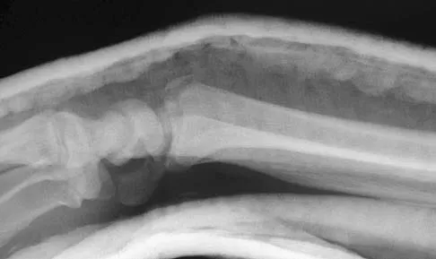
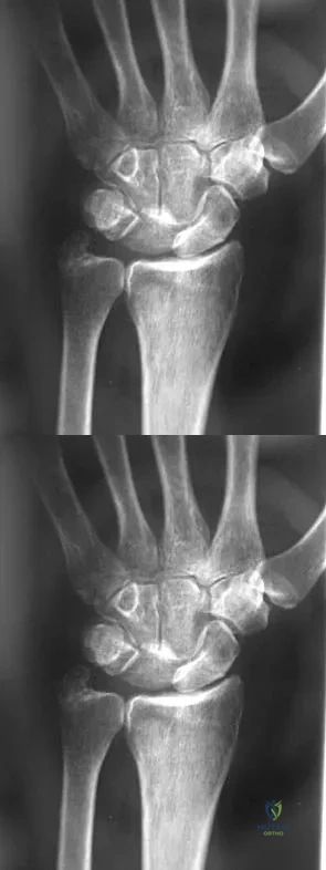
A 67-year-old woman is seen in the emergency department after falling at home. Radiographs before and after treatment are shown in Figures 49a and 49b, respectively. Which of the following best explains the 8-week postinjury clinical findings seen in Figure 49c?


Explanation

49b 49c Patients older than age 40 years at the time of initial anterior dislocation have low rates of redislocation; however, 15% of these patients experience a rotator cuff tear. Moreover, there is a dramatic increase (up to 40%) in the incidence of rotator cuff tears in patients older than age 60 years. Axillary nerve injury may occur but is less common than rotator cuff tear. Koval KJ (ed): Orthopaedic Knowledge Update 7. Rosemont, IL, American Academy of Orthopaedic Surgeons, 2002, pp 273-284.

Question 4

What is the most common complaint in patients with a developmental radial head dislocation?


Explanation

Developmental dislocation of the radial head most frequently presents as a painless mass over the posterior aspect of the elbow. Patients do not have feelings of elbow subluxation but may report pain or clicking. Limitation of motion is most frequently found in the pronation and supination arc rather than in flexion and extension. Lloyd-Roberts GC, Bucknill TM: Anterior dislocation of the radial head in children-etiology: Natural history and management. J Bone Joint Surg Am 1977;58:402.

Question 5

Which of the following has been associated with a decreased rate of glenoid component radiolucent lines?


Explanation

According to a recent study, cemented pegged glenoid components had fewer radiolucent lines initially and at 2-year follow-up when compared to a cemented keeled design. Curve-backed designs have also shown fewer radiolucent lines when compared to flat-backed designs. Oversizing the glenoid can lead to impaired rotator cuff function and decreased range of motion. An off-centered glenoid can lead to early loosening. Gartsman GM, Elkousy HA, Warnock KM, et al: Radiographic comparison of pegged and keeled glenoid components. J Shoulder Elbow Surg 2005;14:252-257. Szabo I, Buscayret F, Edwards TB, et al: Radiographic comparison of flat-back and convex-back glenoid components in total shoulder arthroplasty. J Shoulder Elbow Surg 2005;14:636-642.

Question 6

What neurovascular structure is in closest proximity to the probe in the arthroscopic view of the elbow shown in Figure 50?


Explanation

The image shows a view of the radiocapitellar joint from an anterior medial portal. The radial nerve lies on the elbow capsule at the midportion of the capitellum. It is at risk for injury when capsular excision is performed in this region. Field LD, Altchek DW, Warren RF, et al: Arthroscopic anatomy of the lateral elbow: A comparison of three portals. Arthroscopy 1994;10:602-607.

Question 7

Figure 51 shows the radiograph of a 42-year-old construction worker who has pain and limited motion in his dominant elbow. Management consisting of nonsteroidal anti-inflammatory drugs and cortisone has failed to provide relief. What is the next most appropriate step in treatment?


Explanation

The patient has symptomatic primary osteoarthritis of the elbow with multiple loose bodies. Given his age and occupation, an elbow arthroplasty is not an option. Arthroscopic debridement and removal of loose bodies has been shown to be effective for osteoarthritis of the elbow. Gramstad GD, Galatz LM: Management of elbow osteoarthritis. J Bone Joint Surg Am 2006;88:421-430.

Question 8

A 35-year-old man sustained the closed injury shown in Figure 52 in his dominant extremity. Neurologic function is normal. Treatment should consist of


Explanation

Functional bracing has been demonstrated to have a very high rate of healing without any functional limitations in a large series of patients. Surgery is reserved for "floating elbows," open injuries, neurovascular injuries, and those fractures that go on to nonunion. Sarmiento A, Zagorski JB, Zych GA, et al: Functional bracing for the treatment of fractures of the humeral diaphysis. J Bone Joint Surg Am 2000;82:478-486.

Question 9

A 74-year-old man has had worsening left shoulder pain for the past 3 years. Extensive nonsurgical management has provided only minimal relief. Examination reveals limitations in motion due to pain but good rotator cuff strength. Radiographs are shown in Figures 53a and 53b. What surgical procedure is most appropriate?


Explanation

53b The patient has end-stage shoulder arthritis with posterior glenoid erosion and large humeral osteophyte formation. Since the rotator cuff is likely intact, the reverse total shoulder arthroplasty is unnecessary. All the remaining procedures may provide symptomatic relief in appropriate patients; however, for most patients, total shoulder arthroplasty has been associated with the most predictive pain relief and functional improvements. Bryant D, Litchfield R, Sandow M, et al: A comparison of pain, strength, range of motion, and functional outcomes after hemiarthroplasty and total shoulder arthroplasty in patients with osteoarthritis of the shoulder: A systemic review and meta-analysis. J Bone Joint Surg Am 2005;87:1947-1956. Edwards TB, Kadakia NR, Boulahia A, et al: A comparison of hemiarthroplasty and total shoulder arthroplasty in the treatment of primary glenohumeral osteoarthritis: Results of a multicenter study. J Shoulder Elbow Surg 2003;12:207-213.

Question 10

The radiograph shown in Figure 54 reveals that the plate on the second metacarpal is acting in what manner?


Explanation

There are four ways in which a plate acts: compression, tension bend, bridge or spanning, and buttress. Since there is no cortical contact with the large span of comminution, this plate is acting as a bridge plate. A bridge plate is defined as when the plate is used as an extramedullary splint attached to the two main fragments, leaving the comminution untouched.

Question 11

Which of the following antibiotics is contraindicated in children?


Explanation

The tetracycline family of medications can stain teeth and bone in skeletally immature patients and as a result should be avoided in those patients. The remaining antibiotics have no known specific contraindication to use in children.

Question 12

Which of the following conditions is considered a relative contraindication to interscalene nerve block for patients scheduled to undergo shoulder surgery?


Explanation

A common side effect of interscalene nerve block for shoulder surgery is the blockade of the ipsilateral phrenic nerve. This, in turn, results in paresis of the diaphragm and up to a 30% reduction in pulmonary function volumes. Therefore, interscalene nerve block generally is not recommended for patients whose respiratory function is compromised. Other relative and absolute contraindications for interscalene nerve blocks include allergy to local anesthetics, infection at the injection site, uncontrolled seizure disorder, coagulation abnormality, and preexisting neurologic injury. Chelly JE: Indications for upper extremity blocks, in Chelly JE (ed): Peripheral Nerve Blocks, ed 2. Philadelphia, PA, Lippincott Williams & Wilkins, 2004, pp 19-27.

Question 13

Figure 55 shows the radiograph of a 30-year-old man who sustained a closed comminuted fracture of the right clavicle. Examination reveals decreased sensation in the radial nerve distribution. Weakness is noted with shoulder abduction, internal rotation, and wrist extension. A displaced bone fragment is most likely pressing on what portion of the brachial plexus?


Explanation

Clavicular fractures are occasionally complicated by injury to the brachial plexus. A displaced bone fragment pressing on the posterior cord proximal to the upper subscapularis nerve would account for these findings. Jobe CM, Coen MJ: Gross anatomy of the shoulder, in Rockwood CA, Matsen FA, Wirth MA, et al (eds): The Shoulder. Philadelphia, PA, WB Saunders, 2004, vol 2, pp 1078-1079.

Question 14

Which of the following characteristics is seen in patients with osteochondritis dissecans of the elbow?


Explanation

Osteochondritis dissecans occurs in the older child or adolescent (typically older than age 13 years). It involves the lateral compartment. The etiology is felt to be microtraumatic vascular insufficiency from repetitive rotatory and compressive forces. MRI typically shows separation of cartilage from the capitellum and chondral fissuring. Panner's disease is usually seen in children younger than age 10 years, involves the entire capitellar ossific nucleus, and resolves typically with no residual deformity or late sequelae. There is no evidence of ligamentous injury. Defelice GS, Meunier MJ, Paletta GA: Elbow injury in the adolescent athlete, in Altchek DW, Andrews JR (eds): The Athlete's Elbow. New York, NY, Lippincott Williams & Wilkins, 2001, pp 231-248.

Question 15

Which of the following is considered an important component in treating the lesion shown in Figure 56?


Explanation

Mucoid cysts are commonly associated with DIP joint arthritis. Two treatment options are commonly used: (1) aspiration/drainage and injection of corticosteroid and (2) surgical excision. When performing the surgery, excision of the bony osteophytes about the DIP joint is helpful in achieving a cure. There are no reports of significant benefit with nail removal or partial ligament or extensor tendon resection. Some authors have advocated skin excision and rotational flaps for wound coverage, but this is somewhat controversial. Rizzo M, Beckenbaugh RD: Treatment of mucous cysts of the fingers: Review of 134 cases with minimum 2-year follow-up evaluation. J Hand Surg Am 2003;28:519-524.

Question 16

A 22-year-old female collegiate javelin thrower has shoulder pain. She notes that her pain is primarily located in the posterior aspect of her shoulder, is exacerbated with throwing, and she experiences maximal tenderness in the extreme cocking phase of the throwing cycle. On examination, she reports deep posterior shoulder pain when the arm is abducted 90 degrees and maximally externally rotated to 110 degrees. This reproduces her symptoms precisely. Shoulder radiographs are normal. What is the most likely diagnosis?


Explanation

The patient has internal impingement. Internal impingement is commonly seen in overhead throwing athletes. When positioned in the extreme cocking phase of the throwing cycle, the posterior glenoid impacts the articular surface of the infraspinatus and posterior fibers of the supraspinatus tendon. This impact can cause partial-thickness rotator cuff tearing and posterosuperior labral lesions. She has no evidence of anterior shoulder instability, and her range of motion is excellent which rules out adhesive capsulitis. Subacromial impingement is identified with anterolateral shoulder pain with internal rotation in the abducted position. A full-thickness rotator cuff tear in a 22-year-old individual would require significant trauma and would likely result in pain at rest and with lifting. Meister K, Buckley B, Batts J: The posterior impingement sign: Diagnosis of rotator cuff and posterior labral tears secondary to internal impingement in overhand athletes. Am J Orthop 2004;33:412-415.

Question 17

A patient with rheumatoid arthritis has a rupture of the extensor digitorum communis to 4 and 5. You are planning to perform an extensor indicis proprius (EIP) tendon transfer. What effect will this have on index finger extension?


Explanation

EIP transfer results in no functional deficit. If the tendon is cut proximal to the sagittal band, there will be no extensor deficit. Browne EX, Teague MA, Snyder CC: Prevention of extensor lag after indicis proprius transfer. J Hand Surg Am 1979;4:168-172.

Question 18

What is the most common complication following interscalene nerve block for shoulder surgery?


Explanation

All of these complications have been documented after interscalene nerve block. Other serious complications such as cardiac arrest and respiratory distress have also been noted. However, the most common complication after interscalene nerve block appears to be temporary paresthesia to the hand that can occur in up to 2.3% of the patients. Bishop JY, Sprague M, Gelber J, et al: Interscalene regional anesthesia for shoulder surgery. J Bone Joint Surg Am 2005;87:974-979.

Question 19

A 61-year-old woman with a long-standing history of rheumatoid arthritis reports progressive elbow pain for the past 12 months. She denies any recent trauma to the elbow; however, she notes increasing pain and decreased joint motion that are now compromising her function. Radiographs are shown in Figures 57a and 57b. What is the most appropriate treatment at this time?


Explanation

57b The patient has end-stage arthritis of the elbow with advanced joint destruction. At this point, nonsurgical management is unlikely to provide much relief of symptoms. Arthroscopic procedures can provide relief, but it is likely to be incomplete and unpredictable. The most reliable surgical option is total elbow arthroplasty. Currently, semiconstrained components are generally preferred because constrained components have been associated with a high rate of early prosthetic loosening. Little CP, Graham AJ, Karatzas G, et al: Outcomes of total elbow arthroplasty for rheumatoid arthritis: Comparative study of three implants. J Bone Joint Surg Am 2005;87:2439-2448.

Question 20

What neurovascular structure is at greatest risk when creating a proximal anterolateral elbow arthroscopy portal?


Explanation

The radial nerve is 4 to 7 mm from the anterolateral portal, which is placed 1 cm anterior and 3 cm proximal to the lateral epicondyle. The posterior interosseous nerve can lie 1 to 14 mm from the portal site. Andrews JR, Carson WG: Arthroscopy of the elbow. Arthroscopy 1985;1:97-107.

Question 21

A 52-year-old woman reports mild pain localized to the left sternoclavicular joint. History is notable for chronic renal failure requiring dialysis for the last 5 years. A clinical photograph, chest radiograph, and bone scan are shown in Figures 58a through 58c. What is the most likely diagnosis?


Explanation

58b 58c Spontaneous swelling with the appearance of joint subluxation may be associated with an acute, subacute, or chronic bacterial infection of the sternoclavicular joint. Common causes of infection include bacteremia, rheumatoid arthritis, alcoholism, intravenous drug use, and chronic debilitating diseases. Subclavian vein catheterization and renal dialysis can predispose patients to sepsis and osteomyelitis of the sternoclavicular joint. Renoult B, Lataste A, Jonon B, et al: Sternoclavicular joint infection in hemodialysis patients. Nephron 1990;56:212-213.

Question 22

A 22-year-old college quarterback is tackled and sustains a reducible first carpometacarpal dislocation. What is the recommended treatment?


Explanation

When comparing closed reduction and pinning to ligament reconstruction, the reconstruction group had slightly better abduction and pinch strength. The volar oblique ligament usually tears off the first metacarpal in a subperiosteal fashion. In this young patient, motion-sparing procedures are preferred. Simonian PT, Trumble TE: Traumatic dislocation of the thumb carpometacarpal joint: Early ligamentous reconstruction versus closed reduction and pinning. J Hand Surg Am 1996;21;802-806.

Question 23

What is the most important stabilizing mechanism in the midrange of motion of the glenohumeral joint?


Explanation

Concavity compression is a stabilizing mechanism by which muscular compression of the humeral head into the glenoid fossa stabilizes the glenohumeral joint against shear forces. This is dependent on the depth of the concavity and the magnitude of the compressive force. Lee SB, Kim KJ, O'Driscoll SW, et al: Dynamic glenohumeral stability provided by the rotator cuff muscles in the mid-range and end-range of motion: A study in cadavera. J Bone Joint Surg Am 2000;82:849-857.

Question 24

In a locking plate screw construct, axial forces are borne by which of the following?


Explanation

In a traditional plate system, fracture security depends on the friction between the plate and the underlying bone. Bicortical fixation will decrease the toggle and improve stability. Locking plates absorb axial forces transmitted from the screws. Such plates do not require plate compression against the bone, thus preserving periosteal blood supply. Nana AD, Joshi A, Lichtman DM: Plating of the distal radius. J Am Acad Orthop Surg 2005;13:159-171.

Question 25

What structure provides the most static stability for valgus restraint in the elbow?


Explanation

The anterior band of the ulnar collateral ligament provides the greatest restraint to valgus stress in the elbow. The posterior band is taut in flexion and resists stress between 60 degrees and full flexion. The annular ligament stabilizes the radial head. The flexor/pronator mass are important dynamic stabilizers of the medial elbow. Ahmad CS, ElAttrache NS: Elbow valgus instability in the throwing athlete. J Am Acad Orthop Surg 2006;14:693-700. Regan WD, Korinek SL, Morrey BF, et al: Biomechanical study of ligaments around the elbow joint. Clin Orthop Relat Res 1991;271:170-179.

Question 26

A 42-year-old man falls on an outstretched hand and sustains a 'terrible triad' injury of the elbow. Surgical fixation is planned. To optimize stability and follow standard surgical principles, what is the most appropriate sequence of repair for the injured structures?


Explanation

The terrible triad of the elbow consists of an elbow dislocation, a radial head fracture, and a coronoid process fracture. The standard surgical algorithm follows a 'deep to superficial' and 'medial to lateral' approach. The typical sequence is: 1) Coronoid fracture fixation or anterior capsule repair, 2) Radial head repair or replacement, 3) Lateral ulnar collateral ligament (LUCL) repair, and 4) Medial collateral ligament (MCL) repair (only if the elbow remains unstable after the first three steps).

Question 27

A 45-year-old male presents with chronic wrist pain. Radiographs demonstrate a scaphoid nonunion advanced collapse (SNAC) pattern with arthritic changes present at the radioscaphoid and capitolunate joints. The radiolunate joint is well preserved. What is the most appropriate definitive surgical treatment?


Explanation

The patient has Stage III SNAC wrist (arthritis of the radioscaphoid and capitolunate joints, with a preserved radiolunate joint). A four-corner arthrodesis with scaphoid excision relies on a preserved radiolunate articulation and is the procedure of choice. Proximal row carpectomy is contraindicated in this patient due to the presence of capitolunate arthritis, as the procedure requires a pristine capitate head to articulate with the lunate fossa of the radius.

Question 28

A 28-year-old professional volleyball player complains of vague posterior shoulder pain and weakness with overhead serving. Physical examination reveals isolated weakness in external rotation with the arm at the side and noticeable atrophy of the infraspinatus. Supraspinatus strength is normal. An MRI reveals a paralabral cyst. Where is the cyst most likely located?


Explanation

Isolated atrophy and weakness of the infraspinatus indicate compression of the suprascapular nerve after it has innervated the supraspinatus. This occurs at the spinoglenoid notch. A paralabral cyst at the spinoglenoid notch (often associated with posterior labral tears) compresses the distal branch of the suprascapular nerve. Compression at the suprascapular notch would affect both the supraspinatus and infraspinatus.

Question 29

A 65-year-old woman sustains a 3-part proximal humerus fracture and is managed non-operatively. At her 6-week follow-up, she demonstrates profound weakness in shoulder abduction and reports decreased sensation over the lateral aspect of her shoulder. Injury to which of the following nerves is most likely responsible for her symptoms?


Explanation

The axillary nerve courses close to the inferior capsule and surgical neck of the humerus, making it highly susceptible to injury during proximal humerus fractures or shoulder dislocations. Axillary nerve injury results in denervation of the deltoid and teres minor muscles, leading to profound weakness in shoulder abduction, as well as numbness over the lateral shoulder (regimental badge area) supplied by the superior lateral cutaneous nerve of the arm, a branch of the axillary nerve.

Question 30

A 50-year-old man presents with a 4-month history of numbness in his ring and small fingers, accompanied by weakness in his hand grip. Examination reveals a positive Froment's sign. Which of the following physical examination findings would best differentiate the site of ulnar nerve compression as being at the cubital tunnel rather than Guyon's canal?


Explanation

The dorsal ulnar cutaneous nerve branches from the ulnar nerve approximately 5-8 cm proximal to the wrist (proximal to Guyon's canal). Therefore, decreased sensation over the dorsal ulnar aspect of the hand indicates a lesion proximal to the wrist, such as at the cubital tunnel. Findings like Froment's sign, Wartenberg's sign, and interosseous weakness result from motor deficits of the deep branch of the ulnar nerve and can be seen in both distal and proximal compression.

Question 31

A 40-year-old man undergoes a single-incision anterior approach for a distal biceps tendon repair. Postoperatively, he notes weakness in extending his thumb and fingers. When asked to extend his wrist, his wrist deviates radially. Sensation in the hand and forearm is entirely intact. Which nerve was most likely injured during the procedure?


Explanation

The posterior interosseous nerve (PIN) is at risk during the anterior single-incision approach to the distal biceps, particularly during forceful radial retraction. PIN palsy results in motor weakness of the thumb and finger extensors, as well as the extensor carpi ulnaris (ECU). Because the extensor carpi radialis longus (ECRL) is innervated by the radial nerve proximal to the PIN branch, wrist extension is preserved but deviates radially due to the unopposed ECRL overpowering the paralyzed ECU. There is no sensory deficit in isolated PIN palsy.

Question 32

A 55-year-old woman is 6 months status post volar locking plate fixation for a comminuted distal radius fracture. She complains of a sudden inability to actively flex the interphalangeal joint of her thumb. Lateral radiographs demonstrate the distal edge of the volar plate is positioned prominently volar to the watershed line. What is the most likely cause of her current presentation?


Explanation

Positioning a volar plate distal to the watershed line of the distal radius places the flexor tendons at high risk for frictional wear against the plate. The flexor pollicis longus (FPL) tendon is most commonly affected due to its anatomic position directly over the distal radius. This attritional wear can lead to sudden, painless rupture of the FPL, presenting as a loss of active IP joint flexion of the thumb.

Question 33

A 30-year-old construction worker presents with chronic dorsal wrist pain and stiffness. Radiographs reveal sclerosis and early collapse of the lunate, consistent with Kienböck's disease. Measurement of ulnar variance shows ulnar minus 3 mm. The articular cartilage of the radiocarpal and midcarpal joints appears well-preserved on MRI. Which of the following is the most appropriate initial surgical intervention to decompress the lunate?


Explanation

In early Kienböck's disease (Lichtman Stage II or IIIA) with negative ulnar variance and preserved cartilage, joint-leveling procedures are indicated to mechanically unload the lunate. A radial shortening osteotomy (or ulnar lengthening osteotomy) decreases the compressive forces on the lunate by transferring loads to the ulnocarpal joint. Ulnar shortening is indicated for ulnar impaction syndrome, not Kienböck's.

Question 34

A 22-year-old mountain biker falls over his handlebars and sustains a midshaft clavicle fracture. Which of the following findings is considered an absolute indication for acute open reduction and internal fixation?


Explanation

Absolute indications for operative fixation of a clavicle fracture include open fractures, fractures with associated neurovascular compromise, and severe skin tenting that threatens the integrity of the overlying skin. Relative indications include shortening > 2 cm (20 mm), 100% displacement, symptomatic nonunions, and multiple injuries (e.g., floating shoulder with a displaced scapular neck fracture).

Question 35

You are discussing the biomechanics of a reverse total shoulder arthroplasty (RTSA) with a resident.

Which of the following best describes the primary biomechanical advantage conferred by the medialized and distalized center of rotation typical of a Grammont-style reverse shoulder implant?


Explanation

The key biomechanical principle of the Grammont-style reverse total shoulder arthroplasty is moving the center of rotation medially and distally compared to the native anatomy. This distalization increases the resting tension of the deltoid, while the medialization increases the deltoid's lever arm and recruits more deltoid muscle fibers for forward elevation and abduction, compensating for the absent or deficient rotator cuff. Medialization increases shear forces at the baseplate, which is a known disadvantage counteracted by using inferior tilt or a larger glenosphere.

Question 36

A 45-year-old manual laborer presents with chronic, progressive radial-sided wrist pain. He has a history of a fall on an outstretched hand 10 years ago. Radiographs demonstrate a scaphoid nonunion with advanced collapse (SNAC). There is significant joint space narrowing and sclerosis at the radioscaphoid and capitolunate joints, but the radiolunate articulation is well preserved. Which of the following is the most appropriate surgical treatment?


Explanation

The patient has Stage 2/3 Scaphoid Nonunion Advanced Collapse (SNAC), characterized by radioscaphoid and capitolunate arthritis with a spared radiolunate joint. Scaphoid excision and four-corner fusion is the treatment of choice. Proximal row carpectomy (PRC) is contraindicated in this scenario because it relies on a healthy capitolunate articulation; if the capitate head is arthritic (as in SNAC Stage 2/3 or SLAC Stage 3), PRC will result in painful articulation with the lunate fossa. Total wrist arthrodesis is reserved for pancarpal arthritis or failed partial fusions. Scaphoid ORIF is not indicated once advanced degenerative changes have occurred.

Question 37

A 38-year-old male weightlifter undergoes repair of a complete distal biceps tendon rupture via a classic two-incision approach. During his postoperative course, what complication is significantly more frequent with this surgical approach compared to a single-incision anterior approach?


Explanation

The two-incision approach for distal biceps repair was historically developed to avoid the radial nerve (PIN) injuries associated with a single anterior incision. However, it carries a significantly higher risk of heterotopic ossification and proximal radioulnar synostosis due to the dissection through the interosseous membrane and around the ulna. The single-incision anterior approach carries a higher risk of lateral antebrachial cutaneous nerve (LABCN) neuropraxia and posterior interosseous nerve (PIN) injury.

Question 38

A 22-year-old rugby player with recurrent anterior shoulder instability is scheduled for an open Latarjet procedure (coracoid transfer) after a CT scan demonstrates 28% anterior glenoid bone loss. During the osteotomy of the coracoid and its subsequent transfer through the split in the subscapularis tendon, which of the following nerves is at greatest risk of iatrogenic injury?


Explanation

The musculocutaneous nerve typically enters the coracobrachialis muscle 3 to 8 cm distal to the tip of the coracoid process. During the Latarjet procedure, the coracoid process (with the attached conjoined tendon) is osteotomized and transferred to the anterior glenoid. The mobilization and retraction of the conjoined tendon place the musculocutaneous nerve at high risk for stretch or direct transection injury.

Question 39

A 65-year-old woman is seen 9 months after undergoing open reduction and internal fixation of a distal radius fracture with a volar locking plate. She reports the sudden onset of an inability to actively flex the interphalangeal joint of her thumb. Passive motion is intact. What is the most likely etiology of this complication?


Explanation

Sudden loss of active IP joint flexion of the thumb following volar plating of the distal radius is highly characteristic of a flexor pollicis longus (FPL) tendon rupture. This complication most commonly occurs when the volar plate is placed distal to the watershed line of the distal radius, leading to prominent hardware that causes attritional wear and eventual rupture of the overlying FPL tendon. EPL ruptures (Option 0) result in loss of thumb extension and are more commonly associated with prominent dorsal screws penetrating the dorsal cortex.

Question 40

A 55-year-old woman falls and sustains a displaced 4-part proximal humerus fracture.

According to Hertel's criteria, which of the following radiographic findings is the most reliable predictor of subsequent avascular necrosis (AVN) of the humeral head?


Explanation

Hertel et al. described reliable predictors for ischemia and subsequent AVN in proximal humerus fractures. The most critical predictors of ischemia are a medial calcar hinge disruption greater than 2 mm, a metaphyseal head extension (calcar length attached to the articular segment) of less than 8 mm, and an anatomic neck fracture pattern. Metaphyseal head extension greater than 8 mm (Option 3) is actually protective against AVN, as it indicates better preservation of the blood supply.

Question 41

A 42-year-old male falls from a height and sustains a 'terrible triad' injury of the elbow, which includes an elbow dislocation, a comminuted radial head fracture, and a Type II coronoid fracture. Operative intervention is planned. To optimize stability, what is the most widely accepted surgical sequence for addressing these lesions?


Explanation

The 'terrible triad' of the elbow includes an elbow dislocation, radial head fracture, and coronoid fracture. The standard surgical algorithm follows a deep-to-superficial repair sequence to restore stability. First, the anterior capsule and coronoid are fixed to restore the anterior buttress. Second, the radial head is repaired or replaced. Finally, the lateral ulnar collateral ligament (LUCL/LCL complex) is repaired. MCL repair is rarely necessary unless the elbow remains unstable after the standard sequence.

Question 42

A 50-year-old man presents with profound weakness in external rotation and elevation of his right shoulder following a massive, irreparable posterosuperior rotator cuff tear. Examination reveals a positive Hornblower's sign and intact subscapularis function. There is no evidence of glenohumeral arthritis. Which of the following tendon transfers is biomechanically most appropriate to restore external rotation in this patient?


Explanation

The lower trapezius transfer is increasingly preferred for massive irreparable posterosuperior rotator cuff tears, particularly when the primary deficit is profound external rotation weakness (indicated by a positive Hornblower's sign). The lower trapezius line of pull closely mimics that of the native infraspinatus, making it biomechanically superior to the latissimus dorsi for restoring external rotation. Latissimus dorsi transfers are traditionally used but have a vector that is less ideal for pure external rotation restoration.

Question 43

A 45-year-old carpenter presents with numbness and tingling in the small and ring fingers of his right hand, along with weakness in grip strength. On examination, when asked to hold a piece of paper between his thumb and radial side of his index finger against resistance, the interphalangeal joint of his thumb hyperflexes (positive Froment sign). Which muscle is the patient substituting with to maintain hold of the paper?


Explanation

A positive Froment sign is indicative of ulnar nerve palsy. The primary muscle responsible for adduction of the thumb is the adductor pollicis, which is innervated by the ulnar nerve. When this muscle is weak or paralyzed, the patient involuntarily compensates by using the flexor pollicis longus (FPL), innervated by the anterior interosseous nerve (a branch of the median nerve), causing hyperflexion at the interphalangeal joint of the thumb.

Question 44

A 32-year-old bodybuilder feels a tearing sensation in his anterior chest wall while bench pressing a heavy weight. Examination reveals loss of the anterior axillary fold and significant ecchymosis. MRI confirms a rupture of the sternocostal head of the pectoralis major muscle. Which of the following describes the anatomic footprint of the sternocostal head at its humeral insertion relative to the clavicular head?


Explanation

The pectoralis major tendon undergoes a 180-degree twist before inserting on the lateral lip of the bicipital groove of the humerus. Due to this twist, the inferiorly originating fibers (sternocostal head) form the posterior lamina of the tendon and insert proximal and deep to the superiorly originating fibers (clavicular head), which insert more distally and superficially.

Question 45

A 50-year-old woman presents with persistent ulnar-sided wrist pain that worsens with pronation and gripping, 1 year after non-operative management of a distal radius fracture.

Radiographs demonstrate a healed distal radius with 4 mm of radial shortening, resulting in positive ulnar variance. MRI reveals degenerative tearing of the triangular fibrocartilage complex (TFCC) and cystic changes in the lunate. The distal radioulnar joint (DRUJ) is congruous without advanced arthritis. What is the most appropriate definitive surgical management?


Explanation

The patient has ulnar impaction syndrome secondary to a malunited distal radius fracture with significant positive ulnar variance (4 mm). Ulnar shortening osteotomy is the treatment of choice as it decompresses the ulnocarpal joint while maintaining the congruous DRUJ. The arthroscopic wafer procedure is typically reserved for positive ulnar variance of 2 mm or less. Sauvé-Kapandji and Darrach procedures are salvage operations indicated for DRUJ arthritis, which this patient does not have.

Question 46

A 45-year-old competitive weightlifter undergoes surgical repair of a distal biceps tendon rupture using a single-incision anterior approach. On his first postoperative visit, he complains of numbness and tingling along the lateral aspect of his forearm. Motor function of the hand and wrist is completely intact. Which of the following anatomical structures was most likely injured or stretched during the surgical exposure?


Explanation

The lateral antebrachial cutaneous nerve (LABC) is the terminal sensory branch of the musculocutaneous nerve. It exits the deep fascia just lateral to the biceps tendon and is highly vulnerable to traction neuropraxia or transection during a single-incision anterior approach for distal biceps repair. Injury results in numbness along the lateral aspect of the forearm. The posterior interosseous nerve (PIN) is more commonly at risk during a two-incision approach (particularly if the forearm is not kept in supination during the posterolateral exposure) or if retractors are placed too deep radially.

Question 47

A 32-year-old man presents with chronic, progressive wrist pain. Radiographs reveal a scaphoid nonunion advanced collapse (SNAC). Imaging demonstrates moderate degenerative changes at the radioscaphoid joint and the scaphocapitate joint, while the radiolunate articulation is entirely spared. However, there is marked cartilage loss and degenerative cyst formation on the proximal head of the capitate. Which of the following is the most appropriate surgical treatment?


Explanation

This patient has a SNAC wrist. The decision between a proximal row carpectomy (PRC) and a scaphoid excision with four-corner arthrodesis depends heavily on the condition of the lunate fossa of the radius and the proximal head of the capitate. Because a PRC relies on a new articulation between the capitate head and the lunate fossa, arthritis of the capitate head is an absolute contraindication to PRC. Therefore, scaphoid excision and four-corner arthrodesis is the most appropriate motion-preserving procedure in this scenario.

Question 48

A 40-year-old man falls from a ladder and sustains a 'terrible triad' injury of the elbow. Intraoperatively, the surgeon fixes the coronoid fracture, replaces the highly comminuted radial head with an arthroplasty, and robustly repairs the lateral collateral ligament (LCL) complex to the lateral epicondyle. Upon testing stability, the elbow remains congruous in flexion but persistently subluxates posteriorly when extended beyond 30 degrees. What is the most appropriate next step in management?


Explanation

The standard surgical protocol for a terrible triad injury of the elbow involves sequential restoration of stabilizers: coronoid fixation (or anterior capsule repair), radial head fixation or replacement, and LCL repair. If the elbow remains unstable in extension after these lateral and anterior structures are restored, the medial collateral ligament (MCL) should be repaired. Hinged external fixation is typically reserved for residual instability after all primary ligamentous repairs, including the MCL, have been completed.

Question 49

A 72-year-old woman with pseudoparalysis of the shoulder due to massive rotator cuff tear arthropathy is scheduled for a reverse total shoulder arthroplasty (RTSA). Which of the following best describes the fundamental biomechanical advantage conferred by the implant design of an RTSA compared to an anatomic total shoulder arthroplasty?


Explanation

The fundamental biomechanical principle of the reverse total shoulder arthroplasty (Grammont design) is the medialization and distalization of the glenohumeral center of rotation. Medialization recruits more anterior and posterior deltoid fibers for elevation, while distalization tensions the deltoid and significantly increases its moment arm, allowing the deltoid to compensate for the deficient rotator cuff.

Question 50

A 6-year-old child sustains a widely displaced, extension-type supracondylar humerus fracture.

On initial presentation in the emergency department, the child's hand is pink and well-perfused, but the radial pulse is non-palpable. The patient is taken to the operating room for closed reduction and percutaneous pinning. Post-reduction, the fracture is anatomically aligned, but the radial pulse remains absent. The hand remains warm and pink, with a capillary refill time of 2 seconds. What is the most appropriate next step?


Explanation

The management of a 'pulseless, pink' hand following reduction and pinning of a pediatric supracondylar humerus fracture is observation and close monitoring. Because the hand remains well-perfused (pink, brisk capillary refill), collateral circulation is sufficient. Vascular exploration or routine angiography is only indicated if the hand becomes 'pulseless and white' (poorly perfused) after closed reduction, which signifies inadequate collateral flow and impending ischemia.

Question 51

An 18-year-old high school football player is driven into the turf directly onto his lateral shoulder. He presents to the emergency department supporting his injured arm across his chest. He reports worsening shortness of breath, mild dysphagia, and a choking sensation.

A CT scan of the chest confirms a posterior sternoclavicular dislocation. What is the most appropriate initial management of this injury?


Explanation

Posterior sternoclavicular dislocations are orthopedic emergencies, especially when accompanied by symptoms of mediastinal compression (shortness of breath, dysphagia, venous congestion). Due to the proximity of the great vessels, trachea, and esophagus, reduction should be performed urgently in the operating room under general anesthesia, with a cardiothoracic surgeon available on standby in case of catastrophic hemorrhage or great vessel injury during the reduction maneuver.

Question 52

A 55-year-old woman sustained a nondisplaced fracture of the distal radius 6 weeks ago, which was managed conservatively in a short-arm cast. Two days after cast removal, she suddenly loses the ability to actively extend the interphalangeal joint of her thumb.

Radiographs show healing of the distal radius fracture with no displacement. What is the gold standard surgical intervention for this complication?


Explanation

The patient has sustained a spontaneous rupture of the extensor pollicis longus (EPL) tendon, a known complication following distal radius fractures (even nondisplaced ones) due to mechanical attrition and hypovascularity within the third dorsal compartment (Lister's tubercle). Primary end-to-end repair is generally impossible due to tendon retraction and degeneration. The gold standard treatment is an extensor indicis proprius (EIP) to EPL tendon transfer.

Question 53

A 48-year-old male with severe cubital tunnel syndrome is undergoing an anterior transposition of the ulnar nerve. To prevent secondary compression post-transposition, the surgeon must systematically release all potential sites of ulnar nerve entrapment around the elbow. Which of the following structures represents the most proximal potential site of compression for the ulnar nerve in this region?


Explanation

The Arcade of Struthers is a fascial band extending from the medial head of the triceps to the medial intermuscular septum, located approximately 8 cm proximal to the medial epicondyle, and represents the most proximal potential site of ulnar nerve compression around the elbow. Note that the Ligament of Struthers (option E) is associated with the supracondylar process and compresses the median nerve, not the ulnar nerve. Osborne's ligament and the FCU fascia are located distally.

Question 54

A 35-year-old mechanic presents to the emergency department complaining of a severely swollen, throbbing index finger 3 days after sustaining a minor puncture wound with a wire.

You suspect pyogenic flexor tenosynovitis. According to Kanavel's criteria, which of the following is considered the earliest and most sensitive clinical sign of this condition?


Explanation

Kanavel's four cardinal signs for pyogenic flexor tenosynovitis are: 1) fusiform swelling of the digit, 2) flexed resting posture, 3) tenderness along the flexor tendon sheath, and 4) disproportionate pain with passive extension. Among these, pain with passive extension of the digit is consistently recognized as the earliest and most sensitive clinical sign for diagnosing pyogenic flexor tenosynovitis.

Question 55

A 30-year-old manual laborer presents with progressive dorsal wrist pain and weakened grip strength over the past 6 months. Radiographs demonstrate increased sclerosis and early collapse of the lunate, without fixed scaphoid rotation or adjacent carpal arthritis. Ulnar variance is measured at -3 mm.

The patient is diagnosed with Lichtman Stage II Kienbock's disease. Which of the following surgical interventions is the most appropriate primary joint leveling procedure to unload the lunate in this patient?


Explanation

This patient has Lichtman Stage II Kienbock's disease (lunate sclerosis without fixed collapse/scaphoid rotation) and negative ulnar variance. A joint leveling procedure is indicated to unload the radiolunate articulation. Radial shortening osteotomy is the gold standard in this scenario. Ulnar lengthening is theoretically possible but is associated with a significantly higher rate of nonunion and hardware complications, making radial shortening the preferred procedure.

Question 56

A 42-year-old male weightlifter feels a sudden pop in his anterior elbow during a heavy deadlift. Clinical examination reveals a positive hook test. He undergoes surgical repair via a single-incision anterior approach. Postoperatively, he complains of numbness and paresthesias over the lateral aspect of his forearm. Which of the following nerves was most likely injured or retracted excessively during the surgical exposure?


Explanation

The lateral antebrachial cutaneous nerve (LABCN) is the sensory continuation of the musculocutaneous nerve. It exits the deep fascia lateral to the biceps tendon and is the most commonly injured nerve during a single-incision anterior approach for distal biceps tendon repair. Injury results in numbness or paresthesias over the lateral forearm.

Question 57

A 28-year-old elite volleyball player complains of vague posterior shoulder pain and weakness in external rotation. Clinical examination reveals isolated atrophy of the infraspinatus muscle, while supraspinatus strength and bulk are normal. An MRI confirms the presence of a paralabral cyst. At which of the following anatomic locations is the nerve compression most likely occurring?


Explanation

The suprascapular nerve innervates the supraspinatus muscle before passing through the spinoglenoid notch to innervate the infraspinatus. Compression at the spinoglenoid notch (often due to a paralabral cyst associated with a posterior labral tear) results in isolated infraspinatus denervation, leading to atrophy and isolated external rotation weakness. Compression at the suprascapular notch would affect both the supraspinatus and infraspinatus.

Question 58

A 35-year-old man falls from a ladder and sustains an elbow dislocation associated with a radial head fracture and a coronoid fracture. He is taken to the operating room for surgical reconstruction. To optimally restore elbow stability in this 'terrible triad' injury, what is the generally recommended sequence of repair?


Explanation

The standard surgical algorithm for a 'terrible triad' injury of the elbow involves repairing the deep structures first, progressing from medial to lateral and deep to superficial. The typical sequence is: 1) Coronoid fracture fixation or anterior capsule repair, 2) Radial head fixation or replacement, and 3) Lateral collateral ligament (LCL) complex repair.

Question 59

A 72-year-old female with severe, painful rotator cuff arthropathy and pseudoparalysis of the shoulder undergoes a reverse total shoulder arthroplasty (RTSA). Which of the following best describes the fundamental biomechanical alteration provided by this specific prosthesis compared to native shoulder anatomy?


Explanation

A reverse total shoulder arthroplasty (RTSA) biomechanically medializes and distalizes the center of rotation of the glenohumeral joint. Medialization recruits more anterior and posterior deltoid fibers for elevation, while distalization tensions the deltoid and increases its moment arm. This allows the deltoid to effectively compensate for the deficient rotator cuff and restores active elevation.

Question 60

A 45-year-old manual laborer presents with chronic, progressive wrist pain. He sustained an untreated wrist sprain 10 years ago. Radiographs demonstrate a scaphoid nonunion with advanced arthritic changes at the radioscaphoid and capitolunate joints; however, the radiolunate joint is completely spared.

Which of the following salvage procedures is most appropriate for this patient?


Explanation

The patient has Stage 3 Scaphoid Nonunion Advanced Collapse (SNAC), characterized by radioscaphoid and capitolunate arthritis with sparing of the radiolunate joint. Scaphoid excision with four-corner fusion (capitate, lunate, hamate, triquetrum) is the procedure of choice. Proximal row carpectomy (PRC) relies on a preserved capitate head to articulate with the lunate fossa; because the capitolunate joint is arthritic in Stage 3 SNAC, PRC is contraindicated.

Question 61

A 24-year-old female sustains a severe fall onto an outstretched hand. Imaging reveals a coronal shear fracture of the capitellum that extends medially into the lateral trochlear ridge (Type IV capitellar fracture). She is scheduled for open reduction and internal fixation. Which surgical approach provides the most optimal visualization for addressing this specific fracture pattern?


Explanation

Coronal shear fractures of the distal humerus involving the capitellum and extending into the trochlea (McKee modification Type IV) are best addressed via an extensile lateral approach. This allows excellent exposure of the anterior capitellum and lateral trochlea for placement of headless compression screws from anterior to posterior.

Question 62

A 65-year-old woman is 6 weeks status post nonoperative cast management of a minimally displaced distal radius fracture. She suddenly loses the ability to actively extend her thumb interphalangeal joint, though she denies any new trauma. What is the gold standard surgical management for this specific complication?


Explanation

This patient has experienced an Extensor Pollicis Longus (EPL) tendon rupture, a classic complication following nondisplaced or minimally displaced distal radius fractures. It is caused by mechanical attrition over Lister's tubercle or vascular ischemia within the third dorsal compartment. Because the tendon ends are typically frayed and retracted, primary repair is rarely feasible. An EIP to EPL tendon transfer is the procedure of choice.

Question 63

A 50-year-old man presents with chronic numbness in his small and ring fingers, accompanied by intrinsic muscle wasting and a positive Froment's sign. Electromyography confirms severe compression of the ulnar nerve at the elbow. During an in situ ulnar nerve decompression, the surgeon must ensure all potential sites of compression are released. Which of the following is the most common anatomical site of ulnar nerve compression in this region?


Explanation

While the ulnar nerve can be compressed at multiple sites around the elbow (including the arcade of Struthers, the medial intermuscular septum, and the fascial bands between the two heads of the FCU), the most common site of compression is Osborne's ligament, which forms the roof of the cubital tunnel spanning between the medial epicondyle and the olecranon.

Question 64

A 32-year-old male wrestler sustains a closed, spiral fracture of the distal third of the humeral shaft. On presentation in the emergency department, he is unable to extend his wrist or fingers and has numbness in the first dorsal web space.

According to the American Academy of Orthopaedic Surgeons (AAOS) guidelines, what is the most appropriate initial management for this nerve injury?


Explanation

A primary radial nerve palsy associated with a closed humeral shaft fracture (even a Holstein-Lewis distal third spiral fracture) is not an absolute indication for immediate surgical exploration. The vast majority of these lesions are neurapraxias or axonotmeses that will recover spontaneously. The standard of care is functional bracing or splinting with observation. Exploration is indicated if the fracture is open, if the palsy develops secondarily after a closed reduction, or if there is no clinical or EMG sign of recovery at 3 to 4 months.

Question 65

A 28-year-old male powerlifter feels a tearing sensation in his anterior chest wall while performing a heavy bench press. Examination reveals an asymmetric loss of the anterior axillary fold and marked weakness in internal rotation. MRI confirms a complete rupture of the pectoralis major tendon at its humeral insertion. Based on the biomechanics of the bench press exercise, which specific fibers of the pectoralis major are typically under the greatest tension and tear first?


Explanation

The pectoralis major tendon twists 180 degrees before inserting on the proximal humerus, meaning the inferior fibers of the sternal head insert most superiorly and proximally. During a bench press (arm extended, abducted, and externally rotated), these inferior sternal fibers are placed under maximal stretch and have a mechanical disadvantage, making them the most common starting point for a pectoralis major tendon rupture.

Question 66

A 45-year-old man falls from a height and sustains an intra-articular distal radius fracture. CT scan demonstrates a 3-mm displaced, 4-mm wide volar ulnar corner (lunate facet) fragment. He undergoes open reduction and internal fixation with a standard volar locking plate. Two weeks postoperatively, radiographs reveal acute volar subluxation of the carpus. Which of the following is the most likely cause of this complication?


Explanation

The volar ulnar corner (volar lunate facet) is a critical structure for radiocarpal stability, as the short radiolunate ligament originates here. Standard volar locking plates often do not sit sufficiently distal or ulnar to capture this small but crucial fragment. Failure to specifically secure it (via fragment-specific fixation, customized plates, or wire/suture techniques) leads to loss of the volar buttress and subsequent volar radiocarpal subluxation.

Question 67

A 32-year-old woman sustains a 'terrible triad' injury to her right elbow. She is taken to the operating room for surgical stabilization. To optimally restore elbow stability, what is the most widely accepted and biomechanically sound sequence of structural reconstruction?


Explanation

The standard 'inside-out' surgical sequence for a terrible triad injury (elbow dislocation, radial head fracture, coronoid fracture) is: 1) Repair or fixation of the coronoid to restore the anterior buttress, 2) Repair or replacement of the radial head to restore the anterior and valgus buttress, and 3) Repair of the lateral collateral ligament (LCL) complex. Routine MCL repair is generally unnecessary if these structures are adequately stabilized and the elbow is concentrically reduced.

Question 68

A 55-year-old manual laborer presents with chronic wrist pain and weakness. Radiographs reveal a scaphoid nonunion with advanced radioscaphoid arthritis and narrowing of the capitolunate joint space, but the radiolunate joint is preserved. Which of the following is the most appropriate surgical salvage procedure for this patient?


Explanation

The patient has a Stage III Scaphoid Nonunion Advanced Collapse (SNAC) wrist, characterized by radioscaphoid and capitolunate arthritis with a preserved radiolunate joint. Proximal row carpectomy (PRC) is contraindicated because the capitate head articulates with the lunate fossa; in Stage III, the proximal capitate is arthritic. Therefore, scaphoid excision and four-corner fusion (capitate, lunate, triquetrum, hamate) is the most appropriate motion-preserving salvage procedure.

Question 69

A 28-year-old carpenter presents with an inability to make an 'OK' sign with his right hand. On examination, his thumb interphalangeal joint and index finger distal interphalangeal joint remain extended when attempting to pinch. He has no sensory deficits. Which of the following anatomical variants is a well-known cause of this specific nerve compression syndrome?


Explanation

The patient presents with Anterior Interosseous Nerve (AIN) syndrome, a pure motor palsy affecting the flexor pollicis longus (FPL), flexor digitorum profundus (FDP) to the index/middle fingers, and the pronator quadratus. Gantzer's muscle is an accessory head of the FPL and is a classic anatomical variant that can compress the AIN. Struthers' ligament compresses the main median nerve, anconeus epitrochlearis compresses the ulnar nerve, Martin-Gruber is a normal variant median-to-ulnar nerve anastomosis in the forearm, and Linburg-Comstock is a tendinous interconnection between the FPL and FDP.

Question 70

A 68-year-old woman sustains a 3-part proximal humerus fracture after a fall.

When evaluating the initial trauma radiographs, which of the following findings is the strongest independent predictor for the subsequent development of avascular necrosis (AVN) of the humeral head?


Explanation

According to Hertel's criteria, the most reliable radiographic predictors for ischemia and subsequent AVN of the humeral head after a proximal humerus fracture are a posteromedial hinge disruption, a metaphyseal head extension (calcar length) of less than 8 mm, and an anatomic neck fracture pattern. The disruption of the medial hinge and short calcar signify profound disruption of the ascending branch of the anterior humeral circumflex artery and intraosseous collateral blood supply.

Question 71

A 42-year-old recreational weightlifter undergoes a classic two-incision approach (Boyd-Anderson) for a distal biceps tendon rupture repair. Six months postoperatively, he complains of severe stiffness in forearm rotation. Examination reveals a hard block with only 10 degrees of pronation and 15 degrees of supination. Which of the following technical errors during the index procedure is most likely responsible for this complication?


Explanation

The patient has developed a proximal radioulnar synostosis (heterotopic ossification bridging the radius and ulna), which is a devastating complication of the two-incision distal biceps repair. This typically occurs due to subperiosteal stripping or inappropriate exposure of the ulna during the posterior approach, leading to bleeding and cross-union. Modern modifications (such as Morrey's muscle-splitting approach) specifically avoid exposing the ulna to prevent this complication.

Question 72

A 72-year-old man undergoes a reverse total shoulder arthroplasty (rTSA) for massive, irreparable rotator cuff tear arthropathy. Two years later, routine follow-up radiographs reveal grade 3 scapular notching. Which of the following surgical techniques or implant choices would have most likely decreased the incidence of this specific complication?


Explanation

Scapular notching is a frequent complication after reverse total shoulder arthroplasty, caused by mechanical impingement of the medial humeral metaphysis against the inferior scapular neck during arm adduction. Techniques proven to decrease scapular notching include: inferior translation of the glenosphere (creating an overhang over the inferior rim), inferior tilt of the glenosphere, lateralization of the center of rotation (e.g., BIO-RSA or lateralized baseplates), and using a larger diameter glenosphere.

Question 73

A 6-year-old boy presents to the emergency department after falling off playground equipment. Radiographs demonstrate an isolated plastic deformation of the ulnar shaft and an anteriorly dislocated radial head. Which of the following is the most critical step in the initial management to ensure a stable reduction of the radial head?


Explanation

This is a Bado Type I Monteggia equivalent fracture (plastic deformation of the ulna with anterior radial head dislocation). In pediatric patients, the absolute key to reducing and maintaining the radial head is restoring the exact anatomic length and alignment (the normal bow) of the ulna. If the ulnar plastic deformity is not corrected, the radial head will remain unstable or completely irreducible. Annular ligament reconstruction is rarely needed in acute pediatric cases.

Question 74

A 30-year-old tennis player complains of dorsal, central wrist pain and a subjective 'clunk' 4 weeks after falling on an outstretched hand. Examination reveals tenderness over the dorsal scapholunate interval. Radiographs show a scapholunate gap of 4 mm and a radiolunate angle of 25 degrees. Which of the following is the most appropriate management for this patient?


Explanation

The patient has an acute/subacute scapholunate (SL) dissociation with dynamic or static dorsal intercalated segment instability (DISI), indicated by a gap >3 mm and a radiolunate angle >15 degrees. In a young, active patient with a repairable SL tear and no evidence of osteoarthritis, open reduction, direct repair of the SL ligament (often augmented with a dorsal capsulodesis), and temporary K-wire fixation is the standard of care. Salvage procedures (PRC, STT fusion) are reserved for chronic, unrepairable tears or arthritic wrists (SLAC).

Question 75

A 40-year-old diabetic patient presents with a severely swollen, painful index finger 3 days after sustaining a puncture wound.

Examination reveals the finger is held in slight flexion, uniform volar swelling, exquisite pain on passive extension, and tenderness along the entire flexor tendon sheath. He requires emergent surgical irrigation and debridement. Which of the following annular pulleys MUST be preserved during the surgical approach to prevent postoperative bowstringing of the flexor tendons?


Explanation

The patient presents with Kanavel's four cardinal signs of purulent flexor tenosynovitis. When performing surgical drainage (via open or dual-incision technique), it is critical to preserve the biomechanically essential A2 (located over the proximal phalanx) and A4 (located over the middle phalanx) pulleys. Loss of these critical pulleys leads to flexor tendon bowstringing, drastically reducing the mechanical advantage and excursion of the tendons, leading to severe functional impairment.

Question 76

A 62-year-old man presents to the clinic 4 weeks after an anterior shoulder dislocation that was successfully reduced in the emergency department. He reports persistent weakness in the shoulder, specifically when trying to lift his arm away from his body and externally rotate. Plain radiographs show concentric reduction with no fractures. What is the most appropriate next step in management?


Explanation

Older patients (>40 years) with an anterior shoulder dislocation have a high incidence of associated rotator cuff tears. Persistent weakness, specifically in abduction and external rotation, after a shoulder dislocation in this age group should raise high suspicion for a massive or full-thickness rotator cuff tear. An MRI is the most appropriate next step to evaluate the rotator cuff and other soft tissue structures to guide operative management.

Question 77

A 45-year-old woman falls on an outstretched hand and sustains a 'terrible triad' injury of the elbow. She undergoes operative management. During the procedure, the surgeon decides to repair the lateral ulnar collateral ligament (LUCL) to the lateral epicondyle using a suture anchor. Where is the precise isometric origin of the LUCL on the lateral epicondyle?


Explanation

The isometric point for LUCL reconstruction or repair on the lateral epicondyle is located at the center of the axis of rotation of the capitellum. Placing the anchor or graft at this exact point ensures uniform tension on the ligament throughout the elbow's full range of motion, which is crucial for restoring posterolateral rotatory stability in a terrible triad injury.

Question 78

A 28-year-old laborer presents with chronic wrist pain and is diagnosed with a scaphoid nonunion with a humpback deformity and early radiocarpal arthrosis limited to the radial styloid (SNAC stage I). He has minimal symptoms at rest but pain with heavy gripping. Which of the following is the most appropriate surgical treatment?


Explanation

In a young, active patient with a scaphoid nonunion, humpback deformity, and SNAC stage I (arthrosis limited to the radial styloid-scaphoid articulation), the primary goal is to correct the deformity, achieve union, and address the localized arthritis. Volar wedge grafting restores the scaphoid anatomy (correcting the DISI deformity), rigid fixation stabilizes it, and a radial styloidectomy addresses the localized arthrosis. Four-corner fusion or PRC are salvage procedures reserved for more advanced SNAC wrists (Stages II/III).

Question 79

A 70-year-old woman undergoes reverse total shoulder arthroplasty (RTSA) for massive cuff tear arthropathy. Postoperatively, she has active forward elevation to 135 degrees but reports difficulty eating, drinking, and washing her opposite shoulder. Examination reveals a positive hornblower's sign. Which of the following muscles is most likely deficient and responsible for her functional limitations?


Explanation

A positive hornblower's sign indicates a deficiency of the teres minor. In patients undergoing RTSA, an intact teres minor is critical for providing the external rotation necessary for activities of daily living that require bringing the hand to the mouth or face (e.g., eating, drinking) while the arm is elevated in space. If the teres minor is irreparably damaged or absent, a latissimus dorsi transfer combined with the RTSA may be indicated to restore external rotation.

Question 80

A 32-year-old woman sustains a coronal shear fracture of the distal humerus extending medially to include the lateral aspect of the trochlea (McKee modification of Bryan and Morrey Type IV). She is scheduled for open reduction and internal fixation. Which of the following structures is most commonly injured with this fracture pattern and must be carefully evaluated for repair during surgery?


Explanation

Coronal shear fractures of the distal humerus that extend medially to include the lateral trochlea (McKee modification of a Type IV capitellum fracture) are frequently associated with injuries to the lateral collateral ligament complex, particularly the LUCL. The extensor origin and LUCL may be avulsed from the lateral epicondyle by the trauma itself or may need to be elevated to adequately access and fix the fracture. Repairing the LUCL at the conclusion of the case is critical to prevent posterolateral rotatory instability.

Question 81

A 24-year-old professional tennis player complains of ulnar-sided wrist pain worsening with forearm rotation and ulnar deviation. MRI reveals a peripheral tear of the triangular fibrocartilage complex (TFCC) at its foveal attachment. Nonoperative management has failed. During arthroscopic repair, which of the following is the most appropriate technique for a Palmer Class 1B tear?


Explanation

A Palmer Class 1B tear represents a traumatic avulsion of the peripheral attachment of the TFCC to the ulnar fovea (involving the radioulnar ligaments). Because the peripheral zone of the TFCC is well-vascularized, it is highly amenable to primary repair. Reattachment of the TFCC to its anatomic footprint at the fovea using transosseous sutures or a bone anchor restores stability to the distal radioulnar joint (DRUJ). Debridement (Option A) is indicated for central, avascular tears (Class 1A).

Question 82

A 29-year-old female presents with right shoulder pain and weakness after a prolonged backpacking trip. On examination, medial winging of the scapula is prominent when she performs a wall push-up. EMG confirms a severe, isolated long thoracic nerve injury. After 15 months of physical therapy and observation, there is no clinical or electromyographic improvement. Which of the following tendon transfers is the most appropriate surgical option?


Explanation

Medial winging of the scapula is caused by paralysis of the serratus anterior muscle, innervated by the long thoracic nerve. When nonoperative management fails after a period of 12 to 24 months, the gold standard surgical procedure is the transfer of the sternal head of the pectoralis major (often utilizing a fascia lata autograft extension) to the lower pole of the scapula. The Eden-Lange procedure is indicated for lateral winging caused by a spinal accessory nerve palsy (trapezius deficiency).

Question 83

A 42-year-old male heavy laborer feels a 'pop' in his anterior elbow while lifting a 50-lb box. Clinical examination demonstrates a reverse Popeye deformity and weakness in forearm supination. He undergoes an anatomic single-incision repair of the distal biceps tendon using a cortical button. Which of the following nerve complications is most classically associated with the anterior single-incision approach to the distal biceps?


Explanation

The single-incision anterior approach for distal biceps repair is most commonly associated with lateral antebrachial cutaneous nerve (LABCN) neuropraxia due to the nerve's superficial location crossing the surgical field near the cephalic vein. While posterior interosseous nerve (PIN) injury can occur with aggressive radial retraction, LABCN injury is the most frequent neurologic complication. Heterotopic ossification and radioulnar synostosis are historically more common with the two-incision (Boyd-Anderson) approach.

Question 84

A 35-year-old assembly line worker reports a 4-month history of aching pain in the proximal volar forearm and numbness in the thumb, index, and middle fingers. Symptoms worsen with resisted forearm pronation and elbow flexion, but not with prolonged wrist flexion. Electrodiagnostic studies of the median nerve at the wrist are normal. Which of the following physical exam findings best differentiates this condition from Carpal Tunnel Syndrome?


Explanation

The patient's clinical presentation is classic for Pronator Syndrome, a proximal compressive neuropathy of the median nerve in the forearm. A key differentiating factor from Carpal Tunnel Syndrome (CTS) is the presence of sensory disturbances over the thenar eminence. The palmar cutaneous branch of the median nerve provides sensation to the thenar eminence and branches off proximal to the carpal tunnel, traveling superficial to the flexor retinaculum. Therefore, thenar sensation is preserved in CTS but diminished in Pronator Syndrome.

Question 85

A 55-year-old man falls while skiing, forcibly externally rotating his right arm. He exhibits increased passive external rotation and tests positive on the belly-press test. MRI confirms an isolated, full-thickness tear of the subscapularis tendon with medial retraction. During arthroscopic repair, which anatomic landmark is most reliable for identifying the superior border of the retracted subscapularis tendon?


Explanation

The 'comma sign' is a critical arthroscopic landmark for identifying retracted subscapularis tears. It is formed by the avulsed superior glenohumeral ligament (SGHL) and coracohumeral ligament (CHL) complex, which remains attached to the superomedial corner of the subscapularis tendon. As the subscapularis retracts medially, this tissue forms a distinct comma-shaped arc. Tracing the comma sign distally and laterally guides the surgeon directly to the superior, lateral edge of the retracted subscapularis tendon for mobilization and repair.

Question 86

A 72-year-old woman with pseudoparalysis of the shoulder secondary to a massive, irreparable rotator cuff tear undergoes a reverse total shoulder arthroplasty. How does this specific implant design alter the biomechanics of the shoulder joint to restore active elevation?


Explanation

The reverse total shoulder arthroplasty (RTSA) is designed to medialise the center of rotation (placing it at the glenoid bone-implant interface) and distalize the humerus. Medializing the center of rotation recruits more deltoid fibers for elevation and increases the deltoid's moment arm. Distalizing the humerus tensions the deltoid, further increasing its efficiency to compensate for the absent rotator cuff.

Question 87

A 45-year-old man falls on an outstretched hand and sustains a 'terrible triad' injury of the elbow. Imaging confirms a posterior elbow dislocation, a comminuted radial head fracture, and a Regan-Morrey Type 2 coronoid fracture. During surgical reconstruction, after addressing the radial head and repairing the anterior capsule/coronoid, the elbow remains unstable to varus stress and tends to subluxate posterolaterally. Which of the following structures must be repaired next to restore stability?


Explanation

The standard surgical sequence for a terrible triad injury of the elbow involves: 1) fixing or replacing the radial head, 2) repairing the coronoid fracture or anterior capsule, 3) repairing the lateral collateral ligament (specifically the LUCL) to the lateral epicondyle, and 4) evaluating and repairing the medial collateral ligament only if the elbow remains unstable in extension after the lateral side is fixed. Posterolateral rotatory instability is prevented by restoring the LUCL.

Question 88

A 55-year-old woman undergoes volar locked plating for a displaced intra-articular distal radius fracture. Six months postoperatively, she presents to the clinic with a sudden inability to actively flex the interphalangeal joint of her thumb. She reports no new trauma. Which of the following technical errors during the index procedure is the most likely cause of this complication?


Explanation

The patient has suffered a flexor pollicis longus (FPL) tendon rupture, which is a known complication of volar plating of the distal radius. This typically occurs due to attritional wear of the tendon over a prominent volar plate that is placed too far distal, specifically distal to the 'watershed line' (the bony prominence on the volar aspect of the distal radius). Prominent dorsal pegs would cause extensor tendon irritation or rupture.

Question 89

A 38-year-old weightlifter undergoes an anatomic repair of a distal biceps tendon rupture via a single-incision anterior approach. Postoperatively, he notes numbness and paresthesias along the radial and volar aspect of his forearm. Which of the following nerves was most likely injured or subjected to excessive traction during the surgical approach?


Explanation

The lateral antebrachial cutaneous nerve (LABCN) is a continuation of the musculocutaneous nerve and exits the deep fascia just lateral to the biceps tendon. It is highly susceptible to traction or iatrogenic transection during the anterior single-incision approach for distal biceps repair, leading to sensory deficits in the lateral forearm. The posterior interosseous nerve (PIN) is more at risk during a two-incision approach.

Question 90

A 28-year-old manual laborer presents with chronic dorsal wrist pain. Radiographs reveal sclerosis and collapse of the lunate with a fixed flexion deformity of the scaphoid (radioscaphoid angle of 65 degrees) and proximal migration of the capitate. Ulnar variance is neutral. Based on the Lichtman classification, what is the most appropriate definitive surgical intervention?


Explanation

This patient has Stage IIIB Kienböck disease, defined by lunate collapse, carpal height collapse, and fixed scaphoid rotary subluxation. Joint-leveling procedures (like radial shortening) and vascularized bone grafts are generally indicated for Stage I, II, or IIIA (prior to carpal collapse). For Stage IIIB, salvage procedures such as an STT fusion, scaphocapitate fusion, or proximal row carpectomy (PRC) are indicated to address the altered carpal kinematics and prevent further collapse.

Question 91

A 75-year-old active woman sustains a 4-part proximal humerus fracture. Because of the risk of avascular necrosis and severe comminution, she undergoes a shoulder hemiarthroplasty. What is the most critical prognostic factor determining the long-term functional outcome of active forward elevation in this patient?


Explanation

In proximal humerus fractures treated with hemiarthroplasty, the most critical determinant of functional success, particularly for active forward elevation and overhead function, is the anatomical healing of the greater tuberosity to the humeral shaft and the prosthesis. Failure of the greater tuberosity to heal, or its superior migration, leads to profound rotator cuff dysfunction and poor outcomes.

Question 92

A 32-year-old skier falls and sustains a forced hyperabduction injury to his right thumb. Clinical examination reveals 40 degrees of radial deviation laxity at the metacarpophalangeal (MCP) joint with no distinct endpoint. A discrete, tender mass is palpable just proximal to the MCP joint. Which of the following structures is interposed, preventing anatomic healing of the injured ligament and necessitating surgical repair?


Explanation

The patient has an acute rupture of the ulnar collateral ligament (UCL) of the thumb MCP joint (Skier's thumb). The clinical presentation of a palpable mass indicates a Stener lesion, which occurs when the torn UCL displaces proximal to the adductor pollicis aponeurosis. The aponeurosis becomes interposed between the ruptured ligament and its insertion on the proximal phalanx, preventing primary healing and establishing an absolute indication for operative repair.

Question 93

A 24-year-old male presents with radial-sided wrist pain after a fall. Initial radiographs are negative, but an MRI demonstrates a nondisplaced fracture of the proximal pole of the scaphoid. The patient is at high risk for avascular necrosis due to the unique vascular anatomy of the scaphoid. The predominant blood supply to the proximal pole enters through which of the following regions?


Explanation

The major blood supply to the scaphoid (approximately 70-80%) arises from the radial artery and enters via the dorsal ridge. This dorsal supply perfuses the proximal pole in a retrograde fashion. Consequently, fractures at the proximal pole have a very high risk of avascular necrosis and nonunion due to interruption of this tenuous retrograde blood flow. A minor blood supply (20-30%) enters via the volar tubercle, supplying only the distal pole.

Question 94


A 6-year-old boy presents with a completely displaced, extension-type supracondylar humerus fracture (Gartland Type III). On initial examination, the hand is pink but the radial pulse is non-palpable. The patient is taken emergently to the operating room. After closed reduction and percutaneous pinning, the fracture is anatomically aligned. The hand remains pink with brisk capillary refill, but the radial pulse remains absent. What is the most appropriate next step in management?


Explanation

The scenario describes a 'pink, pulseless' hand following reduction and pinning of a pediatric supracondylar humerus fracture. The current standard of care dictates that if the hand is well-perfused (pink, warm, brisk capillary refill <2 seconds, detectable pulse oximetry waveform) after fracture reduction, even in the absence of a palpable radial pulse, the appropriate management is close observation and monitoring. Open exploration is indicated only if the hand is 'white and pulseless' (ischemic) after reduction.

Question 95

A 25-year-old carpenter sustains a volar laceration to his index finger at the level of the proximal phalanx, transecting both the FDS and FDP tendons (Zone II). During the primary surgical repair, optimizing flexor tendon gliding and preventing bowstringing is paramount. Preserving or reconstructing which of the following annular pulley combinations is most critical to ensure maximal biomechanical efficiency of the digit?


Explanation

The flexor tendon sheath consists of a series of annular (A) and cruciate (C) pulleys. The A2 pulley (located over the proximal phalanx) and the A4 pulley (located over the middle phalanx) are the major biomechanical pulleys. They are critical for preventing tendon bowstringing and maintaining the appropriate moment arm for digit flexion. Loss of both A2 and A4 results in significant mechanical disadvantage, loss of active range of motion, and fixed flexion contractures.

Question 96

A 55-year-old woman presents with the inability to actively flex the interphalangeal joint of her right thumb. She underwent open reduction and internal fixation of a distal radius fracture with a volar locking plate 8 months ago. Radiographs reveal that the plate is positioned distal to the watershed line of the distal radius. What is the most likely etiology of her current deficit?


Explanation

Flexor pollicis longus (FPL) rupture is a known complication of volar plating of the distal radius. It typically occurs when the plate is placed distal to the watershed line, which is the most prominent volar margin of the distal radius. Implants placed distal to this line can impinge on the flexor tendons, causing attritional wear and eventual rupture of the FPL tendon. An anterior interosseous nerve palsy would also cause loss of thumb IP joint flexion, but given the timeline and radiographic findings of a prominent volar plate, attritional tendon rupture is the most likely diagnosis. Extensor pollicis longus (EPL) rupture is more commonly associated with nondisplaced distal radius fractures or dorsal prominent screws.

Question 97

A 40-year-old man falls from a ladder and sustains an elbow injury. Imaging reveals a posterior elbow dislocation, a comminuted radial head fracture, and a type II coronoid fracture. The patient is taken to the operating room for surgical stabilization. What is the generally recommended sequence of reconstruction for this specific pattern of injury?


Explanation

The 'terrible triad' of the elbow consists of an elbow dislocation, a radial head fracture, and a coronoid fracture. The standard surgical protocol established to restore stability typically progresses from deep to superficial, moving from the inside out. The sequence generally recommended is: 1) Fixation of the coronoid fracture (restoring the anterior buttress), 2) Repair or replacement of the radial head (restoring the anterior/valgus buttress), and 3) Repair of the lateral collateral ligament (LCL) complex. MCL repair is only considered if the elbow remains unstable in extension after these three steps have been completed.

Question 98

A 35-year-old male presents with chronic wrist pain and weakness, noting he sustained a 'sprain' during a football game 5 years ago that was never formally evaluated. Radiographs reveal a scaphoid waist nonunion with advanced degenerative changes at the radioscaphoid and capitolunate joints. The radiolunate articulation is well-preserved. Which of the following is the most appropriate surgical treatment?


Explanation

This patient has Stage III Scaphoid Nonunion Advanced Collapse (SNAC). The staging of SNAC is as follows: Stage I involves arthritis isolated to the radial styloid; Stage II involves the entire radioscaphoid joint; Stage III involves the radioscaphoid and capitolunate joints; Stage IV is pancarpal arthritis. Importantly, the proximal pole of the scaphoid and the radiolunate joint are typically spared until very late in the disease process. Because the capitolunate joint is involved (Stage III), proximal row carpectomy (PRC) is contraindicated. PRC requires a pristine capitate head and lunate fossa to form a new articulation. Therefore, four-corner arthrodesis (fusion of the capitate, lunate, hamate, and triquetrum) with scaphoid excision is the standard and most appropriate treatment for this patient.

Question 99

A 78-year-old right-hand-dominant woman sustains a closed right proximal humerus fracture after a mechanical fall. Radiographs demonstrate a 4-part fracture pattern with a head-split component, severe comminution of the tuberosities, and significant osteopenia. She lives independently but leads a sedentary lifestyle. What is the most appropriate surgical management to provide the most predictable pain relief and restoration of forward elevation?


Explanation

In elderly patients with poor bone stock (osteopenia/osteoporosis) and complex 4-part proximal humerus fractures, reverse total shoulder arthroplasty (RTSA) has been shown to provide more predictable outcomes regarding pain relief and functional recovery (specifically forward elevation) compared to hemiarthroplasty or ORIF. The functional success of hemiarthroplasty relies heavily on anatomic tuberosity healing, which is highly unpredictable in the elderly with comminuted, osteoporotic bone. ORIF in this demographic carries an unacceptably high rate of screw cutout, avascular necrosis, and hardware failure. RTSA allows the deltoid to elevate the arm even in the absence of a functioning rotator cuff or healed tuberosities.

Question 100

During surgical decompression and anterior transposition of the ulnar nerve for severe cubital tunnel syndrome, the surgeon meticulously releases all potential sites of compression. From proximal to distal, which of the following structures represents the most proximal potential site of ulnar nerve compression in the arm?


Explanation

The Arcade of Struthers is a fascial band located approximately 8 cm proximal to the medial epicondyle, extending from the medial head of the triceps to the medial intermuscular septum. It is the most proximal site of potential ulnar nerve compression. As the ulnar nerve courses distally, other potential compression sites include the medial intermuscular septum, the medial epicondyle itself, Osborne's ligament (the fascial band connecting the humeral and ulnar heads of the flexor carpi ulnaris), and the deep flexor-pronator aponeurosis. Notably, the 'Ligament of Struthers' is a structure associated with a supracondylar process that can compress the median nerve and brachial artery, not the ulnar nerve.

None

Clinic OS
Medically Verified Content by
Prof. Clinic OS
Consultant Orthopedic & Spine Surgeon
Chapter Index