العربية
Part of the Master Guide

General Orthopedics 2026 Practice Questions: Set 13 (Solved)

Orthopedic Upper Extremity 2026 MCQs: Board Review Questions & Answers (Part 3)

23 Apr 2026 83 min read 74 Views
Figure for Upper Extremity 2008 MCQs - Part 3 - Question 51

Key Takeaway

This article provides essential research regarding Orthopedic Upper Extremity 2026 MCQs: Board Review Questions & Answers (Part 3). Top-rated Orthopedic Upper Extremity 2026 MCQs bank. Practice with clinical case questions, orthopedic surgery board review, and evidence-based answers updated for 2026.

Orthopedic Upper Extremity 2026 MCQs: Board Review Questions & Answers (Part 3)

Comprehensive 100-Question Exam


00:00

Start Quiz

Question 1

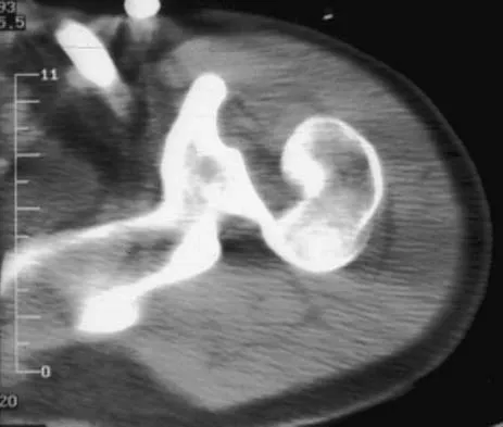
A 22-year-old right hand-dominant man who fell off his motorcycle onto the tip of his right shoulder 2 weeks ago now reports pain and difficulty raising his right arm. Examination reveals tenderness and gross movement over the lateral scapular spine and severe weakness during resisted abduction. A radiograph and 3D-CT scan are shown in Figures 24a and 24b. What is the next most appropriate step in management?


Explanation

24b The patient has a displaced scapular spine fracture that has resulted in shoulder weakness from a poor deltoid lever arm. The downward tilt may lead to subacromial impingement and rotator cuff dysfunction. Open reduction and internal fixation would best allow normal deltoid and shoulder function. Bone stimulators and abduction bracing may lead to healing but in a malunited position. Arthroscopic acromioplasty and fragment excision should be avoided. Ogawa K, Naniwa T: Fractures of the acromion and the lateral scapular spine. J Shoulder Elbow Surg 1997;6:544-548.

Question 2

A 20-year-old minor league baseball pitcher is diagnosed with a symptomatic torn ulnar collateral ligament (UCL) in his pitching elbow. Nonsurgical management consisting of rest and physical therapy aimed at elbow strengthening has failed to provide relief. He has concomitant cubital tunnel symptoms that worsen while throwing. What is his best surgical option?


Explanation

High-level pitchers with symptomatic UCL tears require reconstruction, with autograft being the best studied graft selection. With concomitant ulnar nerve symptoms, a simultaneous ulnar nerve transposition provides good results. Ligament "repairs" and allograft reconstructions have not shown good long-term results. Azar FM, Andrews JR, Wilk KE, et al: Operative treatment of ulnar collateral ligament injuries of the elbow in athletes. Am J Sports Med 2000;28:16-23.

Question 3

A 30-year-old man has pain in the left arm after a motor vehicle accident. His neurovascular examination is intact, and radiographs are shown in Figures 25a and 25b. What is the best course of management?


Explanation

25b The floating elbow is best managed with early open reduction and internal fixation of the humeral and forearm fractures, followed by early range of motion. These fractures predispose the elbow to stiffness, and early range of motion is recommended. Solomon HB, Zadnik M, Eglseder WA: A review of outcomes in 18 patients with floating elbow. J Orthop Trauma 2003;17:563-570.

Question 4

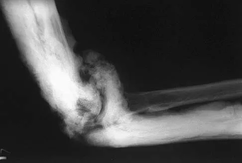
A patient who underwent open reduction and internal fixation of an olecranon fracture 2 months ago now reports painless limitation of motion. Examination reveals a well-healed incision and a flexion-extension arc from 40 degrees to 80 degrees. The patient has been performing home exercises. Radiographs are shown in Figures 26a and 26b. What is the most appropriate treatment?


Explanation

26b The radiographs do not show an articular malunion. Treatment is directed at the soft-tissue contracture and should begin with formal physical therapy and static progressive splinting. Radiation therapy is effective in the perioperative period and is indicated when ectopic bone formation is a concern. Morrey BF: The posttraumatic stiff elbow. Clin Orthop Relat Res 2005;431:26-35.

Question 5

A 23-year-old professional baseball pitcher reports shoulder pain and decreased velocity while pitching. Physical examination reveals a side-to-side internal rotation deficit of 25 degrees. The O'Brien sign is negative; Neer and Hawkins signs are negative. Rotator cuff strength is full. Radiographs are unremarkable. What is the next step in management?


Explanation

Throwing athletes with symptomatic internal rotation deficits often benefit from an intensive posterior capsular stretching program. Patients that fail to respond to nonsurgical management may benefit from an arthroscopic posterior capsular release. Wilk KE, Meister K, Andrews JR: Current concepts in rehabilitation of the overhead throwing athlete. Am J Sports Med 2002;30:136-151.

Question 6

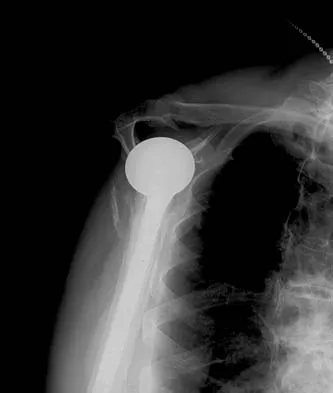
A 72-year-old woman who is right hand-dominant has severe pain in the right shoulder that has failed to respond to nonsurgical management. She reports night pain and significant disability. Examination reveals 30 degrees of active forward elevation. An AP radiograph is shown in Figure 27. Which of the following treatment options will provide the best functional improvement?


Explanation

The patient has end-stage rotator cuff tear arthropathy. The radiograph shows complete proximal humeral migration (acromiohumeral interval of 0 mm), severe glenohumeral arthritis, and acetabularization of the acromion. In addition, she has "pseudoparalysis" with active elevation of only 30 degrees. Reverse shoulder arthroplasty affords her the best opportunity for pain relief and functional improvement. The other procedures have mixed results but typically are better for pain relief than they are for functional gains. Frankle M, Siegal S, Pupello D, et al: The reverse shoulder prosthesis for glenohumeral arthritis associated with severe rotator cuff deficiency: A minimum two-year follow-up study of sixty patients. J Bone Joint Surg Am 2005;87:1697-1705.

Question 7

A healthy 64-year-old man just underwent an uncomplicated shoulder arthroplasty for severe glenohumeral osteoarthritis. Intraoperatively, 60 degrees of external rotation was obtained. Postoperatively, he starts on a range-of-motion program. What limitations are recommended?


Explanation

The patient needs restrictions on his external rotation to allow healing of the subscapularis tendon repair. Limitation to 60 degrees is common if the tendon repair is robust and shows no evidence of tension on range-of-motion testing during the surgery. Restriction from external rotation stretching for even 3 weeks would compromise his ultimate functional recovery. Boardman ND III, Cofield RH, Bengston KA, et al: Rehabilitation after total shoulder arthroplasty. J Arthroplasty 2001;16:483-486.

Question 8

A 64-year-old man who was involved in a high-speed motor vehicle accident 6 weeks ago has been in the ICU with a closed head injury. Examination reveals that his range of motion for external rotation to the side is -30 degrees. Radiographs are shown in Figures 28a and 28b. What is the most likely diagnosis?


Explanation

28b The patient has a posterior shoulder dislocation. The AP radiograph shows overlapping of the humeral head on the glenoid. The scapular Y view shows his humeral articular surface posterior to the glenoid. The posterior shoulder dislocation is frequently missed because the patient is comfortable in the "sling" position with the arm adducted and internally rotated across the abdomen. The marked restriction in external rotation on examination raises the suspicion of a posterior dislocation, adhesive capsulitis, or glenohumeral osteoarthritis. The posterior dislocation is diagnosed based on the radiographic findings. An axillary view or CT is recommended to better evaluate the dislocation. Robinson CM, Aderinto J: Posterior shoulder dislocations and fracture-dislocations. J Bone Joint Surg Am 2005;87:639-650.

Question 9

A 17-year-old high school football player reports wrist pain 5 months after the conclusion of the football season. A radiograph and MRI scan are shown in Figures 29a and 29b. What is the recommended intervention?


Explanation

29b The patient has a nonunion of the proximal pole of the scaphoid. Acutely, this can be repaired with a screw alone, but as a nonunion the proximal pole has very poor healing potential. Vacularized bone grafts have been successful for these challenging nonunions, particularly in adolescents. A cast can be used for nondisplaced acute waist fractures, and corticocancellous grafts can be used for nonunions of the waist. Waters PM, Stewart SL: Surgical treatment of nonunion and avascular necrosis of the proximal part of the scaphoid in adolescents. J Bone Joint Surg Am 2002;84:915-920.

Question 10

A 58-year-old woman with a history of severe asthma and long-term prednisone use reports a progression of chronic shoulder pain for the past 6 months. Radiographs and MRI scans are shown in Figures 30a through 30d. What is the most likely diagnosis?


Explanation

30b 30c 30d The patient has osteonecrosis of the humeral head. The radiographs show increased density in the superior subchondral region of the humeral head. The MRI scans reveal a central collapse of the humeral head. The patient's history of severe asthma and long-term prednisone use predisposes her to this condition. The MRI scans show no evidence of a full- or partial-thickness rotator cuff tear. Without a history of fevers, chills, or other systemic signs or symptoms, there is no indication of septic arthritis. The radiographs do not reveal periarticular erosions, commonly seen in rheumatoid arthritis. Matsen FA III, Rockwood CA Jr, Wirth MA, et al: Glenohumeral arthritis and its management, in Rockwood CA Jr, Matsen FA III (eds): Rockwood and Matsen The Shoulder, ed 2. Philadelphia, PA, WB Saunders, 1998, pp 871-874.

Question 11

A 28-year-old man sustained a shoulder dislocation 2 years ago. It remained dislocated for 3 weeks and required an open reduction. He now reports constant pain and has only 60 degrees of forward elevation and 10 degrees of external rotation. He desires to return to some sporting activities. An AP radiograph and intraoperative photograph (a view of the humeral head through a deltopectoral approach) are shown in Figures 31a and 31b. What is the best treatment option to decrease pain and improve function?


Explanation

31b The radiograph and intraoperative photograph show osteonecrosis with near complete head loss/collapse. A stemmed implant is more appropriate in this patient because there is very little bone to support a resurfacing implant. In a younger patient, a glenoid implant should be delayed as long as possible because of the eventual need for revision secondary to glenoid loosening and wear, especially in a young active male. The hemiarthroplasty may be converted to a total shoulder arthroplasty in the future. Levy O, Copeland SA: Cementless surface replacement arthroplasty of the shoulder: 5- to 10-year results with the Copeland mark-2 prosthesis. J Bone Joint Surg Br 2001;83:213-221.

Question 12

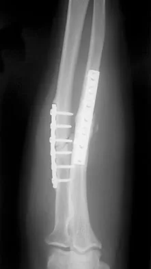
A 34-year-old man underwent open reduction and internal fixation of a closed both bones forearm fracture 11 months ago. The radiographs shown in Figures 32a and 32b reveal a 3-mm gap and loose screws. What is the best treatment option?


Explanation

32b In an atrophic nonunion with a good soft-tissue envelope, adequate plating with cancellous bone graft can be used to span defects of up to 6 cm. Cortical graft from the fibula or iliac crest is not necessary. BMP-7 is a bone graft substitute and should not be used alone in this patient because the hardware is loose.

Question 13

A football lineman who sustained a traumatic injury while blocking during a game now reports that his shoulder is slipping while pass blocking. Examination reveals no apprehension in abduction and external rotation; however, he reports pain with posterior translation of the shoulder. He has full strength in external rotation, internal rotation, and supraspinatus testing. What is the pathology most likely responsible for his symptoms?


Explanation

Traumatic posterior instability is a common finding in football players, especially in the blocking positions as well as in the defensive linemen and linebackers. A traumatic blow to the outstretched arm results in posterior glenohumeral forces. Labral detachment at the glenoid rim is common. Patients report slipping or pain with posteriorly directed pressure. Rarely do these patients have true dislocations that require reduction; however, recurrent episodes of subluxation or pain are not uncommon. Posterior repair has been shown to be successful in the treatment of traumatic instability. Bottoni CR, Franks BR, Moore JH, et al: Operative stabilization of posterior shoulder instability. Am J Sports Med 2005;33:996-1002. Williams RJ III, Strickland S, Cohen M, et al: Arthroscopic repair for traumatic posterior shoulder instability. Am J Sports Med 2003;31:203-209.

Question 14

A 17-year-old girl has multidirectional instability of the shoulder. What is the most appropriate initial management?


Explanation

Multidirectional instability of the shoulder is defined as symptomatic instability in two or more directions (anterior, posterior) but must include a component of inferior instability. Initial treatment should always include physical therapy and instruction in a home exercise program that emphasizes periscapular and rotator cuff strengthening to improve the dynamic stability of the glenohumeral joint. Immobilization has not been shown to be effective. Open capsular shift and arthroscopic capsular plication remain the surgical options when appropriate nonsurgical management fails (typically a minimum of 6 months of dedicated therapy and home program). Thermal capsulorrhaphy remains controversial but is not recommended by many clinicians because of reported complications including recurrent instability, axillary nerve injury, chondrolysis, and capsular injury. Neer CS II, Foster CR: Inferior capsular shift for involuntary inferior and multidirectional instability of the shoulder: A preliminary report. J Bone Joint Surg Am 1980;62:897-908. D'Alessandro DF, Bradley JP, Fleischli JE, et al: Prospective evaluation of thermal capsulorrhaphy for shoulder instability: Indications and results, two- to five-year follow-up. Am J Sports Med 2004;32:21-33. Levine WN, Clark AM Jr, D'Alessandro DF, et al: Chondrolysis following arthroscopic thermal capsulorrhaphy to treat shoulder instability: A report of two cases. J Bone Joint Surg Am 2005;87:616-621.

Question 15

In surgically treating hand and finger infections in patients with diabetes mellitus, what factor is associated with higher amputation rates?


Explanation

Patients with diabetes mellitus are prone to infection, and surgical treatment of their infections frequently requires multiple procedures. The triad of poor wound healing, chronic neuropathy, and vascular disease contributes to the increased infection rate. Studies have demonstrated increased amputation rates in patients with diabetes mellitus who have renal failure or deep polymicrobial or gram-negative infections. Gonzalez MH, Bochar S, Novotny J, et al: Upper extremity infections in patients with diabetes mellitus. J Hand Surg Am 1999;24:682-686. Trumble TE (ed): Hand Surgery Update 3: Hand, Elbow, & Shoulder. Rosemont, IL, American Society for Surgery of the Hand, 2003, pp 433-457.

Question 16

A 40-year-old unrestrained passenger reports chest wall pain after a motor vehicle accident. Which of the following structures is most important in preventing the injury shown in Figure 33?


Explanation

Through cadaveric study, Spencer and associates measured anterior and posterior translation of the sternoclavicular joint. The study demonstrated that the posterior sternoclavicular joint capsule is the most important structure for preventing both anterior and posterior translation of the sternoclavicular joint. Gilot GJ, Wirth MA, Rockwood CA: Injuries to the sternoclavicular joint, in Bucholz RW, Heckman JD, Court-Brown C (eds): Fractures in Adults. Philadelphia, PA, Lippincott, Williams and Wilkins, 2006, vol 2, pp 1373-1374.

Question 17

Figures 34a and 34b show the axial and sagittal MRI scans of a 36-year-old man who reports the insidious onset of pain in the right shoulder. What is the most appropriate description of the acromial morphology?


Explanation

34b The MRI scans reveal a meso os acromiale with edema at the site in a skeletally mature patient. Sher JS: Anatomy, biomechanics, and pathophysiology of rotator cuff disease, in Iannotti JP, Williams GR (eds): Disorders of the Shoulder: Diagnosis and Management. Philadelphia, PA, Lippincott Williams & Wilkins, 1999, p 23.

Question 18

What is the primary indication for performing a total wrist arthroplasty in a patient with painful rheumatoid arthritis?


Explanation

The most conservative indications for a total wrist arthroplasty are to spare motion on one side and to improve activities of daily living. Component loosening, dislocation, and wound problems are frequent. Suitable patients can be of various ages, wrist motion, and radiographic stages of arthritis. Ipsilateral total elbow arthroplasty, type III degenerative changes of the wrist, age older than 55, and limited range of motion are neither primary indications nor contraindications to a total wrist arthroplasty. Divelbiss BJ, Sollerman C, Adams BD: Early results of the universal total wrist arthroplasty in rheumatoid arthritis. J Hand Surg Am 2002;27:195-204. Vicar AJ, Burton RI: Surgical management of rheumatoid wrist-fusion or arthroplasty. J Hand Surg Am 1986;11:790-797.

Question 19

What is the most likely cause of the lesion shown in Figures 35a and 35b?


Explanation

35b The most common cause of myositis ossificans is contusion. Certain regions, including the quadriceps and brachialis, are more commonly affected. The mechanisms of development have not been clearly established. Beiner JM, Jokl P: Muscle contusion injuries: Current treatment options. J Am Acad Orthop Surg 2001;9:227-237.

Question 20

During treatment of rupture of the subscapularis tendon with associated biceps instability, treatment of the biceps tendon should include which of the following?


Explanation

With subscapularis tendon ruptures that have biceps tendon pathology, treatment with tenodesis or tenotomy has improved clinical results. Subluxation or dislocation of the biceps tendon is common with subscapularis rupture. Dislocation of the biceps can occur either beneath the tendon, within the tendon, or extra-articularly. In all cases, the restraints to medial translations of the biceps have been disrupted. Attempts at recentering the biceps have not been successful, and clinical results appear to be improved when tenodesis or tenotomy is employed in the treatment of the unstable biceps associated with subscapularis tears. Edwards TB, Walch G, Sirvenaux F, et al: Repair of tears of the subscapularis: Surgical technique. J Bone Joint Surg Am 2006;88:1-10. Deutsch A, Altchek DW, Veltri DM, et al: Traumatic tears of the subscapularis tendon: Clinical diagnosis, magnetic resonance imaging findings, and operative treatment. Am J Sports Med 1997;25:13-22.

Question 21

What is the most common bacteria cultured from dog and cat bites to the upper extremity?


Explanation

To define bacteria responsible for dog and cat bite infections, a prospective study yielded a median of five bacterial isolates per culture. Pasteurella is most common from both dog bites (50%) and cat bites (75%). Pasteurella canis was the most frequent pathogen of dog bites, and Pasteurella multocida was the most common isolate of cat bites. Other common aerobes included streptococci, staphylococci, moraxella, and neisseria.

Question 22

A previously healthy 65-year-old woman has a closed fracture of the right clavicle after falling down the basement stairs. Examination reveals good capillary refill in the digits of her right hand. Radial and ulnar pulses are 1+ at the right wrist compared with 2+ on the opposite side. In the arteriogram shown in Figure 36, the arrow is pointing at which of the following arteries?


Explanation

The axillary artery commences at the first rib as a direct continuation of the subclavian artery and becomes the brachial artery at the lower border of the teres major. The arteriogram reveals a nonfilling defect in the third portion of the artery just distal to the subscapular artery. The complex arterial collateral circulation in this region often permits distal perfusion of the extremity despite injury.

Question 23

Which of the following structures may help maintain radial length after a radial head fracture?


Explanation

Essex-Lopresti injuries affect axial stability of the forearm. Injury to the interosseous membrane or the triangular fibrocartilage complex can result in proximal migration of the radius. Morrey BF, Chao EY, Hui FC: Biomechanical study of the elbow following excision of the radial head. J Bone Joint Surg Am 1979;61:63-68.

Question 24

An adult patient has a closed humeral fracture that was treated nonsurgically and a concomitant radial nerve injury. Six weeks after injury, electromyography shows no evidence of recovery. Management should now consist of


Explanation

In patients with radial nerve injuries with closed humeral fractures, it has been reported that 85% to 95% spontaneously recover. Based on this premise, most surgeons favor expectant management of these injuries. Even if there is no evidence of recovery at 6 weeks, repeat electromyography at 12 weeks is advocated. If there is no clinical or electromyographic signs of recovery at 6 months, exploration is recommended. If the nerve is in continuity at the time of exploration, nerve action potentials are useful in helping determine the need for neurolysis, excision, and grafting, or if excision and repair is the best option. Pollock FH, Drake D, Bovill EG, et al: Treatment of radial neuropathy associated with fractures of the humerus. J Bone Joint Surg Am 1981;63:239-243.

Question 25

A 55-year-old man who works as a carpenter reports chronic right anterior shoulder pain and weakness. Examination reveals 90 degrees of external rotation (with the arm at the side) compared to 45 degrees on the left side. His lift-off examination is positive, along with a positive belly press finding. An MRI scan reveals a chronic, retracted atrophied subscapularis tendon. What is the most appropriate management of his shoulder pain and weakness?


Explanation

Chronic subscapularis tendon ruptures preclude primary repair. In such instances, subcoracoid pectoralis major tendon transfers may improve function and diminish pain. The subcoracoid position of the transfer allows redirection of the pectoralis major in a direction recreating the vector of the subscapularis tendon. Shoulder fusion is a salvage procedure, and corticosteroid injection may reduce pain but will not improve function. Jost B, Puskas GJ, Lustenberger A, et al: Outcome of pectoralis major transfer for the treatment of irreparable subscapularis tears. J Bone Joint Surg Am 2003;85:1944-1951.

Question 26

A 45-year-old competitive weightlifter feels a sudden, painful 'pop' in his anterior elbow while performing heavy bicep curls. Examination reveals a 'Popeye' deformity and significant weakness in forearm supination. He undergoes a distal biceps tendon repair using a traditional two-incision technique. Compared to a single anterior incision approach, which of the following complications is significantly more likely with the two-incision technique?


Explanation

The traditional two-incision approach for distal biceps repair (extensile anterior incision and posterolateral incision) carries a higher risk of heterotopic ossification and radioulnar synostosis compared to a single anterior incision. A single anterior incision, however, carries a higher risk of nerve injury, specifically to the lateral antebrachial cutaneous (LABC) nerve and the posterior interosseous nerve (PIN), due to the vigorous retraction required to visualize the radial tuberosity. Rerupture rates are generally low and comparable between both techniques.

Question 27

A 35-year-old woman falls on an outstretched hand and sustains a complex elbow injury. Radiographs and CT scan demonstrate a posterior elbow dislocation, a comminuted radial head fracture, and a Type II coronoid fracture. Following closed reduction, the elbow remains grossly unstable. During open surgical reconstruction, what is the most widely accepted sequence of fixation to restore elbow stability?


Explanation

The injury described is the 'terrible triad' of the elbow. The standard surgical sequence advocated by Pugh and colleagues follows an 'inside-out' or deep-to-superficial approach. The coronoid is addressed first (via the anterior capsule/brachialis), followed by the radial head (fixation or arthroplasty), and finally the lateral collateral ligament (LCL) complex is repaired to the lateral epicondyle. MCL repair or hinged external fixation is only added if the elbow remains unstable after these three steps.

Question 28

A 74-year-old woman presents with severe shoulder pain after a mechanical fall. Radiographs demonstrate a displaced 4-part proximal humerus fracture with severe comminution of the tuberosities. Her medical history is significant for chronic pseudoparalysis of the affected shoulder secondary to a known, massive, irreparable rotator cuff tear. What is the most appropriate surgical management?


Explanation

Reverse total shoulder arthroplasty (RTSA) is the treatment of choice for elderly patients with complex 4-part proximal humerus fractures in the setting of pre-existing massive rotator cuff tears (cuff tear arthropathy) or pre-existing pseudoparalysis. Hemiarthroplasty relies heavily on tuberosity healing for functional outcome, which is highly unpredictable in the elderly and impossible in the setting of a massive, pre-existing cuff tear. ORIF in osteoporotic bone with poor cuff tissue has a high failure rate. Anatomic total shoulder arthroplasty is contraindicated in the absence of a functioning rotator cuff.

Question 29

A 48-year-old manual laborer complains of a 5-year history of progressive, aching right wrist pain. Radiographs reveal a scaphoid nonunion with advanced collapse (SNAC). There is prominent arthritic change at the radioscaphoid joint and the capitolunate joint, but the radiolunate articulation is completely spared. Which of the following is the most appropriate surgical treatment?


Explanation

The patient has Stage III SNAC wrist, characterized by radioscaphoid and midcarpal (capitolunate) arthritis, with preservation of the radiolunate joint. Proximal row carpectomy (PRC) is contraindicated because the capitate head is arthritic and would articulate with the lunate fossa, leading to rapid failure. Scaphoid excision with a four-corner fusion removes the arthritic radioscaphoid joint and fuses the arthritic capitolunate joint while preserving some wrist motion through the pristine radiolunate joint. Total wrist arthrodesis is reserved for pan-carpal arthritis (Stage IV).

Question 30

A 22-year-old collegiate rugby player undergoes a Latarjet procedure for recurrent anterior shoulder instability associated with 28% anterior glenoid bone loss. The procedure involves transferring the coracoid process to the anterior glenoid neck. Which muscular structure is transferred along with the coracoid to provide a dynamic 'sling' effect across the anteroinferior capsule?


Explanation

The Latarjet procedure involves osteotomizing the coracoid process and transferring it, along with the attached conjoined tendon (composed of the short head of the biceps and the coracobrachialis), to the anterior glenoid. This provides a 'triple blocking effect': 1) the bone block itself, 2) the dynamic sling effect of the conjoined tendon reinforcing the inferior capsule when the arm is abducted and externally rotated, and 3) the repair of the capsule to the stump of the coracoacromial ligament (if preserved).

Question 31

Six weeks after nonoperative management of a nondisplaced distal radius fracture, a 62-year-old woman presents with the sudden inability to actively extend the interphalangeal joint of her thumb. An extensor indicis proprius (EIP) to extensor pollicis longus (EPL) tendon transfer is planned. At the level of the metacarpophalangeal joint of the index finger, where is the EIP tendon located relative to the extensor digitorum communis (EDC) tendon?


Explanation

EPL rupture is a known complication of nondisplaced distal radius fractures, often secondary to ischemia or attrition over a bony spike at Lister's tubercle. The standard treatment is an EIP to EPL tendon transfer. To harvest the EIP correctly, the surgeon must remember the anatomic relationship: at the level of the metacarpophalangeal joint, the EIP tendon consistently lies ulnar and volar to the EDC tendon of the index finger.

Question 32

A 29-year-old elite volleyball attacker reports vague posterior shoulder pain and progressive weakness. Physical examination reveals isolated weakness in external rotation and profound atrophy of the infraspinatus fossa. Abduction strength and supraspinatus bulk are entirely normal. An MRI confirms a paralabral cyst compressing a peripheral nerve. At what specific anatomic location is the nerve compression occurring?


Explanation

The patient has isolated infraspinatus atrophy and external rotation weakness, which indicates compression of the suprascapular nerve distal to the innervation of the supraspinatus. This occurs at the spinoglenoid notch. Compression at the suprascapular notch (proximally) would affect both the supraspinatus (abduction) and the infraspinatus (external rotation). Quadrilateral space syndrome involves the axillary nerve.

Question 33

A 27-year-old woman complains of progressive dorsal wrist pain. Radiographs demonstrate sclerosis, fragmentation, and flattening of the lunate without carpal collapse. Her ulnar variance is determined to be -3 mm. Based on the most likely diagnosis, what is the preferred initial surgical intervention?


Explanation

The clinical picture describes Kienböck's disease (avascular necrosis of the lunate), Lichtman Stage II or IIIA (fragmentation/flattening without fixed carpal collapse). In a patient with negative ulnar variance, a joint-leveling procedure such as a radial shortening osteotomy is the treatment of choice. It unloads the lunate by shifting compressive forces to the radioulnar joint and the scaphoid. Ulnar lengthening is theoretically similar but associated with higher complication rates (nonunion). Proximal row carpectomy is reserved for advanced stages (Stage IIIB or IV).

Question 34

A 52-year-old man with severe, electromyographically confirmed cubital tunnel syndrome is undergoing an anterior subcutaneous transposition of the ulnar nerve. During the transposition, a specific structural band extending from the medial epicondyle to the medial humerus must be resected to prevent secondary kinking of the nerve as it is moved anteriorly. What is this structure?


Explanation

When performing an anterior transposition of the ulnar nerve, the medial intermuscular septum must be excised. If left intact, the ulnar nerve can tether or kink sharply over the edge of the septum as it routes anteriorly, leading to persistent or worsened iatrogenic compression. The Arcade of Struthers is a proximal site of primary compression but kinking during transposition is classic for the medial intermuscular septum. The Arcade of Frohse relates to the posterior interosseous nerve (PIN).

Question 35

A 38-year-old man falls from a height of 10 feet, sustaining a severely comminuted, unsalvageable radial head fracture. The surgeon performs an isolated radial head excision. Six months later, the patient develops severe, progressive ulnar-sided wrist pain. Radiographs reveal 5 mm of positive ulnar variance. What concurrent injury was missed at the time of the initial trauma?


Explanation

The scenario describes an Essex-Lopresti lesion, which consists of a radial head fracture, rupture of the forearm interosseous membrane (IOM), and disruption of the distal radioulnar joint (DRUJ). If the radial head is simply excised without recognizing the IOM injury, there is no proximal block to radius migration. The radius migrates proximally, causing dynamic positive ulnar variance, severe ulnar impaction syndrome, and wrist pain. In these injuries, the radial head must be replaced with an arthroplasty to maintain radial length.

Question 36

A 55-year-old woman sustained a distal radius fracture treated with a volar locking plate. Three months postoperatively, she presents with inability to actively flex the interphalangeal joint of her thumb. Radiographs show plate placement distal to the watershed line. Which tendon is most commonly ruptured in this scenario?


Explanation

Volar plating of distal radius fractures is associated with flexor tendon rupture if the plate is placed distal to the watershed line. The flexor pollicis longus (FPL) tendon is most commonly affected due to its direct proximity to the volar margin of the radius and the plate edge. Extensor pollicis longus (EPL) ruptures are more frequently associated with dorsal screw prominence or nonoperative management of nondisplaced fractures.

Question 37

A 45-year-old manual laborer presents with chronic shoulder pain, profound weakness in external rotation, and a positive hornblower's sign. MRI demonstrates a massive, retracted, and irreparable tear of the supraspinatus and infraspinatus with Goutallier stage 4 fatty infiltration. The subscapularis is intact, and there is no glenohumeral arthritis. Which of the following is the most appropriate tendon transfer to restore active external rotation?


Explanation

In a young, active patient with an irreparable posterosuperior rotator cuff tear (supraspinatus/infraspinatus) without arthritis, a tendon transfer is indicated. Lower trapezius transfer is increasingly favored and highly tested for external rotation deficits (loss of infraspinatus/teres minor) because its force vector more closely replicates the infraspinatus. While latissimus dorsi transfer has historically been used, its primary vector is adduction and internal rotation, requiring significant cortical retraining. Pectoralis major transfer is indicated for irreparable subscapularis tears.

Question 38

A 35-year-old man falls from a height and sustains a traumatic elbow dislocation. After closed reduction, radiographs reveal a displaced radial head fracture, a small type 1 coronoid tip fracture, and a lateral collateral ligament (LCL) tear. He is scheduled for operative fixation. What is the standard and most appropriate sequence of surgical repair for this 'terrible triad' injury?


Explanation

The standard surgical protocol for a terrible triad injury of the elbow proceeds from deep to superficial. The typical sequence is: 1) Coronoid fixation or anterior capsule repair, 2) Radial head fixation or replacement, 3) Lateral collateral ligament (LCL) repair. If the elbow remains unstable after these steps, the medial collateral ligament (MCL) may be repaired or a hinged external fixator applied.

Question 39

A 72-year-old woman with a history of osteoporosis sustains a severely comminuted 4-part proximal humerus fracture. A reverse total shoulder arthroplasty (RTSA) is planned. To optimize outcomes and restore appropriate deltoid tension, which of the following landmarks is most critical for determining the correct height of the humeral prosthesis?


Explanation

When performing an RTSA or hemiarthroplasty for a proximal humerus fracture, restoring humeral length and retroversion is critical for proper soft-tissue tensioning, especially of the deltoid. The superior border of the pectoralis major tendon insertion is a reliable landmark. The average distance from the superior border of the pectoralis major insertion to the top of the native humeral head is approximately 5.6 cm.

Question 40

A 24-year-old man presents with chronic wrist pain 8 months after a fall. Radiographs show a scaphoid proximal pole fracture nonunion. MRI demonstrates low T1 and T2 signal intensity throughout the proximal pole, indicating avascular necrosis (AVN). There is no radioscaphoid arthritis. Which of the following is the most appropriate surgical treatment?


Explanation

In the presence of a proximal pole scaphoid nonunion with avascular necrosis (AVN), indicated by low T1 and T2 MR signal without punctate bleeding intraoperatively, vascularized bone grafting is required. Free vascularized grafts like the medial femoral condyle (MFC) graft or pedicled grafts (1,2-ICSRA) are indicated because non-vascularized grafts have a much lower healing rate when AVN is present. Salvage procedures (four-corner fusion, PRC) are reserved for cases with established arthritis.

Question 41

A 50-year-old manual worker presents with dorsal wrist pain and decreased range of motion. Radiographs demonstrate advanced narrowing of the radioscaphoid joint and midcarpal (capitolunate) joint, while the radiolunate joint is perfectly preserved. Which of the following procedures is strictly contraindicated in this patient?


Explanation

The patient has a stage III Scapholunate Advanced Collapse (SLAC) wrist (radioscaphoid and midcarpal arthritis, capitate migration). Proximal row carpectomy (PRC) involves excising the scaphoid, lunate, and triquetrum, which leaves the capitate to articulate with the lunate fossa of the radius. Therefore, PRC requires a preserved capitate head and lunate fossa. In stage III SLAC wrist, the capitate head is arthritic, making PRC contraindicated. Four-corner fusion is the preferred motion-preserving salvage procedure.

Question 42

A 42-year-old bodybuilder feels a 'pop' in his anterior elbow while lifting a heavy object, presenting with an abnormal contour of the biceps muscle belly and weakness in supination. He undergoes a 2-incision surgical repair of the distal biceps tendon. Which of the following complications is most specifically associated with the 2-incision technique compared to a single anterior incision technique?


Explanation

The 2-incision technique (Boyd-Anderson or Morrey modification) for distal biceps repair avoids the radial nerve (PIN) injuries associated with a single anterior incision. However, because it involves dissecting between the radius and ulna and exposing the ulna during the posterolateral approach, it carries a higher risk of heterotopic ossification and potentially radioulnar synostosis. The single anterior incision carries a higher risk of lateral antebrachial cutaneous nerve (LABCN) neuropraxia and PIN injury.

Question 43

A 21-year-old collegiate rugby player is undergoing a Latarjet procedure for recurrent anterior shoulder instability. The coracoid process is osteotomized and transferred to the anterior glenoid neck. Which of the following structures creates the 'sling effect' that provides dynamic stability in this procedure?


Explanation

The Latarjet procedure provides stability through the 'triple-blocking' effect: 1) The bone block effect from the transferred coracoid; 2) The dynamic 'sling effect' of the conjoined tendon (short head of biceps and coracobrachialis) acting as a sling across the inferior subscapularis and anterior-inferior capsule when the arm is abducted and externally rotated; 3) Capsule repair to the stump of the coracoacromial ligament.

Question 44

A 32-year-old carpenter presents with insidious onset of dorsal wrist pain. Radiographs demonstrate sclerosis and fragmentation of the lunate, but carpal height is maintained, and there is no fixed scaphoid rotation. The patient has negative ulnar variance. Which of the following surgical interventions is most appropriate?


Explanation

The patient has Lichtman Stage IIIa Kienböck's disease (lunate collapse/fragmentation, but normal carpal height and no fixed scaphoid rotation). For early and intermediate stages (I, II, and IIIa) in patients with negative ulnar variance, a joint-leveling procedure such as a radial shortening osteotomy is the treatment of choice to decrease radiolunate contact stresses and offload the lunate. Salvage procedures are reserved for stage IIIb or IV.

Question 45

A 19-year-old pitcher presents with medial elbow pain during the late cocking phase of throwing. MRI arthrogram shows a partial tear of the ulnar collateral ligament (UCL) anterior bundle. After failing nonoperative management, he undergoes surgical reconstruction. What is the precise anatomical insertion site for the graft on the ulna during a standard UCL reconstruction?


Explanation

The anterior bundle of the ulnar collateral ligament (UCL) is the primary restraint to valgus stress at the elbow. Its anatomical origin is the anteroinferior aspect of the medial epicondyle, and its anatomical insertion is the sublime tubercle on the medial aspect of the coronoid process of the ulna. Graft fixation at the sublime tubercle is critical to restoring native biomechanics.

Question 46

A 45-year-old heavy laborer undergoes a single-incision anterior approach for the repair of a retracted acute distal biceps tendon rupture. Two weeks postoperatively, he complains of numbness over the lateral aspect of his forearm. His motor function in the hand and wrist is completely intact. Which of the following structures was most likely injured during the procedure?


Explanation

The lateral antebrachial cutaneous (LABC) nerve is the most commonly injured nerve during a single-incision anterior repair of the distal biceps tendon. It courses between the biceps and brachialis muscles before piercing the fascia lateral to the biceps tendon. Injury results in sensory deficits (numbness or paresthesia) over the lateral forearm. The posterior interosseous nerve (PIN) injury is a devastating potential complication more commonly associated with two-incision approaches or over-retraction laterally, but it would present with motor deficits in wrist and finger extension.

Question 47

A 38-year-old woman falls on an outstretched hand and sustains a 'terrible triad' injury to her right elbow. Which of the following represents the most appropriate sequence of surgical reconstruction to effectively restore joint stability?


Explanation

The standard surgical protocol for a terrible triad injury of the elbow (elbow dislocation, radial head fracture, coronoid fracture) typically proceeds from deep to superficial and medial to lateral (if approached from a single lateral incision). The widely accepted sequence is: 1) fixation of the coronoid process to restore the anterior buttress, 2) repair or replacement of the radial head to restore the anterior and valgus buttress, and 3) repair of the lateral ulnar collateral ligament (LUCL) to restore posterolateral rotatory stability. If the elbow remains unstable after these steps, the medial collateral ligament (MCL) may be repaired or a hinged external fixator applied.

Question 48

A 28-year-old male falls from a height and sustains a highly comminuted radial head fracture, diffuse forearm tenderness, and distal radioulnar joint (DRUJ) instability, consistent with an Essex-Lopresti injury. He undergoes prompt radial head replacement to restore length. Intraoperatively, following the radial head replacement, the DRUJ remains grossly unstable in neutral rotation. What is the next best step in management?


Explanation

An Essex-Lopresti injury involves a radial head fracture, disruption of the central band of the interosseous membrane (IOM), and DRUJ instability. The primary treatment in the acute setting involves restoring the radiocapitellar contact and length with rigid radial head fixation or arthroplasty. If the DRUJ remains unstable after restoring the radial column, it should be closed reduced and pinned with K-wires in a stable position (typically supination) for 4 to 6 weeks to allow the IOM and DRUJ ligaments to heal. Acute open repair of the IOM is rarely performed, and distal ulna resection is contraindicated as it will result in proximal migration of the radius.

Question 49

A 45-year-old male presents with chronic, progressive wrist pain. Radiographs reveal a scaphoid nonunion with advanced radioscaphoid arthritis and capitolunate arthritis. The radiolunate joint is well-preserved. This is consistent with a Stage III Scaphoid Nonunion Advanced Collapse (SNAC). Which of the following is the most appropriate motion-preserving surgical treatment?


Explanation

SNAC stage III involves degenerative arthritis of both the radioscaphoid and capitolunate joints, with preservation of the radiolunate joint. Proximal row carpectomy (PRC) is contraindicated in this stage because it relies on placing the head of the capitate into the lunate fossa; since the proximal capitate is arthritic, a PRC would result in a painful articulation. The most appropriate motion-preserving salvage procedure is scaphoid excision and a four-corner fusion (capitate, lunate, triquetrum, hamate), which preserves the healthy radiolunate articulation. Radial styloidectomy is reserved for SNAC I.

Question 50

A 29-year-old elite volleyball player presents with insidious onset of posterior right shoulder pain and weakness. On physical examination, there is noticeable atrophy of the infraspinatus muscle fossa, but the bulk of the supraspinatus is normal. She demonstrates significant weakness in external rotation, while her abduction strength is fully preserved. Which of the following is the most likely anatomic location of the nerve compression?


Explanation

The suprascapular nerve passes through the suprascapular notch (innervating the supraspinatus) and then continues distally through the spinoglenoid notch to innervate the infraspinatus. Compression at the suprascapular notch affects both muscles, causing weakness in both abduction and external rotation. Compression at the spinoglenoid notch, often due to a paralabral cyst (associated with posterior labral tears) or repetitive traction in overhead athletes, affects only the terminal branch to the infraspinatus. This causes isolated external rotation weakness and isolated infraspinatus atrophy.

Question 51

A 72-year-old right-hand-dominant woman sustains a 4-part proximal humerus fracture after a ground-level fall. Radiographs show a valgus impacted fracture pattern with disruption of the medial hinge and 1.5 cm of medial translation of the humeral shaft. She has pre-existing advanced glenohumeral osteoarthritis and a documented massive, irreparable rotator cuff tear. What is the most reliable surgical option for pain relief and functional restoration in this patient?


Explanation

Reverse total shoulder arthroplasty (RTSA) is strongly indicated for elderly, lower-demand patients presenting with complex proximal humerus fractures (e.g., poor bone quality, disrupted medial hinge) who also have pre-existing glenohumeral arthritis and massive rotator cuff tears (cuff tear arthropathy). Hemiarthroplasty outcomes are heavily dependent on tuberosity healing, which is unpredictable in older patients with poor bone stock and compromised cuff function. Anatomic TSA is contraindicated in the setting of a deficient rotator cuff, as it will lead to 'rocking horse' loosening of the glenoid component.

Question 52

A 22-year-old collegiate baseball pitcher presents with vague posterior shoulder pain during the late cocking and early acceleration phases of throwing. Examination reveals a 25-degree Glenohumeral Internal Rotation Deficit (GIRD) compared to the non-throwing shoulder, and positive apprehension. MRI arthrography demonstrates a posterosuperior labral tear and a partial articular-sided supraspinatus tendon avulsion (PASTA) lesion. Which of the following pathophysiologic mechanisms is most directly responsible for this specific cascade of pathology?


Explanation

The clinical scenario perfectly describes internal impingement, highly prevalent in overhead throwing athletes. It is characterized by pathologic contact between the posterior-superior glenoid labrum and the articular surface of the rotator cuff during maximal abduction and external rotation (late cocking phase). This condition is heavily driven by a posterior capsular contracture, which alters glenohumeral kinematics, shifts the center of rotation posterosuperiorly, and clinically manifests as Glenohumeral Internal Rotation Deficit (GIRD). This leads to 'peel-back' of the superior labrum and articular-sided cuff fraying (PASTA lesions).

Question 53

A 31-year-old male bodybuilder feels a sudden, painful 'pop' in his anterior chest while performing a heavy bench press. He presents with extensive ecchymosis over the anterior axilla, swelling, and a loss of the normal anterior axillary fold contour. Examination reveals severe weakness in adduction and internal rotation of the humerus. Which of the following anatomical locations is the most common site of rupture for this injury?


Explanation

Pectoralis major ruptures almost exclusively occur in males lifting heavy weights, particularly during the eccentric phase of the bench press. The most common site of rupture is an avulsion of the tendon from its insertion on the proximal humerus (specifically, the lateral lip of the bicipital groove). The sternocostal head is most frequently involved, as it is subjected to maximal stretch during eccentric loading. Surgical repair via anatomic reattachment to the humerus using suture anchors or cortical buttons provides superior functional and cosmetic outcomes in young, active individuals compared to nonoperative treatment.

Question 54

A 25-year-old man complains of persistent dorsal wrist pain and a clicking sensation 3 months after falling on an extended, ulnar-deviated wrist. Physical examination reveals focal tenderness over the dorsal scapholunate interval and a positive Watson scaphoid shift test. Radiographs show a scapholunate gap of 4 mm and a cortical 'ring sign' of the scaphoid. The radiolunate angle measures 20 degrees of dorsal tilt. Based on these radiographic findings, what carpal instability pattern has developed?


Explanation

Scapholunate dissociation disrupts the scapholunate interosseous ligament. Left unchecked, the scaphoid naturally flexes volarly (creating the 'signet ring' sign on PA radiographs as the scaphoid is viewed end-on). Conversely, the lunate falls into extension dorsally, dragged by the intact lunotriquetral ligament and the natural tendency of the triquetrum. This uncoupling creates a Dorsal Intercalated Segment Instability (DISI) deformity. Radiographically, a DISI is confirmed by a radiolunate angle of >15 degrees of dorsal tilt (extension) and a scapholunate angle >60 degrees.

Question 55

A 40-year-old carpenter presents with 6 months of persistent numbness in his small and ring fingers, and subjective clumsiness in his right hand. Examination shows a positive Tinel's sign at the cubital tunnel, weak pinch strength, and a positive Froment's sign. Electrodiagnostic studies confirm severe ulnar neuropathy at the elbow with active denervation in the first dorsal interosseous muscle. Notably, his right elbow demonstrates a 15-degree cubitus valgus deformity from a malunited pediatric supracondylar fracture. What is the most appropriate surgical intervention?


Explanation

While in situ decompression of the ulnar nerve is highly effective and widely utilized for primary, idiopathic cubital tunnel syndrome, an anterior transposition (whether subcutaneous, intramuscular, or submuscular) is specifically indicated when there is tension on the nerve, subluxation of the nerve over the medial epicondyle during flexion, or a significant valgus deformity of the elbow (cubitus valgus leading to tardy ulnar nerve palsy). In a patient with a malunited pediatric fracture and cubitus valgus, in situ decompression fails to address the underlying traction neuritis, making anterior transposition the procedure of choice.

Question 56

A 40-year-old patient undergoes surgical management for a 'terrible triad' injury of the elbow. A lateral approach is utilized. The coronoid fracture (Type 1) is repaired with a capsule suture lasso, the comminuted radial head is replaced, and the lateral ulnar collateral ligament (LUCL) is repaired to the lateral epicondyle. Following these steps, fluoroscopic evaluation reveals that the elbow remains persistently unstable and subluxates symmetrically at 45 degrees of flexion. What is the most appropriate next step in management?


Explanation

The standard surgical sequence for a terrible triad injury involves addressing the radial head, coronoid, and LUCL. If the elbow remains unstable with a symmetrical joint space widening after restoring the anterior and lateral restraints, this indicates severe medial sided insufficiency. The most appropriate next step is to repair the medial ulnar collateral ligament (MUCL). If the elbow remains unstable even after MUCL repair, the application of a hinged external fixator is indicated.

Question 57

A 65-year-old woman with a long-standing history of rheumatoid arthritis sustains a severe intra-articular, comminuted distal humerus fracture (AO/OTA 13-C3). Radiographs demonstrate severe osteopenia and preexisting radiocapitellar joint space narrowing. Which of the following is the most appropriate surgical treatment?


Explanation

Total elbow arthroplasty (TEA) is the treatment of choice for elderly patients with complex, comminuted intra-articular distal humerus fractures, particularly in the setting of poor bone quality or preexisting inflammatory arthritis (such as rheumatoid arthritis). TEA allows for immediate weight-bearing as tolerated and early range of motion, providing a more reliable outcome than internal fixation in this specific patient demographic.

Question 58

A 25-year-old man presents with chronic wrist pain 1 year after falling on an outstretched hand. Imaging reveals a scaphoid nonunion with a humpback deformity and avascular necrosis (AVN) of the proximal pole. There is no significant radiocarpal arthritis. Which of the following vascularized bone grafts is most appropriate for addressing this specific injury?


Explanation

For a scaphoid proximal pole nonunion complicated by avascular necrosis (AVN) and structural collapse (humpback deformity), a structural vascularized bone graft is required. The medial femoral condyle (MFC) free vascularized bone graft is the treatment of choice, as it provides robust blood supply and the structural integrity needed to correct the deformity. The 1,2-ICSRA graft often fails in the setting of true AVN with structural collapse.

Question 59

A 30-year-old man sustains a hyperextension wrist injury, resulting in a volar perilunate dislocation. During surgical reconstruction via a combined dorsal and volar approach, which of the following ligaments must be repaired to restore the primary stabilizer of the proximal carpal row and prevent a dorsal intercalated segment instability (DISI) deformity?


Explanation

The scapholunate interosseous ligament (SLIL), specifically its dorsal band, is the primary stabilizer of the scapholunate articulation. Failure to appropriately repair the SLIL during perilunate dislocation reconstruction will reliably lead to a dorsal intercalated segment instability (DISI) deformity and eventual Scapholunate Advanced Collapse (SLAC) arthritis.

Question 60

A 19-year-old male is brought to the emergency department after a high-impact rugby tackle. He complains of shortness of breath, dysphagia, and severe pain over his medial clavicle. Physical examination reveals a palpable depression over the medial aspect of the affected clavicle. After ensuring a stable airway, what is the most appropriate next step in management?


Explanation

The patient's clinical presentation (shortness of breath, dysphagia, and palpable medial depression) is classic for a posterior sternoclavicular joint dislocation. Because the displaced medial clavicle can compress critical mediastinal structures (trachea, esophagus, great vessels), a CT scan is the diagnostic modality of choice to accurately assess the dislocation and its proximity to these structures prior to attempting reduction, which should ideally be done with thoracic surgery backup.

Question 61

A 72-year-old right-hand-dominant woman sustains a 4-part proximal humerus fracture. Her medical history includes severe rotator cuff arthropathy with a massive, irreparable rotator cuff tear diagnosed prior to the injury. Which of the following is the most appropriate surgical treatment?


Explanation

Reverse total shoulder arthroplasty (rTSA) is indicated in elderly patients with a 4-part proximal humerus fracture when there is preexisting severe rotator cuff dysfunction or arthropathy. It is also favored when the bone quality is poor or tuberosity healing is unreliable. Hemiarthroplasty relies on a functional rotator cuff and anatomic healing of the tuberosities for a good outcome, making it inappropriate in this patient.

Question 62

A 6-year-old boy sustains a Bado Type I Monteggia fracture-dislocation. Radiographs show a plastic deformation of the ulna and an anterior dislocation of the radial head. During closed reduction, the radial head reduces when the forearm is supinated and the elbow is flexed, but it repeatedly subluxates upon pronation. What is the most critical step to ensure stable maintenance of the radial head reduction?


Explanation

In pediatric Monteggia fractures, the stability of the radial head is entirely dependent on the length and anatomic alignment of the ulna. If the radial head subluxates or fails to remain reduced, it is almost always due to incomplete correction of the ulnar deformity (including plastic deformation). Operative correction of the ulna is required to restore length and alignment, which will spontaneously stabilize the radial head.

Question 63

A 45-year-old avid cyclist complains of numbness and tingling in his right ring and small fingers, along with weakness in gripping. Examination reveals clawing of the small and ring fingers, a positive Froment's sign, but normal sensation over the dorso-ulnar aspect of the hand. Where is the most likely site of ulnar nerve compression?


Explanation

The patient has both motor (weakness, clawing, positive Froment's sign) and sensory (volar ring and small finger numbness) deficits of the ulnar nerve, but spared dorsal sensation. The dorsal ulnar cutaneous nerve branches proximal to the wrist; its sparing rules out a lesion at the elbow (cubital tunnel). Within Guyon's canal, Zone 1 is located proximal to the bifurcation and contains both motor and sensory fibers. Therefore, compression in Zone 1 causes mixed symptoms.

Question 64

A 55-year-old manual laborer presents with progressive wrist pain and stiffness. Radiographs show severe joint space narrowing between the scaphoid and the radial styloid, as well as between the lunate and capitate. The radiolunate joint space is completely preserved. This radiographic appearance is most consistent with which stage of Scapholunate Advanced Collapse (SLAC)?


Explanation

SLAC wrist follows a predictable pattern of progressive arthritis. Stage I involves the radial styloid and distal scaphoid. Stage II involves the entire radioscaphoid fossa. Stage III progresses to involve the capitolunate joint. A hallmark of SLAC wrist is the sparing of the radiolunate joint, due to its congruent spherical articulation and lack of cartilage defect, differentiating it from generalized wrist osteoarthritis.

Question 65

A 40-year-old weightlifter feels a 'pop' in his anterior elbow during a heavy deadlift, followed by ecchymosis and weakness in forearm supination. He undergoes surgical repair of a distal biceps tendon rupture using a single-incision anterior approach. Postoperatively, he reports isolated numbness over the lateral aspect of his forearm. Which of the following nerves was most likely injured during the surgical exposure?


Explanation

The lateral antebrachial cutaneous nerve (LABCN) is the terminal sensory branch of the musculocutaneous nerve. It emerges lateral to the biceps tendon in the antecubital fossa and supplies sensation to the lateral forearm. It is the most frequently injured nerve during the single-incision anterior approach for distal biceps tendon repair.

Question 66

A 65-year-old woman presents to the emergency department after falling on an outstretched hand. Radiographs demonstrate a displaced volar shear fracture of the distal radius (volar Barton's fracture).

Which of the following is the most appropriate surgical approach and fixation strategy to prevent displacement?


Explanation

Volar shear fractures of the distal radius (volar Barton's fractures) are highly unstable due to the deforming forces of the robust radiocarpal ligaments and flexor tendons pulling the carpus and volar rim proximally. The most biomechanically sound method of fixation is a volar approach utilizing a volar buttress plate to counteract these shearing forces. External fixation or dorsal plating does not adequately buttress the volar articular fragment and risks subluxation.

Question 67

A 40-year-old man sustains a terrible triad injury to his left elbow following a fall from a height. Intraoperatively, through a lateral approach, the radial head is replaced and the coronoid fracture is anatomically fixed. However, during range of motion testing, the elbow tends to subluxate posteriorly when brought into extension. Which of the following structures must be addressed next to restore stability?


Explanation

The standard surgical algorithm for terrible triad injuries of the elbow involves stabilizing the coronoid, restoring the radial head, and repairing the lateral ligamentous complex, specifically the lateral ulnar collateral ligament (LUCL). The LUCL is the primary lateral stabilizer against posterolateral rotatory instability. If the elbow remains unstable after coronoid, radial head, and LUCL repair, then repair of the medial collateral ligament (MCL) or application of a hinged external fixator should be considered.

Question 68

A 45-year-old construction worker presents with chronic wrist pain and limited range of motion. Radiographs demonstrate a long-standing scaphoid nonunion with arthritic changes at the radioscaphoid and capitolunate joints, while the radiolunate articulation remains preserved.

What is the most appropriate definitive surgical management?


Explanation

The patient has Scaphoid Nonunion Advanced Collapse (SNAC) Stage III, characterized by arthrosis extending to the capitolunate joint but sparing the radiolunate joint. Because the capitate head is arthritic, a proximal row carpectomy (PRC) is contraindicated (as the capitate must articulate smoothly with the lunate fossa). Therefore, scaphoid excision combined with a four-corner fusion (capitate, lunate, triquetrum, and hamate) is the most appropriate motion-preserving salvage procedure.

Question 69

A 68-year-old osteoporotic woman undergoes open reduction and internal fixation with a locking plate for a displaced 3-part proximal humerus fracture.

Postoperatively, what technical error during fixation most significantly increases her risk of varus collapse and subsequent superior screw cutout?


Explanation

In the fixation of proximal humerus fractures with locking plates, restoring medial column support is critical to prevent varus collapse and superior screw cutout. This is best achieved by accurately placing inferomedial screws (calcar screws) into the inferomedial quadrant of the humeral head. Failure to place these screws is a major independent risk factor for construct failure.

Question 70

A 50-year-old man presents with persistent ulnar neuropathy 14 months after undergoing an in situ cubital tunnel release. He reports progressive intrinsic weakness. Dynamic ultrasound reveals the ulnar nerve is encased in thick scar tissue and subluxates over the medial epicondyle during elbow flexion. What is the most appropriate surgical intervention?


Explanation

In the revision setting for recurrent or persistent cubital tunnel syndrome, especially when the nerve is subluxating or encased in heavy scar tissue, an anterior submuscular transposition is the preferred technique. It places the nerve in a healthy, well-vascularized muscular bed away from the scarred bed and eliminates the dynamic friction over the medial epicondyle. Subcutaneous transpositions have a higher failure rate in revision scenarios due to the poor quality of the subcutaneous tissue bed.

Question 71

A 42-year-old recreational tennis player has persistent, severe anterior shoulder pain. Nonoperative management, including physical therapy and injections, has failed. MRI arthrogram reveals a Type II SLAP tear. Diagnostic arthroscopy confirms a detached superior labrum and an unstable biceps anchor. Based on current orthopedic literature, what is the best management strategy for this patient?


Explanation

In patients older than 35-40 years with symptomatic Type II SLAP tears, biceps tenodesis is highly recommended over SLAP repair. Studies show that SLAP repairs in this age demographic have significantly higher rates of postoperative stiffness, persistent pain, and need for revision surgery compared to primary biceps tenodesis.

Question 72

A 45-year-old man undergoes repair of an acute distal biceps tendon rupture via a single-incision anterior approach.

Postoperatively, he notes a patch of numbness on the radial aspect of his forearm. Which nerve was most likely injured or stretched during the surgical exposure?


Explanation

The lateral antebrachial cutaneous nerve (LABCN) is the most commonly injured nerve during a single-incision anterior approach for distal biceps repair. It runs in the subcutaneous tissue near the cephalic vein in the lateral aspect of the antecubital fossa and is highly vulnerable during superficial dissection and retraction. While the PIN is at risk during deep retractor placement radially or in a two-incision approach, the LABCN is overall the most frequently affected.

Question 73

A 28-year-old carpenter presents with worsening dorsal wrist pain. Radiographs demonstrate sclerosis and early fragmentation of the lunate, without carpal collapse. Ulnar variance is measured at -3 mm. What is the most appropriate surgical treatment to halt disease progression?


Explanation

The patient has Lichtman Stage IIIa Kienböck's disease (fragmentation of the lunate, normal carpal height/alignment) with negative ulnar variance. A joint-leveling procedure is the treatment of choice to decrease compressive loads across the radiolunate joint. Radial shortening osteotomy is technically preferred over ulnar lengthening due to lower rates of nonunion and hardware complications, effectively offloading the lunate.

Question 74

A 24-year-old sustains a clean laceration to the volar index finger in Zone II. Surgical exploration reveals >60% laceration of both the FDS and FDP tendons. Following an optimal 4-strand core suture repair with a continuous epitendinous stitch, what is the best postoperative rehabilitation protocol?


Explanation

For Zone II flexor tendon repairs with a robust core suture technique (4-strand or greater), early active motion (EAM) protocols utilizing a dorsal block splint are currently the standard of care. EAM improves tendon excursion, reduces adhesion formation, and yields better functional range of motion compared to strict immobilization or purely passive protocols, without significantly increasing the risk of rupture.

Question 75

A 72-year-old woman with advanced cuff tear arthropathy and pseudoparalysis undergoes a reverse total shoulder arthroplasty (RTSA).

To minimize the risk of inferior scapular notching postoperatively, how should the glenoid baseplate and glenosphere be positioned?


Explanation

Scapular notching is a frequent complication of RTSA resulting from the medial and inferior aspect of the humeral tray impinging against the scapular neck during adduction. The widely accepted technique to minimize this mechanical conflict is positioning the glenoid baseplate with an inferior tilt and inferior translation, allowing the glenosphere to overhang the inferior rim of the native glenoid.

Question 76

A 45-year-old man sustains a terrible triad injury to his left elbow after falling from a ladder. He undergoes operative management including radial head replacement, lateral collateral ligament (LCL) repair, and coronoid fracture fixation. Postoperatively, he is engaged in a supervised rehabilitation protocol. What is the most common complication this patient is likely to experience following surgical management of this injury?


Explanation

The most common complication following operative management of a terrible triad injury (elbow dislocation, radial head fracture, and coronoid fracture) is elbow stiffness, specifically a loss of terminal extension. While recurrent instability, heterotopic ossification, and ulnar neuropathy can occur, some degree of extension loss is nearly universal, with studies showing an average loss of 10 to 15 degrees of terminal extension even with optimal surgical fixation and early rehabilitation.

Question 77

A 42-year-old right-hand-dominant carpenter presents with chronic wrist pain 6 years after an untreated fall on his outstretched hand. Radiographs reveal a scaphoid nonunion with arthritic changes involving the radioscaphoid joint and the capitolunate joint. The radiolunate articulation is entirely spared. Based on this Scaphoid Nonunion Advanced Collapse (SNAC) pattern, which of the following is the most appropriate surgical intervention?


Explanation

The patient has Stage III Scaphoid Nonunion Advanced Collapse (SNAC), which is characterized by arthritis involving the radioscaphoid and capitolunate joints, while sparing the radiolunate joint. Proximal row carpectomy (PRC) is contraindicated in SNAC III because it relies on a pristine capitate head to articulate with the lunate fossa; here, the capitolunate joint is already arthritic. Therefore, a four-corner arthrodesis (capitate, hamate, lunate, triquetrum) with scaphoid excision is the motion-preserving procedure of choice.

Question 78

A 72-year-old woman with a massive, irreparable rotator cuff tear and pseudoparalysis is scheduled to undergo a reverse total shoulder arthroplasty (RTSA). Scapular notching is a well-documented complication of this procedure. Which of the following surgical modifications or implant design choices is most effective in decreasing the incidence of scapular notching?


Explanation

Scapular notching occurs when the humeral component impinges against the inferior neck of the scapula during adduction. Placing the glenosphere with an inferior translation (overhanging the inferior glenoid rim by 2 to 4 mm) limits this mechanical impingement. Additionally, lateralization of the glenosphere, a smaller neck-shaft angle (e.g., 135 vs 155 degrees), and a larger glenosphere diameter have also been shown to reduce the risk of scapular notching.

Question 79

A 65-year-old woman sustains a severely displaced 4-part proximal humerus fracture after a motor vehicle collision. A standard AP radiograph

is reviewed. According to the Hertel criteria, which of the following radiographic findings is the most accurate predictor of humeral head ischemia?


Explanation

Hertel et al. described specific criteria highly predictive of humeral head ischemia following proximal humerus fractures. The most critical predictors are a metaphyseal head extension (calcar length) of less than 8 mm, a disrupted medial hinge (>2 mm displacement), and an anatomic neck fracture pattern. When all three are present, the positive predictive value for ischemia is 97%.

Question 80

A 48-year-old man presents with severe recurrence of right ulnar neuropathy symptoms 18 months after an in situ ulnar nerve decompression at the cubital tunnel. Electrodiagnostic studies confirm a conduction block at the elbow. Intraoperatively, the ulnar nerve is found to be encased in thick perineural scar tissue within the postcondylar groove. What is the most appropriate management for this revision procedure?


Explanation

In the setting of revision cubital tunnel surgery where the nerve bed is heavily scarred, leaving the nerve in the same scarred bed (in situ or epicondylectomy) is generally avoided. An anterior submuscular transposition is widely considered the gold standard for revision procedures with a scarred bed, as it places the ulnar nerve in a well-vascularized, healthy muscular environment free from the prior scar tissue.

Question 81

A 28-year-old male athlete presents with dorsal wrist pain 3 weeks after falling on a hyperextended wrist. Imaging confirms an isolated, complete rupture of the scapholunate interosseous ligament (SLIL). Anatomically, which component of the SLIL is the thickest, strongest, and acts as the primary restraint to palmar flexion of the scaphoid?


Explanation

The scapholunate interosseous ligament (SLIL) is a C-shaped ligament divided into three anatomic regions: dorsal, proximal (membranous), and volar. The dorsal band is the thickest and strongest portion, providing the primary mechanical restraint to translation and palmar flexion of the scaphoid relative to the lunate. The volar band provides secondary restraint, while the proximal membranous portion has negligible biomechanical strength but prevents fluid leakage between the radiocarpal and midcarpal joints.

Question 82

A 14-year-old elite gymnast presents with lateral elbow pain and catching that worsens during weight-bearing activities. Radiographs demonstrate a radiolucent defect in the capitellum. An MRI is obtained. Which of the following MRI findings is an absolute indication for surgical intervention rather than nonoperative management?


Explanation

Capitellar osteochondritis dissecans (OCD) typically affects adolescent athletes involved in repetitive upper extremity weight-bearing (gymnasts) or throwing. Nonoperative management (rest, cessation of the offending activity) is indicated for stable lesions with open physes. However, fluid tracking deep to the lesion on T2-weighted MRI indicates instability (the fragment is loose or detached), which is a clear indication for surgical management, such as internal fixation or osteochondral autograft transfer.

Question 83

A 55-year-old woman was treated nonoperatively in a short-arm cast for a nondisplaced distal radius fracture. Eight weeks post-injury, she returns to the clinic reporting a sudden inability to actively lift her thumb off a flat table. Physical examination demonstrates intact IP joint flexion but absent active IP joint extension of the thumb. Which of the following is the most appropriate surgical treatment?


Explanation

The patient has sustained a delayed rupture of the extensor pollicis longus (EPL) tendon, a known complication of nondisplaced distal radius fractures due to vascular watershed ischemia and mechanical attrition at the Lister tubercle. Because the tendon ends are typically frayed and retracted, primary repair is almost always impossible. The gold standard treatment is an extensor indicis proprius (EIP) to EPL tendon transfer, which restores independent thumb extension with predictable outcomes and minimal donor site morbidity.

Question 84

A 30-year-old rock climber presents with acute pain and swelling at the volar base of his right ring finger after hearing a 'pop' while using a crimp grip. Ultrasound confirms bowstringing of the flexor tendons over the proximal phalanx. Which pulley has been ruptured, and what is its precise anatomical origin?


Explanation

Bowstringing over the proximal phalanx indicates rupture of the A2 pulley, which is the most commonly injured pulley in rock climbers. The flexor tendon sheath consists of annular and cruciate pulleys. The A2 and A4 pulleys are the critical biomechanical restraints to bowstringing and arise directly from the periosteum of the proximal and middle phalanges, respectively. In contrast, the A1, A3, and A5 pulleys originate from the volar plates of the MCP, PIP, and DIP joints.

Question 85

A 40-year-old avid cyclist presents with intrinsic muscle weakness in his right hand. He reports numbness on the volar aspect of his small finger and the ulnar half of his ring finger. Sensation on the dorsum of his right hand is completely normal. Tinel's sign is positive at the wrist but negative at the cubital tunnel. Compression of the ulnar nerve is most likely occurring at which anatomical location?


Explanation

The clinical presentation is consistent with ulnar nerve compression at Guyon's canal. Guyon's canal is divided into three zones. Zone 1 is proximal to the nerve bifurcation; compression here causes mixed motor (intrinsic weakness) and sensory (volar ring/small fingers) deficits. Sensation to the dorsal ulnar hand is preserved because the dorsal ulnar cutaneous nerve branches off approximately 5-8 cm proximal to the wrist. Zone 2 compression is purely motor (deep branch), and Zone 3 compression is purely sensory (superficial branch).

Question 86

A 28-year-old man presents with chronic radial-sided wrist pain 18 months after a fall onto an outstretched hand. Radiographs reveal a scaphoid proximal pole nonunion with a humpback deformity, but no evidence of radiocarpal arthritis. An MRI confirms avascular necrosis (AVN) of the proximal pole. Which of the following is the most appropriate surgical treatment?


Explanation

In the setting of a scaphoid nonunion with avascular necrosis (AVN) of the proximal pole and a humpback deformity (carpal collapse), a free vascularized bone graft, such as the medial femoral condyle (MFC) free flap, is the treatment of choice. The MFC provides structural support capable of correcting the humpback deformity while delivering a robust vascular supply to heal the AVN. The 1,2 ICSRA pedicled graft is an option for proximal pole AVN without collapse, but it is typically inadequate for correcting a structural humpback deformity and has higher failure rates in the presence of AVN. Salvage procedures (PRC or four-corner fusion) are reserved for cases with established scaphoid nonunion advanced collapse (SNAC) arthritis, which this patient does not have.

Question 87

A 22-year-old collegiate rugby player presents with recurrent anterior shoulder instability. He has had four anterior dislocations over the past season. A 3D-CT scan reveals 25% anterior glenoid bone loss, and MRI shows an engaging Hill-Sachs lesion. What is the most appropriate surgical management?


Explanation

This patient has 'critical' anterior glenoid bone loss (>20-25%). In high-demand contact athletes with critical bone loss, isolated soft tissue procedures (such as a Bankart repair, even with Remplissage) have an unacceptably high failure rate. The open Latarjet procedure (coracoid transfer to the anterior glenoid) is the gold standard for restoring stability. It works through a 'triple effect': the bone graft restores the glenoid arc, the conjoint tendon provides a dynamic sling across the anterior capsule when the arm is abducted and externally rotated, and the capsule is repaired to the stump of the coracoacromial ligament.

Question 88

Scapular notching is a well-recognized complication following reverse total shoulder arthroplasty (RTSA). Which of the following technical adjustments regarding glenosphere positioning most effectively minimizes the risk of inferior scapular notching?


Explanation

Scapular notching in RTSA occurs when the medial aspect of the humeral tray or liner impinges against the inferior scapular neck during arm adduction. To minimize this, biomechanical and clinical studies have shown that placing the glenosphere low on the glenoid (inferior placement) with an inferior tilt (typically 0 to 10 degrees) increases the impingement-free range of motion. Lateralization of the center of rotation also decreases notching, whereas medialization and superior placement increase the risk of impingement and subsequent notching.

Question 89

A 42-year-old woman sustains a 'terrible triad' injury of the elbow (elbow dislocation, radial head fracture, and coronoid fracture).

Following an initial closed reduction in the emergency department, the elbow remains persistently unstable in extension. During operative management, what is the generally recommended sequence of reconstruction to restore elbow stability?


Explanation

The standard surgical protocol for a terrible triad injury of the elbow progresses from deep to superficial and from inside to outside. The generally accepted sequence is: 1) Fixation of the coronoid process (or repair of the anterior capsule to the coronoid base) to restore the anterior buttress; 2) Repair or replacement of the radial head; 3) Repair of the lateral collateral ligament (LCL) complex to the lateral epicondyle. If the elbow remains unstable after these three steps, the MCL may be repaired, or a hinged external fixator may be applied.

Question 90

A 38-year-old male weightlifter undergoes surgical repair of a complete distal biceps tendon rupture via a single-incision anterior approach. Postoperatively, he complains of numbness and tingling over the anterolateral aspect of his forearm. Motor function of the hand and wrist is completely intact. Which nerve is most likely injured, and what is its anatomic relationship to the biceps tendon?


Explanation

The lateral antebrachial cutaneous nerve (LABCN), which is the terminal sensory branch of the musculocutaneous nerve, is the most commonly injured nerve during a single-incision anterior approach for distal biceps repair. It emerges from beneath the lateral edge of the biceps muscle, passing between the biceps and brachialis, making it highly vulnerable during retraction. The posterior interosseous nerve (PIN) is more commonly injured during a two-incision approach or if retractors are placed blindly around the radial neck.

Question 91

A 27-year-old professional volleyball player presents with an insidious onset of right shoulder pain and weakness. Physical examination reveals isolated atrophy of the infraspinatus with profound weakness in external rotation, while abduction strength is completely normal. MRI of the shoulder is most likely to show a paralabral cyst in which of the following locations, and what labral pathology is typically associated with this finding?


Explanation

Isolated infraspinatus weakness and atrophy indicate entrapment of the suprascapular nerve at the spinoglenoid notch, as the branches to the supraspinatus innervate that muscle more proximally. Spinoglenoid notch cysts are strongly associated with posterior or posterosuperior labral tears, which allow joint fluid to track extra-articularly and form a ganglion cyst. Entrapment at the suprascapular notch would typically affect both the supraspinatus and infraspinatus.

Question 92

A 32-year-old male competitive weightlifter felt a sudden 'pop' in his anterior chest wall while performing a heavy bench press. Examination reveals loss of the normal anterior axillary fold contour and weakness with resisted internal rotation and adduction. In a complete rupture of the pectoralis major tendon, which portion of the muscle typically ruptures first, and where does it normally insert anatomically on the humerus?


Explanation

The pectoralis major tendon undergoes a 180-degree twist before inserting on the lateral lip of the bicipital groove. Because of this twist, the sternal head inserts most distal and deep, while the clavicular head inserts most proximal and superficial. During heavy lifting (like a bench press in the extended and externally rotated position), the inferior fibers of the sternal head are under maximal tension and typically rupture first, with tears progressing superiorly.

Question 93

An 18-year-old male is brought to the emergency department after a high-speed motor vehicle collision. He complains of severe chest pain, shortness of breath, and difficulty swallowing. Physical examination reveals a palpable depression at the right medial clavicle. A CT scan of the chest confirms a posterior sternoclavicular dislocation. What is the most appropriate next step in management?


Explanation

Posterior sternoclavicular (SC) joint dislocations are true orthopedic emergencies due to the risk of compression or injury to posterior mediastinal structures, including the trachea, esophagus, and great vessels (subclavian vein/artery, brachiocephalic vein). The standard of care is an urgent attempt at closed reduction in the operating room under general anesthesia. A cardiothoracic surgeon must be readily available because the reduction maneuver itself can dislodge a fractured clavicle from a great vessel, leading to catastrophic hemorrhage. Kirschner wires are strictly contraindicated in the SC joint due to the high risk of fatal migration into the heart or great vessels.

Question 94

A 62-year-old woman sustained a nondisplaced distal radius fracture treated nonoperatively with a short arm cast for 6 weeks. Two weeks after cast removal, she reports a sudden, painless inability to actively extend her thumb interphalangeal joint. Radiographs confirm a healed distal radius fracture in anatomic alignment. Which of the following is the most appropriate surgical treatment for this specific complication?


Explanation

This patient has experienced an extensor pollicis longus (EPL) tendon rupture, a known complication of nondisplaced or minimally displaced distal radius fractures. The rupture is typically secondary to mechanical attrition over the fracture callus or ischemic watershed necrosis within the third extensor compartment. Because the tendon ends are usually degenerated and retracted, primary end-to-end repair is rarely possible. The gold standard surgical treatment is an extensor indicis proprius (EIP) to EPL tendon transfer, which provides excellent function and appropriately matches the required excursion.

Question 95

A 21-year-old rugby player sustained an injury to his right ring finger when he grasped an opponent's jersey. Physical examination reveals an inability to actively flex the distal interphalangeal (DIP) joint. A lateral radiograph demonstrates a small bony avulsion fragment located volar to the proximal interphalangeal (PIP) joint. According to the Leddy and Packer classification, what type of injury is this, and what is its blood supply status?


Explanation

This is a flexor digitorum profundus (FDP) avulsion ('jersey finger'). According to the Leddy and Packer classification: Type I injuries involve retraction of the tendon into the palm, rupturing the vincula, rendering it avascular (must be repaired <7-10 days). Type II injuries involve retraction to the level of the PIP joint; a small cortical fragment may be present, and the tendon is held by an intact vinculum longum, preserving blood supply (can be repaired up to 6 weeks). Type III injuries feature a large bony avulsion fragment that catches at the A4 pulley, keeping the tendon at the DIP joint level. Because the fragment is at the PIP joint, this is a Type II injury.

Question 96

A 74-year-old right-hand-dominant woman sustains a closed 4-part proximal humerus fracture after a mechanical fall. Her medical history is notable for severe glenohumeral osteoarthritis and a known massive, irreparable rotator cuff tear with preoperative pseudoparalysis. What is the most appropriate surgical management?


Explanation

In an elderly patient with a 4-part proximal humerus fracture and a history of rotator cuff tear arthropathy (indicated by severe osteoarthritis and pseudoparalysis), reverse total shoulder arthroplasty (rTSA) is the treatment of choice. Hemiarthroplasty and anatomic total shoulder arthroplasty rely on functional tuberosity healing and an intact, functioning rotator cuff for good outcomes, which this patient lacks. ORIF has a high failure rate in osteoporotic bone with 4-part fractures and does not address the preexisting symptomatic arthritis and cuff deficiency.

Question 97

Six months after undergoing volar locking plate fixation for a distal radius fracture, a 58-year-old woman reports the sudden inability to actively flex the interphalangeal joint of her right thumb. Radiographs confirm that the fracture is fully healed, but the plate is noted to be placed prominent and distal to the watershed line. Which of the following is the most likely cause of her current symptoms?


Explanation

Flexor pollicis longus (FPL) tendon rupture is a well-documented complication of volar plating of distal radius fractures, particularly when the plate is positioned distal to the watershed line (the distal margin of the pronator fossa). The prominent hardware causes frictional attrition and subsequent rupture of the FPL tendon. Extensor pollicis longus (EPL) rupture is more commonly associated with nonoperative management of distal radius fractures or dorsally prominent screws. AIN syndrome could cause a lack of FPL function, but it typically presents as a neuropathy earlier in the clinical course, lacking the sudden 'snapping' history typical of an attrition rupture at 6 months.

Question 98

A 48-year-old manual laborer presents with chronic right wrist pain. Radiographs reveal advanced degenerative changes at the radioscaphoid and capitolunate articulations, while the radiolunate joint is completely spared. Based on these findings, which of the following surgical interventions is most appropriate?


Explanation

The patient's radiographic findings describe Stage III Scapholunate Advanced Collapse (SLAC), characterized by osteoarthritis involving the radioscaphoid and capitolunate joints with preservation of the radiolunate joint. Scaphoid excision and four-corner fusion (capitate-lunate-triquetrum-hamate) is the treatment of choice. Proximal row carpectomy (PRC) is contraindicated in Stage III SLAC because PRC relies on a pristine capitate head and lunate fossa to form a functional new articulation. In Stage III SLAC, the capitate head is already arthritic.

Question 99

A 35-year-old man falls from a height and sustains a 'terrible triad' injury of the elbow. During surgical reconstruction, the surgeon successfully fixes the coronoid fracture and replaces the highly comminuted radial head. However, the elbow remains persistently unstable in extension and supination. Which of the following is the most critical next step to restore stability?


Explanation

The terrible triad of the elbow consists of an elbow dislocation, a radial head fracture, and a coronoid fracture. The standard surgical sequence involves addressing the coronoid, fixing or replacing the radial head, and repairing the lateral collateral ligament (LCL) complex—specifically the lateral ulnar collateral ligament (LUCL)—to its isometric origin on the lateral epicondyle. The LUCL is the primary restraint to posterolateral rotatory instability and is virtually always torn in this injury mechanism. Repairing the MUCL or applying an external fixator is generally reserved for residual instability after the LUCL has been properly repaired.

Question 100

A 42-year-old bodybuilder undergoes a single-incision anterior approach for the repair of a complete acute distal biceps tendon rupture. In the recovery room, he complains of numbness and tingling along the lateral (radial) border of his forearm. Motor function is fully intact. Which nerve was most likely injured or compressed by retractors during the surgical exposure?


Explanation

The lateral antebrachial cutaneous nerve (LABCN) is the terminal sensory branch of the musculocutaneous nerve. It exits the deep fascia lateral to the biceps tendon and is the most commonly injured nerve during a single-incision anterior approach for distal biceps repair due to retraction or direct trauma. Injury results in paresthesias or numbness along the lateral aspect of the forearm. While the posterior interosseous nerve (PIN) is the most dreaded motor nerve injury (causing weak finger/thumb extension), the patient's intact motor function and specific sensory distribution point to the LABCN.

None

Clinic OS
Medically Verified Content by
Prof. Clinic OS
Consultant Orthopedic & Spine Surgeon
Chapter Index