العربية
Part of the Master Guide

General Orthopedics 2026 Practice Questions: Set 13 (Solved)

Orthopedic Basic 2026 MCQs: Board Review Questions & Answers (Part 3)

23 Apr 2026 83 min read 87 Views
Figure for Mtd 2008 MCQs - Part 3 - Question 51

Key Takeaway

Here are the crucial details you must know about Orthopedic Basic 2026 MCQs: Board Review Questions & Answers (Part 3). Top-rated Orthopedic Basic 2026 MCQs bank. Practice with clinical case questions, orthopedic surgery board review, and evidence-based answers updated for 2026.

Orthopedic Basic 2026 MCQs: Board Review Questions & Answers (Part 3)

Comprehensive 100-Question Exam


00:00

Start Quiz

Question 1

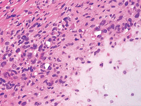
A 7-year-old girl has had a painful forearm for the past 2 months. Examination reveals fullness on the volar aspect of the forearm. Radiographs and an MRI scan are shown in Figures 42a through 42c. Biopsy specimens are shown in Figures 42d and 42e. What is the most likely diagnosis?


Explanation

42b 42c 42d 42e The radiographs reveal phleboliths on the volar side of the forearm consistent with hemangioma. The MRI scan reveals a rather well-circumscribed in size, irregular in shape, intramuscular soft-tissue mass in the volar aspect of the distal right forearm within the flexor group musculature. The mass demonstrates heterogeneous mixed signal intensity in both T1- and T2-weighted sequences with increased signal intensity on the T1, suggesting fat within the tumor, typical of hemangioma. The postgadolinium-enhanced sequences demonstrate heterogeneous enhancement. The MRI findings are consistent with a soft-tissue hemangioma. Garzon M: Hemangiomas: Update on classification, clinical presentation and associate anomalies. Cutis 2000;66:325-328.

Question 2

Which of the following is an important factor in performing a proper biopsy?


Explanation

There are a number of important technical details in performing a biopsy. Incisions should always be longitudinal in the extremity. Good hemostasis is important in avoiding contamination from hematoma. The approach should avoid neurovascular structures, and go through a single muscle belly when possible. Although a frozen section should be obtained to ensure adequate viable tissue has been obtained, definitive diagnosis is not necessary at the time of the frozen section. Vaccaro AR (ed): Orthopaedic Knowledge Update 8. Rosemont, IL, American Academy of Orthopaedic Surgeons, 2005, pp 197-215.

Question 3

A 16-year-old girl has had painless swelling in her posterior left arm for the past 4 months. A radiograph, MRI scans, and an incisional biopsy specimen are shown in Figures 43a through 43d. What is the cytogenetic translocation most commonly associated with this tumor?


Explanation

43b 43c 43d This is a case of synovial sarcoma. The radiograph shows some soft-tissue swelling in the upper arm. The MRI scans show a lesion that has increased signal on T2-weighted images and low signal on T1-weighted images. There is a suggestion of a large cystic component to this lesion. The pathology shows a biphasic population of cells, a spindle cell component, and an epithelioid component. Up to 20% of synovial cell sarcomas have areas of cyst formation. The most common cytogenetic translocation with synovial cell sarcoma is X; 18. The 11; 22 translocation is most commonly associated with Ewing's sarcomas; the 12; 22 translocation is most commonly associated with clear cell sarcomas; the 2; 13 translocation is most commonly associated with alveolar rhabdomyosarcomas, and the 12; 16 translocation is most commonly associated with myxoid liposarcomas. Kawai A, Woodruff J, Healey JH, et al: SYT-SSX gene fusion as a determinant of morphology and prognosis in synovial sarcoma. New Engl J Med 1998;338:153-160.

Question 4

A 43-year-old woman is referred after excisional biopsy of a cutaneous soft-tissue mass from her left shoulder. Based on the biopsy specimens shown in Figures 44a and 44b, what is the best course of action?


Explanation

44b Dermatofibrosarcoma protuberans (DFSP) is a rare superficial sarcoma that is frequently misdiagnosed at presentation. It is frequently excised prior to suspecting that the lesion is a sarcoma and if not appropriately treated with tumor bed resection to obtain wide margins, these lesions have a high incidence of local recurrence. It is recommended that the wide excision include the deep fascia and a 2.5- to 3-cm cuff of normal-appearing skin. Distant disease spread is rare and usually occurs in the face of a multiply recurrent lesion. Despite the apparent gross circumscription of these lesions, the tumor diffusely infiltrates the dermis and subcutaneous tissues. A characteristic histologic finding can be seen in the deep margins of the tumor where it intricately interdigitates with normal fat. Lindner NJ, Scarborough MT, Powell GJ, et al: Revision surgery in dermatofibrosarcoma protuberans of the trunk and extremities. Eur J Surg Oncol 1999;25:392-397.

Question 5

A 33-year-old man reports an enlarging painful soft-tissue mass in his right forearm. A radiograph and MRI scans are shown in Figures 45a through 45c. Treatment should consist of


Explanation

45b 45c An intramuscular lipoma is a benign soft-tissue lesion that can grow and has a small risk of progressing to a liposarcoma. Radiographs usually show a globular radiolucent mass adjacent to higher-density muscle tissue shadows. When the patient has symptoms and reports an increase in size of the mass, the treatment of choice after appropriate radiographic analysis is complete excision of the mass with marginal resection. Sampling error is a problem with fatty lesions and core or incisional biopsies are frequently unnecessary, especially if an MRI scan of the lesion shows signal intensity that matches subcutaneous fat on all sequences. Damron TA: What to do with deep lipomatous tumors. Instr Course Lect 2004;53:651-655. Gaskin CM, Helms CA: Lipomas, lipoma variants, and well-differentiated liposarcomas (atypical lipomas): Results of MRI evaluations of 126 consecutive fatty masses. Am J Roentgenol 2004;182:733-739.

Question 6

A 20-year-old woman has had wrist pain for the past 5 months. A radiograph, MRI scans, and biopsy specimen are shown in Figures 46a through 46d. The patient is then treated with intralesional surgery. The patient should be counseled that her risk of developing lung metastasis is approximately what percent?


Explanation

46b 46c 46d Giant cell tumor of bone has about a 2% risk of benign pulmonary metastasis in all cases and 6% risk in recurrent cases. The radiograph and MRI scans show a lytic destructive lesion in the distal radius with no matrix mineralization. The lesion extends up to the subchondral bone. In a young woman, the most likely diagnosis is giant cell tumor of bone, which is supported by the pathology results that show monotonous fibrovascular stroma with numerous multinucleated giant cells where the nuclei that make up the giant cells are identical to the nuclei that make up the background stromal cells. Athanasian EA, Wold LE, Amadio PC: Giant cell tumors of the bones in the hand. J Hand Surg Am 1997;22:91-98.

Question 7

What is the most common location for localized pigmented villonodular synovitis (PVNS) to occur?


Explanation

Localized PVNS is a form of the disease in which synovial proliferation is restricted to one area of a joint and causes the formation of a small mass-like lesion. The true incidence of this is unknown but is probably less common than the diffuse form of the disease. PVNS presents as a usually painful discrete mass. The anterior compartment of the knee is the most common location. Tyler WK, Vidal AF, Williams RJ, et al: Pigmented villonodular synovitis. J Am Acad Orthop Surg 2006;14:376-385.

Question 8

A 45-year-old man reports right shoulder pain with overhead activities only. Figures 47a through 47d show the radiographs, bone scan, and MRI scan of a lesion of the proximal shoulder. What is the most appropriate treatment?


Explanation

47b 47c 47d The figures show a lesion of the proximal humerus consistent with an enchondroma. The lesion is calcified on the radiographs. There is no cortical destruction, significant endosteal scalloping, or soft-tissue mass. The bone scan shows mild uptake in the area of the proximal humerus, and the T2-weighted MRI scan shows a lesion with high uptake, suggesting a lesion with high water content. A CT scan could also be obtained to rule out bone destruction or periosteal reaction. Pain with overhead activities is likely related to the rotator cuff. A biopsy is unlikely to add information because of inherent difficulties interpreting low-grade cartilaginous lesions. Curettage and grafting and en bloc resection are excessive treatments for a benign lesion that is apparently asymptomatic. Observation with a follow-up radiograph in 3 to 6 months is appropriate. Menendez LR (ed): Orthopaedic Knowledge Update: Musculoskeletal Tumors. Rosemont, IL, American Academy of Orthopaedic Surgeons, 2002, pp 103-111.

Question 9

What is the second most common primary bone malignancy in children?


Explanation

Ewing's sarcoma is the second most common bone tumor in children with an incidence of three per one million Caucasian children younger than 21 years of age. Ewing's sarcoma is rare in African Americans. Osteosarcoma is the most common bone tumor in children. Rhabdomyosarcoma is the most common soft-tissue sarcoma in children. Fibrosarcoma is a rare primary bone tumor most commonly seen in adults. Adamantinoma is a rare primary bone malignancy also most commonly seen in adults in the tibia. Menendez LR (ed): Orthopaedic Knowledge Update: Musculoskeletal Tumors. Rosemont, IL, American Academy of Orthopaedic Surgeons, 2002, p 195.

Question 10

An 11-year-old boy sustained an injury to his arm in gym class. He denies prior pain in the arm. Radiographs are shown in Figures 48a and 48b. What is the next most appropriate step in the management of this lesion?


Explanation

48b This radiolucent lesion with a "fallen leaf sign" is typical for a unicameral bone cyst(UBC). The most appropriate treatment is to allow the fracture to heal with clinical and radiographic observation. Curettage and bone grafting is not the best initial management for UBC. Wide resection is not indicated for UBC. The proximal humerus is the most common site for UBC. While staging studies consisting of MRI, bone scan, and CT of the chest are appropriate for lesions suspected of being malignant, the classical appearance of this UBC is such that this work-up is not necessary initially. Following fracture healing, aspiration and injection of the cyst may be indicated. Dormans JP, Pill SG: Fractures through bone cysts: Unicameral bone cysts, aneurysmal bone cysts, fibrous cortical defects, and nonossifying fibromas. Instr Course Lect 2002;51:457-467.

Question 11

An 83-year-old woman reports pain in her left middle finger after a minor injury. Laboratory studies show a WBC count of 7,000/mm3, an erythrocyte sedimentation rate of 3 mm/h, a uric acid of 10.4 mg/dL, and a normal serum protein electrophoresis. Radiographs are shown in Figures 49a and 49b. A core biopsy specimen is shown is Figure 49c. In addition to treatment of the finger fracture, treatment should include


Explanation

49b 49c This clinical picture is most consistent with periarticular erosions from gout. The patient has multiple periarticular lytic lesions in the hand. The laboratory studies show an elevated serum uric acid level, and the biopsy specimen demonstrates acute and chronic inflammation with prominent clefts. Therefore, the preferred treatment is systemic control of her gout. Radiation therapy, chemotherapy, and/or amputation should be considered for a malignancy; however, the pathology does not demonstrate any evidence of pleomorphism, high nuclear-to-cytoplasmic ratio, nuclear atypia, or mitotic activity. Antibiotics for an infectious process is a consideration, but the minimal elevation in the WBC count and erythrocyte sedimentation rate does not support an infectious process. Wise CM: Crystal-associated arthritis in the elderly. Clin Geriatr Med 2005;21:491-511.

Question 12

A 21-year-old man has had right groin pain for the past year. A radiograph, CT scan, MRI scans, and a biopsy specimen are shown in Figures 50a through 50e. What is the most likely diagnosis?


Explanation

50b 50c 50d 50e The pathology demonstrates a very cellular chondroid matrix with multinucleated forms, atypia, and myxomatous regions. This is most consistent with a myxoid chondrosarcoma. The radiograph shows a well-circumscribed lesion in the superior and medial aspect of the right acetabulum. The CT and MRI scans confirm these same findings with no evidence of matrix mineralization or significant surrounding edema. Unfortunately, in this location with this appearance, the radiographic differential diagnosis includes all the diagnoses listed. Terek RM: Recent advances in the basic science of chondrosarcoma. Orthop Clin North Am 2006;37:9-14. Donati D, El Ghoneimy A, Bertoni F, et al: Surgical treatment and outcome of conventional pelvic chondrosarcoma. J Bone Joint Surg Br 2005;87:1527-1530.

Question 13

A healthy 16-year-old boy has had increasing pain in the right knee for the past 3 months. Examination reveals warmth and swelling around the distal femur. Radiographs and an MRI scan are shown in Figures 51a through 51c, and a biopsy specimen is shown in Figure 51d. What is the most likely diagnosis?


Explanation

51b 51c 51d The radiographs show a bone-producing lesion in the distal femoral metaphysis in this case of classic osteosarcoma presenting in the most common location, the distal femur. The coronal MRI scan reveals a marrow-occupying lesion with extension into the soft tissues. The histology shows osteoid production by pleomorphic cells consistent with an osteosarcoma. Ewing's sarcoma is a bone tumor characterized by uniform small blue cells on histology. Rhabdomyosarcoma is the most common childhood soft-tissue sarcoma. Osteomyelitis has an inflammatory appearance on histology. Malignant fibrous histiocytoma of bone has a lytic radiographic appearance and a pleomorphic storiform pattern without osteoid on histology. Wold LE, Adler CP, Sim FH, et al: Atlas of Orthopedic Pathology, ed 2. Philadelphia, PA, WB Saunders, 2003, p 179.

Question 14

A 10-year-old boy has had wrist pain for the past 3 months. He denies any history of trauma. He reports mild tenderness associated with a palpable mass. A radiograph and biopsy specimens are shown in Figures 52a through 52c. What is the most likely diagnosis?


Explanation

52b 52c The radiograph shows a benign-appearing cortically based lesion eroding the underlying cortex, producing a saucer-shaped defect typical of a periosteal chondroma. The histology shows benign-appearing neoplastic cartilage. Although enchondroma would have the same histologic appearance, radiographs generally show a lesion with a central medullary epicenter. The benign-appearing histology does not support chondrosarcoma. Chondromyxoid fibroma will generally show histologic elements of its fibrous and myxoid components. Chondroblastoma typically demonstrates histologic findings of polyhedral cells separated by a chondroid matrix with pericellular, lattice-like "chicken wire" calcification. Schajowicz F: Tumors and Tumorlike Lesions of Bone: Pathology, Radiology, and Treatment, ed 2. Berlin, Springer-Verlag, 1994, pp 147-151.

Question 15

A 29-year-old woman reports shoulder pain after sustaining a minor fall 6 weeks ago. She has a history of celiac sprue. Radiographs of the forearm and shoulder are shown in Figures 53a and 53b. Which of the following serum abnormalities would be expected?


Explanation

53b Celiac sprue results in rapid gastrointestinal transit and fatty stools that impair the absorption of calcium and vitamin D and result in nutritional-deficiency osteomalacia with secondary hyperparathyroidism. The radiographs show marked osteopenia with brown tumors. A pathologic fracture is seen in the proximal humerus through a large brown tumor. Serum findings include low or normal calcium, low phosphate, elevated alkaline phosphatase, low 1,25(OH)2D, and increased PTH levels. Secondary hyperparathyroidism is associated with a variety of conditions including malabsorption syndromes. Potts JT: Parathyroid hormone: Past and present. J Endocrinol 2005;187:311-325. Corazza GR, Di Stefano M, Maurino E, et al: Bones in coeliac disease: Diagnosis and treatment. Best Pract Res Clin Gastroenterol 2005;19:453-465.

Question 16

A 73-year-old man stepped off a street curb and felt a crack in his left hip. He is now unable to bear weight. A radiograph is shown in Figure 54a. Biopsy specimens are shown in Figures 54b and 54c. What is the most likely diagnosis?


Explanation

54b 54c The biopsy specimens reveal a high-grade spindle cell lesion adjacent to an area of benign cartilage. This is consistent with a dedifferentiated chondrosarcoma. The radiograph shows a pathologic fracture through a lesion characterized by calcification within the left greater trochanter. Distal to the area of calcification, there is a more osteolytic, destructive appearance. Synovial sarcoma has a biphasic appearance histologically with areas of glandular differentiation that stain positive with keratin. Metastatic prostate cancer, although osteoblastic in appearance, would have a glandular histologic appearance. There is no cartilage in these lesions. Classic low-grade chondrosarcoma does not have an area of high-grade pleomorphic spindle cells within the lesion. A periosteal osteosarcoma is a surface-based lesion with a sunburst radiographic pattern. Although there may be cartilage in the lesion histologically, there are also malignant cells producing osteoid. Dedifferentiated chondrosarcoma is an aggressive, high-grade variant of chondrosarcoma. Wold LE, Adler CP, Sim FH, et al: Atlas of Orthopedic Pathology, ed 2. Philadelphia, PA, WB Saunders, 2003, p 269.

Question 17

The biopsy specimens seen in Figures 55a and 55b are from a lytic lesion in the sacrum of a 58-year-old man. What is the most likely diagnosis?


Explanation

55b The lesion is a chordoma and the other listed choices can be eliminated based on the histology. Many tumors can occur in the sacrum including chordoma, multiple myeloma, giant cell tumor, aneurysmal bone cyst, and metastatic disease. The histology in this patient shows a lobulated lesion on low power with fibrous septae separating the lobules. At higher magnification, the cells have eosinophilic vacuolated cytoplasm and are called physaliferous cells. Chordoma is a low-grade neoplasm that most commonly occurs in the sacrum and rarely in the base of the skull. The diagnosis is often delayed. Chordoma is thought to originate from notochordal remnants. Chordoma typically occurs in the midline and has an associated soft-tissue mass. Wold LE, Adler CP, Sim FH, et al: Atlas of Orthopedic Pathology, ed 2. Philadelphia, PA, WB Saunders, 2003, p 372. Fuchs B, Dickey ID, Yaszemski MJ, et al: Operative management of sacral chordoma. J Bone Joint Surg Am 2005;87:2211-2216.

Question 18

A 65-year-old man has a painful right hip mass that has been growing for several years. A radiograph, CT scan, and photomicrograph are shown in Figures 56a through 56c. What is the most appropriate treatment?


Explanation

56b 56c This is a conventional chondrosarcoma. The radiograph and the CT scan show a lesion arising from the inferior pubic ramus with a large soft-tissue mass. Abundant punctate, stippled, or "popcorn-like" calcification is present. The photomicrograph demonstrates hypercellular cartilage. Surgical resection is the only effective treatment. Whereas chemotherapy might play a role in the treatment of a dedifferentiated chondrosarcoma, it has no role in the treatment of a conventional chondrosarcoma. Chondrosarcomas are relatively radioresistant. Donati D, El Ghoneimy A, Bertoni F, et al: Surgical treatment and outcome of conventional pelvic chondrosarcoma. J Bone Joint Surg Br 2005;87:1527-1530. Lee FY, Mankin HJ, Fondren G, et al: Chondrosarcoma of bone: An assessment of outcome. J Bone Joint Surg Am 1999;81:326-338.

Question 19

An 8-year-old boy is diagnosed with acute onset cauda equina syndrome. A radiograph, MRI scans, and a biopsy specimen are shown in Figures 57a through 57d. What is the most appropriate treatment?


Explanation

57b 57c 57d The findings are consistent with an aneurysmal bone cyst. The MRI scan demonstrates a lesion involving the posterior elements of the vertebrae with fluid-fluid levels and neural compression. Fibrovascular tissue with multinucleated giant cells surrounding a vascular lake is seen on the histology. The most appropriate treatment is a marginal resection of the involved posterior elements. Although the recurrence rate can be as high as 25% to 30%, wide surgical resection could result in permanent neurologic injury and is not necessary. Aspiration and steroid injection have been advocated but would not relieve the nerve compression in this patient. Radiation therapy and chemotherapy are not indicated. Mankin HJ, Hornicek FJ, Ortiz-Cruz E, et al: Aneurysmal bone cyst: A review of 150 patients. J Clin Oncol 2005;23:6756-6762.

Question 20

A 19-year-old man has had pain and swelling in his left forearm for the past 8 months. Laboratory studies show a mildly elevated WBC count and erythrocyte sedimentation rate. Radiographs are shown in Figures 58a and 58b, a CT scan is shown in Figure 58c, and T1- and T2-weighted MRI scans are shown in Figures 58d and 58e, respectively. A biopsy specimen is shown in Figure 58f. Immunohistochemistry demonstrates that the lesion is negative for leukocyte common antigen (CD34). What is the most common cytogenetic translocation associated with this lesion?


Explanation

58b 58c 58d 58e 58f The imaging studies show a permeative lytic destructive lesion in the proximal radius with "hair-on-end" periosteal reaction and a large soft-tissue mass most consistent with Ewing's sarcoma. The pathology reveals monotonous sheets of "round blue" cells. This limits the differential diagnosis to primary lymphoma of bone versus Ewing's sarcoma. These are best differentiated by immunohistochemistry, cytogenetics, and flow cytometry. Lymphoma of bone is typically CD34 positive and CD99 negative; whereas, the reverse is true of Ewing's sarcoma, CD34 negative and CD99 positive. The most common cytogenetic translocation with Ewing's sarcoma is 11; 22; 21; 22 and 7; 22 translocations have also been reported in Ewing's sarcomas. The X; 18 translocation is most commonly associated with synovial cell sarcomas; the 12; 22 translocation is most commonly associated with clear cell sarcomas; the 2; 13 translocation is most commonly associated with alveolar rhabdomyosarcomas, and the 12; 16 translocation is most commonly associated with myxoid liposarcomas. Flow cytometry is used to characterize the cell types of lymphomas. Womer R: The cellular biology of bone tumors. Clin Orthop Relat Res 1991;262:12-21. Yamaguchi U, Hasegawa T, Morimoto Y, et al: A practical approach to the clinical diagnosis of Ewing's sarcoma/primitive neuroectodermal tumour and other small round cell tumours sharing EWS rearrangement using new fluorescence in situ hybridisation probes for EWSR1 on formalin fixed, paraffin wax embedded tissue. J Clin Pathol 2005;58:1051-1056.

Question 21

A 9-year-old girl reports progressive right knee pain. Radiographs are shown in Figures 59a and 59b. Work-up reveals no other sites of disease. Low- and high-power photomicrographs are shown in Figures 59c and 59d. What is the most appropriate treatment?


Explanation

59b 59c 59d This classic case of osteosarcoma illustrates the typical radiographic and histologic characteristics of this disease. The radiographs show an aggressive-appearing lesion of the distal femur. The lesion has both lytic and blastic areas. Periosteal reaction is present in the form of a Codman's triangle. The radiographs are highly suggestive of osteosarcoma. The photomicrographs show malignant spindle cells that produce osteoid, thus confirming the diagnosis of osteosarcoma. Treatment of osteosarcoma is multimodal including multi-agent chemotherapy and surgery (wide resection or amputation). Bacci G, Ferrari S, Bertoni F, et al: Long-term outcome for patients with nonmetastatic osteosarcoma of the extremity treated at the Instituto Ortopedico Rizzoli according to the Instituto Ortopedico Rizzoli/osteosarcoma-2 protocol: An updated report. J Clin Oncol 2000;18:4016-4037. Bielack SS, Kempf-Bielack B, Delling G, et al: Prognostic factors in high-grade osteosarcoma of the extremities or trunk: An analysis of 1,702 patients treated on neoadjuvant cooperative osteosarcoma study group protocols. J Clin Oncol 2002;20:776-790.

Question 22

What is the most significant factor affecting long-term survival for a patient with bone sarcoma?


Explanation

The most significant impact on long-term survival is the presence or absence of identifiable metastatic disease on initial presentation. All of these factors have been shown to be predictive of long-term survival to varying degrees. Enneking WF, Spanier SS, Goodman MA: A system for the surgical staging of musculoskeletal sarcoma. Clin Orthop Relat Res 1980;153:106-120.

Question 23

A 75-year-old woman notes a slowly enlarging mass in the right anterior thigh. Her medical history is significant only for hypertension. An MRI scan of her thigh is shown in Figures 60a through 60d. Which of the following surgical margins is the most appropriate for removal of this lesion?


Explanation

60b 60c 60d The patient has a large deep anterior thigh mass that has imaging characteristics of mature fat. Intramuscular lipomas are effectively treated with marginal resections with very low recurrence rates. Large lipomas often have small amounts of intralesional signal changes frequently representing trapped muscle fibers and do not necessitate more extensive margins. Gaskin CM, Helms CA: Lipomas, lipoma variants, and well-differentiated liposarcomas (atypical lipomas): Results of MRI evaluations of 126 consecutive fatty masses. Am J Roentgenol 2004;182:733-739.

Question 24

Figures 61a and 61b show the CT and MRI scans of a 40-year-old man who has hip pain. He undergoes total hip arthroplasty and curettage and cementation of the lesion as shown in Figure 61c. Histopathologic photomicrographs of the curettage specimen are shown in Figures 61d and 61e. What is the best course of treatment?


Explanation

61b 61c 61d 61e The definitive surgery would be removal of the entire resection bed, and in this case of dedifferentiated chondrosarcoma, a hemipelvectomy was performed. The MRI and CT scans show an aggressive cartilage lesion. The histology, representative of a dedifferentiated chondrosarcoma, shows a bimorphic low-grade cartilage lesion with high-grade spindle cell sarcoma. The cartilage lesion is usually an enchondroma or low-grade chondrosarcoma. The dedifferentiated portion is typically a malignant fibrous histocytoma, osteosarcoma, or fibrosarcoma. Weber KL, Pring ME, Sim FH: Treatment and outcome of recurrent pelvic chondrosarcoma. Clin Orthop Relat Res 2002;397:19-28.

Question 25

Compared to postoperative radiation therapy, preoperative radiation therapy has a higher rate of what complication?


Explanation

Radiation therapy is commonly used as an adjuvant in the treatment of soft-tissue sarcomas, but a controversy exists whether it should be preoperative or postoperative. Radiation therapy can be given prior to or following resection of the tumor. Postoperative radiation is usually given in a higher dose to a larger treatment field. This commonly results in a higher incidence of fibrosis and lymphedema. There is no statistical difference in local recurrence rate between the two radiation treatment plans. Neuropathy is more commonly a complication of chemotherapy. Preoperative radiation therapy has been shown to have a higher wound complication rate than postoperative radiation. Vaccaro AR (ed): Orthopaedic Knowledge Update 8. Rosemont, IL, American Academy of Orthopaedic Surgeons, 2005, pp 197-215.

Question 26

A 24-year-old man presents with a slow-growing, painful mass around his knee. An MRI reveals a soft tissue mass near the joint but not within the joint cavity. Core needle biopsy demonstrates a biphasic pattern consisting of epithelial and spindle cells. Which of the following cytogenetic translocations is most characteristic of this tumor?


Explanation

The patient has a synovial sarcoma, which typically presents in young adults as a slow-growing soft tissue mass near large joints, most commonly the knee. The characteristic genetic abnormality is the translocation t(X;18)(p11;q11), resulting in the SS18-SSX fusion gene. This fusion protein disrupts gene expression and drives oncogenesis. The other translocations are associated with Ewing sarcoma (t(11;22)), myxoid liposarcoma (t(12;16)), alveolar rhabdomyosarcoma (t(2;13)), and extraskeletal myxoid chondrosarcoma (t(9;22)).

Question 27

A 45-year-old woman is incidentally found to have a destructive lesion in her distal femur on plain radiographs. She is scheduled to undergo a core needle biopsy. Which of the following is an essential orthopedic oncology principle when performing a biopsy for a suspected primary bone sarcoma?


Explanation

A critical principle of biopsy for suspected sarcomas is that the biopsy tract is considered contaminated with tumor cells. Therefore, it must be carefully planned longitudinally so that the entire tract can be completely excised en bloc with the tumor during the definitive surgical resection. Transverse incisions complicate future en bloc resection. Exsanguination with an Esmarch bandage should be avoided to prevent tumor embolization; if a tourniquet is used, the limb should only be elevated. Core needle biopsies are the standard of care due to high accuracy and minimal morbidity.

Question 28

A 14-year-old boy is diagnosed with high-grade conventional osteosarcoma of the proximal tibia. He completes neoadjuvant chemotherapy and undergoes wide surgical resection. Which of the following represents the most reliable prognostic factor for his overall long-term survival?


Explanation

The most important prognostic factor for overall survival in patients with high-grade conventional osteosarcoma is the histological response to neoadjuvant chemotherapy, which is measured by the percentage of tumor necrosis in the resected specimen (Huvos grading system). Greater than 90% necrosis indicates a good chemotherapeutic response and is associated with significantly better long-term survival rates. While surgical margin status is critical for local tumor control, chemotherapy response is the primary predictor of systemic microscopic disease eradication and overall survival.

Question 29

A 35-year-old man presents with chronic hip pain. Plain radiographs reveal a well-defined lytic lesion localized to the proximal femoral epiphysis. A biopsy demonstrates sheets of cells with clear, vacuolated cytoplasm and distinct cell membranes, interspersed with prominent trabeculae of reactive woven bone. What is the most appropriate definitive management for this lesion?


Explanation

The clinical presentation (epiphyseal lesion in an adult) and histology (clear cells, woven bone) are classic for clear cell chondrosarcoma. Unlike conventional chondrosarcomas, which rarely involve the epiphysis, clear cell chondrosarcomas are typically epiphyseal. They are low-grade malignant bone tumors. The treatment of choice is wide en bloc resection. Intralesional curettage has an unacceptably high local recurrence rate for clear cell chondrosarcoma. Like most chondrosarcomas, they are relatively resistant to both systemic chemotherapy and radiation therapy.

Question 30

A 28-year-old woman presents with knee pain. Radiographs show an eccentric, lytic lesion in the distal femoral epiphysis extending precisely to the subchondral bone. Biopsy confirms a giant cell tumor of bone. Which of the following molecular targets is specifically responsible for the aggressive osteolysis in this tumor and is targeted in its medical management?


Explanation

Giant cell tumor of bone (GCT) is primarily driven by neoplastic mononuclear stromal cells that overexpress RANKL. This overexpression recruits and stimulates normal osteoclast precursors to fuse into reactive, multinucleated giant cells, leading to aggressive and rapid bone resorption. Denosumab, a monoclonal antibody that binds to and neutralizes RANKL, is utilized in the medical management of advanced, unresectable, or recurrent GCTs to inhibit bone destruction and induce ossification of the tumor matrix.

Question 31

A 40-year-old man undergoes excision of a deep, painless soft tissue mass in his medial thigh. Pathological analysis reveals a prominent myxoid stroma, a delicate arborizing 'chicken-wire' capillary network, and scattered lipoblasts. Which of the following genetic abnormalities is most characteristic of this soft tissue tumor?


Explanation

The histological description of a myxoid stroma, chicken-wire vascular pattern, and lipoblasts is pathognomonic for myxoid liposarcoma. The hallmark translocation for myxoid liposarcoma is t(12;16)(q13;p11), which results in the FUS-DDIT3 (formerly FUS-CHOP) fusion gene. MDM2 amplification is characteristic of well-differentiated and dedifferentiated liposarcomas. The other translocations correspond to Ewing sarcoma (EWSR1-FLI1), synovial sarcoma (SS18-SSX), and alveolar rhabdomyosarcoma (PAX3-FOXO1).

Question 32

A 12-year-old girl presents with a rapidly enlarging mass in her proximal humerus. Plain radiographs show an expansile, multiloculated, radiolucent lesion with a 'soap bubble' appearance and a thin cortical shell. A biopsy is performed. Which of the following histological findings is most characteristic of this specific lesion?


Explanation

The clinical and radiographic presentation is highly consistent with an aneurysmal bone cyst (ABC). Histologically, ABCs are characterized by cavernous, blood-filled spaces that are separated by fibrous septa containing fibroblasts, osteoclast-like giant cells, and reactive woven bone. Crucially, these blood-filled spaces lack a true endothelial lining, which differentiates them morphologically from true vascular tumors such as hemangiomas or angiosarcomas.

Question 33

A 55-year-old man presents with generalized diffuse bone pain and profound proximal muscle weakness. Laboratory studies show severe hypophosphatemia, low 1,25-dihydroxyvitamin D levels, and elevated alkaline phosphatase. Serum calcium and parathyroid hormone levels are normal. A full-body MRI reveals a small, benign-appearing soft tissue mass in the plantar aspect of his foot. Excision of this mass is most likely to result in which of the following physiological changes?


Explanation

The patient is presenting with tumor-induced osteomalacia (TIO), an acquired paraneoplastic syndrome typically caused by a small, benign phosphaturic mesenchymal tumor (PMT). These tumors autonomously secrete fibroblast growth factor 23 (FGF-23). Excess FGF-23 inhibits renal proximal tubule phosphate reabsorption (causing phosphaturia and hypophosphatemia) and suppresses 1-alpha-hydroxylase (decreasing 1,25-dihydroxyvitamin D synthesis). Surgical excision of the causative tumor leads to a rapid decline in FGF-23, which results in the prompt resolution of hypophosphatemia and normalization of bone mineralization.

Question 34

A 10-year-old boy presents with multiple painless, hard bony bumps around his knees and ankles. Radiographs demonstrate multiple sessile and pedunculated osteochondromas pointing away from the adjacent joints. He is clinically diagnosed with multiple hereditary exostoses (MHE). Loss-of-function mutations in which of the following genes are responsible for this autosomal dominant condition?


Explanation

Multiple hereditary exostoses (MHE), also known as multiple osteochondromatosis, is an autosomal dominant skeletal disorder caused by loss-of-function mutations in the tumor suppressor genes EXT1 or EXT2. These genes encode glycosyltransferases that are essential for the synthesis of heparan sulfate. A deficiency in heparan sulfate disrupts the Indian hedgehog (Ihh) signaling pathway at the physis, leading to ectopic cartilage growth and osteochondroma formation. NF genes cause neurofibromatosis, RB1/TP53 mutations are seen in osteosarcoma, GNAS mutations cause fibrous dysplasia, and APC mutations cause familial adenomatous polyposis.

Question 35

Denosumab has revolutionized the non-surgical management of advanced giant cell tumors of bone. What is the precise cellular mechanism of action of this targeted medication?


Explanation

Denosumab is a fully human monoclonal antibody that specifically targets and binds to RANK Ligand (RANKL). By binding to RANKL, denosumab prevents the ligand from interacting with its designated receptor, RANK, which is located on the surface of osteoclasts and osteoclast precursors. This blockade effectively halts osteoclast formation, function, and survival, thereby rapidly decreasing tumor-induced bone resorption. In contrast, bisphosphonates work by inhibiting farnesyl pyrophosphate synthase, leading to osteoclast apoptosis.

Question 36

A 32-year-old woman presents with knee pain. Radiographs reveal an eccentric, lytic epiphyseal lesion extending to the subchondral bone of the distal femur. Biopsy reveals multinucleated giant cells in a background of mononuclear stromal cells. She is prescribed a medication that binds to a specific ligand to prevent osteoclast activation. What is the target of this medication?


Explanation

The clinical and radiographic presentation is classic for a Giant Cell Tumor (GCT) of bone. The neoplastic mononuclear stromal cells of GCT express RANKL, which recruits and activates the reactive multinucleated osteoclast-like giant cells that express the RANK receptor. Denosumab is a fully human monoclonal antibody that directly targets and binds to RANK ligand (RANKL), thereby preventing osteoclast activation and subsequent bone destruction. Osteoprotegerin (OPG) is the body's natural decoy receptor for RANKL.

Question 37

A 15-year-old boy is diagnosed with high-grade intramedullary osteosarcoma of the proximal tibia. He undergoes 10 weeks of neoadjuvant chemotherapy with methotrexate, doxorubicin, and cisplatin, followed by wide surgical resection. Pathologic evaluation of the resected specimen is performed. Which of the following is the most important prognostic factor for his overall survival?


Explanation

The most important prognostic factor for overall survival in a patient with high-grade osteosarcoma treated with neoadjuvant chemotherapy is the histologic response to the chemotherapy. This is measured as the percentage of tumor necrosis in the definitive resection specimen. A "good response" is typically defined as greater than 90% tumor necrosis (Huvos grade III or IV) and correlates with significantly improved disease-free and overall survival rates.

Question 38

A 28-year-old man presents with a slow-growing, deep-seated soft tissue mass near his knee joint. An MRI demonstrates a heterogeneous mass adjacent to the capsule but not within the joint space. Core needle biopsy shows a biphasic tumor with both epithelial and spindle cell components. Which of the following cytogenetic translocations is most characteristic of this diagnosis?


Explanation

The patient's clinical presentation, MRI findings, and biphasic histology (epithelial and spindle cells) are highly characteristic of Synovial Sarcoma. Synovial sarcoma is characterized by the cytogenetic translocation t(X;18)(p11;q11), which results in the SYT-SSX fusion gene. Option A refers to Ewing Sarcoma. Option B is Myxoid Liposarcoma. Option C is Extraskeletal Myxoid Chondrosarcoma. Option D is Alveolar Rhabdomyosarcoma.

Question 39

A 14-year-old boy with multiple painless bony protuberances around his knees and ankles presents with a new onset of pain and rapid growth of a lesion on his proximal humerus over the last 3 months. His father has a similar history. Genetic testing of this patient is most likely to reveal a mutation in a gene responsible for which of the following cellular processes?


Explanation

The patient has Multiple Hereditary Exostoses (MHE), an autosomal dominant condition characterized by multiple osteochondromas. MHE is primarily caused by mutations in the EXT1 or EXT2 genes. These genes encode glycosyltransferases that are essential for the biosynthesis of heparan sulfate proteoglycans. Disruption of this pathway leads to abnormal chondrocyte proliferation and bone growth. FGFR3 mutations cause achondroplasia, while Gs-alpha mutations are associated with fibrous dysplasia.

Question 40

A 16-year-old boy complains of chronic, aching shoulder pain. Radiographs show a well-circumscribed lytic lesion in the epiphysis of the proximal humerus with a thin sclerotic rim. MRI reveals extensive surrounding bone marrow edema. Histological examination of a biopsy specimen demonstrates sheets of mononuclear cells with grooved nuclei and areas of pericellular 'chicken-wire' calcification. What is the most appropriate definitive treatment for this lesion?


Explanation

The clinical, radiographic, and histologic findings (epiphyseal location, grooved 'coffee-bean' nuclei, and 'chicken-wire' calcification) are pathognomonic for a Chondroblastoma. Chondroblastoma is a benign but locally aggressive cartilaginous tumor. The gold standard treatment is extended intralesional curettage (often utilizing a high-speed burr or chemical adjuvants) followed by bone grafting or PMMA cementation. Wide resection is overly aggressive for this benign lesion, and observation is inappropriate given its locally destructive nature.

Question 41

A 19-year-old man presents with severe mid-back pain that awakens him at night and is dramatically relieved by ibuprofen. A CT scan of the spine shows a 1.2-cm radiolucent nidus with surrounding sclerosis in the posterior elements of L3. The exquisite pain caused by this lesion is primarily mediated by the local overproduction of which of the following substances?


Explanation

The clinical presentation of severe night pain relieved by NSAIDs and the CT finding of a sclerotic lesion with a central radiolucent nidus in the posterior elements of the spine are classic for an Osteoid Osteoma. The profound pain associated with this benign bone tumor is due to the overproduction of prostaglandins, specifically Prostaglandin E2 (PGE2), by the nidus via increased expression of cyclooxygenase-2 (COX-2).

Question 42

A 45-year-old woman is found to have an aggressive, bone-destructive lesion in the distal femur. A biopsy is planned. Which of the following statements regarding the principles of orthopedic oncologic biopsy is most accurate?


Explanation

Strict adherence to oncologic biopsy principles is mandatory to avoid tumor dissemination and limb compromise. The biopsy tract must be placed longitudinally and in line with the planned definitive resection so that it can be completely excised. Transverse incisions contaminate multiple compartments. Exsanguination with an Esmarch bandage is contraindicated as it may push tumor cells systemically; only elevation should be used before tourniquet inflation. Drains should exit near the wound, in line with the incision. Cortical windows should be plugged (e.g., with PMMA) to prevent tumor hematoma tracking.

Question 43

A 50-year-old man presents with a large, painless mass in his posterior thigh. MRI shows a multilobulated soft-tissue mass with high signal intensity on T2-weighted images and a cystic-like appearance, though it enhances with gadolinium. Biopsy reveals a prominent plexiform capillary network and lipoblasts in a myxoid stroma. Which of the following features is most unique to the management or behavior of this specific sarcoma subtype?


Explanation

The pathology describes a Myxoid Liposarcoma, classically presenting with a prominent plexiform capillary network ('crow's feet' vessels) and t(12;16) FUS-DDIT3 translocation. A unique behavioral characteristic of myxoid liposarcoma is its strong propensity to metastasize to extrapulmonary sites, particularly the skeletal system (especially the spine), often before pulmonary metastases occur. Consequently, staging typically requires full-spine MRI. It is also exceptionally sensitive to radiation therapy, contrary to Option A.

Question 44

A 40-year-old man undergoes curettage of a lytic lesion in the proximal femoral epiphysis. The lesion was initially thought to be a chondroblastoma based on its location. However, definitive histopathology reveals large cells with abundant clear cytoplasm, distinct cytoplasmic membranes, and central nuclei, interspersed with areas of conventional hyaline cartilage and reactive woven bone. What is the most appropriate next step in management?


Explanation

The histopathology and clinical scenario describe a Clear Cell Chondrosarcoma. This low-grade malignant cartilage tumor frequently involves the epiphysis, mimicking a benign chondroblastoma radiographically. However, clear cell chondrosarcoma tends to occur in an older age group (30-50 years) compared to chondroblastoma (teens). Because it is a malignant tumor, intralesional curettage alone results in a very high recurrence rate. The standard of care is wide en bloc surgical resection.

Question 45

A 25-year-old woman presents with a slow-growing, painless mass deep in the thigh. MRI shows a highly vascular lesion with prominent peritumoral flow voids. Chest CT reveals bilateral small pulmonary nodules, and a brain MRI shows a single contrast-enhancing lesion. Biopsy of the thigh mass reveals large, polygonal cells arranged in a nested pattern with central loss of cellular cohesion. What is the characteristic chromosomal translocation associated with this condition?


Explanation

The clinical picture describes Alveolar Soft Part Sarcoma (ASPS). ASPS typically presents in young adults as a deep-seated thigh mass. It is notorious for its highly vascular nature and an extremely high rate of early metastases to the lungs and brain, underscoring the need for brain MRI during staging. Pathologically, it features nested, polygonal cells with loss of central cohesion (pseudoalveolar pattern). It is characterized by the unbalanced translocation der(17)t(X;17)(p11;q25), resulting in the ASPSCR1-TFE3 fusion.

Question 46

A 14-year-old boy presents with a painful, swollen distal femur. Imaging shows a sunburst periosteal reaction. Biopsy reveals malignant spindle cells producing osteoid. Mutation of which of the following tumor suppressor genes is most commonly implicated in the pathogenesis of this disease?


Explanation

The patient has osteosarcoma, which is characterized by the production of malignant osteoid. The pathogenesis is strongly associated with mutations in the tumor suppressor genes TP53 (associated with Li-Fraumeni syndrome) and RB1 (associated with hereditary retinoblastoma). EXT1 and EXT2 mutations are seen in multiple hereditary exostoses. GNAS mutations are associated with fibrous dysplasia. RUNX2 is a transcription factor essential for osteoblast differentiation, frequently involved in cleidocranial dysplasia.

Question 47

When performing an incisional biopsy for a suspected primary malignant bone tumor in the distal femur, which of the following principles must be strictly adhered to?


Explanation

A properly planned biopsy is crucial in orthopedic oncology. The biopsy incision should be longitudinal and placed perfectly in line with the definitive surgical incision, ensuring that the entire biopsy tract can be excised en bloc during the tumor resection. Transverse incisions contaminate wide tissue planes. The procedure should remain within a single compartment. While a tourniquet may be used, the limb should not be exsanguinated (e.g., with an Esmarch bandage) to avoid pushing tumor cells centrally. Drains, if used, must exit directly in line with the surgical incision so their tract can also be excised.

Question 48

A 12-year-old boy presents with a painful, swollen mid-shaft femur. Radiographs demonstrate a permeative, destructive lesion with an 'onion skin' periosteal reaction. Histological evaluation shows uniform small, round blue cells. The t(11;22)(q24;q12) translocation associated with this tumor results in which of the following fusion products?


Explanation

The clinical presentation and histology describe Ewing sarcoma. Over 90% of Ewing sarcomas are characterized by the t(11;22)(q24;q12) chromosomal translocation, which results in the EWS-FLI1 fusion transcript. SYT-SSX represents the t(X;18) translocation seen in synovial sarcoma. TLS-CHOP (FUS-DDIT3) is seen in myxoid liposarcoma via t(12;16). PAX3-FKHR is seen in alveolar rhabdomyosarcoma via t(2;13). COL1A1-PDGFB is characteristic of dermatofibrosarcoma protuberans.

Question 49

A 28-year-old man undergoes excision of a slowly growing, painful soft tissue mass in his popliteal fossa. Histology demonstrates a biphasic pattern of spindle cells and epithelial-like gland structures. Cytogenetic analysis reveals a t(X;18) translocation. Which of the following is the most appropriate next step in staging this patient?


Explanation

The patient's histology and genetic profile (t(X;18)) confirm the diagnosis of synovial sarcoma. Like most high-grade soft tissue sarcomas, synovial sarcoma preferentially metastasizes hematogenously to the lungs. Therefore, a high-resolution Chest CT is the standard and most critical imaging modality for staging. Although synovial sarcoma has a slightly higher rate of regional lymph node metastasis compared to other sarcomas (along with clear cell, epithelioid, angiosarcoma, and rhabdomyosarcoma), routine sentinel lymph node biopsy is not considered the standard initial staging test over a chest CT.

Question 50

A 45-year-old woman presents with severe generalized bone pain and proximal muscle weakness. Laboratory studies show severe hypophosphatemia, normal serum calcium, normal parathyroid hormone, normal vitamin D, and elevated alkaline phosphatase. A skeletal survey reveals multiple pseudofractures (Looser zones). A small, benign-appearing soft tissue mass is noted on the plantar aspect of her foot. Which of the following substances is most likely being pathologically secreted by this soft tissue mass?


Explanation

The patient has tumor-induced osteomalacia (oncogenic osteomalacia), a rare paraneoplastic syndrome typically caused by benign phosphaturic mesenchymal tumors. These tumors secrete Fibroblast Growth Factor 23 (FGF-23). Excess FGF-23 acts on the kidneys to severely decrease phosphate reabsorption and inhibit 1-alpha-hydroxylase, reducing the production of active 1,25-dihydroxyvitamin D. This leads to profound hypophosphatemia, causing osteomalacia. Excision of the tumor is curative.

Question 51

A 40-year-old man presents with chronic, dull shoulder pain. Radiographs demonstrate a radiolucent, expansile lesion strictly confined to the epiphysis of the proximal humerus with focal intralesional calcifications. Histological sections reveal lobules of cells with abundant clear cytoplasm embedded in a chondroid matrix, without significant mitotic activity. Which of the following statements regarding this condition is true?


Explanation

The clinical and microscopic findings are classic for clear cell chondrosarcoma. This rare, low-grade malignant cartilaginous tumor typically involves the epiphysis of long bones (most commonly the proximal femur and proximal humerus) in adults aged 20-50. Because it is a malignant lesion with a high rate of local recurrence and potential for metastasis if treated inadequately, intralesional curettage is contraindicated. The standard of care is wide en bloc surgical resection. Like other conventional chondrosarcomas, it is generally resistant to chemotherapy and radiotherapy.

Question 52

A 15-year-old boy presents with progressive knee pain. Radiographs show a 2-cm lytic lesion with a thin sclerotic margin located in the proximal tibial epiphysis. MRI confirms the purely epiphyseal location and reveals extensive surrounding bone marrow edema. Histology reveals mononuclear cells with prominent longitudinal nuclear grooves ('coffee bean' nuclei) and dispersed multinucleated giant cells within areas of 'chicken-wire' calcification. What is the most appropriate management for this lesion?


Explanation

This patient has a chondroblastoma, a benign but locally aggressive epiphyseal tumor characteristically found in skeletally immature individuals. The classic histological features include mononuclear cells with 'coffee-bean' nuclei (longitudinal grooves) and fine, pericellular 'chicken-wire' calcification. Since it is a benign lesion, the standard treatment is thorough intralesional curettage with local adjuvant application (such as high-speed burring, phenol, or cryotherapy) followed by bone grafting or cementation. Wide resection is unnecessary and overly morbid.

Question 53

A 9-year-old girl is evaluated for an antalgic gait and leg length discrepancy. Radiographs of her right femur reveal an expansive, 'ground-glass' intramedullary lesion with cortical thinning. On physical examination, she has large, irregularly bordered hyperpigmented macules ('coast of Maine') and signs of precocious puberty. This clinical syndrome is caused by a somatic, activating postzygotic mutation in which of the following genes?


Explanation

The clinical presentation describes McCune-Albright syndrome, a classic triad of polyostotic fibrous dysplasia, cafe-au-lait spots with irregular borders ('coast of Maine'), and peripheral precocious puberty (or other endocrinopathies). The syndrome is driven by a somatic, activating postzygotic missense mutation in the GNAS1 gene. This mutation causes a constituent activation of the stimulatory G-protein (Gs-alpha), leading to unregulated, sustained production of intracellular cyclic AMP (cAMP) and subsequent abnormal cell proliferation and differentiation.

Question 54

A 70-year-old woman is prescribed denosumab for the treatment of severe postmenopausal osteoporosis. Which of the following best describes the exact molecular mechanism of action of this pharmacological agent?


Explanation

Denosumab is a fully human monoclonal antibody designed to mimic the physiological effects of osteoprotegerin (OPG). It specifically binds to Receptor Activator of Nuclear Factor Kappa-B Ligand (RANKL). By neutralizing RANKL, it prevents RANKL from binding to the RANK receptor situated on the surface of osteoclasts and their precursors. This blockade dramatically reduces osteoclast differentiation, function, and survival, leading to decreased bone resorption. Romosozumab binds to Sclerostin, and Odanacatib (investigational) inhibits Cathepsin K.

Question 55

Recombinant human bone morphogenetic protein-2 (rhBMP-2) is utilized in complex spine fusion surgery to induce osteoinduction. Bone morphogenetic proteins are potent growth factors belonging to the transforming growth factor-beta (TGF-β) superfamily. Which of the following best describes the principal intracellular signaling cascade directly activated following BMP binding to its specific cellular receptor?


Explanation

Bone Morphogenetic Proteins (BMPs) bind to cell surface type I and type II serine/threonine kinase receptors. Receptor binding induces cross-phosphorylation, which directly activates the intracellular canonical Smad signaling cascade. Specifically, for osteogenic BMPs like BMP-2, the activated receptors phosphorylate the receptor-regulated Smads (R-Smads): Smad 1, Smad 5, and Smad 8. These phosphorylated Smads then bind to the common-partner Smad (Co-Smad), Smad 4, and the entire complex translocates into the nucleus to act as transcription factors for osteogenic genes (like Runx2).

Question 56

A 25-year-old male presents with a slow-growing, painful soft tissue mass in his foot. An MRI reveals a heterogeneous mass adjacent to the plantar fascia. Biopsy shows a biphasic histology with both spindle and epithelial cells. Molecular testing reveals a specific cytogenetic translocation. Which of the following is the most likely translocation associated with this patient's diagnosis?


Explanation

The clinical presentation and biphasic histology (spindle and epithelial cells) strongly suggest a synovial sarcoma. Synovial sarcoma classically presents in young adults, often in the soft tissues of the extremities (especially foot and ankle). The hallmark translocation is t(X;18)(p11;q11), which results in the SYT-SSX fusion gene. Ewing sarcoma is characterized by t(11;22); myxoid liposarcoma by t(12;16); alveolar rhabdomyosarcoma by t(2;13); and extraskeletal myxoid chondrosarcoma by t(9;22).

Question 57

An orthopedic oncologist is planning an incisional biopsy of a suspected high-grade soft tissue sarcoma located in the anterior compartment of the thigh. Which of the following is the most critical technical principle to minimize the risk of local recurrence following definitive resection?


Explanation

The most critical principle of an open incisional biopsy for a suspected malignancy is that the entire biopsy tract (incision, subcutaneous tissue, and deeper tissues) must be cleanly excisable en bloc with the tumor during definitive surgery. Transverse incisions in the extremity must be avoided as they contaminate multiple compartments and require a much larger resection block. If a tourniquet is used, it should be deflated before closure to ensure meticulous hemostasis; post-operative hematoma can spread tumor cells extensively. Elevating large flaps contaminates normal tissue planes. Biopsying the necrotic core yields non-diagnostic, dead tissue.

Question 58

A 45-year-old patient requires a bone graft for a highly comminuted tibial nonunion. The surgeon chooses a graft that provides a structural physical scaffold for host osteogenic cells to migrate into, but the graft itself lacks living cells and does not contain intrinsic growth factors to stimulate stem cell differentiation. Which of the following best describes this graft's primary property and a classic example?


Explanation

Osteoconduction refers to the provision of a passive structural scaffold (matrix) for host osteoprogenitor cells and blood vessels to migrate into and form new bone. Cancellous allografts and synthetic ceramics are primarily osteoconductive. Osteogenesis requires the presence of living, viable osteoprogenitor cells capable of forming new bone (e.g., fresh autograft). Osteoinduction is the stimulation of host mesenchymal stem cells to differentiate into osteoblasts, primarily mediated by growth factors such as bone morphogenetic proteins (BMPs); DBM and recombinant BMP-2 are osteoinductive.

Question 59

When inserting a fully threaded cortical screw to provide interfragmentary compression across an oblique fracture, what mechanical parameter is directly optimized by overdrilling the near cortex to create a 'glide hole'?


Explanation

Creating a glide hole in the near cortex (overdrilling so the hole is the size of the screw's outer thread diameter) prevents the threads from engaging the near fragment. As the screw head engages the near cortex, the threads engaging the far cortex pull the far fragment towards the near fragment, generating interfragmentary compression. This mechanism is known as the lag effect. If the near cortex is not overdrilled, the threads engage both cortices simultaneously, maintaining the gap without generating compression.

Question 60

Articular (hyaline) cartilage has a highly specialized extracellular matrix that relies heavily on its composition for biomechanical resilience. Which of the following best describes the structural and biochemical composition of normal adult articular cartilage?


Explanation

Normal adult articular cartilage is avascular, aneural, and alymphatic. It has a very low cell density (chondrocytes make up <5% of tissue volume). The predominant collagen is Type II (comprising 90-95% of the collagen), which provides tensile strength. The primary proteoglycan is aggrecan, which interacts with hyaluronic acid to trap water, providing compressive stiffness. Type I collagen is the primary collagen found in fibrocartilage (e.g., meniscus) and bone.

Question 61

Physeal fractures and specific pediatric orthopedic conditions tend to preferentially involve distinct histologic zones of the physis. Through which zone of the physis does the failure typically occur in a slipped capital femoral epiphysis (SCFE)?


Explanation

Slipped capital femoral epiphysis (SCFE) and most Salter-Harris physeal fractures typically occur through the zone of hypertrophy (specifically the primary spongiosa region). In this zone, the chondrocytes swell and undergo apoptosis, and the extracellular matrix is sparse and lacks structural collagen, making it the mechanically weakest point of the physis. The reserve zone is affected in Gaucher disease; the proliferative zone in achondroplasia; and the zone of provisional calcification in rickets.

Question 62

Following a complete peripheral nerve transection (neurotmesis), Wallerian degeneration occurs in the distal stump to prepare the environment for potential axonal regeneration. Which of the following cells are primarily responsible for clearing myelin debris and forming bands to guide regenerating axons in the peripheral nervous system?


Explanation

In the peripheral nervous system, Schwann cells play a crucial role in nerve regeneration. Following injury, Schwann cells undergo dedifferentiation and proliferation. Along with recruited macrophages, they phagocytose myelin debris and form longitudinal arrays known as bands of Büngner, which provide physical and molecular guidance for regenerating axonal sprouts. Oligodendrocytes and microglia serve related functions in the central nervous system, where regeneration is typically abortive.

Question 63

A 70-year-old woman is treated with alendronate for postmenopausal osteoporosis. Bisphosphonates primarily reduce fracture risk by decreasing bone turnover. What is the precise molecular mechanism by which nitrogen-containing bisphosphonates inhibit osteoclast function?


Explanation

Nitrogen-containing bisphosphonates (e.g., alendronate, zoledronate, risedronate) inhibit bone resorption by inhibiting farnesyl pyrophosphate (FPP) synthase, a key enzyme in the mevalonate pathway. This prevents the prenylation of small regulatory GTPases (like Ras, Rho, Rac) essential for normal osteoclast function, cytoskeletal arrangement (ruffled border formation), and cell survival, ultimately inducing osteoclast apoptosis. Denosumab inhibits RANKL. Odanacatib inhibits cathepsin K. Romosozumab binds sclerostin.

Question 64

Tendons transmit high tensile forces from muscle to bone, but specific anatomical regions are predisposed to degeneration and spontaneous rupture due to relative hypovascularity. Where is the classic 'watershed area' of the Achilles tendon located?


Explanation

The Achilles tendon has a relative zone of hypovascularity, often referred to as a 'watershed area,' located approximately 2 to 6 cm proximal to its insertion on the calcaneus. This region relies heavily on diffusion for nutrition and is the most common site for degenerative tendinopathy and acute ruptures. The myotendinous and osteotendinous junctions generally possess a more robust local blood supply derived from the adjacent muscle belly and bony attachments, respectively.

Question 65

A 15-year-old boy presents with severe night pain in his right mid-thigh that is dramatically relieved within 30 minutes of taking ibuprofen. Radiographs demonstrate a 10 mm radiolucent nidus surrounded by dense, sclerotic reactive bone in the femoral diaphysis. What biochemical mediator is produced in high amounts by this tumor, directly responsible for the intense localized pain?


Explanation

The patient's clinical presentation (severe night pain dramatically relieved by NSAIDs) and radiographic findings are pathognomonic for an osteoid osteoma. The tumor cells within the nidus produce extremely high levels of cyclooxygenase-2 (COX-2), leading to the robust local production of Prostaglandin E2 (PGE2). PGE2 causes local vasodilation and intense, characteristic pain. The rapid and dramatic relief of symptoms following NSAID administration is directly due to the pharmacological inhibition of COX enzymes and subsequent suppression of PGE2 synthesis.

Question 66

A 45-year-old man presents with a 6-cm painless, deep-seated soft tissue mass in the anterior thigh. An MRI confirms a heterogeneous, enhancing lesion within the quadriceps muscle. Which of the following is the most appropriate principle when performing a core needle or incisional biopsy of this mass?


Explanation

A core or incisional biopsy of a suspected sarcoma must be carefully planned. The most critical principle is that the biopsy tract must be considered contaminated with tumor cells and should be excised en bloc with the definitive specimen. Incisions should be longitudinal (in line with the extremity), strict hemostasis must be achieved to prevent a contaminated hematoma, and uninvolved compartments must be strictly avoided.

Question 67

A 28-year-old woman presents with a slowly enlarging, painful mass around her knee, which she first noticed 6 months ago. MRI shows a well-circumscribed soft tissue mass near the joint line. Core needle biopsy reveals a biphasic tumor with both epithelial and spindle cell components. Which of the following cytogenetic translocations is diagnostic of this tumor?


Explanation

The patient has a synovial sarcoma, which classically presents near a joint in young adults and often demonstrates biphasic histology (epithelial and spindle cells). It is genetically characterized by the t(X;18)(p11;q11) translocation, which results in the SYT-SSX fusion gene. Translocation t(11;22) is seen in Ewing sarcoma, t(12;16) in myxoid liposarcoma, and t(2;13) in alveolar rhabdomyosarcoma.

Question 68

A 35-year-old man presents with knee pain. Radiographs demonstrate an eccentric, lytic lesion extending into the epiphysis of the distal femur. Biopsy confirms a giant cell tumor of bone. Neoadjuvant treatment with denosumab is planned. What is the mechanism of action of this medication?


Explanation

Denosumab is a human monoclonal antibody that binds to Receptor Activator of Nuclear factor Kappa-B Ligand (RANKL), thereby preventing it from activating RANK. In Giant Cell Tumor (GCT) of bone, the neoplastic mononuclear stromal cells express RANKL, which recruits and activates the osteoclast-like multinucleated giant cells that cause bone destruction. Denosumab inhibits this process.

Question 69

A 14-year-old boy presents with pain and swelling in his left shoulder. Radiographs show an expansile, lytic lesion in the proximal humerus. Magnetic resonance imaging demonstrates multiple 'fluid-fluid levels'. A core needle biopsy confirms an aneurysmal bone cyst. Which of the following genetic abnormalities is considered the primary driver of this neoplastic lesion?


Explanation

Primary aneurysmal bone cysts (ABCs) were historically considered reactive lesions but are now recognized as true neoplasms driven by specific genetic alterations. Up to 70% of primary ABCs harbor rearrangements of the USP6 gene, most commonly t(16;17). GNAS mutations are associated with fibrous dysplasia, and EXT1 mutations are seen in multiple hereditary exostoses.

Question 70

A 40-year-old man presents with a lytic lesion in the proximal femoral epiphysis. Radiographs show a well-defined lucency with central calcifications. Histology reveals sheets of cells with abundant clear cytoplasm, distinct cell membranes, and interspersed areas of chondroid matrix. What is the most appropriate management for this condition?


Explanation

The clinical and histological presentation is highly characteristic of clear cell chondrosarcoma. Unlike chondroblastoma (which is typically seen in younger patients and treated with curettage), clear cell chondrosarcoma is a low-grade malignant bone tumor. It generally does not respond to radiation or chemotherapy, and intralesional curettage carries a high risk of recurrence. Wide en bloc resection is the standard of care.

Question 71

A 45-year-old man presents with a painless, deep mass in his posterior thigh. MRI reveals a large, multilobulated mass that is hyperintense on T2-weighted images with nodular enhancement. A core biopsy shows a prominent myxoid stroma with a 'plexiform' capillary network and lipoblasts. Molecular testing confirms a t(12;16) translocation. What is the most common site of metastasis for this specific tumor type, dictating specialized staging imaging?


Explanation

Myxoid liposarcoma is characterized by the t(12;16) translocation resulting in the FUS-DDIT3 fusion gene. Unlike most high-grade soft tissue sarcomas which primarily metastasize to the lungs, myxoid liposarcoma has a strong predilection for extrapulmonary metastasis, particularly to the bone and spine. A whole-spine MRI is typically recommended for staging.

Question 72

A 14-year-old girl recently underwent wide resection of a conventional high-grade osteosarcoma of the distal femur following a standard 10-week course of neoadjuvant chemotherapy. Which of the following factors is the most significant prognostic indicator for her overall survival?


Explanation

In patients with conventional high-grade osteosarcoma, the degree of tumor necrosis in the resected surgical specimen following neoadjuvant chemotherapy is the single most important prognostic factor for overall survival. A good response is typically defined as 90% or greater tumor necrosis.

Question 73

A 29-year-old woman presents with a slowly growing, firm mass in her right shoulder girdle. Core biopsy reveals a proliferation of uniform spindle cells in a collagenous stroma with a lack of significant atypia or mitoses. Immunohistochemistry is strongly positive for nuclear beta-catenin. According to current evidence-based guidelines, what is the most appropriate initial management for this patient?


Explanation

The diagnosis is a desmoid tumor (aggressive fibromatosis), characterized by uniform spindle cells and nuclear beta-catenin positivity. Because desmoid tumors do not metastasize, can regress spontaneously, and have high recurrence rates even with negative surgical margins, current guidelines recommend 'active surveillance' or a 'wait-and-see' approach as the first line of management for asymptomatic or mildly symptomatic lesions.

Question 74

A 35-year-old woman with recurrent, diffuse tenosynovial giant cell tumor (pigmented villonodular synovitis) of the knee has failed multiple extensive open synovectomies. Her disease is driven by a specific genetic translocation [t(1;2)] causing overexpression of a particular cytokine. Which of the following targeted systemic therapies is most appropriate to manage her severe, symptomatic disease?


Explanation

Diffuse tenosynovial giant cell tumor (TGCT / PVNS) is driven by a chromosomal translocation involving the CSF1 gene, leading to excessive production of Colony-Stimulating Factor 1 (CSF-1). Pexidartinib is a CSF-1 receptor (CSF1R) inhibitor that specifically targets this pathway and is approved for adults with symptomatic TGCT not amenable to improvement with surgery.

Question 75

A 30-year-old man with a known history of neurofibromatosis type 1 (NF1) presents with rapid enlargement and new onset of severe pain in a pre-existing mass in his sciatic nerve. MRI demonstrates a large, heterogeneous mass with perilesional edema. A core needle biopsy is consistent with a high-grade malignant peripheral nerve sheath tumor (MPNST). The underlying syndrome is caused by a mutation in a gene that encodes a protein responsible for negatively regulating which of the following signaling pathways?


Explanation

Neurofibromatosis type 1 (NF1) is caused by mutations in the NF1 gene, which encodes neurofibromin. Neurofibromin functions as a GTPase-activating protein (GAP) that negatively regulates the RAS signaling pathway. Loss of neurofibromin leads to hyperactive RAS/MAPK signaling, predisposing patients to neurofibromas and MPNSTs.

Question 76

A 25-year-old male presents with a slowly enlarging, painless mass around his left knee. MRI shows a well-defined, multilobulated soft-tissue mass adjacent to the joint. Biopsy reveals a biphasic pattern of spindle cells and epithelial cells. What is the most common cytogenetic abnormality associated with this condition?


Explanation

The clinical and histologic findings are characteristic of synovial sarcoma, which frequently features a biphasic pattern of spindle and epithelial cells. The characteristic cytogenetic abnormality is t(X;18)(p11;q11), resulting in the SYT-SSX fusion gene. t(11;22) is associated with Ewing sarcoma. t(9;22) is seen in extraskeletal myxoid chondrosarcoma. t(12;16) is seen in myxoid liposarcoma. t(2;13) is seen in alveolar rhabdomyosarcoma.

Question 77

Which of the following is considered a critical error and potential absolute contraindication when planning and performing a biopsy for a suspected primary malignant bone tumor of the distal femur?


Explanation

The biopsy tract must be excised en bloc with the tumor during the definitive resection. Therefore, placing the biopsy tract outside the planned definitive resection bed is a major error, potentially leading to tumor seeding into normal tissues and making limb salvage impossible. Longitudinal incisions should always be used. Tourniquets should not be exsanguinated with an Esmarch to prevent embolization, and should be deflated prior to closure to achieve meticulous hemostasis. Core needle biopsy is typically the preferred initial method.

Question 78

A new orthopedic polymer implant is being evaluated for use in load-bearing applications. When subjected to a constant load over a prolonged period of several months, the material demonstrates a progressive increase in strain and deformation. Which of the following viscoelastic properties best describes this phenomenon?


Explanation

Creep is the progressive deformation of a material when it is subjected to a constant force or stress over time. Stress relaxation is the decrease in stress over time when a material is held at a constant deformation. Hysteresis refers to the energy lost (usually as heat) during the loading and unloading cycle of a viscoelastic material. Fatigue failure occurs from repetitive cyclical loading below the ultimate tensile strength. Anisotropy means a material's mechanical properties are dependent on the direction of loading.

Question 79

A 32-year-old woman presents with knee pain. Radiographs reveal an eccentric, lytic epiphyseal lesion in the proximal tibia extending to the subchondral bone. Biopsy demonstrates numerous multinucleated giant cells in a background of mononuclear stromal cells. If medical therapy is considered to downstage this unresectable tumor, what is the mechanism of action of the most appropriate targeted agent?


Explanation

The lesion described is a giant cell tumor (GCT) of bone. Denosumab is frequently used for unresectable or metastatic GCTs, or to downstage tumors before surgery. Denosumab is a fully human monoclonal antibody that binds to RANKL, preventing it from activating RANK on the surface of osteoclasts and their precursors, thereby inhibiting osteoclast-mediated bone destruction. In GCT, the neoplastic mononuclear stromal cells express RANKL, which recruits the non-neoplastic multinucleated giant cells (osteoclasts) that cause bone lysis.

Question 80

A 45-year-old man undergoes open reduction and internal fixation of a transverse radial shaft fracture using absolute stability techniques with a dynamic compression plate. Six weeks postoperatively, radiographs show a well-reduced fracture with no visible callus formation. Which of the following is the predominant mechanism of bone healing occurring in this scenario?


Explanation

When absolute stability is achieved (e.g., with compression plating) and the fracture gap is minimal, primary bone healing occurs. This process bypasses callus formation. Instead, osteoclasts at the head of cutting cones cross the fracture site, followed closely by osteoblasts that lay down new lamellar bone (Haversian remodeling). Endochondral ossification is the hallmark of secondary bone healing, which involves a cartilaginous intermediate and callus formation, typical of non-rigid fixation (e.g., intramedullary nailing or cast immobilization).

Question 81

You are designing a new cortical screw to maximize pullout strength in osteoporotic bone. According to the formula for screw pullout strength, which of the following modifications to the thread geometry will result in the greatest proportional increase in pullout strength, assuming bone failure is the limiting factor?


Explanation

Screw pullout strength can be approximated by the formula: Pullout strength = S × L × π × D_major × TSF (where S is the shear strength of bone, L is the length of thread engagement, D_major is the outer thread diameter, and TSF is the thread shape factor). The outer diameter has the most direct linear relationship with pullout strength. Increasing the outer diameter significantly increases the volume of bone interposed between the threads. Decreasing pitch increases pullout strength but has limits before threads strip bone. Increasing the inner root diameter increases bending strength but decreases thread depth, potentially reducing pullout strength.

Question 82

A 14-year-old boy presents with progressive knee pain and swelling. Radiographs demonstrate a mixed lytic and sclerotic lesion in the distal femoral metaphysis with a Codman's triangle and 'sunburst' periosteal reaction. Biopsy confirms high-grade conventional osteosarcoma. Following neoadjuvant chemotherapy, what is the most important prognostic factor for long-term overall survival in this patient?


Explanation

In high-grade conventional osteosarcoma, the degree of tumor necrosis following neoadjuvant chemotherapy (evaluated by the Huvos grading system) is the single most significant prognostic indicator for long-term overall survival. Greater than 90% necrosis indicates a good response and is associated with significantly higher survival rates compared to less than 90% necrosis (poor responder). While achieving clear surgical margins is essential for minimizing local recurrence, systemic response to chemotherapy dictates overall survival.

Question 83

Articular cartilage is a highly specialized, avascular tissue composed of specific structural zones. In which zone of articular cartilage are the type II collagen fibers predominantly oriented perpendicular to the joint surface to provide maximum resistance to compressive forces?


Explanation

Articular cartilage is structurally divided into four distinct zones. In the superficial (tangential) zone, collagen fibers are oriented parallel to the joint surface to resist shear forces. In the middle (transitional) zone, collagen fibers are organized obliquely. In the deep (radial) zone, the collagen fibers are oriented perpendicular to the joint surface, anchoring the cartilage to the underlying subchondral bone and providing high resistance to compressive forces. The calcified zone anchors cartilage to bone, separated from the deep zone by the tidemark.

Question 84

A randomized controlled trial is conducted to compare a new implant against a standard implant for total hip arthroplasty. The study concludes that there is no statistically significant difference in the complication rates between the two implants (p = 0.15). However, five years later, a large, well-powered national registry demonstrates that the new implant actually has a significantly higher complication rate. The initial study's erroneous conclusion is an example of which of the following?


Explanation

A Type II error (beta error) occurs when a study fails to reject a false null hypothesis—meaning the study concludes there is no difference between groups when a true difference actually exists. This most commonly happens when a study is underpowered due to an insufficient sample size. A Type I error (alpha error) occurs when a study incorrectly rejects a true null hypothesis (finding a false-positive difference). In this scenario, the initial RCT failed to detect the higher complication rate that actually existed, making it a Type II error.

Question 85

A 55-year-old man has an incidental finding of a proximal humerus lesion. Radiographs show a lytic lesion with 'popcorn' calcifications. MRI demonstrates endosteal scalloping greater than 2/3 of the cortical thickness and soft tissue extension. Core needle biopsy confirms a grade II (intermediate-grade) conventional chondrosarcoma. What is the most appropriate management strategy for this patient?


Explanation

Intermediate (Grade II) and high-grade (Grade III) conventional chondrosarcomas are typically treated with wide surgical resection. They are relatively avascular and have poor replication rates, making them highly resistant to both standard chemotherapy and radiation. Intralesional curettage with local adjuvants (like cryotherapy, phenol, or bone cement) may be appropriate for selected low-grade (Grade I) chondrosarcomas of the appendicular skeleton. However, grade II tumors require en bloc resection with negative margins to prevent local recurrence.

Question 86

A 14-year-old boy presents with a painful mass in the diaphysis of his femur. Imaging shows a permeative lesion with a distinct 'onion-skin' periosteal reaction. Biopsy reveals uniform small round blue cells. Cytogenetic analysis identifies a t(11;22) translocation. Which of the following fusion proteins is most likely produced as a result of this translocation?


Explanation

Ewing sarcoma is classically associated with the t(11;22)(q24;q12) translocation, which leads to the formation of the EWS-FLI1 fusion protein in approximately 85-90% of cases. The SYT-SSX fusion protein is associated with synovial sarcoma [t(X;18)]. TLS-CHOP (also known as FUS-DDIT3) is seen in myxoid liposarcoma [t(12;16)]. PAX3-FKHR is seen in alveolar rhabdomyosarcoma [t(2;13)]. COL1A1-PDGFB is associated with dermatofibrosarcoma protuberans [t(17;22)].

Question 87

When performing an incisional biopsy for a suspected malignant primary bone tumor of the distal femur, which of the following technical principles must be strictly adhered to in order to avoid compromising definitive limb-salvage surgery?


Explanation

When performing an open biopsy for a suspected bone sarcoma, the biopsy tract must be planned so it can be completely excised en bloc during definitive resection. This requires a longitudinal incision (not transverse), avoiding neurovascular bundles, and performing the biopsy directly through a single muscle compartment (muscle belly) rather than contaminating intermuscular planes. Exsanguination with an Esmarch before tourniquet inflation is contraindicated as it may embolize tumor cells into the systemic circulation; gravity elevation should be used instead. The cortical window should be oval or perfectly round to minimize stress risers.

Question 88

A 15-year-old boy with a primary osteosarcoma of the proximal tibia undergoes 10 weeks of neoadjuvant chemotherapy followed by wide resection. What is the most important histologic prognostic factor evaluated at the time of definitive surgical resection?


Explanation

The most important histologic prognostic factor for overall survival in osteosarcoma after neoadjuvant chemotherapy is the percentage of tumor necrosis in the resected specimen, as described by the Huvos grading system. Greater than 90% necrosis (Huvos grade III or IV) indicates a good response to chemotherapy and is associated with a significantly better prognosis.

Question 89

A 65-year-old man presents with severe back pain and fatigue. Radiographs demonstrate multiple punched-out lytic lesions in his skull and vertebral bodies. Laboratory studies show hypercalcemia, an elevated serum creatinine, and an abnormal M-spike on serum protein electrophoresis. A bone marrow biopsy reveals sheets of plasma cells. Which of the following is the most appropriate initial medical management directly targeting his skeletal disease?


Explanation

The patient has multiple myeloma, characterized by the CRAB criteria (HyperCalcemia, Renal failure, Anemia, Bone lesions). The bony destruction in multiple myeloma is heavily mediated by increased osteoclast activity stimulated by RANKL and MIP-1 alpha produced by myeloma cells. Intravenous bisphosphonates (like zoledronic acid) are the standard of care to reduce skeletal-related events (SREs), treat hypercalcemia, and directly inhibit osteoclast-mediated bone resorption. Teriparatide is contraindicated in patients with an active bone malignancy.

Question 90

In healthy normal articular cartilage, the highest concentration of water is found in which of the following histological zones?


Explanation

Normal articular cartilage is highly hydrated. The water content is highest at the superficial zone (approximately 80%) and decreases linearly as one moves toward the deep zone (approximately 65%). In osteoarthritis, the overall water content of the cartilage paradoxically increases, which is accompanied by a decrease in proteoglycan content and disruption of the collagen network.

Question 91

A 28-year-old woman presents with a slowly enlarging, painless mass deep in her thigh. MRI shows a well-circumscribed, multilobulated mass with heterogeneous signal intensity on T2-weighted images ('triple signal' sign). Biopsy demonstrates a biphasic spindle cell and epithelial tumor. Which of the following immunohistochemical markers is most likely to be positive in this lesion?


Explanation

The clinical and histologic description is highly characteristic of synovial sarcoma, which frequently presents as a deep soft-tissue mass in young adults, often near a joint (though rarely intra-articular). Histologically, it can be biphasic (spindle cells and epithelial cells) or monophasic. Both forms characteristically stain positive for epithelial markers such as cytokeratin (CK) and epithelial membrane antigen (EMA), distinguishing it from other sarcomas. S-100 is typically used for nerve sheath tumors; desmin and myogenin for rhabdomyosarcoma; and CD34 for solitary fibrous tumors.

Question 92

Osteoclasts resorb bone by secreting hydrogen ions and proteolytic enzymes into the sealed Howship's lacuna. Which enzyme plays a critical role in the generation of the intracellular hydrogen ions necessary for this process?


Explanation

Osteoclasts produce hydrogen ions intracellularly through the action of carbonic anhydrase II, which catalyzes the hydration of carbon dioxide to form carbonic acid, which then dissociates into protons (H+) and bicarbonate. The protons are actively pumped into the resorption pit via a V-type H+-ATPase. Cathepsin K is a protease responsible for degrading the organic bone matrix (collagen) once it is demineralized. Mutations in carbonic anhydrase II cause osteopetrosis with renal tubular acidosis.

Question 93

A 32-year-old woman presents with a painful mass in the distal radius. Radiographs reveal an eccentrically located, purely lytic lesion in the epiphysis extending to the subchondral bone. A biopsy demonstrates multinucleated giant cells in a stroma of mononuclear cells. Treatment with denosumab is considered. What is the specific cellular or molecular target of denosumab in the treatment of this condition?


Explanation

Giant cell tumor of bone (GCTB) is characterized by neoplastic mononuclear stromal cells that express high levels of RANK Ligand (RANKL). This overproduction of RANKL recruits and activates normal osteoclast precursors to form the characteristic multinucleated giant cells, leading to aggressive bone resorption. Denosumab is a monoclonal antibody that specifically binds to RANKL, preventing it from interacting with the RANK receptor on osteoclasts and their precursors, thereby halting bone destruction.

Question 94

Distinguishing a low-grade chondrosarcoma from a benign enchondroma can be challenging on imaging and histology. Which of the following clinical or radiographic features is most suggestive of a low-grade chondrosarcoma rather than an enchondroma?


Explanation

Differentiating an enchondroma from a low-grade chondrosarcoma is notoriously difficult. Clinical and radiographic features that strongly favor chondrosarcoma include the presence of unremitting pain (especially at night) unrelated to mechanical activity, rapid growth, large size (>5 cm), deep endosteal scalloping (>2/3 of cortical thickness), cortical breakthrough, and a soft tissue mass. Enchondromas are typically asymptomatic (unless associated with a pathologic fracture), small, heavily calcified, and commonly found in the hands and feet.

Question 95

A 45-year-old patient with Neurofibromatosis type 1 (NF1) presents with rapid enlargement and new onset of pain in a long-standing thigh mass. MRI reveals a large, heterogeneous mass with irregular margins. A biopsy reveals high-grade spindle cells with increased mitoses and focal necrosis. Which of the following cellular mechanisms is most closely associated with the patient's underlying genetic syndrome?


Explanation

The patient has Neurofibromatosis type 1 (NF1), an autosomal dominant condition caused by mutations in the NF1 gene on chromosome 17q11.2. The NF1 gene encodes neurofibromin, a tumor suppressor protein that acts as a GTPase-activating protein (GAP) to negatively regulate the Ras signaling pathway. Loss of neurofibromin leads to continuous Ras activation and uncontrolled cell proliferation. The rapid enlargement and pain in a pre-existing neurofibroma in an NF1 patient strongly suggests malignant peripheral nerve sheath tumor (MPNST) transformation.

Question 96

A 14-year-old boy presents with an enlarging, painful mass in his left thigh over the past 3 months. Radiographs demonstrate a diaphyseal permeative lytic lesion with a lamellated periosteal reaction. Biopsy reveals uniform, small round blue cells with scant cytoplasm. Immunohistochemistry is strongly positive for CD99. Which of the following is the most common cytogenetic abnormality associated with this tumor?


Explanation

The patient's clinical presentation, imaging, and histology are highly characteristic of Ewing sarcoma, a malignant small round blue cell tumor. The most common chromosomal translocation associated with Ewing sarcoma is t(11;22)(q24;q12), which results in the EWSR1-FLI1 fusion gene (present in ~85% of cases). CD99 (MIC2) is a sensitive but non-specific surface marker strongly expressed in Ewing sarcoma. Translocation t(X;18) is associated with synovial sarcoma; t(9;22) with extraskeletal myxoid chondrosarcoma; t(12;16) with myxoid liposarcoma; and t(2;13) with alveolar rhabdomyosarcoma.

Question 97

A 45-year-old man has a large, deep soft-tissue mass in his anterior thigh. He is scheduled for an incisional biopsy. Which of the following is a critical principle to follow when performing an orthopedic oncology biopsy?


Explanation

Meticulous hemostasis is a paramount principle of oncologic biopsies. A post-operative hematoma can spread tumor cells into adjacent tissue planes or compartments, thereby necessitating a much wider definitive resection. A longitudinal incision (not transverse) must be used so that the entire biopsy tract can be easily excised en bloc with the tumor during definitive surgery. If a tourniquet is used, it must be deflated prior to closure to ensure complete hemostasis. Biopsies should sample the periphery of the tumor because central areas are frequently necrotic and non-diagnostic. Any drains used should exit directly in line with the surgical incision to allow for subsequent en bloc excision of the drain tract.

Question 98

A 42-year-old man presents with a deep, painless, slow-growing mass in his thigh. MRI reveals a well-circumscribed, multilobular intramuscular mass with high signal intensity on T2-weighted images and interspersed thin fatty septa. Histologic evaluation of a biopsy specimen reveals proliferating lipoblasts and a prominent, delicate 'chicken-wire' capillary network within a myxoid background. Which of the following translocations is most characteristic of this lesion?


Explanation

The clinical, radiographic, and histologic findings describe a myxoid liposarcoma. This tumor is histologically characterized by a myxoid matrix, a plexiform ('chicken-wire') capillary network, and lipoblasts. The pathognomonic cytogenetic abnormality for myxoid liposarcoma is t(12;16)(q13;p11), which results in the FUS-DDIT3 fusion protein. Options A, C, D, and E refer to Ewing sarcoma, synovial sarcoma, extraskeletal myxoid chondrosarcoma, and alveolar rhabdomyosarcoma, respectively.

Question 99

A 15-year-old boy is diagnosed with high-grade, conventional, intramedullary osteosarcoma of the distal femur. He undergoes a standard protocol of neoadjuvant chemotherapy followed by surgical resection. Which of the following factors is considered the most significant prognostic indicator for his overall, long-term survival?


Explanation

The percentage of tumor necrosis following neoadjuvant chemotherapy is the single most important prognostic factor for long-term survival in high-grade conventional osteosarcoma. A 'good response' is typically defined as >=90% tumor necrosis (Huvos Grade III or IV), which strongly correlates with improved disease-free and overall survival. While tumor size and elevated alkaline phosphatase have some prognostic value, histologic response to induction chemotherapy remains the most powerful predictor of outcome. The type of local reconstruction does not influence overall survival provided wide surgical margins are achieved.

Question 100

A 34-year-old woman presents with knee pain. Radiographs reveal an eccentric, purely lytic lesion in the distal femur extending to the subchondral bone without a sclerotic rim. Biopsy confirms a giant cell tumor of bone. Due to extensive joint involvement and a high risk of morbidity with immediate curettage, neoadjuvant pharmacological therapy is planned to consolidate the lesion prior to joint-salvage surgery. Which of the following describes the mechanism of action of the most appropriate medical therapy?


Explanation

The diagnosis is a giant cell tumor (GCT) of bone, which typically presents in young adults as an eccentric, lytic epiphyseal/metaphyseal lesion. The neoplastic stromal cells in GCT express high levels of RANKL, which recruits and activates the multinucleated giant cells (osteoclast-like cells) responsible for the aggressive osteolysis. Denosumab is a human monoclonal antibody that specifically binds to and inhibits RANKL, thereby halting osteoclastogenesis and reducing osteolysis. It is highly effective as a neoadjuvant therapy in advanced GCTs to induce a thick peripheral rim of bone, facilitating joint-salvage procedures such as extended curettage.

None

Clinic OS
Medically Verified Content by
Prof. Clinic OS
Consultant Orthopedic & Spine Surgeon
Chapter Index